HAL Id: tel-01376497
https://tel.archives-ouvertes.fr/tel-01376497
Submitted on 5 Oct 2016HAL is a multi-disciplinary open access archive for the deposit and dissemination of sci-entific research documents, whether they are pub-lished or not. The documents may come from teaching and research institutions in France or abroad, or from public or private research centers.
L’archive ouverte pluridisciplinaire HAL, est destinée au dépôt et à la diffusion de documents scientifiques de niveau recherche, publiés ou non, émanant des établissements d’enseignement et de recherche français ou étrangers, des laboratoires publics ou privés.
for a proliferative, neurogenic and neuroprotective action
Mona Karout
To cite this version:
Mona Karout. Multidisciplinary analysis of biological effects of novel analogs of the neurosteroid allopregnanolone : evidence for a proliferative, neurogenic and neuroprotective action. Neurobiol-ogy. Université de Strasbourg; Albert-Ludwigs-Universität (Freiburg im Breisgau, Allemagne), 2015. English. �NNT : 2015STRAJ056�. �tel-01376497�
ÉCOLE DOCTORALE des Sciences de la vie et de la santé (ED 414)
INSERM UMR_S U1119 Biopathologies de la Myéline, Neuroprotection et
Stratégies Thérapeutiques
THÈSE
En cotutelle avec l´Albert-Ludwigs-Universität Freiburg, Allemagne
présentée par :
Mona KAROUT
soutenue le : 30 Septembre 2015
pour obtenir le grade de :
Docteur de l’université de Strasbourg
Discipline / Spécialité : Neurosciences
Multidisciplinary analysis of biological
effects of novel analogs of the
neurosteroid allopregnanolone:
evidence for a proliferative, neurogenic
and neuroprotective action
THÈSE dirigée par :
M. MENSAH-NYAGAN Ayikoé, Guy Professeur, Université de Strasbourg, France
M. HOFMANN Hans-Dieter Professeur, Albert-Ludwigs-Universität Freiburg, Allemagne
RAPPORTEURS :
M. FISCHBACH Karl-Friedrich Professeur, Albert-Ludwigs-Universität Freiburg, Allemagne
M. SCHWEMMLE Martin Professeur, Albert-Ludwigs-Universität Freiburg, Allemagne
AUTRES MEMBRES DU JURY :
M. DUFOUR André Professeur, Université de Strasbourg, France
!"# $%&'(%#
To my parents
A mes parents
ACKNOWLEDGEMENT
First of all, I would like to express my appreciation and gratitude to all members of my thesis committee, Prof. Karl-Friedrich Fischbach, Prof. Martin Schwemmle, Prof. André Dufour and Dr. Christine Patte-Mensah for taking the time to evaluate my thesis.
Furthermore, I would like to express my deepest gratitude to my two PhD supervisors Prof. Ayikoé Guy Mensah-Nyagan and Prof. Hans-Dieter Hofmann.
I am very thankful to you Prof. Mensah-Nyagan for giving me a great opportunity for performing this joint French-German PhD. Thank you very much for the continuous support, for your patience, enthusiasm, passion for research and immense knowledge. Thank you for pushing me to give my best. Your guidance helped me a lot during my thesis. Thank you for always caring on academic and personal level; I really appreciate your concern and compassion towards me especially when I was at the hospital. It meant so much to me!
I am very thankful to you Prof. Hofmann for welcoming me in your lab and especially for the switch in the language! Thank you a lot for your help with all my administrative papers when I came to Freiburg, for finding me a place to live and even going with me to open a bank account. I will never forget your kindness and your unlimited help. Thank you for your patience, understanding, discussions and encouragement. I am very grateful for all the effort, time, support and help during my thesis writing. And once again I am very sorry for all the stress!
Dear Matthias, I am deeply thankful for all the help, knowledge and guidance during these three years. Thank you for being available and willing to listen and help every day. Thank you for teaching me all the methods. This work would never be done without you. I guess we will never forget all the animals we had to perfuse! Thank you a lot for your time and all the trips to Strasbourg.
This PhD work is a part of an international research program, INTERREG IV Upper Rhine, within the framework of the Offensive Sciences on neuroprotection and neurogenesis led by the consortium Neuro-Rhine which includes laboratories from the three neighboring countries (France, Germany and Switzerland) of the Upper Rhine Valley. I gratefully acknowledge the funding sources for my scholarship and the grant of our collaborative project: INTERREG, European Regional Development Fund (ERDF), Offensive Sciences, Région Alsace and Baden Württemberg region.
I would like to thank all the members of the consortium Neuro-Rhine for the scientific exchange. It was a great pleasure to meet and to work with all of you. A special thanks to Dr. Michel Miesch and Dr. Philippe Geoffroy, our drug dealers! Thanks to Dr. Chantal Mathis and Aurélia Ces for all the behavioral studies and for great communication and super planning.
I would like to thank Stephanie Kraft for being a great colleague and a nice friend. You are the best German-English translator! Thank you for all the help in everything: scientific experiments and all the German documents. Thanks to you, I learned a lot of new techniques. Good luck in everything and I am sure you will be a very bright scientist in the near future.
I would like also to thank Imane Lejri for her friendship, support and the many discussions (scientific or not) that we had during the past three years.
In addition, I would like to thank all the members of Prof. Mensah Nyagan´s lab in Strasbourg and Prof. Hofmann´s lab in Freiburg (Sarah, Judith and Gaby).
Life would not be the same without great friends which became family to me. I am grateful to Strasbourg that made me meet people like you guys: André, Yara, Ranine and Laurene. I will never forget our funny, crazy and amazing time together. You were there for me in my happy and sad moments. THANK YOU!
Andy, thank you for simply being there in the past 8 years. Your support helped me to make it through it all especially when I doubted myself.
Last but not the least, I am so lucky to have such a wonderful family: my adorable parents Wajih and Vera and my charming sisters Zeinab, Jeannette and Vika. I cannot find enough words to thank you. I appreciate that you all bore my bad temper and helped me to overcome my stress in the past three years. Despite the distance, you made me feel so close to you!
Dear Dad and mum, thank you for everything: your love, kindness, encouragement and especially the unconditional support in all my decisions. And most importantly for the fate and the trust you have in me.
I am here today because of an extraordinary family. You were the source of my motivation and a reminder of what is truly important in life. I LOVE YOU!
Table of contents
FIGURES AND TABLES LISTS ... 1
1.Figures list ... 1 2.Tables list ... 3 ABREVIATIONS ... 4 INTRODUCTION ... 9 1.Neurodegenerative diseases ... 9 2.Alzheimer´s disease ... 10 2.1. Neurofibrillary tangles ... 11 2.2. Amyloid peptide ... 12
2.2.1. The non-amyloidogenic pathway ... 12
2.2.2. The amyloidogenic pathway ... 13
2.2.3. Enzymes implicated in proteolytic processing of APP ... 14
2.2.4. The amyloid cascade hypothesis and its evolution ... 14
2.2.5. Toxicity of amyloid beta oligomers ... 15
2.2.6. Amyloid beta induced apoptotic pathways ... 17
3.Adult neurogenesis in the rodent brain ... 20
3.1. Neural stem cells in the adult mammalian brain ... 21
3.2. The Subventricular Zone (SVZ) ... 22
3.3. The Subgranular Zone (SGZ) ... 23
3.4. Immunohistological markers for proliferation events, gliogenesis and neurogenesis in the adult hippocampus ... 26
3.4.1. Markers for neural stem cells ... 26
3.4.2. Markers for proliferatively active cells ... 26
3.4.3. Markers for the neuronal lineage ... 27
3.4.4. Markers for the glial lineage ... 28
3.5. Functional significance of adult neurogenesis ... 28
3.6. Adult neurogenesis during aging and neurodegenerative diseases ... 29
3.7. Contribution of neural stem/progenitor cells to brain repair ... 31
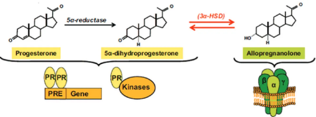
4.Neurosteroids ... 32 4.1. Definition ... 32 4.2. Biosynthesis ... 33 4.3. Mechanisms of action ... 34 5.Allopregnanolone (AP) ... 36 5.1. Biosynthesis ... 36 5.2. Mechanisms of action ... 38
5.3. Role of allopregnanolone in psychiatric disorders ... 42
5.4. Neuroprotective effects of allopregnanolone in neurodegenerative disorders ... 44
5.5. Allopregnanolone as a therapeutic candidate for the treatment of neuropathic pain ... 45
5.6. Allopregnanolone as a regenerative therapeutic in Alzheimer´s disease ... 46
5.6.1. Allopregnanolone promotes neurogenesis in vitro ... 46
5.6.2. Allopregnanolone reverses neurogenic and cognitive deficits in aged and Alzheimer´s disease mutant mice ... 47
6.PhD project ... 50
6.1. Hypothesis ... 50
6.2. Objectives ... 51
6.3. Experimental models... 51
MATERIALS AND METHODS ... 53
1. In vitro experiments ... 53
1.1. Adult neural stem cell cultures (Neurosphere culture) ... 53
1.2. Primary hippocampal cell cultures ... 55
1.3. Human neural stem cells (HNSC 100) ... 55
1.4. Material ... 56
1.4.1. Antibodies ... 56
1.4.2. RNA oligonucleotides ... 56
1.4.3. Reagents, chemicals and material ... 57
1.5. Cell culture ... 60
1.5.1. Preparation of primary neurosphere cultures ... 60
1.5.2. Passaging of neurosphere cultures ... 62
1.5.3. Proliferation and differentiation of aNSC cultures ... 62
1.5.4. Isolation, culturing and proliferation of primary hippocampal cell cultures… ... 63
1.5.5. Human neural stem cell cultures ... 65
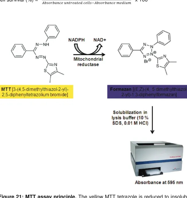
1.6. MTT cell viability assay ... 66
1.7. Immunocytochemistry ... 68
1.7.1. Coating of coverslips ... 68
1.7.2. Cell fixation ... 68
1.7.3. Immunocytochemical staining (ICC) ... 68
1.7.4. Identification of proliferating cells by incorporation of BrdU and EdU . 69 1.7.5. Caspase-3/7 assay, quantification of apoptotic cells by cell counting . 71 1.8. Caspase-3/7 assay, quantification by fluorometric measurement ... 71
1.9. BrdU cell proliferation ELISA ... 72
1.10. Real-time reverse transcription polymerase chain reaction (qRT-PCR) ... 73
1.10.1. RNA extraction ... 73
1.10.2. RNA concentration measurement and quality determination ... 74
1.10.3. Reverse transcription ... 74
1.10.4. Real-time reverse transcription polymerase chain reaction ... 75
1.11. Cell counting and statistical analysis ... 76
2.In vivo experiments ... 76
2.1. Animals ... 76
2.2. Anesthesia and perfusion of the animals ... 78
2.3. Brain freezing ... 78
2.4. Morphological analysis ... 79
2.4.1. Vibratome sectioning ... 79
2.4.2. Immunohistochemistry (IHC) ... 79
2.4.3. Quantification of immunolabeled cells in hippocampal sections ... 81
2.5. Determination of A)40 and A)42 concentration by ELISA ... 81
2.5.1. Sample preparation ... 81
2.5.2. Human A)40and A)42 ELISA... 82
2.6. Statistical analysis ... 83
RESULTS ... 84
1.Proliferation-stimulating effects of AP analogs in aNSCs ... 84
1.1. Growth of aNSC neurospheres in the presence of neurosteroids ... 84
1.2. Effects of AP analogs on aNSCs proliferation: EdU incorporation ... 86
1.2.1. 12 oxo-AP and O-allyl-AP stimulate proliferation of aNSCs ... 86
1.2.2. Phenotype of proliferating cells ... 88
1.2.3. Proliferation-promoting effects of AP analogs are mediated via L-type calcium channels ... 90
1.2.4. Effects of AP analogs on Tuj-1 and GFAP expression in aNSCs ... 91
2.Stimulatory effect of O-allyl-AP on neuronal differentiation in aNSC cultures ... 92
3.Protective effects of AP analogs against beta amyloid peptide 1-42 toxicity in aNSCs ... 95
3.1. Effect of monomeric A)42 on aNSCs viability ... 95
3.2. A)42 induces apoptosis in aNSC cultures... 96
3.3. Protective effects of AP analogs against A)42-induced toxicity ... 97
3.4. Mechanisms of action of the protective effect of AP analogs against A)42-induced toxicity ... 101
4.Proliferation-promoting effects of AP and its analogs in rat and mouse
primary hippocampal cell cultures ... 102
4.1. Proliferation-promoting effect of AP analogs in postnatal hippocampal cultures ... 103
4.2. Phenotype of proliferating cells in rat primary hippocampal cell cultures . 105 4.3. Is AP analogs-induced neural progenitor cell proliferation in mouse primary hippocampal cell cultures mediated via GABAARs? ... 107
5.Proliferation-promoting effects of O-allyl-AP on human neural stem cells ... 108
6.Effects of O-allyl-AP on proliferation and neurogenesis in the aged adult brain ... 109
6.1. Effect of O-allyl-AP on neural stem and progenitor cells ... 110
6.1.1. Sox2 labelling ... 110
6.1.2. BLBP labelling ... 111
6.2. O-allyl-AP effect on proliferating cells ... 112
6.3. O-allyl-AP effect on newborn neurons... 113
7.Effects of AP analogs in an AD mouse model (Tg2576) ... 115
7.1. Effects on proliferation ... 115
7.1.1. O-allyl-AP treatment ... 115
7.1.2. O-allyl-epiAP treatment ... 117
7.2. Effects of AP analogs on A) burden in Tg2576 mutant mice ... 119
DISCUSSION ... 122
1.Effect of AP analogs on proliferation and differentiation ... 123
2.Protective effects of AP analogs against A)42-induced toxicity on aNSCs ... 126
3.The differential pattern of biological activities efficacy of AP analogs ... 128
4.Mechanisms of action of AP analogs ... 129
5.Effects of O-allyl-AP on neurogenesis in the aged brain ... 133
6.Effects of O-allyl-AP and O-allyl-epiAP in Tg2576 ... 135
CONCLUSIONS AND PERSPECTIVES ... 139
REFERENCES ... 143
DESCRIPTIF SYNTHÉTIQUE EN FRANÇAIS DES TRAVAUX DE LA THÈSE ... 165
1
FIGURES AND TABLES LISTS
1. Figures list
Figure 1: Amylpoid plaques and neurofibrillary tangles in a section from the
hippocampus of an AD patient. ... 11
Figure 2: Formation of neurofibrillary tangles (NFTs) ... 12
Figure 3: Amyloid precursor protein (APP) proteolytic pathways. ... 13
Figure 4: Proposed molecular mechanisms of amyloid beta oligomer toxicity ... 17
Figure 5: A)-induced apoptotic pathways ... 19
Figure 6: Schematic representation of neurogenic regions in the adult mammalian brain... 21
Figure 7: Adult neurogenesis in the subventricular zone of the lateral ventricle and olfactory bulb ... 23
Figure 8: Adult neurogenesis in the subgranular zone of the dente gyrus of the hippocampus ... 25
Figure 9: Biosynthetic pathways for neurosteroids, exemplified by progesterone- related steroids ... 34
Figure 10: General structure of nuclear hormone receptors. ... 35
Figure 11: Biosynthetic pathway of allopregnanolone ... 37
Figure 12: The GABAA receptor and its various binding sites ... 39
Figure 13: Model for the mechanism of AP-induced stimulation of proliferation in neural stem cells ... 41
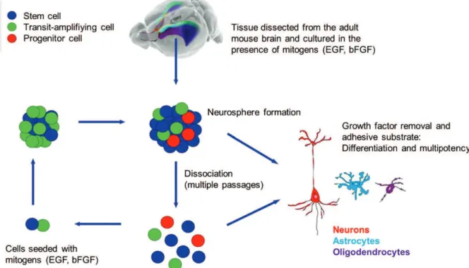
Figure 14: Schematic representation of the in vitro neurosphere culture system ... 54
Figure 15: In vitro adult neural stem cell cultures ... 54
Figure 16: Primary hippocampal cell cultures from postnatal mice ... 55
Figure 17: Chemical structures of allopregnanolone analogs investigated in this study. ... 58
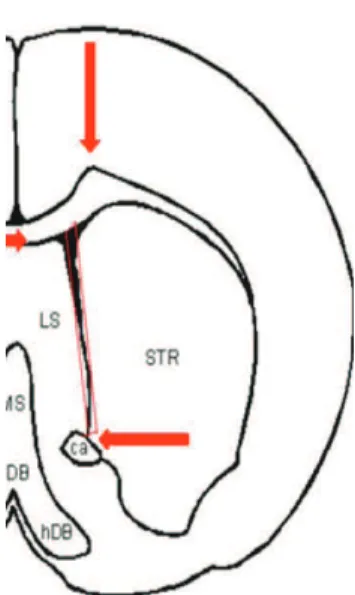
Figure 18: Coronal section through the adult mouse brain. The red arrows indicate the incision sites. ... 61
Figure 19: Illustration of the technique to remove brains from P2 mice ... 63
Figure 20: Steps for dissection of the hippocampus from the intact brain. ... 64
2
Figure 22: Principle of the indirect immunofluorescence labelling technique ... 68
Figure 23: Detection of incorporated EdU with the Alexa Fluor® azide versus incorporated BrdU with an anti-BrdU antibody ... 70
Figure 24: Experimental setups used for quantification of the protective effect of analogs of allopregnanolone by fluorometric measurement of caspase-3/7 activity . 72 Figure 25: Schematic representation of the major steps for TRIzol RNA extraction. 73 Figure 26: Photograph of a coronal section stained for Neuronal differentiation marker NeuroD ... 81
Figure 27: Growth of neurospheres cultures in the presence of neurosteroids: AP (A), O-allyl-epiAP (B) and O-allyl-AP (C) ... 85
Figure 28: Proliferation-promoting effects of neurosteroids on aNSCs ... 87
Figure 29: Phenotype of proliferating cells following treatment with AP analogs ... 90
Figure 30: Proliferation promoting effects of analogs on aNSCs are mediated via L-type calcium channels ... 91
Figure 31: Neurosteroids effects on Tuj-1 (A) and GFAP (B) mRNA expression levels in aNSCs ... 92
Figure 32: Effect of neurosteroids on neuronal differentiation in dissociated aNSC cultures ... 93
Figure 33: Stimulatory effect of O-allyl-AP on neuronal differentiation ... 94
Figure 34: Dose-dependent toxic effect of A)42... 96
Figure 35: Induction of apoptosis by A)42 in aNSC cultures ... 97
Figure 36: Anti-apoptotic effects of AP analogs in A)42-treated dissociated neurosphere cells ... 99
Figure 37: Neurosteroids reduce the Bax/Bcl-2 ratio in aNSC cultures ... 100
Figure 38: Neuroprotective activity of AP analogs is not mediated via GABAARs (A), intracellular progesterone receptors (B) or conversion back to 5*-forms of the neurosteroids (C) ... 102
Figure 39: Proliferation promoting effect of AP analogs on rat (A,B) and mouse (C) primary hippocampal cell cultures... 104
Figure 40: Phenotype of proliferating cells in AP analogs-treated hippocampal cell cultures ... 106
Figure 41: Effect of bicuculline on neurosteroid-induced proliferation ... 107
3 Figure 43: Effect of O-allyl-AP on the number of Sox2-expressing cells in the
SGZ ... 111
Figure 44: Effect of O-allyl-AP on the number of BLBP-immunoreactive radial glia-like stem cells ... 112
Figure 45: Effect of O-allyl-AP on proliferative activity in the SGZ of adult mice ... 113
Figure 46: Effect of O-allyl-AP on generation of NeuroD-immunoreactive immature neurons ... 114
Figure 47: O-allyl-AP effects on proliferating cells and newborn neurons in Tg2576 mice. ... 116
Figure 48: Effects of O-allyl-epiAP on the expression and neurogenesis markers in Tg2576 mice ... 118
Figure 49: Amyloid plaques are surrounded by activated astrocytes in Tg2576 mutant mice. ... 120
Figure 50: Effects of AP analogs on amyloid beta peptide levels in the brain of Tg2576 mutants ... 121
2. Tables list
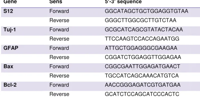
Table 1: Antibodies used in the immunocytochemistry. ... 56Table 2: Oligonucleotide sequences. ... 57
Table 3: Neurosteroids used in this study. ... 58
Table 4: Chemicals, kits and devices. ... 60
Table 5: Click-iT reaction cocktail. ... 70
Table 6: Blocking solutions used in immunohistochemistry. ... 79
4
ABREVIATIONS
[Ca2+]i Intracellular calcium concentration
12 oxo-AP 12 oxo-allopregnanolone, Pregnane-12,20 dione 3-hydroxy (3*, 5*)
12 oxo-epiAP 12 oxo-epiallopregnanolone, Pregnane-12,20 dione 3-hydroxy (3), 5*)
3*-HSD 3*-hydroxysteroid dehydrogenase 3)-HSD 3)-hydroxysteroid dehydrogenase 5*-DHP 5*-dihydroprogesterone
AD Alzheimer´s disease
ADAM A disintegrin and metalloprotease
AEBSF 4-(2-Aminoethyl) benzenesulfonyl fluoride hydrochloride AF-1 and -2 Activation function 1 and 2
AMP Adenosine monophosphate ANOVA Analysis of variance
ANS Analogs of allopregnanolone aNSCs Adult neural stem cells AP Allopregnanolone
Apaf-1 Apoptotic protease activating factor-1 APH-1 Anterior pharynx-defective-1
APOE Apolipoprotein E
APP Amyloid precursor protein Ascl1 Achaete-scute homolog 1 ATP Adenosine triphosphate A) Amyloid )-peptide B Bicuculline
BACE1 )-site APP cleaving enzyme 1 Bax Bcl-2 associated X protein BCA Bicinchoninic acid
Bcl-2 B-cell Lymphoma 2
BDNF Brain-derived neurotrophic factor bFGF Basic fibroblast growth factor BLBP Brain lipid-binding protein
5 BMP Bone morphogenetic protein
BrdU 5-bromo-2´-deoxyuridine BSA Bovine serum albumin
CA1 and CA3 Cornu Ammonis area 1 and 3 CalB Calbindin
CaMK IV Calcium/calmodulin-dependent protein kinase type IV Caspase Cysteine-dependent aspartate-directed proteases CDK1 Cyclic dependent kinase 1
cDNA Complementary DNA CGC Cerebellar granule cells CNS Central nervous system CR Calretinin
CREB1 Cyclin AMP-responsive element-binding protein Ct Threshold cycle DAB 3,3+-diaminobenzidine DBD DNA-binding function DCX Doublecortin DG Dentate gyrus DHEA Dehydroepiandrosterone DIV Days in vitro
Dlx-2 Distal-less gene 2
DMEM Dubelcco´s modified eagle medium DMSO Dimethyl sulfoxide
DNA Deoxyribonucleic acid dNTP Nucleoside triphosphate
EC50 Half maximal effective concentration EdU 5-ethynyl-2´-deoxyuridine
EGF Epidermal growth factor
ELISA Enzyme-linked immunosorbent assay
epiAP Epiallopregnanolone, 3)-Hydroxy-5*-pregnan-20-one ES Embryonic stem
FAD Familial Alzheimer disease
FADD Fas-associated protein with death domain FCS Fetal calf serum
6 FGF2 Fibroblast growth factor 2
GABA ,-Aminobutyric acid
GABAAR ,-Aminobutyric acid type A receptor GCL Granular cell layer
GFAP Glial fibrillary acidic protein H region Hinge region
HBSS Hanks Buffered salt solution
HEPES 4-(2-hydroxyethyl)-1-piperazineethanesulfonic acid HMG-CoA 3-hydroxy-3-methylglutaryl-coenzyme A
HNSC Human neural stem cells HRP Horse radish peroxidase HS Horse serum
Hu A)40, A)42 Human A)40, A)42
Iba1 Ionized calcium binding adaptor molecule 1 IL6 Interleukin 6
IPSC Induced pluripotent stem cells LV Lateral wall of the lateral ventricle LXR liver X receptor
M Mifepristone
MAPs Microtubule-associated proteins MCI Mild cognitive impairment
MEM Minimum essential medium MMA Methylazoxymethanol acetate mPR Membrane progesterone receptors
MPTP 1-methyl-4-phenyl-1,2,3,6-tetrahydropyridine mRNA Messenger ribonucleic acid
MTT 3-[4,5-dimethylthiazol-2-yl]-2,5 diphenyltetrazolium bromide
N Nifedipine
NaN3 Sodium azide
NDS Normal donkey serum NeuN Neuronal nuclear antigen
NeuroD Neurogenic differentiation factor
NF--B Nuclear factor 'kappa-light-chain-enhancer' of activated B-cells NFTs Neurofibrillary tangles
7 NGF Nerve growth factor
NGS Normal goat serum NHS Normal horse serum NMDA N-methyl-D-aspartate NRS Normal rabbit serum NSCs Neural stem cells
nSR Nuclear steroid receptors NTg Non-transgenic
O-allyl-AP O-allyl-allopregnanolone, Pregnan-20 one 3-(2-propen-1-yloxy) (3 *, 5*)
O-allyl-epiAP O-allyl-epiallopregnanolone, Pregnan-20 one 3-(2-propen-1-yloxy) (3), 5*)
OB Olfactory bulb OD Optical density
P Provera, Medroxyprogesterone 17-acetate P450c21 Cytochrome P450c21 or steroid 21-hydroxylase P450scc Cytochrome P450side-chain-cleavage
Pax6 Paired box protein 6 PB Phosphate Buffer
PBS Phosphate Buffered Saline PCNA Proliferating cell nuclear antigen PCR Polymerase chain reaction PFA Paraformaldehyde
PNS Peripheral nervous system PR Progesterone receptors PrPC Cellular prion protein
Prox1 Prospero-related homeobox gene 1 PSEN1 and 2 Presenilin 1 and Presenilin 2
PTSD Post-traumatic stress disorder PXR Pregnane xenobiotic receptor
qPCR Quantitative polymerase chain reaction
qRT-PCR Real-time reverse transcription polymerase chain reaction RAGE Receptor for advanced glycation end products
8 RMS Rostral migratory stream
RNA Ribonucleic acid
ROS Reactive oxygen species RT Room temperature S12 Ribosomal protein S12 SDS Sodium dodecyl sulfate SEM Standard error of the means SGZ Subgranular zone
Shh Sonic hedgehog
siRNA Small interfering ribonucleic acid Sox2 SRY-related HMG-box gene 2
SSRIs Selective serotonin reuptake inhibitors StAR Steroidogenic acute regulatory protein SVZ Subventricular zone
Tg Transgenic
TH Tyrosine hydroxylase TMB Tetramethylbenzidine TNF* Tumor necrosis factor alpha
TRIzol Guanidinium thiocyanate-phenol-chloroform TSPO Translocator protein 18 kDa
Tuj-1 Class III beta-tubulin
VDLCC Voltage-dependent L-type calcium channels Wnt Wingless
9
INTRODUCTION
1. Neurodegenerative diseases
Neurodegenerative diseases are common disorders of the nervous system that affect millions of people worldwide and generate significant suffering for patients as well as medical and social costs of several billion euros per year.
Neurodegenerative diseases such as Alzheimer´s disease (AD), Parkinson´s disease, Huntington´s disease, amyotrophic lateral sclerosis, multiple sclerosis, frontotemporal dementia, spinocerebellar ataxia, motor neuron diseases, prion disease, peripheral neuropathic pain, dementia with Lewy bodies and many others present a common feature which is the deregulation of processes controlling the protection and survival of nerve cells, which leads to the degeneration of a part of the central nervous system (CNS) or peripheral nervous system (PNS) and neural death. In this thesis we will mainly focus on Alzheimer´s disease.
Neurological disorders caused by neurodegenerative diseases, depending on the brain region and the neuron type concerned, affect memory, cognition, language, perception and sensitivity to pain, locomotion and realization of motor actions. For example, loss of memory and cognitive functions are the hallmark of AD, whereas involuntary tremor and the loss of control of motor activities are the main symptoms of Parkinson’s disease.
Aging is considered the primary risk factor for most neurodegenerative disorders. However, the cellular and molecular mechanisms involved in neurodegenerative diseases are still poorly understood, which explains the absence of effective treatment, despite the large number of affected people.
Interestingly, many forms of neurodegenerative diseases share a common pathological phenotype which is the accumulation of misfolded and aggregated proteins in the brain. Therefore, a lot of studies aim to better understand the processes which lead to generation of these protein aggregates in order to elaborate new therapeutic approaches (Ciechanover and Kwon, 2015).
10
2. Alzheimer´s disease
AD was first described in 1906 by Alois Alzheimer, a German psychiatrist and neuropathologist (Alzheimer et al., 1995), as a form of dementia that was subsequently named after him. His study was based on the observation of a 51-year-old female patient, Augusta Deter, who presented a progressive cognition impairment, disorientation, aphasia, hallucinations, mental confusion, and paranoid delusions (Maurer et al., 1997). From the post-mortem study of her brain, Alois Alzheimer was able to identify neuritic plaques and neurofibrillary tangles.
AD is the most common form of dementia and the first leading cause of death according to the 2015 Alzheimer’s Disease Facts and Figures (Alzheimer´s Association). It accounts for an estimated 70 % of cases of dementia and affects nearly 44 million people worldwide. Age is the main risk factor of AD, the incidence of AD increases rapidly with age: from 0.5 % among 65-70 years old to 15 % of individuals over the age of 80. With the continuous growth of the proportion of the older population due to medical progress, the number of patients is expected to increase to nearly 76 million by 2030 if breakthroughs in prevention or treatment are not achieved. Therefore, AD represents a major health concern and became a research priority worldwide.
AD typically progresses slowly in three clinical stages: preclinical, mild cognitive impairment (MCI) and dementia (Sperling et al., 2011). Changes in the brain related to Alzheimer's begin years before any symptoms of the disease.
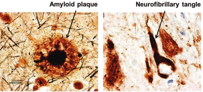
AD leads to the progressive loss of mental and behavioral capabilities and of the ability to learn (Anand et al., 2014). The neuropathology of AD is characterized by abnormal protein deposits (Fig. 1): hyperphosphorylated tau proteins, which assemble into intraneuronal neurofibrillary tangles (NFTs) (Ballatore et al., 2007) and amyloid )-peptide (A)), which accumulates in the extracellular space of the brain as senile plaques (Glenner and Wong, 1984; Masters et al., 1985). The functional relationship between these two processes is heavily investigated (Ittner et al., 2010; Vossel et al., 2010).
11
2.1. Neurofibrillary tangles
Neurofibrillary tangles are filamentous inclusions, consisting of paired helical filaments, which accumulate in the cell bodies and processes of neurons. These intraneuronal lesions are formed by highly phosphorylated forms of the microtubule-associated protein Tau (Fig. 1 and 2) (Goedert et al., 1989; Buée et al., 2000; Mandelkow and Mandelkow, 2012). The disequilibrium between the activities of protein kinases and phosphatases acting on tau leads to an early accumulation of phosphorylated tau proteins which later on form neurofibrillary tangles (Fig. 2). Tau protein, belonging to the family of microtubule-associated proteins (MAPs), is highly expressed in neurons. In physiological conditions, Tau is a soluble protein that participates in microtubule assembly and in their stabilization. Its activity is regulated by phosphorylation (Köpke et al., 1993). In pathological conditions, tau loses its solubility, forms filamentous structures and is abnormally phosphorylated on certain residues. As a consequence, Tau is no longer able to bind to microtubules, which leads to microtubule instability and axonal degeneration (Fig. 2) (Bramblett et al., 1993; Crespo-Biel et al., 2012).
Neurofibrillary tangles are not specific to AD. They were also identified in fronto-temporal dementia and in Parkinson´s disease (Poorkaj et al., 1998; Spillantini and Goedert, 1998).
Figure 1: Amylpoid plaques and neurofibrillary tangles in a section from the hippocampus of an AD patient (Images taken from coloradodementia.org).
12 Figure 2: Formation of neurofibrillary tangles (NFTs) (Mokhtar et al., 2013).
2.2. Amyloid peptide
Amyloid )-peptide (A)) is the primary component of the senile plaques found in AD patient brain tissue (Masters et al., 1985). A) (4-kDa) is a proteolytic cleavage product of amyloid precursor protein (APP). APP, a type I transmembrane protein, can be cleaved by different proteolytic enzymes: *- )- and ,-secretases and processed through two different pathways: the amyloidogenic pathway and the non-amyloidogenic pathway (Eggert et al., 2004) (Fig. 3). Under physiological conditions, these two pathways are balanced as there is an equilibrium between the production of A) peptides and their clearance from the brain (Vetrivel and Thinakaran, 2006). In AD, there is a metabolic shift favoring the amyloidogenic cleavage of APP which, along with a reduction of A) clearance, leads to the accumulation of A) within the brain (Kunjathoor et al., 2004).
2.2.1. The non-amyloidogenic pathway
In the non-amyloidogenic pathway, the extracellular region of APP is cleaved by the metalloprotease *-secretase, releasing a soluble N-terminal fragment (sAPP-*) and a C-terminal fragment 83 (APP-CTF83) (Fig. 3). The sAPP-* domain is secreted into the extracellular space and is thought to have neuroprotective and neurotrophic effects (Furukawa et al., 1996; Kojro and Fahrenholz, 2005). The C83 fragment is subsequently cleaved by ,-secretase, releasing an APP intracellular domain (AICD) and a P3 fragment that is not toxic; in contrast it is believed to exert a neuroprotective
13 effect (Kojro and Fahrenholz, 2005; Dulin et al., 2008). Enzymes with *-secretase activity have been identified as members of the ADAM (A Disintegrin And Metalloprotease) family. The major members of the *-secretase family are ADAM9, ADAM10 and the tumor necrosis factor converting enzyme ADAM17 (Allinson et al., 2003; De Strooper, 2010).
2.2.2. The amyloidogenic pathway
The amyloidogenic pathway, the less abundant pathway for APP cleavage, leads to generation of A) peptides. In this pathway, )-secretase cleaves APP at the N-terminal boundary of the A) peptide domain and produces a smaller soluble N-terminal fragment (sAPP-)) and the C-N-terminal fragment 99 (APP-CTF99) (Fig. 3) (Haass, 2004). C99 fragment produces AICD and the full-length A) peptides upon the subsequent cleavage by ,-secretase. The length of A) peptides varies between 37 and 43 amino acids (Qi-Takahara et al., 2005). The biological functions of APPs-), AAPPs-), and the AICD are not yet fully understood, although A) release has been shown to be associated with reduced synaptic activity and abnormal Figure 3: Amyloid precursor protein (APP) proteolytic pathways. The transmembrane protein APP (membrane indicated in blue) can be processed by two pathways, the nonamyloidogenic *-secretase pathway and the amyloidogenic )-secretase pathway (Wang et al., 2012).
14 neurotransmission (Kamenetz et al., 2003). The major A) species are A)40 and A)42 (Korczyn, 2008).
2.2.3. Enzymes implicated in proteolytic processing of APP
)-secretase activity is mainly due to the )-site APP cleaving enzyme 1 (BACE1), a type 1 transmembrane aspartic protease, that is related to pepsin and retroviral aspartic protease families (Hussain et al., 1999). BACE1 is the rate-limiting enzyme in the proteolytic processing of APP and is required for the production of A). BACE1-processing in APP mutants can lead to a dramatic increase (Citron et al., 1992) or decrease (Jonsson et al., 2012) in amyloidogenic processing and to altered AD risks. ,-secretase has been identified as a multi-subunit aspartyl protease which is composed of presenilin 1 or 2 (PSEN1 or PSEN2) forming the catalytic core of ,-secretase, and three accessory proteins: nicastrin, anterior pharynx-defective 1 (APH-1) and presenilin enhancer 2 (PEN2) (De Strooper, 2003; Edbauer et al., 2003; Takasugi et al., 2003). These three proteins appear to be involved in the maturation and stability of the complex. ,-secretase is heterogeneous with respect to its protein composition and expression as six different functional ,-secretase complexes have been recognized which allow several cleavage possibilities of APP (Hébert et al., 2004; Shirotani et al., 2004).
2.2.4. The amyloid cascade hypothesis and its evolution
There are several A) peptide species, the most abundant ones produced by the amyloidogenic pathway are A)40 and A)42 (Korczyn, 2008). A) peptides are released as monomers which progressively self-aggregate into oligomers, protofibrils and fibrils, and finally are deposited and mature to amyloid plaques (Roberts et al., 1994). A)42, a hydrophobic peptide, has a strong tendency to aggregate and fibrilize which leads to the formation of amyloid fibrils and plaques (Jarrett et al., 1993a, b). Therefore, it is considered as a major cause of neurotoxicity and plays a crucial role in AD pathogenesis (Butterfield, 2002; Walsh and Selkoe, 2007; Yankner and Lu, 2009). A)42 was first described in familial AD forms (FAD) and an elevated ratio of A)42 to A)40 was detected in AD brains (Hardy and Selkoe, 2002). Interestingly, recent studies showed that A) oligomers, rather than plaques are the most toxic form of the A) peptide (Haass and Selkoe, 2007; Walsh and Selkoe, 2007).
15 The hypothesis that AD may arise from the accumulation of misfolded )-sheet proteins in a manner analogous to systemic amyloidoses was proposed after the isolation and identification of A) peptides in the brain of AD patients (Glenner and Wong, 1984). This hypothesis was supported by several studies; and in 1992, Hardy and Higgins named it the amyloid cascade hypothesis, which became the basis for work on AD pathogenesis (Hardy and Allsop, 1991; Hardy and Higgins, 1992; Tanzi and Bertram, 2005). The amyloid cascade hypothesis proposed that accumulation of )-amyloid peptides into neuritic and senile plaques in the brain, due to an imbalance between A) production and A) clearance, is the key pathogenic feature of AD; whereas the rest of the disease process, including the formation of neurofibrillary tangles is a consequence of this imbalance (Hardy and Selkoe, 2002). In the past few years, the amyloid cascade hypothesis has been modified because of a non-linear correlation between the number of amyloid plaques in the brain and the progression of AD pathology especially the degree of cognitive impairment (Games et al., 1995; Price et al., 2009). Results from recent studies now suggest a correlation between the concentration of soluble A) species and the degree of pathology (Giannakopoulos et al., 2003). Therefore, a modified amyloid cascade hypothesis has been proposed, suggesting that soluble A) oligomers are the major cause of synaptic dysfunction, neuron damage and memory impairment in early AD (Scheff and Price, 2006; Scheff et al., 2007; Selkoe, 2008).
On the other hand, Braak and Braak demonstrated a clear correlation between the degree of neurofibrillary tangle pathology and cognitive impairment in AD patients (Braak and Braak, 1996). These observations have been proven by additional studies (Nelson et al., 2012; Jack and Holtzman, 2013). Moreover, intra-neuronal hyperphosphorylated Tau was found in brains of subjects with mild dementia, unaccompanied by )-amyloid pathology (Mazanetz and Fischer, 2007; Grinberg et al., 2009). Therefore an alternative hypothesis started to develop recently, suggesting that dysfunction in Tau homeostasis may be an early event in the physiopathology of AD, whereas A) overproduction and oxidative stress might be resulting consequences of this dysfunction in neuronal homeostasis.
2.2.5. Toxicity of amyloid beta oligomers
After the discovery that the most toxic form of the A) peptide are the A) oligomers (Haass and Selkoe, 2007), it has been shown that A) oligomers are produced by the
16 cooperative activity of both neurons and associated astrocytes (Dal Prà et al., 2014). A) oligomers induce toxic effects on synapses and mitochondria such as oxidative stress (Sultana et al., 2009) and tau hyperphosphorylation (De Felice et al., 2008). Oligomers are found both extracellularly and intracellularly and are capable of moving between the interior of the cell and the extracellular space (Gaspar et al., 2010). However, the exact mechanisms of amyloid oligomers formation and toxicity are still not clear. Several mechanisms have been proposed (Fig. 4) (Kayed and Lasagna-Reeves, 2013): (i) “A) receptors”, a number of A)-binding proteins have been identified on the cell surface of neurons which could mediate A)-induced neurotoxicity (Fig. 4A). These proteins include the nerve growth factor (NGF) receptor, N-methyl-D-aspartate (NMDA) receptor, insulin receptor, Frizzled receptor and cellular prion protein (PrPC) receptor (Costantini et al., 2005; Snyder et al., 2005; Magdesian et al., 2008; Zhao et al., 2008b; Laurén et al., 2009). (ii) Membrane permeabilization and A)-channels formation, which lead to disturbed homeostasis of calcium and other ions and subsequent promotion of free radical formation and Tau hyperphosphorylation (Fig. 4B) (Takashima et al., 1993; Yatin et al., 1998; Kagan et al., 2002; Kagan et al., 2004). (iii) Internalization and intracellular accumulation of A) by binding to several receptors such as scavenger receptor for advanced glycation end products (RAGE), *7 nicotinic acetylcholine receptor (*7nAChR) and apolipoprotein E receptor (APOE) (Fig. 4C) (Yan et al., 1996; Wang et al., 2000a; Bu et al., 2006). Intracellular accumulation of the oligomeric A) leads to proteasome impairment, mitochondrial dysfunction and disturbance of autophagy (Caspersen et al., 2005; Nixon et al., 2005; Mousnier et al., 2007).
17
2.2.6. Amyloid beta induced apoptotic pathways
Su and colleagues were the first to suggest that apoptosis is a physiopathological process involved in AD (Su et al., 1994). Since then, other studies have shown that increased production and accumulation of A) peptides induce neurotoxicity by activating apoptotic processes (LaFerla et al., 1995; Li et al., 1996; Cotman, 1998; Eckert et al., 2001). For example, exposure of PC12 cells and hippocampal neurons in culture to A)42 peptide activates two parallel apoptotic pathways which lead to cell death (Jordán et al., 1997; Troy et al., 2000). A)42 is able to activate the intrinsic
A
B
C
Figure 4: Proposed molecular mechanisms of amyloid beta oligomer toxicity. A) A) oligomers may bind to multiple receptors leading to the activation of various signaling pathways. B) A) oligomers insert into the membrane and subsequently form ion channels or pores which lead to neurodegenerative processes. C) The intracellular accumulation of A) oligomers and other aggregates causes many key pathological events of AD, including proteasome impairment, mitochondrial dysfunction and breakdown of many cellular processes (Kayed and Lasagna-Reeves, 2013).
18 pathway of apoptosis (Fig. 5). A)42 downregulates bcl-2, a key anti-apoptotic protein, and upregulates bax, a protein known to promote apoptotic cell death induced by a decrease in mitochondrial membrane potential. Subsequently, cytochrome c is released by dysfunctional mitochondria. Cytochrome c, together with the adapter protein apoptotic protease-activating factor-1 (Apaf-1) and pro-caspase-9 forms an apoptosome (Shiozaki et al., 2002; Degli Esposti and Dive, 2003). The initiator caspase-9 becomes active and cleaves the effector pro-caspase-3 into its active form which initiates apoptosis leading to protein and DNA cleavage and cell death.
In addition, A)42 has been shown to have an effect on the extrinsic pathway of apoptosis (Fig. 5). A)42 may lead to the cross-linking and activation of death receptors, such as Fas receptor, resulting in activation of the initiator caspase-8 (Rohn et al., 2001). Caspase-8 then mediates the cleavage of the pro-caspase-3. Activated caspase-3 is the common, cell death initiating effector enzyme of both pathways.
Based on the strong evidence in favor of the amyloid cascade hypothesis and the toxic effect of A) oligomers, AD drug development was focused on molecules targeting A) generation and aggregation. Most drug candidates aimed to inhibit A) toxicity by reducing further A) aggregation and plaque formation. These drug candidates include secretase inhibitors to lower A) production from APP, A) aggregation inhibitors to inhibit A) oligomerization or fibrillization, A) degradation inducers to increase A) clearance, as well as active and passive A) immunotherapies designed to capture either soluble or aggregated A), which then can be either degraded or cleared from the brain (Blennow et al., 2014).
19 It is important that therapeutic approaches aim at inhibiting disease progression but also at improving cognitive impairment due to neuronal loss. Therefore, in search of an effective treatment, it is essential to take into consideration two important aspects: (i) that of therapeutic neurogenesis which relies on the ability to stimulate the generation of new neurons in the nervous system to compensate for cell loss caused by neural damage, trauma, neurotoxic substances, oxidative stress, senescence or even brain aging; (ii) that of neuroprotection which aims to protect neurons against cell death and thus preserves their functions. Consequently, the exploitation of these
Figure 5: A -induced apoptotic pathways. A) oligomers exert their toxic effects
via two apoptotic pathways: the extrinsic and the intrinsic pathways. Both pathways lead to neuronal loss and neurodegeneration. Adapted from (Hersey and Zhang, 2001; Eckert et al., 2003).
20 two aspects in the adult brain is a very promising avenue for research of new therapies against neurodegenerative diseases. Neurosteroids, endogenous steroidal compounds, have been found to positively influence both of these processes, and therefore, are promising therapeutic candidates. They are synthesized locally within the nervous system by both neurons and glial cells and have been shown to exert several effects in brain including neuroprotection (see parts 4 and 5, pages 33-50).
3. Adult neurogenesis in the rodent brain
Neurogenesis is defined as the process of generating functional neurons from stem and precursor cells. This process was believed to occur only during embryonic and perinatal stages of development in the mammalian brain (Ming and Song, 2005). In 1965, Altman and colleagues provided the first anatomical evidence for the presence of newly generated dentate granule cells in the postnatal rat hippocampus (Altman and Das, 1965). After several studies in different experimental models, it became accepted that mammalian brains retain the capacity to generate new neurons throughout life (Kempermann and Gage, 1999; Gross, 2000; Lie et al., 2004). In the mammalian CNS, active adult neurogenesis occurs in two specific “neurogenic” brain regions under normal conditions: (i) the subventricular zone (SVZ) lining the lateral ventricles, where new neurons are generated and then migrate via the rostral migratory stream (RMS) to the olfactory bulb (OB) to become interneurons and integrate into the neuronal circuit; (ii) the subgranular zone (SGZ) located in the dentate gyrus of the hippocampus, where new dentate granule cells are generated (Fig. 6). However, generation of new neurons was also observed in the hypothalamus (Kokoeva et al., 2005; Lee et al., 2012). It is still controversial whether neurogenesis occurs in neocortical areas (Bhardwaj et al., 2006; Zhao et al., 2008a). Knowledge regarding neurogenesis in the adult human brain is limited in comparison to that in rodents. However, it has been shown that new neurons are generated in the dentate gyrus of the hippocampus in the adult human brain in comparable numbers to those observed in rodents (Eriksson et al., 1998; Spalding et al., 2013). In the human olfactory system neurogenesis however, is extremely low even though quiescent neural stem cells and progenitor cells may also persist in the human SVZ (Sanai et al., 2004; Curtis et al., 2007). Recently, it has been reported that the human striatum is capable of generating striatal interneurons throughout life (Ernst et al.,
21 2014). Novel methods to measure levels of neurogenesis in the human brain, like the non-invasive magnetic resonance imaging technique, are needed to overcome the existing gap between animal and human research (Ho et al., 2013).
3.1. Neural stem cells in the adult mammalian brain
Neural stem cells (NSCs) should fulfill two defined characteristics, the capacity for self-renewal through cell division and the capacity for multi-lineage differentiation (multipotency) into neurons, astrocytes and oligodendrocytes (Gage, 2000). Neural progenitors include all dividing cells with some capacity for differentiation.
Radial glia-like cells expressing the glial fibrillary acidic protein (GFAP) are the primary precursors of new neurons in the adult brain. In the SVZ-OB system, these radial glia-like cells are termed “Type B cells” while in SGZ of adult hippocampus, they are termed “Type 1-cells” (Alvarez-Buylla and Lim, 2004). In addition, non-radial stem cells exist in the adult SGZ. They are active neural stem cells expressing SRY-related HMG-box gene 2 (Sox2) and give rise to new neurons and glia in the adult SGZ (Suh et al., 2007).
Figure 6: Schematic representation of neurogenic regions in the adult mammalian brain. The two major neurogenic regions: Subventricular zone (SVZ) and subgranular zone of the dentate gyrus (DG) in the adult mammalian brain are marked in green. Newly generated cells from the SVZ migrate via the rostral migratory stream (RMS) to the olfactory bulb (OB) to become interneurons. Neurogenesis in the human OB is relatively low. Additional sites of neurogenesis including the hypothalamus in the mouse brain and the striatum in the human brain are represented in gray (Braun and Jessberger, 2014).
22
3.2. The Subventricular Zone (SVZ)
The SVZ generates immature neuronal precursors called neuroblasts which migrate through the rostral migratory stream, to the olfactory bulb where they mature into local interneurons (Altman, 1969; Buylla et al., 1994; Lois and Alvarez-Buylla, 1994). The SVZ-OB represents an established system for studying neurogenesis and neuronal replacement as it includes the basic processes of NSCs maintenance, progenitor cell fate specialization, migration, differentiation and survival or death of newly born neurons. The SVZ in the adult mammalian brain contains four main cell types (B cells, A cells, C cells and ependymal cells), which are defined by morphology, ultrastructure and by the expression of various molecular markers (Doetsch et al., 1997) (Fig. 7).
The type B cells, GFAP-expressing radial glia-like cells with ultrastructural characteristics typical of astrocytes, represent the quiescent NSCs within the SVZ neurogenic niche. They are slowly dividing cells (Doetsch et al., 1999) and give rise to highly proliferative intermediate precursor cells termed type C cells. B cells also express the intermediate filament proteins nestin and vimentin (Lendahl et al., 1990).
The type C cells function as transit-amplifying progenitors intermediate between B and A cells. C cells are generated by asymmetric cell division of B cells and divide more rapidly (Doetsch, 2003a).They generate large numbers of neuroblasts, named type A cells. C cells express the progenitor cell marker nestin.
Type A cells or migrating neuroblasts are typically organized as chains of cells throughout the SVZ, indicating that these neuronal precursors migrate closely associated to each other in the form of a chain aligned along the blood vessels (Lois et al., 1996). The immature neurons migrate through the RMS to the OB where they preferentially differentiate into olfactory granule interneurons, expressing calretinin (CR), calbindin (CalB) or tyrosine hydroxylase (TH) and periglomerular neurons expressing CR. They both integrate into local circuits (Alvarez-Buylla and Garcia-Verdugo, 2002; Carleton et al., 2003). Type A cells typically express the immature neuronal marker Tuj-1, doublecortin (DCX) and the differentiation factor Dlx-2.
23 Type E cells are ciliated ependymal cells, building a single layer, which separates the above mentioned cells from the cerebrospinal liquid. They are essential for neuronal fate specification by providing inhibitors of gliogenesis.
Studies in rodents have revealed that this dynamic neurogenic process generates thousands of neuroblasts per day; however, only a small fraction of immature neurons survive and functionally integrate into OB circuits (Carleton et al., 2003).
3.3. The Subgranular Zone (SGZ)
Neurogenesis in the SGZ of the dentate gyrus (DG) of the hippocampus has been extensively studied due to its possible implication in learning and memory (Kempermann et al., 2004a). In the adult SGZ, radial NSCs give rise to intermediate progenitors which in turn generate neuroblasts that migrate into the inner part of the granule cell layer and differentiate into dentate granule cells. Precursor cells in the SGZ seem to be different from the cells found in the SVZ (Seaberg and van der Kooy, 2002). Therefore, an alternative nomenclature, based on numbers, has been
Figure 7: Adult neurogenesis in the subventricular zone of the lateral ventricle and olfactory bulb. Adult SVZ NSCs (type B) reside below the ependymal cell layer
(EC) in the wall of the later ventricle. These cells give rise to rapidly dividing transit-amplifying progenitors (type C) that generate neuroblasts (type A). The newborn immature neurons migrate through the RMS towards the OB where neuroblasts preferentially differentiate into granule neurons and periglomerular neurons that integrate into OB neuronal circuits. Adapted from (Braun and Jessberger, 2014).
24 proposed for the neurogenic cells located in the DG (Kempermann et al., 2004b) instead of the letters that had been originally used to describe the neurogenic cells within the SVZ, mentioned earlier. The SGZ in the adult mammalian brain contains three cell types (Type-1, Type-2 and type-3 cells) (Fig. 8) (Emsley et al., 2005).
Type-1 cells, radial glia-like precursors, are quiescent neural stem cells expressing GFAP, nestin and the brain lipid-binding protein (BLBP).
Type-2 cells, non-radial progenitors, are produced by type-1 cells. They are amplifying neural progenitors which lack glial features and express nestin and sox2 as markers. Type-2 cells can be divided into two subpopulations, both nestin-positive, but one being negative and the other one being positive for the immature neuronal marker DCX. They are named type-2a and type-2b, respectively (Kempermann et al., 2004b).
Type-3 cells are slowly proliferating neuroblasts which express markers of the neuronal lineage such as DCX, NeuroD and Prox1. Immature neurons migrate into the granular layer and differentiate into mature granular neurons (expressing postmitotic neuronal markers such as Prospero-related homeobox gene 1 Prox1 and neuron-specific nuclear protein NeuN). Over a period of 3 weeks newborn granule neurons extend their dendrites towards the molecular layer of the DG and project axons through the hilus towards the CA3 region, and acquire their mature electrophysiological properties. The mature granule cells (expressing NeuN, Prox1 and calbindin) become functionally integrated into the hippocampus. A small proportion of progenitor cells differentiates into glial cells (Hastings and Gould, 1999; Markakis and Gage, 1999; van Praag et al., 2002; Zhao et al., 2006).
In the adult rodent brain, the majority of newborn cells die within the first days after birth (Sierra et al., 2010). The survival rate of newborn neurons that mature and integrate into the pre-existing circuit under normal condition is about 20 %.
25 Since the discovery of neurogenic areas in the adult brain, the molecular mechanisms that regulate the process of persistent formation of new neurons were intensively investigated. Each step of lineage progression is controlled by niche-derived and intrinsic mechanisms, which together ensure appropriate levels of proliferation of NSCs and progenitor cells, their correct differentiation, migration and integration of newborn cells. Sox2, NeuroD, Pax6, Prox1, Sp8, Ascl1 were identified as important transcriptional regulators of neurogenesis in the SVZ and/or SGZ (Zhao et al., 2008a). In addition, neurogenesis levels can be controlled by epigenetic mechanisms, for example through histone modifications or via small non-coding RNAs (Zhao et al., 2008a). Furthermore, neurotransmitters, growth factors and cytokines play a role in controlling NSC and progenitor cell activity and neuronal differentiation; such as ,-aminobutyric acid (GABA), glutamate, brain-derived neurotrophic factor (BDNF), epidermal growth factor (EGF), fibroblast growth factor 2 (FGF2), wingless (Wnt) ligands, sonic hedgehog (Shh), bone morphogenetic proteins
Figure 8: Adult neurogenesis in the subgranular zone of the dente gyrus of the hippocampus. Adult SGZ NSCs give rise to glutamatergic granule cells of the DG
via a well characterized multistep process. Quiescent radial glia-like cells (type-1) enter the cell cycle and divide to produce transient amplifying non-radial glia cells (type-2). These cells form immature neurons (type-3) that integrate into the neural network. Adapted from (Braun and Jessberger, 2014).
26 (BMP), interleukin 6 (IL6) and tumor necrosis factor alpha (TNF*), among others (Zhao et al., 2008a).
3.4. Immunohistological
markers
for
proliferation
events,
gliogenesis and neurogenesis in the adult hippocampus
Hippocampal neurogenesis is divided into different stages: proliferation, differentiation, migration, maturation and synaptic integration. This allows the monitoring of hippocampal neurogenesis in more detail, since the various developmental stages correlate with the expression of different markers.
3.4.1. Markers for neural stem cells
SRY-related HMG-box gene 2 (Sox2)
Sox2, a member of the Sox family, is a transcription factor that is essential for maintaining self-renewal, or pluripotency, of undifferentiated stem cells. Sox2 is expressed in the adult brain in non-radial stem cell-like cells (Ferri et al., 2004; Brazel et al., 2005). Sox2 is expressed mainly by type-2a cells and is rarely observed to be expressed by type -2b or type-3 cells (Steiner et al., 2006).
Brain lipid-binding protein (BLBP)
BLBP is a small nucleocytoplasmic protein expressed by adult radial glia-like cells (Pinto and Götz, 2007). It is expressed by type-1 cells in the DG (Brunne et al., 2010). Because BLBP is not co-expressed with the mature astrocytic marker S100beta nor with the markers of the neuronal lineage, DCX or NeuN, it represents a radial-glia-like progenitor marker (Brunne et al., 2010). BLBP positive radial glia-like cells can divide and thus are positive for Ki-67 (Hartfuss et al., 2001). Under specific circumstances, BLBP can also be expressed by astrocytes (Pinto and Götz, 2007).
3.4.2. Markers for proliferatively active cells
Ki-67
Ki-67, a nuclear protein, is necessary for cellular proliferation and is associated with ribosomal RNA transcription. Ki-67 is expressed during all active phases of the cell cycle (G1, S, G2, and mitosis) except the beginning of the G1 phase and the resting phase G0 (Zacchetti et al., 2003). It has a short half-life of about one hour. Ki-67 is
27 absent in quiescent cells and is not detectable during DNA repair processes. Ki-67 is intrinsically expressed and, therefore, does not exhibit side effects observed with nucleotide analogs that are used to identify dividing cells (Kee et al., 2002). For instance, application of 5-bromo-2´-deoxyuridine (BrdU) can cause cell stress and mutagenesis following incorporation. Therefore, Ki-67 is considered to be the preferred marker to monitor cell division.
Proliferating cell nuclear antigen (PCNA)
PCNA is a subunit of DNA polymerase-delta and is essential for DNA replication and the repair of DNA errors (Zacchetti et al., 2003). PCNA is highly expressed during G1 and S-phases of the cell cycle, and its expression decreases in G2 and M-phases (Linden et al., 1992). Since PCNA has a long half-life of about 8-20 hours, it can also be present in the early G0 phase. PCNA is used as a proliferative marker for adult neurogenesis because of its involvement in DNA replication (Limke et al., 2003).
Both markers PCNA and Ki-67 can label proliferating cells. PCNA is expressed in all phases of the cell cycle including those where Ki-67 is not expressed; however, no significant difference has been found in cell numbers expressing those two markers in the DG of the hippocampus (Jinno, 2011).
3.4.3. Markers for the neuronal lineage
Neurogenic differentiation factor (NeuroD)
The basic helix-loop-helix protein NeuroD has been identified as a differentiation factor in diverse species, ranging from xenopus to humans (Lee et al., 1995; Tamimi et al., 1996). NeuroD has been shown to be important for the proper development of the DG (Miyata et al., 1999; Liu et al., 2000) and to be expressed in NSC progeny in the adult DG (Kawai et al., 2004). Therefore, NeuroD is used as a marker for the early stages of the neuronal lineage in the DG.
Neuronal migration marker doublecortin (DCX)
DCX is a microtubule-associated protein expressed by neuronal precursor cells and immature neurons in embryonic and adult cortical structures. It promotes microtubule polymerization and is present in migrating neuroblasts and young neurons (Francis et al., 1999; Gleeson et al., 1999). Neuronal precursor cells begin to express DCX while
28 still actively dividing, and their neuronal daughter cells continue to express DCX for 2-3 weeks as the cells mature into neurons. After 2 weeks, DCX expression starts to decrease and disappears at the time when these cells begin to express NeuN, a marker for mature neurons (Knoth et al., 2010). Therefore, DCX is used as a marker of immature neurons within the granule layer of the DG.
The differentiation factor NeuroD is used as an early neuronal marker during differentiation and migration stages of adult neurogenesis when cells are still mitotically active, while doublecortin identifies immature neurons during migration (mitotic) and targeting (postmitotic) stages.
3.4.4. Markers for the glial lineage
Glial fibrillary acidic protein (GFAP)
GFAP is an intermediate filament protein. It is widely used as a marker for mature astrocytes in the adult brain. However, as already mentioned, radial glia-like stem cells express GFAP in the neurogenic areas (Eckenhoff and Rakic, 1988; Maslov et al., 2004). Consequently, it is important to differentiate between GFAP as a marker for mature astrocytes and GFAP as a marker for glia-like stem cells. In the context of this project, GFAP was used as a marker for mature astrocytes whereas other markers were used for radial glia-like precursor cells as mentioned above.
The diverse markers, specific for either the neuronal or glial lineage, allow us to monitor the time course and fate of newly generated cells in detail. All available markers have various advantages and disadvantages; therefore it is important to carefully combine them to be useful to examine effects of experimental manipulations or in models of disease on neurogenesis.
3.5. Functional significance of adult neurogenesis
After the discovery of neurogenesis in the postnatal rat hippocampus, Altman (1976) postulated that newborn neurons are crucial for learning and memory. Since then, several studies investigated this hypothesis. To examine the role of new neurons in memory processes, studies have investigated the existence of a correlation between neurogenesis and memory performance. Different approaches were used to increase
29 or decrease neurogenesis. For example, physical activity or enriched environment was found to increase neurogenesis in rodents (Kempermann, 2011) and improve learning in hippocampus-dependent tests, such as spatial tasks (van Praag et al., 2005) and the object recognition task (Bruel-Jungerman et al., 2005). In contrast, stress and aging decreased neurogenesis and was often correlated with memory deficits in tasks depending on hippocampal function (Walter et al., 2011).
Two important behavioral capabilities, pattern separation and pattern completion, depend on information processing in the hippocampus. Several studies showed that pattern separation, which is the ability to distinguish similar stimuli and contexts, is mediated by the DG of the hippocampus (Hunsaker and Kesner, 2013). Pattern completion, on the other hand, which is the reinstatement of activity patterns correlated with complete contexts and association using only partial or degraded information, is mediated by the CA3 region of the hippocampus. Newborn granule neurons play a critical role in mediating pattern separation (Sahay et al., 2011b). Blocking neurogenesis by injecting anti-mitotic agents such as methylazoxymethanol acetate (MMA), irradiation with X-rays, or genetic techniques involving retroviral infection, showed that newborn neurons play a role in storage of long-term memory of hippocampal-dependent spatial information (Dupret et al., 2008). In addition, recent studies revealed that hippocampal neurogenesis plays an important role in spatial and object recognition memory (Jessberger et al., 2009), fear conditioning and synaptic plasticity (Saxe et al., 2006).
3.6. Adult neurogenesis during aging and neurodegenerative
diseases
Several reports showed that adult neurogenesis is not stable in rodents. It is regulated by various physiological and molecular factors which up- or down-regulate neurogenesis by acting on neural stem and progenitor cell proliferation, differentiation and maturation (Ming and Song, 2011). For example, altered levels of neurotransmitter release, higher corticosteroid concentrations and accumulation of proteins that are associated with inflammation are able to inhibit neurogenesis (Cameron and McKay, 1999; Popa-Wagner et al., 2011; Villeda et al., 2011). However, the most potent physiological factor is aging. Adult hippocampal neurogenesis declines dramatically with age; in 21-month-old rats, the number of
30 newborn neurons in the DG is decreased by 90 % in comparison to rats of 6 months of age (Kuhn et al., 1996). Additional studies confirmed the pronounced age-related decrease of hippocampal neurogenesis in rodents, human and non-human primates (Gould et al., 1999; Leuner et al., 2007; Amrein et al., 2011). With aging a decrease in the number of dividing NSCs is also observed (Encinas et al., 2011).
Neurodegenerative disorders also negatively influence adult neurogenesis. Alterations in adult neurogenesis appear in different neurodegenerative diseases including AD, Parkinson’s disease and Huntington’s disease (Braun and Jessberger, 2014; Winner and Winkler, 2015).
In summary, two main effects on neurogenesis were detected in neurodegenerative diseases: (i) a reduction of neural stem and progenitor cell activity or impaired neuronal survival. All these deficits can lead to an overall reduction in the number of newborn cells; (ii) an alteration in the course of neurogenesis which can lead to aberrant maturation and consequently to abnormal integration of newborn neurons.
Adult neurogenesis in Alzheimer´s disease
Contradictory findings were reported with respect to the impact of AD pathology on adult neurogenesis. In a cohort of patients with AD, a post-mortem analysis showed that the expression and the number of cells expressing neurogenesis marker proteins (DCX, NeuroD) are increased in the DG of the hippocampus (Jin et al., 2004). These data suggested the existence of an intrinsic compensatory response, which however was not sufficient to balance the cell loss caused by the disease. More recent data using large cohorts of patients indicate that the number of NSCs (Sox2+) and new hippocampal neurons (DCX+) is reduced in AD patients. The increase observed in previous studies has been explained to result from glial and vasculature-associated changes (Boekhoorn et al., 2006; Crews et al., 2010). Some of the contradictory results could be explained by findings from a gene expression study, showing that AD neuropathology in the prefrontal cortex is preceded by changes in gene expression that point to increased synaptic activity and plasticity in human AD brains at different Braak stages (Bossers et al., 2010).
Studies in different mouse models of AD have reported changes in neurogenesis with very high variability depending on promoters, transgene expression, developing pathology at different ages and amount of overexpression/loss of the