HAL Id: hal-01780367
https://hal-univ-rennes1.archives-ouvertes.fr/hal-01780367v2
Submitted on 14 Sep 2018HAL is a multi-disciplinary open access archive for the deposit and dissemination of sci-entific research documents, whether they are pub-lished or not. The documents may come from teaching and research institutions in France or abroad, or from public or private research centers.
L’archive ouverte pluridisciplinaire HAL, est destinée au dépôt et à la diffusion de documents scientifiques de niveau recherche, publiés ou non, émanant des établissements d’enseignement et de recherche français ou étrangers, des laboratoires publics ou privés.
Aurora A kinase activity is required to maintain an
active spindle assembly checkpoint during prometaphase
Thibault Courtheoux, Alghassimou Diallo, Arun Prasath Damodaran, David
Reboutier, Erwan Watrin, Claude Prigent
To cite this version:
Thibault Courtheoux, Alghassimou Diallo, Arun Prasath Damodaran, David Reboutier, Erwan Wa-trin, et al.. Aurora A kinase activity is required to maintain an active spindle assembly checkpoint during prometaphase. Journal of Cell Science, Company of Biologists, 2018, 131 (7), pp.jcs191353. �10.1242/jcs.191353�. �hal-01780367v2�
Aurora A kinase activity is required to maintain
an active
spindle assembly checkpoint active during pro-metaphase.
Thibault COURTHEOUX*, Alghassimou DIALLO*, Arun Prasath DAMODARAN, David
REBOUTIER, Erwan WATRIN, Claude PRIGENT**.
Univ Rennes, CNRS, IGDR (Institut de Génétique et Développement de Rennes), UMR 6290,
Équipe labellisée Ligue contre le Cancer 2014-2016, F-35000 Rennes, France
* Authors contributed equally
Abstract
During the prometaphase stage of mitosis, the cell builds a bipolar spindle of microtubules that mechanically segregates sister chromatids for two daughter cells in anaphase. The spindle assembly checkpoint (SAC) is a quality control mechanism that monitors proper attachment of microtubules to chromosome kinetochores during prometaphase. Segregation occurs only when each chromosome is bi-oriented with each kinetochore pair attached to microtubules emanating from opposite spindle poles. Overexpression of the protein kinase Aurora A is a feature of various cancers and is thought to enable tumour cells to bypass the SAC leading to aneuploidy. Here, we took advantage of a chemical and chemical-genetic approach to specifically inhibit Aurora A kinase activity in late prometaphase. We observed that a loss of Aurora A activity directly affects SAC function, that Aurora A is essential for maintaining the checkpoint protein Mad2 on unattached kinetochores, and that inhibition of Aurora A leads to SAC extinction, even in the presence of nocodazole or taxol. This is a new finding that should affect the way Aurora A inhibitors are used in cancer treatments.
Introduction
Aurora kinases are key regulators of mitosis that fulfil complementary functions, as suggested by their localisation on mitotic structures; Aurora A localises at the centrosome and spindle poles, whereas Aurora-B and Aurora-C localise on chromosomes and at the midbody (Giet et al., 2005). Aurora-B and Aurora-C belong to the chromosome passenger complex (CPC) (Carmena et al., 2012b); their kinase activities are required for chromosome condensation by phosphorylating histones (Hsu et al., 2000; Wilkins et al., 2014), for the spindle assembly checkpoint (SAC) by phosphorylating and regulating Zwint-1 (Andrews et al., 2004; Kasuboski et al., 2011), for cytokinesis by phosphorylating the central spindlin component (Guse et al., 2005), and for the abscission checkpoint (Mathieu et al., 2013; Norden et al., 2006). Aurora A is involved in G2/M transition (Dutertre et al., 2004; Seki et al., 2008), centrosome maturation (Hannak et al., 2001), and spindle assembly during prometaphase (Roghi et al., 1998). The SAC is a surveillance mechanism that monitors the attachment of kinetochores to microtubules during the process of bipolar spindle assembly (Rieder and Maiato, 2004). Every mitotic chromosome made of two sister chromatids possesses one kinetochore per chromatid. Once every chromosome kinetochores are attached to microtubules, the SAC is switched off, chromosome segregation occurs and the cell enters anaphase (Foley and Kapoor, 2013). The SAC components are localised at the kinetochores, a macromolecular structure organised in different layers of protein complexes. Starting at the level of the centromeric chromatin, the histone H3 variant centromere protein A (CENP-A) defines the localisation of the constitutive centromere-associated network (CCAN), which is composed of 16 CENPs. This complex provides a platform on which to build an interface between the kinetochore, the microtubules and the KMN network, which is made up of Knl1, Mis12, and Ndc80.
At the molecular level, the SAC maintains the ubiquitin ligase Anaphase Promoting Complex/Cyclosome (APC/C) in an inactive state through sequestration of its activator Cdc20 by the protein Mad2 (Nilsson et al., 2008). The MCC (Mitotic Checkpoint Complex) responsible for the SAC signal in human cells is composed of the four proteins Mad2, BubR1, Bub3, Cdc20. Mad2 localises to unattached kinetochores and is released upon attachment by microtubules (Buffin et al., 2005; Chen et al., 1996). When all kinetochores are attached to microtubules, the inhibition of Cdc20 by Mad2 is relieved, allowing Cdc20 to bind to and activate the APC/C, which in turn ubiquitinates cylin B and securin for their degradation by the proteasome. Without securin, the separase becomes active and cleaves the cohesin subunit Scc1/Rad21/Mcd1, triggering sister chromatid separation (Uhlmann et al., 1999). APC/C also targets cyclin B1 for degradation, which induces the inhibition of CDK1 activity and allows the cell to exit mitosis (Thornton and Toczyski, 2003). BubR1 and Mad2 are present on unattached kinetochores and participates in SAC activity (Fang, 2002). Although the regulation of the SAC has been attributed to Aurora-B rather than Aurora A, data suggest that Aurora A might also participate in SAC regulation. For example, phosphorylation of the centromere histone H3 variant CENP-A on serine 7 by Aurora A is required to localise Aurora-B at the kinetochore (Kunitoku et al., 2003). CENP-A is considered as a platform on which to build the kinetochore where the mitotic checkpoint complex (MCC) is assembled. Unfortunately, inhibition of Aurora A kinase leads to defects in spindle assembly, which hinders any study of the role of the kinase in regulating the SAC (Hoar et al., 2007). To overcome this problem, we used two independent approaches that allow Aurora A inhibition in a timely and precise manner (analog-sensitive inhibition and specific inhibitor). Using analog-(analog-sensitive inhibition method, we previously showed that the kinase activity of Aurora A is required for central spindle assembly through the phosphorylation of dynactin subunit p150Glued (Reboutier et al., 2013). Here, we report that, in the absence of Aurora A activity, cells exhibit defective chromosome
congression, premature entry into anaphase and delocalisation of Mad2 from kinetochores to centrosomes, demonstrating for the first time that Aurora A activity is required to maintain an active SAC during prometaphase.
Method
Cell cultureFor analog-sensitive inhibition, three U20S cell lines were used: a normal U2OS cell line as control and two stable cell lines expressing a wild-type version of Aurora A tagged with GFP (wt-Aurora A) and an allele-sensitive version of Aurora A tagged with GFP (as-Aurora A). Both versions of Aurora A were expressed under the control of the Aurora A endogenous promoter (Reboutier et al., 2013). These cell lines were maintained in McCoy's 5A medium containing GluthaMAX (Gibco, Invitrogen) supplemented with 10% FBS (PAA) and 1% penicillin-streptomycin (Gibco, Invitrogen). In addition, the wt-Aurora A and as-Aurora A cells were grown in the presence of 1.25 mg/ml geneticin (G418 sulphate, PAA).
Hela tubulin-GFP and U2OS Aurora A-GFP under endogenous promotor were grown in a humidified incubator at 37°C and 5% CO2 in Dulbecco’s modified Eagle medium (Gibco, Invitrogen, Carlsbad, USA) supplemented with 10% fetal calf serum (PAA Laboratories) and 1% penicillin/streptomycin (Gibco).
Small interfering RNA and transient transfections
The oligonucleotide sequence of the siRNA targeting Aurora A (Qiagen) was 5’-AAATGCCCTGTCTTACTGTCA - 3' (Reboutier et al., 2013). Transfection was performed using JetPRIME (Polyplus transfection). Depletion of endogenous Aurora A kinase was controlled by Western blot in every experiment as described by Reboutier et al. (2013).
Western blot
Cells were harvested by treatment with trypsin-EDTA (Gibco, Invitrogen) and resuspended in McCoy's 5A medium containing GluthaMAX, centrifuged at 1200 rpm at 4°C for 3 min, and washed three times in PBS. The cell pellet was resuspended in Laemmli buffer, sonicated, and incubated for 5 min at 95°C. Proteins in the cell extracts were separated on 12.5% SDS-PAGE and transferred to a nitrocellulose membrane. After transfer, the membrane was blocked with 3% non-fat milk in TBST for 1 h at room temperature followed by an overnight incubation in TBST containing 3% non-fat milk and primary antibodies. The primary antibodies were mouse anti-Aurora A 5C3 (1/100) (Cremet et al. 2003); rabbit anti-Mad2 (1/1000, Covance); rabbit anti-HCAP-D2 (1/10000) (Collas et al. 1999); mouse anti-Cyclin B1 (1/1000, Santa Cruz); mouse anti-HA (1/1000, Covance); mouse anti-β- tubulin (1/2000, Sigma-Aldrich); rabbit anti Phospho-Aurora A/B/C (1/1000, Cell Signaling); rabbit antiphospho-histone H3 (H3S10, 1/1000, Euromedex). The secondary antibodies were anti-mouse or anti-rabbit IgG coupled to peroxidase (1/5000 and 1/110,000, Jackson Laboratories). Finally, the membranes were processed for chemiluminescent enhancement with Dura or Pico (Thermo Fisher Scientific) before film exposure.
Cell synchronisation
MLN8237 approach: The cells were grown on coverslips in 12-well plates. Cells were then synchronized in late G2 (G2/M transition) by treatment with 100 nM nocodazole for 12 hours at 37°C.
Analog sensitive approach: The cells were grown on coverslips in 12-well plates. They were depleted of endogenous Aurora A using siRNA as described by (Reboutier et al., 2012). Cells were then synchronised in late G2 (G2/M transition) by treatment with 2 M CDK1 inhibitor RO3306 (Calbiochem) at 37°C for 5 h. Next, they were released in late prometaphase by
washing out RO3306 three times for 4 min. Finally, the cells were treated with 10 M 1-Na-PP1 to inhibit as-Aurora A, fixed for 10 min at either -20°C in 100% cold methanol or at room temperature in 4% paraformaldehyde, and processed for immuno-fluorescence microscopy.
Immunofluorescence
MLN8237 approach: Cells fixed in –20°C methanol for 5 min and treated as described in (Courthéoux et al., 2016). For GFP stable cell line, GFP nano-booster (chromotek) was used at the secondary antibody incubation step. Images were taken with the new LSM800 Airyscan (Zeiss Inc. and processed with FIJI (imageJ). For figures 3, 4 and 5, the Fast-Airyscan mode was used with optimum parameter settings. Lasers power and acquisition settings are the same for all conditions within an experiment. Enlarged pictures in Figures 3C and 5A are z-projection of 5 z stacks.
Analog sensitive approach: Cells fixed in methanol or paraformaldehyde as described above were washed with PBST buffer (PBS containing 0.1% Triton X100) and permeabilised by incubation in MBS buffer (100 mM PIPES pH 6.8, 1 mM MgCl2, 0.1 mM CaCl2, 0.1% Triton X100). The cells were then incubated in PBST containing 1% BSA for 1 h at room temperature, followed by 1 h incubation at room temperature in PBST containing 1% BSA and primary antibodies. After several washes in PBST containing 1% BSA, the cells were incubated with secondary antibodies. Images were taken using a Coolsnap ES (Photometrics) equipped Leica DMRXA2 microscope and image acquisition software MetaVue (Molecular Devices, Inc.). The images were deconvolved by the Metamorph software (Molecular Devices, Inc.) and edited using Adobe Photoshop CS3.
Antibodies
We used antibodies directed against: mouse -tubulin B-5-1-2 (1/2000, Sigma); rat anti--tubulin (YL1/2 1/500, Millipore), rabbit anti-Mad2 (1/500, Covance), human anti-CREST centromere protein (1/3000, Antibodies Incorporated); mouse anti-Aurora B (1/1000, BD). Secondary antibodies: rabbit and mouse CFTM (1/1000, Biotium). Alexa Fluor 488 donkey
anti-rat IgG (1/1000), Alexa Fluor 555 donkey anti-mouse IgG (1/1000), or Alexa Fluor 555 goat anti-rabbit IgG (1/1000) (Invitrogen). Finally, samples were mounted with Vectashield or Vectashield-DAPI (1/1000, Vector Laboratories) for DNA staining.
Quantification of fluorescence signals
MLN8237 approach: For quantitative analysis in Fig. 3C and Fig. 4C of Aurora A and Mad2 intensities at kinetochore, kinetochore volume was isolated using “3D object counter” from FIJI on thresholded CREST z-stack images. Kinetochore volumes were imported in “3D ROI manager”. Average intensity per kinetochore were collected and normalized to the maximum mean from NOCO (for Mad2) or MLN condition (respectively showing the highest Mad2 or Aurora A mean intensity per kinetochore). Aurora A and Mad2 kinetochore values were pooled per condition and showed as box plots.
Analog sensitive approach: The integrated densities of Mad2 (equatorial plates and bipolar spindle poles) were determined from deconvolved images taken with the Leica DMRXA2 (63°- objective) using the FIJI (ImageJ) 1.46i software (NIH).
Live cell imaging
MLN8237 approach: HeLa cell line expressing Tubulin-GFP (Reboutier et al., 2013) was grown on Zeiss High resolution cover sleeps for 24h then synchronized as describe in Fig. 1A. Video microscopy was performed on Spinning disk CSU-X1 on Nikon Ti-E. Seven z-stack (3 m step) were collected every 3 min using 63X magnification objective. Z-stack were z-projected and movie were analysed using FIJI. Anaphase was detected manually (as 00:30 in control condition Fig. 1B). Timing was collected for experiments, pooled and presented as box plot (Fig. 1C).
Analog sensitive approach: Endogenous Aurora A was depleted from both stable U2OS cell lines expressing wt-Aurora A and as-Aurora A using siRNA as described by (Reboutier et al., 2013). The cells were then incubated in 100 ng/ml (0.2 M) nocodazole or 5 M taxol until the end of the time lapse (16 h). After 13 h incubation in nocodazole, the cells were treated with 10 M 1-Na-PP1 and filmed for 3 h. The addition of 1-Na-PP1 marks T0 for each movie. Video microscopy was performed on a Leica DM IRB microscope equipped with a 63x/1.4 oil-objective and a Coolsnap HQ camera (Photometrics) driven by Metamorph software (Molecular Devices, Inc.). Images were acquired every 5 min and analysed using the same software.
Statistical analysis
All experiments have been performed at least three independent times and T-test on the mean was performed using Igor (Wavemetrics).
Other statistical analyses were performed using Tukey's Honestly Significant Difference (HSD) test in R 2.13.0 software (R Core Group). Values were represented as histograms or box plots.
Results
Aurora A inhibition causes premature exit from mitosis of cells arrested in pro-metaphase due to SAC activation.
We previously developed an allele-sensitive Aurora A (as-Aurora A), of which the activity can be inhibited by an ATP analogue (Reboutier et al., 2013), allowing specific inhibition of Aurora A in a very narrow window of time during cell cycle progression. We took advantage of this system to determine whether Aurora A activity plays a role in the SAC. We used a cell line expressing a GFP-tagged wild-type version of Aurora A (WT-U2OS) and another cell line expressing a GFP-tagged allele-sensitive Aurora A (AS-U2OS). Only as-Aurora A is sensitive to the ATP analogue 1-Na-PP1 (Reboutier et al., 2013). Importantly the ectopic kinase was expressed under the control of its own minimum promoter (Reboutier et al., 2013; Tanaka et al., 2002). We depleted the endogenous Aurora A via RNA interference, controlled the efficiency of the depletion (Fig. 1A and B) and left the cells with ectopic Aurora A. The cells were then treated with the microtubule poisons nocodazole at 100 nM for 13-hours. This compound affects microtubule dynamics and arrests the cells in prometaphase of mitosis for several hours by maintaining the SAC active (Rieder and Maiato, 2004). To directly test whether Aurora A kinase activity was required in this cell cycle arrested cells, each cell line AS-U2OS and WT-U2OS was treated with 10 M 1-Na-PP1 for 30 min in the presence of nocodazole and filmed during 3 hrs. In the presence of the inhibitor only the WT-U2OS cells containing an active Aurora A kinase remain arrested in prometaphase, while the cells containing an inactive Aurora A kinase exit from mitosis as soon as we added the as-Aurora A
inhibitor (Fig. 1A and 1C). To control that Aurora A inhibition did not stabilize kinetochore-microtubule attachments we repeated the experiment with 3.3 M nocodazole that not only maintains the SAC active but also depolymerizes all mitotic microtubules (De Brabander et al., 1981; Jordan et al., 1992). We obtained the same result. When treated with 3.3 M nocodazole or with 3.3 M nocodazole plus 10 M 1NaPP1 the two cell lines AS-U2OS and WT-U2OS behave the same way, they arrest in prometaphase (Fig. 1D). Only when cells treated with nocodazole and 1-Na-PP1 were depleted of endogenous Aurora A, cells expressing wt-Aurora A (insensitive to 1-Na-PP1) remained in mitosis, whereas cells expressing as-Aurora A (inhibited by 1-Na-PP1) exited mitosis in less than 1 h Fig. 1C and D).
We also counted the number of mitotic cells upon depletion of endogenous Aurora A and treated with nocodazole and 1-Na-PP1. The number of cells in mitosis was much higher in cells expressing wt-Aurora A (78.29%, +/- 4.65, n=46) than in cells expressing as-Aurora A and treated with 1-Na-PP1 (9.41%, +/-1.99, n=62): 7-times higher in the presence of nocodazole (Fig. 1E). 21.71% of the cells expressing active wt-Aurora A escape the nocodazole block and exit mitosis within 1 h, whereas the percentage of cells escaping the block reached 91.59 % upon inhibition of Aurora A (Fig. 1E). Similar observations were made when cells were treated with taxol; 40% of cells escaped mitosis in the presence of active Aurora A, whereas 85% escaped mitosis in the absence of active Aurora A (data not shown).
These finding demonstrated that inhibition of Aurora A induces cells to exit mitosis in condition where the SAC should prevent it.
Premature exit and abortive cytokinesis
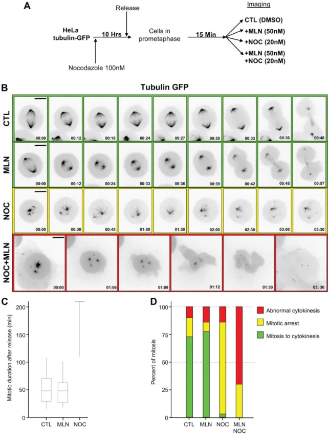
To confirm and further characterized observations above, we simplified the approach and used the Aurora A inhibitor MLN8237 at a concentration (50 nM) that affects only Aurora A and not B and C (Asteriti et al., 2014). We also decided to used low concentration of nocodazole to avoid a complete depolymerisation of microtubules, but a concentration that remains sufficient to keep the SAC active (Wang and Burke, 1995). HeLa cells expressing tubulin-GFP were treated with 100 nM nocodazole for ten hours, released in prometaphase for 15 min, then incubated with 50 nM MLN8237, 20 nM nocodazole or both and filmed (Fig.2A).
As already reported, Aurora A inhibition affects spindle morphology in particular spindle length (Bird and Hyman, 2008), Fig. 2B compare green lines 1 & 2) but does not affect mitotic timing (Fig. 2C and D). Low dose of nocodazole (20 nM), however, affects drastically spindle formation (Fig. 2B yellow line) and as it maintains the SAC active, induces a mitotic arrest (Fig. 2C and D). By adding MLN on these mitotic arrested cells (20 nM nocodazole and 50 nM MLN8237), we confirm our first observation (Fig. 1), almost 75 percent of mitotic cells exit from mitosis (Fig. 2D) without having been able to assemble a proper bipolar spindle structure. Nonetheless, these cells that prematurely exit mitosis do not divide (Fig2B red line). Instead, we observed a deformation of the cell cortex that might reveal cell attempt to complete mitosis and to divide. Cytokinesis eventually aborted and the tetraploid cell re-adhered to the cover slip (Fig. 2B red line).
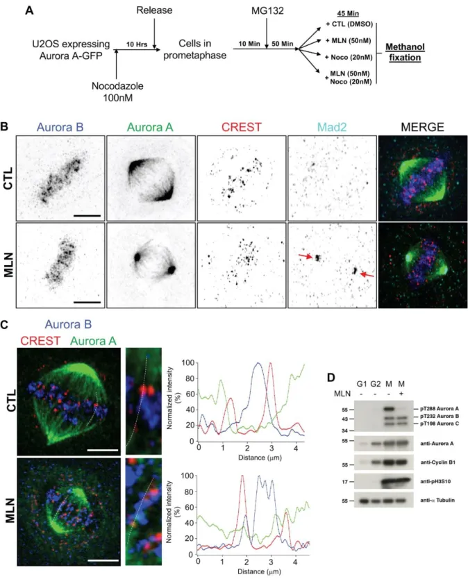
Because these phenotypes were reminiscent to that Aurora-B inhibition, we decided to control
whether Aurora-B could be affected during Aurora A inhibition. Cells were synchronized in
prophase and release in presence on MG132 (50 minutes, a proteasome inhibitor) to inhibit
anaphase execution. Then cells were treated with DMSO, MLN, Nocodazole, MLN and
nocodazole (Fig. 3A and B). In control cells Aurora B signal was detected in between CREST
pairs as previously reported (Fig. 2B and Fig. 3C right graphics, line scans). We did not detect
any overlapping area between Aurora A and Aurora-B. Nor did we detect any modification of
Aurora-B localisation under MLN8237 treatment (line scans, Fig. 3C) or using the as-Aurora
A/1NaPP1 approach (not shown).
To control the specificity of MLN8732 towards Aurora A kinase activity, we compared the
auto-phosphorylation state of each Aurora kinases (A, B and C) in the absence or in the presence
of 50 nM MLN8237 using anti-phosphoserine antibodies in immunoblots (Fig. 3D). Aurora A
auto-phosphorylation was almost completely lost under MLN8237 treatment, demonstrating
that Aurora A was inactivated. In the same condition, we did not observe any change in Aurora
B or C auto-phosphorylation. Additionally, the phosphorylation level of serine10 on histone
H3, an in vivo substrate of Aurora B and C was not affected by MLN8732 (Fig. 3D).
These data indicate that the condition we used to inhibit Aurora A kinase does not affect Aurora
B localisation or activity.
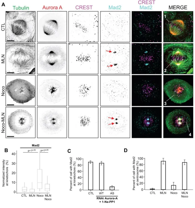
Inactivation of Aurora A disrupts the localisation of Mad2.
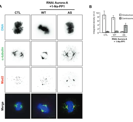
To obtain molecular insight into the role of Aurora A in mitotic exit in the presence of abnormal spindle we investigated the behaviour of Mad2, one major MCC component that localises on unattached kinetochores during prometaphase and that signals the presence of an active SAC. Using the as-Aurora A approach, we observed that without any treatment, 90% of the control cells had Mad2 at the kinetochores in prometaphase. In the presence of the as-Aurora A inhibitor 1-Na-PP1, 87% of cells expressing wt-Aurora A had Mad2 at the kinetochores, whereas only 10% of cells expressing as-Aurora A did (Fig. Sup 1A and B). Western blot analysis revealed that Mad2 protein levels were not affected by Aurora A inhibition, indicating
that the absence of the protein at the kinetochores was not due to a reduced amount of protein (Fig. 1B). Interestingly, in these last cells Mad2 was accumulating at the centrosomes (Fig. Sup
1A) as revealed by a quantification of Mad2 signal at the kinetochore and centrosome (Fig. Sup 1C).
We then used MLN8237 treatment to confirm these data by following the previously described experimental approaches (Fig. 3A). Then in the presence of MG132 the cells were treated with either 50 nM MLN8237 or 20 nM nocodazole or both and images were acquired using super resolution microscopy (Zeiss Airyscan) to evaluate the localisation of Mad2 in late prometaphase (Fig. 4A). Like previously reported by others (Bird and Hyman, 2008), inhibition of Aurora A leads to shorter spindles indicative of a good efficiency of MLN8237. By using super resolution images and quantitative analysis, mean Mad2 intensity per kinetochore (stained with CREST) was calculated, pooled and compared. As expected, we observed a recruitment of Mad2 at kinetochores under nocodazole treatment (Fig. 4B). Interestingly, MLN8237 treatment reduces Mad2 signal levels at kinetochore in nocodazole treated as well as in untreated cells (Fig. 4B). Like observed in the as-Aurora A approach, this decrease of Mad2 at kinetochores is concomitant with a massive recruitment of Mad2 at centrosomes (Fig.
4A red arrows, Fig. 4B and C).
This data indicates that Aurora A activity is required to maintain Mad2 on unattached kinetochores thereby possibly contributing to maintain the SAC active.
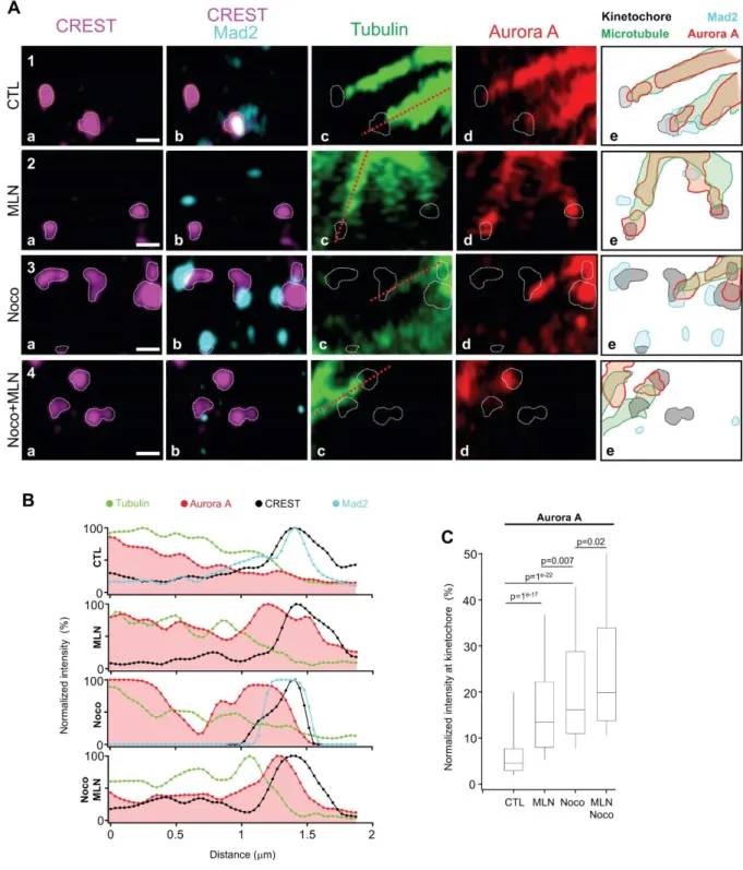
Kinetochore localisation of Aurora A
Because we observed Mad2 localisation at kinetochores is disrupted upon Aurora A inhibition, we investigated whether the Aurora A itself could be detected at or near the kinetochores. Although it is thought that Aurora A localises at the centrosome while Aurora-B at the kinetochores during prometaphase, a presence of the Aurora A at kinetochores has been previously reported (Katayama et al., 2008). Using super resolution, we investigated Aurora A localisation around kinetochores during kinetochores-microtubules attachment process. Like above, we used cells synchronized with nocodazole and blocked in metaphase by 50 nM of the proteasome inhibitor MG132 (Fig. 3A). We investigated the localisation of Mad2 and Aurora A along the axis of microtubules attached to kinetochores (red discontinued lines in c panels of
Fig. 5A). Microtubules are visualised by tubulin staining and kinetochores by CREST staining.
In control cells, we observed Aurora A signal following that of microtubules with an intensity (reflecting protein levels) regularly decreasing until reaching the kinetochore. Two kinetochores are shown in figure 5A in the first row. Mad2 is absent on the first kinetochore on the left but present in the one on the right signalling the presence of an active SAC (Fig. 5A
panel 1b). The drawing scheme representing the area occupied by the CREST signal
(kinetochore) in purple and by Aurora A signal in red reveals an area occupied by both signals at the external surface of the kinetochore where the microtubules attach (Fig. 5A panel 1e). After MLN8237 treatment, as observed above, Mad2 was not found at kinetochores anymore (Fig. 5A panel 2b). Interestingly, MLN8237 treatment also affects Aurora A localisation, the kinase signal accumulated at kinetochores (Fig. 5A panel 2d and 2e, Fig. 5B second row). Nocodazole treatment at low dose on the other hand, induced a massive recruitment of Mad2 at kinetochores (Fig. 4A panel 3b and 3e) but also a recruitment of Aurora A (Fig. 5A panel
In the presence of both nocodazole and MLN8237, only kinetochores still linked to microtubules showed Aurora A accumulation (Fig. 5A panel 4c and 4d, Fig. 5B fourth row), suggesting Aurora A accumulation at kinetochores relies on the presence of microtubules. We tested this hypothesis by investigating the level of Aurora A signal at kinetochores using the same methodology we used to quantify Mad2 (Fig. 3C). We observed an increase of Aurora A signal at kinetochores when cells were treated with either MLN8237 or nocodazole. This indicate that inhibiting Aurora A or maintaining the checkpoint active triggers Aurora A localisation at kinetochores. In cells treated with MLN8237 and nocodazole together, the level of Aurora A to kinetochore further increased again (Fig. 5A panel 4d and 4e, Fig. 5B fourth
row, Fig. 5C). This data strongly suggests that Aurora A uses microtubules to localize at
kinetochores.
Discussion
Until very recently, all of the studies describing the function of Aurora A have used loss of function by RNA interference, mutations in animal models, expression of a dominant negative version of the kinase, or gain of function by over-expressing wild-type Aurora A or a mutated version that become hyperactive or non-degradable (Glover et al., 1995; Littlepage et al., 2002; Sasai et al., 2008; Schumacher et al., 1998; Zhou et al., 1998). Because Aurora A is essential for centrosome maturation, the phenotype observed in all of these cases corresponds to a defect in spindle assembly and the cell never reaches metaphase (Berdnik and Knoblich, 2002; Hannak et al., 2001). We investigated whether Aurora A is involved in the SAC by specifically inhibiting Aurora A activity after the centrosomes have matured and the cell reaches metaphase. We succeeded by taking advantage of the as-Aurora A isoform already described (Reboutier et
al., 2013). And we confirmed our results using the Aurora A specific inhibitor MLN8237. Although Aurora A has never been directly involved in kinetochore functions, it phosphorylates CENP-A in prophase to allow Aurora-B localisation at kinetochores (Kunitoku et al., 2003). Aurora A also phosphorylates Haspin in late G2, participating indirectly to the phosphorylation of Threonine 3 of Histone H3 and to the recruitment of Aurora-B to kinetochores and more largely to the localisation of CPC and SAC proteins at kinetochores (Yu et al., 2017). Aurora A was also recently reported to phosphorylate Hec1 and to associate with the inner centromere Aurora-B partner INCENP, when overexpressed, to localise at the mitotic chromosome kinetochore (DeLuca et al., 2018).
Eventually, Aurora A and Aurora-B share substrates with phosphorylation events occurring where the kinases are located. Aurora A phosphorylates MCAK and PLK1 at the centrosome (Macurek et al., 2008; Seki et al., 2008; Zhang et al., 2008), whereas Aurora-B phosphorylates the same two proteins on the same residues but at the kinetochore (Andrews et al., 2004; Carmena et al., 2012a; Lan et al., 2004). Therefore, both Aurora A and Aurora-B act in concert on the same substrates but at different locations to coordinate mitotic progression. This coordination was demonstrated further in DT40 cells in which Aurora A was depleted (KO) and Aurora-B inhibited. The authors found cooperation between both proteins in the coordination of chromosome segregation in metaphase and microtubule depolymerisation in anaphase (Hégarat et al., 2011).
In the present study and in contrast to Aurora A KO, Aurora A was present and active until very late prometaphase. Its inhibition in that precise window of time led to a premature exit from mitosis with reduced Mad2 at kinetochores. In the presence of nocodazole or paclitaxel, which maintains the SAC active due to Mad2 recruitment at the kinetochores (Rieder and Maiato, 2004), inhibition of Aurora A led to the removal of Mad2 from the kinetochores, illustrating
that Aurora A activity is required to maintain Mad2 at non-attached kinetochores during prometaphase, ensuring that the SAC remains active. Whether this is a direct or indirect effect of Aurora A remains unknown.
Consistent with such role of Aurora A, we also observed the kinase inhibition (MLN8237) leads to its retention at kinetochores. Consistently with a recent report by DeLuca and collaborators (DeLuca et al., 2018) who identified HEC1 (Ndc80 component) as Aurora A kinase substrate, we detected Aurora A at the outer kinetochores. This localisation of Aurora A seems to be very dynamic and the kinase might in fact shuttle at this localisation to phosphorylate substrates. Indeed, we observed that an inhibition of Aurora A increases its localization to kinetochores as
if the inactive kinase remained blocked on its a substrate (Widmann et al., 2012). This localisation of Aurora A under MLN8237 shed light on a possible cooperation between both Aurora A and B in regulating kinetochores bi-orientation leading to SAC silencing. Aurora A localises at the outer kinetochore while Aurora-B at the inner kinetochore (Fig. 6)
Aurora A has been observed at kinetochores where it participates in kinetochore/chromatin microtubule nucleation (Katayama et al., 2001). Consistent with a potential function in the SAC, overexpression of Aurora A has been shown to be sufficient to override the SAC in the presence of taxol (Anand et al., 2003; Dutertre et al., 2004). Increase taxol sensitivity was also observed in pancreatic cancer cell lines upon Aurora A depletion (Hata et al., 2005). More recently, it has been reported that the phosphorylation of p73 by Aurora A contributes to the breakdown of the Mad2-Cdc20 complex releasing Cdc20 to degrade cyclin B and to securin, providing a molecular explanation for the SAC override (Katayama et al., 2012). Although resistance to taxol treatment has been observed in breast cancer cells over-expressing Aurora A, this is only true in estrogen receptor (ER)-positive tumours and not in ER-negative tumours (Noguchi, 2006). The reason for this difference remains to be elucidated, but the
phosphorylation by Aurora A of Ser167 and Ser305 in ER might be involved (Zheng et al., 2014).
Reports have also indicated a bypass of the SAC upon Aurora inhibition, but the inhibitors used were not specific to one Aurora kinase and the bypass was eventually attributed to Aurora-B inhibition. For example, the use of MLN8054, a more specific inhibitor of Aurora A than Aurora-B, revealed that inhibition of Aurora A accelerates mitosis exit in the presence of nocodazole or taxol (Wysong et al., 2009). The concentration of MLN8054 used in the study was optimised for Aurora A inhibition, but the observed phenotype mimicked Aurora-B inhibition (Tyler et al., 2007).
Here, we used two strategies to specifically inhibit Aurora A to clearly demonstrate for the first time that inhibition of Aurora A is sufficient to inhibit SAC activity and exit from mitosis. We demonstrated that the mechanism involved is not SAC override, but SAC inactivation. The molecular mechanism underlying the involvement of Aurora A in maintaining an active SAC during pro-metaphase remains to be elucidated, and a search for Aurora A substrates has begun. This report is the first to demonstrate a role of Aurora A kinase activity at the kinetochore for the SAC maintenance. Aurora A is a major target in cancer therapy with several inhibitors currently in clinical trials (Kollareddy et al., 2012). A precise understanding of the multiple functions of Aurora A during mitosis will undoubtedly help in designing drug associations and increase the efficiency of chemotherapeutic strategies to eliminate cancer cells (Bush et al., 2013; Schmidt et al., 2010).
ACKNOWLEDGMENTS
Thibault Courtheoux was a fellow of the Region Bretagne and FHU CAMIn. Alghassimou Diallo was a fellow of the Guinée government, some data in the paper forms part of his PhD thesis prepared in the Institute of Genetics and Development of Rennes and defended at the University of Rennes 1 in December 18th, 2013. We thank the MRic facility (Microscopy Rennes Imaging Center, Biosit, IBISA), Stéphanie Dutertre (Biosit), and Jacques Pécreaux for helpful advice on integrated density. This work was supported by the Centre National de la Recherche Scientifique, Université de Rennes 1, Institut National du Cancer, Ligue Nationale Contre le Cancer (Equipe Labelisée 2014), and Agence Nationale de la Recherche (Aurora).
Bibliography
Anand, S., Penrhyn-Lowe, S. and Venkitaraman, A. R. (2003). AURORA-A
amplification overrides the mitotic spindle assembly checkpoint, inducing resistance to Taxol. Cancer Cell 3, 51–62.
Andrews, P. D., Ovechkina, Y., Morrice, N., Wagenbach, M., Duncan, K., Wordeman, L. and Swedlow, J. R. (2004). Aurora B regulates MCAK at the
mitotic centromere. Dev Cell 6, 253–268.
Asteriti, I. A., Di Cesare, E., De Mattia, F., Hilsenstein, V., Neumann, B., Cundari, E., Lavia, P. and Guarguaglini, G. (2014). The Aurora-A inhibitor MLN8237
affects multiple mitotic processes and induces dose-dependent mitotic abnormalities and aneuploidy. Oncotarget 5, 6229–6242.
Berdnik, D. and Knoblich, J. A. (2002). Drosophila Aurora-A is required for
centrosome maturation and actin-dependent asymmetric protein localization during mitosis. Curr Biol 12, 640–647.
Bird, A. W. and Hyman, A. A. (2008). Building a spindle of the correct length in
human cells requires the interaction between TPX2 and Aurora A. J. Cell Biol.
182, 289–300.
Buffin, E., Lefebvre, C., Huang, J., Gagou, M. E. and Karess, R. E. (2005).
Recruitment of Mad2 to the kinetochore requires the Rod/Zw10 complex. Curr
Biol 15, 856–861.
Bush, T. L., Payton, M., Heller, S., Chung, G., Hanestad, K., Rottman, J. B., Loberg, R., Friberg, G., Kendall, R. L., Saffran, D., et al. (2013). AMG 900, a
small-molecule inhibitor of aurora kinases, potentiates the activity of microtubule-targeting agents in human metastatic breast cancer models. Mol. Cancer Ther.
12, 2356–2366.
Carmena, M., Pinson, X., Platani, M., Salloum, Z., Xu, Z., Clark, A., MacIsaac, F., Ogawa, H., Eggert, U., Glover, D. M., et al. (2012a). The Chromosomal
Passenger Complex Activates Polo Kinase at Centromeres. PLOS BIOLOGY 10, e1001250.
Carmena, M., Wheelock, M., Funabiki, H. and Earnshaw, W. C. (2012b). The
chromosomal passenger complex (CPC): from easy rider to the godfather of mitosis. Nat Rev Mol Cell Biol 13, 789–803.
Chen, R. H., Waters, J. C., Salmon, E. D. and Murray, A. W. (1996). Association of
Courthéoux, T., Enchev, R. I., Lampert, F., Gerez, J., Beck, J., Picotti, P., Sumara, I. and Peter, M. (2016). Cortical dynamics during cell motility are
regulated by CRL3(KLHL21) E3 ubiquitin ligase. Nat Commun 7, 12810.
De Brabander, M., Geuens, G., Nuydens, R., Willebrords, R. and De Mey, J.
(1981). Taxol induces the assembly of free microtubules in living cells and blocks the organizing capacity of the centrosomes and kinetochores. Proc Natl Acad Sci
USA 78, 5608–5612.
DeLuca, K. F., Meppelink, A., Broad, A. J., Mick, J. E., Peersen, O. B., Pektas, S., Lens, S. M. A. and Deluca, J. G. (2018). Aurora A kinase phosphorylates Hec1
to regulate metaphase kinetochore-microtubule dynamics. J. Cell Biol. 217, 163– 177.
Dutertre, S., Cazales, M., Quaranta, M., Froment, C., Trabut, V., Dozier, C., Mirey, G., Bouché, J.-P., Theis-Febvre, N., Schmitt, E., et al. (2004).
Phosphorylation of CDC25B by Aurora-A at the centrosome contributes to the G2-M transition. Journal of Cell Science 117, 2523–2531.
Fang, G. (2002). Checkpoint protein BubR1 acts synergistically with Mad2 to inhibit
anaphase-promoting complex. Mol Biol Cell 13, 755–766.
Foley, E. A. and Kapoor, T. M. (2013). Microtubule attachment and spindle
assembly checkpoint signalling at the kinetochore. Nat Rev Mol Cell Biol 14, 25– 37.
Giet, R., Petretti, C. and Prigent, C. (2005). Aurora kinases, aneuploidy and cancer,
a coincidence or a real link? Trends Cell Biol 15, 241–250.
Glover, D. M., Leibowitz, M. H., McLean, D. A. and Parry, H. (1995). Mutations in
aurora prevent centrosome separation leading to the formation of monopolar spindles. Cell 81, 95–105.
Green, R. A. and Kaplan, K. B. (2003). Chromosome instability in colorectal tumor
cells is associated with defects in microtubule plus-end attachments caused by a dominant mutation in APC. J. Cell Biol. 163, 949–961.
Guse, A., Mishima, M. and Glotzer, M. (2005). Phosphorylation of ZEN-4/MKLP1
by aurora B regulates completion of cytokinesis. Curr Biol 15, 778–786.
Hannak, E., Kirkham, M., Hyman, A. A. and Oegema, K. (2001). Aurora-A kinase
is required for centrosome maturation in Caenorhabditis elegans. J. Cell Biol.
155, 1109–1116.
Hata, T., Furukawa, T., Sunamura, M., Egawa, S., Motoi, F., Ohmura, N.,
Marumoto, T., Saya, H. and Horii, A. (2005). RNA interference targeting aurora
kinase a suppresses tumor growth and enhances the taxane chemosensitivity in human pancreatic cancer cells. Cancer Res 65, 2899–2905.
Hégarat, N., Smith, E., Nayak, G., Takeda, S., Eyers, P. A. and Hochegger, H.
(2011). Aurora A and Aurora B jointly coordinate chromosome segregation and anaphase microtubule dynamics. J. Cell Biol. 195, 1103–1113.
Hoar, K., Chakravarty, A., Rabino, C., Wysong, D., Bowman, D., Roy, N. and Ecsedy, J. A. (2007). MLN8054, a small-molecule inhibitor of Aurora A, causes
spindle pole and chromosome congression defects leading to aneuploidy. Mol
Cell Biol 27, 4513–4525.
Hsu, J. Y., Sun, Z. W., Li, X., Reuben, M., Tatchell, K., Bishop, D. K., Grushcow, J. M., Brame, C. J., Caldwell, J. A., Hunt, D. F., et al. (2000). Mitotic
phosphorylation of histone H3 is governed by Ipl1/aurora kinase and Glc7/PP1 phosphatase in budding yeast and nematodes. Cell 102, 279–291.
Jordan, M. A., Thrower, D. and Wilson, L. (1992). Effects of vinblastine,
podophyllotoxin and nocodazole on mitotic spindles. Implications for the role of microtubule dynamics in mitosis. Journal of Cell Science 102 ( Pt 3), 401–416.
Kasuboski, J. M., Bader, J. R., Vaughan, P. S., Tauhata, S. B. F., Winding, M., Morrissey, M. A., Joyce, M. V., Boggess, W., Vos, L., Chan, G. K., et al.
(2011). Zwint-1 is a novel Aurora B substrate required for the assembly of a dynein-binding platform on kinetochores. Mol Biol Cell 22, 3318–3330.
Katayama, H., Sasai, K., Kloc, M., Brinkley, B. R. and Sen, S. (2008). Aurora
kinase-A regulates kinetochore/chromatin associated microtubule assembly in human cells. cc 7, 2691–2704.
Katayama, H., Wang, J., Treekitkarnmongkol, W., Kawai, H., Sasai, K., Zhang, H., Wang, H., Adams, H. P., Jiang, S., Chakraborty, S. N., et al. (2012). Aurora
kinase-A inactivates DNA damage-induced apoptosis and spindle assembly checkpoint response functions of p73. Cancer Cell 21, 196–211.
Katayama, H., Zhou, H., Li, Q., Tatsuka, M. and Sen, S. (2001). Interaction and
feedback regulation between STK15/BTAK/Aurora-A kinase and protein phosphatase 1 through mitotic cell division cycle. J Biol Chem 276, 46219– 46224.
Kollareddy, M., Zheleva, D., Dzubak, P., Brahmkshatriya, P. S., Lepsik, M. and Hajduch, M. (2012). Aurora kinase inhibitors: progress towards the clinic. Invest New Drugs 30, 2411–2432.
Kunitoku, N., Sasayama, T., Marumoto, T., Zhang, D., Honda, S., Kobayashi, O., Hatakeyama, K., Ushio, Y., Saya, H. and Hirota, T. (2003). CENP-A
phosphorylation by Aurora-A in prophase is required for enrichment of Aurora-B at inner centromeres and for kinetochore function. Dev Cell 5, 853–864.
Lan, W., Zhang, X., Kline-Smith, S. L., Rosasco, S. E., Barrett-Wilt, G. A., Shabanowitz, J., Hunt, D. F., Walczak, C. E. and Stukenberg, P. T. (2004).
Aurora B phosphorylates centromeric MCAK and regulates its localization and microtubule depolymerization activity. Curr Biol 14, 273–286.
Littlepage, L. E., Wu, H., Andresson, T., Deanehan, J. K., Amundadottir, L. T. and Ruderman, J. V. (2002). Identification of phosphorylated residues that affect
Macurek, L., Lindqvist, A., Lim, D., Lampson, M. A., Klompmaker, R., Freire, R., Clouin, C., Taylor, S. S., Yaffe, M. B. and Medema, R. H. (2008). Polo-like
kinase-1 is activated by aurora A to promote checkpoint recovery. Nature 455, 119–123.
Mathieu, J., Cauvin, C., Moch, C., Radford, S. J., Sampaio, P., Perdigoto, C. N., Schweisguth, F., Bardin, A. J., Sunkel, C. E., McKim, K., et al. (2013). Aurora
B and cyclin B have opposite effects on the timing of cytokinesis abscission in Drosophila germ cells and in vertebrate somatic cells. Dev Cell 26, 250–265.
Nilsson, J., Yekezare, M., Minshull, J. and Pines, J. (2008). The APC/C maintains
the spindle assembly checkpoint by targeting Cdc20 for destruction. Nat Cell Biol
10, 1411–1420.
Noguchi, S. (2006). Predictive factors for response to docetaxel in human breast
cancers. Cancer Sci. 97, 813–820.
Norden, C., Mendoza, M., Dobbelaere, J., Kotwaliwale, C. V., Biggins, S. and Barral, Y. (2006). The NoCut pathway links completion of cytokinesis to spindle
midzone function to prevent chromosome breakage. Cell 125, 85–98.
Reboutier, D., Troadec, M.-B., Cremet, J.-Y., Chauvin, L., Guen, V., Salaun, P. and Prigent, C. (2013). Aurora A is involved in central spindle assembly through
phosphorylation of Ser 19 in P150Glued. J. Cell Biol. 201, 65–79.
Reboutier, D., Troadec, M.-B., Cremet, J.-Y., Fukasawa, K. and Prigent, C.
(2012). Nucleophosmin/B23 activates Aurora A at the centrosome through phosphorylation of serine 89. J. Cell Biol. 197, 19–26.
Rieder, C. L. and Maiato, H. (2004). Stuck in division or passing through: what
happens when cells cannot satisfy the spindle assembly checkpoint. Dev Cell 7, 637–651.
Roghi, C., Giet, R., Uzbekov, R., Morin, N., Chartrain, I., Le Guellec, R.,
Couturier, A., Dorée, M., Philippe, M. and Prigent, C. (1998). The Xenopus
protein kinase pEg2 associates with the centrosome in a cell cycle-dependent manner, binds to the spindle microtubules and is involved in bipolar mitotic spindle assembly. Journal of Cell Science 111 ( Pt 5), 557–572.
Sasai, K., Parant, J. M., Brandt, M. E., Carter, J., Adams, H. P., Stass, S. A., Killary, A. M., Katayama, H. and Sen, S. (2008). Targeted disruption of Aurora
A causes abnormal mitotic spindle assembly, chromosome misalignment and embryonic lethality. Oncogene 27, 4122–4127.
Schmidt, J. C., Kiyomitsu, T., Hori, T., Backer, C. B., Fukagawa, T. and
Cheeseman, I. M. (2010). Aurora B kinase controls the targeting of the Astrin–
SKAP complex to bioriented kinetochores.
Schumacher, J. M., Ashcroft, N., Donovan, P. J. and Golden, A. (1998). A highly
conserved centrosomal kinase, AIR-1, is required for accurate cell cycle
progression and segregation of developmental factors in Caenorhabditis elegans embryos. Development 125, 4391–4402.
Seki, A., Coppinger, J. A., Jang, C.-Y., Yates, J. R. and Fang, G. (2008). Bora and
the kinase Aurora a cooperatively activate the kinase Plk1 and control mitotic entry. Science 320, 1655–1658.
Tanaka, M., Ueda, A., Kanamori, H., Ideguchi, H., Yang, J., Kitajima, S. and Ishigatsubo, Y. (2002). Cell-cycle-dependent regulation of human aurora A
transcription is mediated by periodic repression of E4TF1. J Biol Chem 277, 10719–10726.
Thornton, B. R. and Toczyski, D. P. (2003). Securin and B-cyclin/CDK are the only
essential targets of the APC. Nat Cell Biol 5, 1090–1094.
Tyler, R. K., Shpiro, N., Marquez, R. and Eyers, P. A. (2007). VX-680 inhibits
Aurora A and Aurora B kinase activity in human cells. cc 6, 2846–2854.
Uhlmann, F., Lottspeich, F. and Nasmyth, K. A. (1999). Sister-chromatid
separation at anaphase onset is promoted by cleavage of the cohesin subunit Scc1. Nature 400, 37–42.
Wang, Y. and Burke, D. J. (1995). Checkpoint genes required to delay cell division
in response to nocodazole respond to impaired kinetochore function in the yeast Saccharomyces cerevisiae. Mol Cell Biol 15, 6838–6844.
Widmann, B., Wandrey, F., Badertscher, L., Wyler, E., Pfannstiel, J., Zemp, I. and Kutay, U. (2012). The kinase activity of human Rio1 is required for final
steps of cytoplasmic maturation of 40S subunits. Mol Biol Cell 23, 22–35.
Wilkins, B. J., Rall, N. A., Ostwal, Y., Kruitwagen, T., Hiragami-Hamada, K., Winkler, M., Barral, Y., Fischle, W. and Neumann, H. (2014). A cascade of
histone modifications induces chromatin condensation in mitosis. Science 343, 77–80.
Wysong, D. R., Chakravarty, A., Hoar, K. and Ecsedy, J. A. (2009). The inhibition
of Aurora A abrogates the mitotic delay induced by microtubule perturbing agents. cc 8, 876–888.
Yu, F., Jiang, Y., Lu, L., Cao, M., Qiao, Y., Liu, X., Liu, D., Van Dyke, T., Wang, F., Yao, X., et al. (2017). Aurora-A promotes the establishment of spindle assembly
checkpoint by priming the Haspin-Aurora-B feedback loop in late G2 phase. Cell
Discov 3, 16049.
Zhang, X., Ems-Mcclung, S. C. and Walczak, C. E. (2008). Aurora A
phosphorylates MCAK to control ran-dependent spindle bipolarity. Mol Biol Cell
19, 2752–2765.
Zheng, X. Q., Guo, J. P., Yang, H., Kanai, M., He, L. L., Li, Y. Y., Koomen, J. M., Minton, S., Gao, M., Ren, X. B., et al. (2014). Aurora-A is a determinant of
tamoxifen sensitivity through phosphorylation of ERα in breast cancer. Oncogene
Zhou, H., Kuang, J., Zhong, L., Kuo, W. L., Gray, J. W., Sahin, A., Brinkley, B. R. and Sen, S. (1998). Tumour amplified kinase STK15/BTAK induces centrosome
Figure 1: Aurora A inhibition during mitosis causes SAC override. (A) Methods used to analyse the effect of Aurora A inhibition on the SAC. Cells were first depleted of endogenous Aurora A by RNAi and then treated with nocodazole for 16 h to hold them in mitosis. 13 h later, the cells were treated with 1-Na-PP1 to inhibit as-Aurora A and filmed for 3 hours. (B) Western blots showing the efficiency of endogenous Aurora A depletion and exogenous WT and AS expression in both stable cell lines. Mad2 levels was controlled in Aurora A depleted cells treated with 1-Na-PP1. Cyclin B1 was used as mitotic marker. HCAP-D2 was used as a loading control. (C) Snapshots of movies corresponding to nocodazole-treated stable cell lines depleted of endogenous Aurora A and expressing wt- Aurora A (WT) and as-Aurora A (AS).
(D) The graph represents the kinetics of mitosis exit in the presence of 3.3 μM nocodazole of
stable cell lines expressing either WT or AS. The dark grey lines correspond to mitosis and light grey to interphase (exit from mitosis). The addition of 1-Na-PP1 marks the T0 for each film. *Left*: 1-Na-PP1 treatment of cells depleted of endogenous Aurora A by siRNA. *Middle*: no treatment. *Right*: 1-Na-PP1 treatments. Numbers in the panels indicate the number of cells observed. (E) The percentage of cells in mitosis in both stable cell lines treated as described above (Noc: nWT=199, nAS=250, Noc+RNAi: nWT=66, nAS=70, Noc+ RNAi+1Napp1: nWT=46, nAS=62, error bars: standard deviation).
Figure 2: Aurora A inhibition during mitosis causes SAC override. (A) Methods used for
cell synchronisation, Aurora A is inhibited in late prometaphase using Aurora A specific inhibitor MLN8237. (B) Live cell imaging of synchronized HeLa Tubulin-GFP. Image panel
showing representative case of mitotic progression under CTL, MLN, Nocodazole or Nocodazole and MLN treatment. Scale bar: 5m. (C) and (D) Quantification of mitotic duration (C) and mitotic behaviour (D) after release (CTL n=49, MLN8237 n=49, NOC=49), green: mitosis to cytokinesis, yellow: mitotic delay and red: abnormal cytokinesis.
Figure 3: Aurora A inhibition does not affect Aurora B localisation. (A) Methods used for
cell synchronisation, Aurora A is inhibited in late prometaphase using Aurora A specific inhibitor MLN8237. (B) and (C) Immunostaining of U2OS stable cell line expressing Aurora
Airyscan confocal super-resolution microscope. Red arrows showed Mad2 centrosomal aggregation. Scale bar: 5m. (C) Three z-stacks have been projected to select a kinetochore pair. Graphical representation of fluorescence signal intensity for kinetochore (CREST, red), Aurora B (blue) and Aurora A (green). Scale bar: 5m (D) Western blot of HeLa cells lysis synchronized at different cell cycle stages (G1, G2 and M). Cells were treated with Aurora A inhibitor (MLN8237, 50 nM) during mitosis.
Figure 4: Aurora A Inhibition during mitosis causes Mad2 mis-localisation (A)
Immunostaining of U2OS stable cell line expressing Aurora A-GFP under endogenous promotor. Cells were synchronized as in fig2A and imaged using Airyscan confocal super-resolution microscope. Numbered white boxes on right column are enlarged and analysed in fig 4A. Red arrows showed Mad2 centrosomal aggregation. Scale bar: 5m. (B) Quantitative analysis of Mad2 intensity at kinetochore. Average intensity per kinetochore were collected and
normalized to the maximum mean from nocodazole condition (representative of SAC activation, n=914 kinetochores per condition, error bars = s.d). Mad2 kinetochore values were pooled per condition and showed as box plots. T-test on the means were performed and p-values are presented. (C) Percentage of cells with normal localisation of Mad2 in (Fig. sup1A) during late prometaphase (CTL=14, WT+Na-PP1= 20, AS+Na-PP1= 26). (D) Quantification of cells showing mad2 centrosomal aggregation in percent of cell (CTL n=90 , Noc n=128 , MLN n=139 and Noc with MLN n=94, error bars=s.d).
Figure 5: Mad2 and Aurora A localisation at kinetochore (A) Enlarged pictures from Fig
4A. White circles represent kinetochore positions. Right column, cartoon of different localisations showing overlap. Scale bar: 500 nm. (B) Line scans from red discontinued lines in A. Aurora A signal overlaps with Mad2 signal at kinetochores (C) Quantitative analysis of
normalized to the maximum mean from MLN condition (condition with highest values). Aurora A kinetochore values were pooled per condition and showed as box plot (n=914 kinetochores per condition), T-test on the means were performed and p-values are presented.
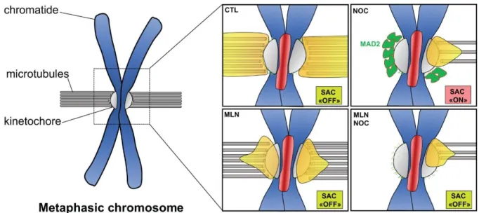
Figure 6: Schematic representation of Aurora A and Mad2 localization at mitotic chromosome. Aurora A localizes to kinetochore microtubules and outer kinetochore in
cells arrested at metaphase (CTL- MG132), the Spindle Assembly Checkpoint is
inactivated (SAC “OFF”). Under Nocodazole (NOC): Mad2 accumulates at kinetochore, the Spindle Assembly Checkpoint is activated (SAC “ON”), Aurora A accumulates at partially attached kinetochore. Under MLN and/or Nocodazole (MLN and MLN+NOC), Aurora A accumulates at outer kinetochore and Mad2 is no longer at kinetochore. The SAC is “OFF”, cells are escaping from mitotic arrest.
α
Figure Sup 1: (A) Stable control, wt-Aurora A (WT), and as-Aurora A (AS) cell lines (as
indicated) immunostained to detect α-tubulin (Green and Kaplan, 2003) and Mad2
(Kollareddy et al., 2012) and counterstained for DNA with Vectashield containing DAPI during
late prometaphase under Aurora A inhibition. Scale bar= 10 μm. (B) The integrated density of
Mad2 at the kinetochores and centrosomes in (A) during late prometaphase (CTL=14,
WT+Na-PP1= 20, AS+Na-WT+Na-PP1= 26, error bar = s.d).