HAL Id: hal-02048154
https://hal.archives-ouvertes.fr/hal-02048154
Submitted on 22 Oct 2020
HAL is a multi-disciplinary open access
archive for the deposit and dissemination of
sci-entific research documents, whether they are
pub-lished or not. The documents may come from
teaching and research institutions in France or
abroad, or from public or private research centers.
L’archive ouverte pluridisciplinaire HAL, est
destinée au dépôt et à la diffusion de documents
scientifiques de niveau recherche, publiés ou non,
émanant des établissements d’enseignement et de
recherche français ou étrangers, des laboratoires
publics ou privés.
Dynamic stability of the actin ecosystem
Julie Plastino, Laurent Blanchoin
To cite this version:
Julie Plastino, Laurent Blanchoin. Dynamic stability of the actin ecosystem. Journal of Cell Science,
Company of Biologists, 2019, 132 (4), pp.jcs219832. �10.1242/jcs.219832�. �hal-02048154�
REVIEW SPECIAL ISSUE: RECONSTITUTING CELL BIOLOGY
Dynamic stability of the actin ecosystem
Julie Plastino1,2,* and Laurent Blanchoin3,4,*ABSTRACT
In cells, actin filaments continuously assemble and disassemble while maintaining an apparently constant network structure. This suggests a perfect balance between dynamic processes. Such behavior, operating far out of equilibrium by the hydrolysis of ATP, is called a dynamic steady state.Thisdynamic steady state confers a high degree of plasticity to cytoskeleton networks that allows them to adapt and optimize their architecture in response to external changes on short time-scales, thus permitting cells to adjust to their environment. In this Review, we summarize what is known about the cellular actin steady state, and what gaps remain in our understanding of this fundamental dynamic process that balances the different forms of actin organization in a cell. We focus on the minimal steps to achieve a steady state, discuss the potential feedback mechanisms at play to balance this steady state and conclude with an outlook on what is needed to fully understand its molecular nature.
KEY WORDS: Actin, Cytoskeleton, Steady state
Introduction
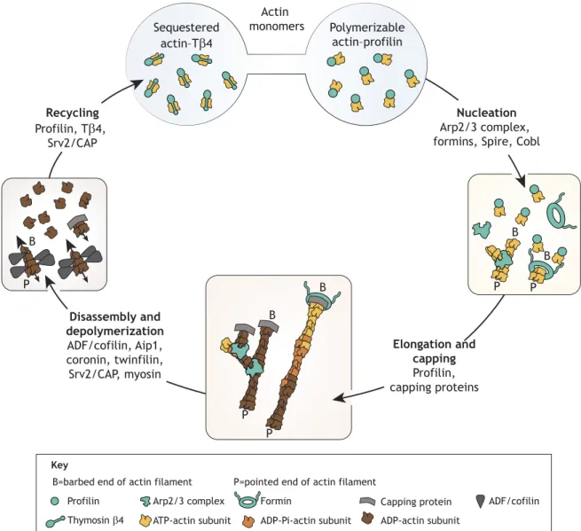
A key property of most living systems is their ability to move and/or change shape according to environmental cues. This is instrumental for the cell or the tissue to carry out its biological program, including cell processes such as division and motility, and developmental processes like morphogenesis. Understanding the dynamic steady state of actin is a major challenge in cell and developmental biology because actin is a key player and driving force in the construction of the complex and dynamic scaffolding that makes up the internal architecture of eukaryotic cells (Blanchoin et al., 2014; Chhabra and Higgs, 2007). Whereas many molecules that are involved in building the actin cytoskeleton are known, the basic rules that control the coordinated dynamics of structures that exist at the same time in the cell, such as branched networks, parallel bundles and antiparallel contractile bundles, are still poorly understood. The dynamic steady state of actin has four facets: nucleation (formation of actin dimers or trimers), elongation and capping (controlled polymer growth), disassembly (breakdown of actin structures) and recycling (replenishment of the pool of actin monomers that are charged with ATP) (Fig. 1). Here,we considereach facet in detail
andhighlight the potential existence of feedback mechanisms that could balance the dynamics of cellular actin, and the need for reconstitution experiments to fully dissect the balance of the steady state of actin.
The pool of actin monomers
The elementary building block for actin assembly is the actin monomer. The concentration of actin monomers is quite variable in different living systems, ranging from potentially as low as 0.01 µM in the yeast Saccharomyces cerevisiae to 300 µM in unactivated platelets (Karpova et al., 1995; Pollard et al., 2000). Since the rate of actin assembly is directly proportional to the concentration of monomers (Pollard et al., 2000), this variability means that the dynamic steady state of actin is not the same in different cell types; actin filaments could potentially grow orders of magnitude faster in platelets than in yeast.
In the cell, most of the pool of polymerizable actin is bound to profilin (Fig. 1 and Kaiser et al., 1999). However, some actin monomers in a given cell type might not be polymerizable as they can be sequestered by proteins such as thymosinβ4 (Tβ4) (Fig. 1 and Pantaloni and Carlier, 1993). In this context, determining the exact concentration of polymerizable actin at a given time is challenging (Raz-Ben Aroush et al., 2017) as the balance between sequestered and polymerizable actin during dynamic actin assembly is not well characterized (Skruber et al., 2018). In addition, Tβ4 has been proposed to play an active role in preventing monomer incorporation into branched networks and in targeting cytosolic actin monomers to formin-mediated assembly at the leading edge of cells (Vitriol et al., 2015). This polymerizable pool of actin monomers needs further characterization at the cellular level, but also at the subcellular level– at sites of active actin assembly– where the pool of polymerizable actin can become depleted (Boujemaa-Paterski et al., 2017). In addition, different actin networks compete for actin monomers, and this is crucial for determining network density and size (Burke et al., 2014; Suarez and Kovar, 2016). To understand the landscape of the monomer pool in a complex, actively polymerizing, network as found in keratocyte fragments (Raz-Ben Aroush et al., 2017), it will be necessary to use methods such as fluorescence recovery after photobleaching (FRAP) or photoactivation and/or photoconversion experiments coupled with mathematical modeling to assess actin monomer dynamics in different in vivo contexts (Skruber et al., 2018) and/or to use cell-size confinement to generate reconstituted systems with well-defined but limited sources of actin monomers. Given the importance of the local polymerizable actin concentration for determining actin dynamics, evaluating the pool of polymerizable actin – and potentially its gradients – in different cellular contexts is one of the key challenges of the coming years.
Nucleation
Profilin prevents the spontaneous association of actin monomers into actin dimers and trimers, which are necessary intermediates prior to the formation of actin filaments (Dominguez, 2009; Sept and McCammon, 2001). These nucleation steps, which are thermodynamically unfavorable, are accelerated by actin nucleators (Fig. 1). Three main classes of actin nucleators have been characterized: the Arp2/3 complex, the formin family and the tandem monomer-binding protein family, including the proteins
1Institut Curie, PSL Research University, CNRS, 75005 Paris, France.2Sorbonne
Université, 75005 Paris, France.3CytomorphoLab, Biosciences & Biotechnology
Institute of Grenoble, Laboratoire de Physiologie Cellulaire & Végétale, Université Grenoble-Alpes/CEA/CNRS/INRA, 38054 Grenoble, France.4CytomorphoLab,
Hô pital Saint Louis, Institut Universitaire d’Hématologie, UMRS1160, INSERM/AP-HP/Université Paris Diderot, 75010 Paris, France
AQ1
¶ .
*Authors for correspondence ( [email protected], [email protected]) L.B., 0000-0001-8146-9254 1 2 3 4 5 6 7 8 9 10 11 12 13 14 15 16 17 18 19 20 21 22 23 24 25 26 27 28 29 30 31 32 33 34 35 36 37 38 39 40 41 42 43 44 45 46 47 48 49 50 51 52 53 54 55 56 57 58 59 60 61 62 63 64 65 66 67 68 69 70 71 72 73 74 75 76 77 78 79 80 81 82 83 84 85 86 87 88 89 90 91 92 93 94 95 96 97 98 99 100 101 102 103 104 105 106 107 108 109 110 111 112 113 114 115 116 117 118 119 120 121 122 123 124
Spire and Cobl, which are proposed to cause nucleation by tethering three or more actin monomers together (Campellone and Welch, 2010). The different mechanisms of action and the cellular localization of these nucleators influence their physiological properties and abilities to build specific actin structures. The Arp2/3 complex and formins are also influenced by external signals as their activity is controlled either directly or indirectly by membrane-bound Rho GTPases (Lawson and Ridley, 2018; Ridley, 2015). In addition, profilin has been shown to favor formin-mediated assembly over Arp2/3-based nucleation, thus modulating homeostasis between different networks (Suarez et al., 2015; Rotty et al., 2015).
Elongation and capping
Pointed ends do not grow in profilin-actin since profilin masks the barbed face of the actin monomer, preventing addition to pointed ends (Pollard et al., 2000). Therefore, in cellular conditions of profilin-actin, networks that are generated by the Arp2/3 complex grow with a rate that depends on the association rate constant for
monomer addition at the barbed ends (which is typically 10 per µM per second, (Pollard, 1986) and the concentration of polymerizable actin monomers. Growth is terminated by a lack of monomers or by capping proteins (Fig. 1). Therefore, the balance between rates of growth and capping needs to be well-adjusted for the formation of defined actin networks (Akin and Mullins, 2008; Blanchoin et al., 2000a; Kawska et al., 2012). This balance is illustrated by a comparison of the dynamics of two similar structures found in different cells, where the actin monomer concentrations are very
different. For example,in yeast AQ2
,actin patches and cell lamellipodia are both generated by the Arp2/3 complex and consist of highly branched and intertangled actin networks (Young et al., 2004; Svitkina and Borisy, 1999). Based on the concentration of available monomers, the growth of a lamellipodium should be orders of magnitude faster than a patch, but this is not the case because capping proteins regulate growth in lamellipodia (Moseley and Goode, 2006). Indeed, capping proteins are in fact necessary for correct lamellipodia formation in motile cells with a high concentration of monomers (Iwasa and Mullins, 2007). In
P B P B P P B B P B Actin monomers Sequestered actin–Tβ4 Polymerizable actin–profilin Nucleation Arp2/3 complex, formins, Spire, Cobl
Elongation and capping Profilin, capping proteins Disassembly and depolymerization ADF/cofilin, Aip1, coronin, twinfilin, Srv2/CAP, myosin Recycling Profilin, Tβ4, Srv2/CAP Key Formin Arp2/3 complex
Profilin Capping protein ADF/cofilin
Thymosin β4
P=pointed end of actin filament B=barbed end of actin filament
ATP-actin subunit ADP-Pi-actin subunit ADP-actin subunit
Fig. 1. The dynamic steady state of actin. Minimal stepsneededto reach a dynamic steady state of different actin architecturesare illustrated.Sequestered and polymerizable actin represent the pool of actin monomers. Nucleation is the formation of actin dimers or trimers. Elongation and capping modulate
controlled growth of the different forms of actin organization (branched networks that are generated by the Arp2/3 complex or bundles generated by formins). Disassembly and depolymerizationresults inthe breakdown of actin structures to monomer subunits. Recyclingrenewsthe pool of actin monomers that are
charged with ATP. The different nucleotide AQ7
states of actin, barbed and pointed ends and different proteins or complexes are represented by the indicated symbols.
REVIEW Journal of Cell Science (2018) 131, jcs219832. doi:10.1242/jcs.219832
125 126 127 128 129 130 131 132 133 134 135 136 137 138 139 140 141 142 143 144 145 146 147 148 149 150 151 152 153 154 155 156 157 158 159 160 161 162 163 164 165 166 167 168 169 170 171 172 173 174 175 176 177 178 179 180 181 182 183 184 185 186 187 188 189 190 191 192 193 194 195 196 197 198 199 200 201 202 203 204 205 206 207 208 209 210 211 212 213 214 215 216 217 218 219 220 221 222 223 224 225 226 227 228 229 230 231 232 233 234 235 236 237 238 239 240 241 242 243 244 245 246 247 248
contrast, inS. cerevisiae, where monomer concentration is very low, capping protein can be removed and actin patches still show a qualitatively normal organization (Young et al., 2004). Growth of actin filaments that is mediated by formin is even more complex because different formins produce different association rate constants at filament barbed ends (Chesarone and Goode, 2009). In addition, formin and capping proteins function antagonistically at the barbed ends of actin filaments to control their length through the formation of a ‘decision complex’, where capping protein and formin are simultaneously bound to a paused barbed end. Depending on how the complex decomposes, growth will resume or the filament will be permanently capped, lending an extra layer of control to the dynamics of the barbed end (Fig. 1, Bombardier et al., 2015; Shekhar et al., 2015). Finally, formins are known to cooperate with the Arp2/3 complex for the elongation of protrusive networks (Block et al., 2012; Kage et al., 2017). Taken together, elongation and capping, in the context of the amount of polymerizable monomeric actin and the nature of the nucleating agent, are what defines the dynamics of actin network growth.
Disassembly
Actin disassembly takes place in two steps: fragmentation of actin networks into small filaments, and depolymerization into monomers of the small actin fragments that were generated in this process (Fig. 1, Blanchoin et al., 2014). Like nucleation or elongation, disassembly depends on the nature of the network (Gressin et al., 2015). A branched network disassembles mainly through debranching that is mediated by the actin depolymerizing factor (ADF)/cofilinfamily proteins [comprising ADF (also known as destrin), cofilin-1 (CFL1) and cofilin-2 (CFL2)] or glia maturation factor-like protein (GMF) (Blanchoin et al., 2000b; Chan et al., 2009; Gandhi et al., 2010; Gressin et al., 2015). For parallel or antiparallel networks of bundled filaments, the severing of filaments by ADF/cofilin on its own is not sufficient to dismantle the structure entirely (Gressin et al., 2015), but enough to maintain a steady state length, at least for parallel bundles that are initiated by formin (Michelot et al., 2007). Actin-interacting protein 1 (Aip1, also known as WDR1) is a necessary cofactor that synergizes with ADF/cofilin and coronin proteins
AQ3
¶ (Nadkarni and Brieher, 2014; Gressin et al., 2015; Jansen et al.,to disassemble actin bundles 2015). For antiparallel contractile actin networks, the contribution of the myosin motor protein during disassembly is unclear, but actin filament buckling that is produced by myosin contraction has been shown to lead to filament breakage (Murrell and Gardel, 2012), and buckling might also favor severing by ADF/cofilin. To be fully efficient, ADF/cofilin must work in concert with capping proteins (Suarez et al., 2011). Indeed, the presence of ATP or ADP-Pi-loaded subunits near a growing barbed end prevents this region of the filament from being decorated by ADF/cofilin (Suarez et al., 2011; Wioland et al., 2017). Barbed-end capping terminates the growth of filaments within a structure, and thus favors decoration of filaments by ADF/cofilin (Suarez et al., 2011). Therefore, the disassembly of actin networks is intricately linked to the growth and capping balance of the actin steady state, as discussed above.
Until recently, a few puzzling questions remained concerning the disassembly step. First, why do fragments that are generated by ADF/cofilin not elongate rapidly until capped, thus reversing the disassembly effect of ADF/cofilin? Second, how does rapid disassembly from capped fragments occur? The rate constant of depolymerization at pointed ends is only 0.27 per second, and depolymerization would be much more efficient if it occurred at the barbed end with a rate constant of 7.2 per second (Pollard, 1986).
Recently, these two questions have been elegantly addressed by Wioland and co-workers, who showed that decoration with ADF/ cofilin– upon nearing the barbed end – dissociates capping protein from that end (Wioland et al., 2017). Even more striking, barbed ends of filaments that are saturated with ADF/cofilin do not grow because ADF/cofilin prevents monomer addition (Wioland et al., 2017). In other words, ADF/cofilin alters barbed end dynamics by binding to the sides of the filament and changing its structure (Tanaka et al., 2018), thus preventing capping protein binding and monomer addition, while still allowing subunit dissociation (Fig. 1). Other proteins, such as twinfilin and Srv2/profilin AQ4
and cyclase-associated protein (CAP), which accelerate depolymerization at the barbed and pointed ends, can also participate in actin disassembly (Johnston et al., 2015). Their collaborative effort depolymerizes a filament of onemicrometerlength in less than a minute (Johnston et al., 2015). How such depolymerization occurs on ADF/cofilin-decorated filaments, or on small fragments that are generated by both ADF/cofilin and Aip1, needs to be investigated.Srv2/CAPhas also been shown to enhance severing by ADF/cofilin (Chaudhry et al., 2013). It is still unclear exactly how small fragments depolymerize into single subunits. Recent advances in time-resolved electron microscopy, combined with static and dynamic light scattering might helpin the capture and identificationof these entities,which fallbelow the diffraction limit (Frank, 2017; Lopez et al., 2016).
Recycling
An assembly-competent actin oligomer pool has previously been proposed (Okreglak and Drubin, 2010; Smith et al., 2013); however, for most reassemblyprocesses, actin filaments must be broken down into their individual monomers. Actin monomers are bound to ADP when they dissociate from a filament (Blanchoin and Pollard, 1999). These subunits therefore need to be reloaded with ATP to reintegrate into the pool of sequestered or polymerizable actin (Fig. 1). As ADF/ cofilin bound to an actin subunit blocks nucleotide exchange (the rate of nucleotide dissociation of ADF/cofilin-bound ADP-actin is 0.006 per second (Blanchoin and Pollard, 1998), profilin or Srv2/CAP act as nucleotide exchanging factors: they dissociate ADF/cofilin from ADP-actin subunits and load subunits with ATP (Blanchoin and Pollard, 1998; Chaudhry et al., 2010; Gurel et al., 2015; Kotila et al., 2018). This replenishes the pool of polymerizable actin (Fig. 1). In the presence of high concentrations of Tβ4, as in platelets, the situation is more complex, because thymosins also block nucleotide exchange (Goldschmidt-Clermont et al., 1992; Xue et al., 2014). However. Tβ4 has a 100-fold higher affinity for ATP-actin compared with ADP-actin monomers (Jean et al., 1994), so nucleotide exchange probably occurs before thymosin binds monomers. This might occur through formation of a transient ternary complex between Tβ4, actin monomer and profilin, or other nucleotide-exchanging factors (Yarmola et al., 2001). Overall, a complex choreography of actin-binding proteins controls the recycling of ADP-actin monomers to the polymerizable or sequestered ATP form.
Feedback
The huge variability in the structure of different forms of actin organization, their growth rates and lifetimes beg the question as to how the perfect match between assembly and disassembly rates and maintenance of the pool of actin monomers is ensured in these different contexts. This must be controlled by asyet unidentified feedback mechanisms. Is it a structural feedback, where network size, structure and filament density affect actin dynamics, amechanical feedback, where tension and pressure regulate dynamics, or evena
249 250 251 252 253 254 255 256 257 258 259 260 261 262 263 264 265 266 267 268 269 270 271 272 273 274 275 276 277 278 279 280 281 282 283 284 285 286 287 288 289 290 291 292 293 294 295 296 297 298 299 300 301 302 303 304 305 306 307 308 309 310 311 312 313 314 315 316 317 318 319 320 321 322 323 324 325 326 327 328 329 330 331 332 333 334 335 336 337 338 339 340 341 342 343 344 345 346 347 348 349 350 351 352 353 354 355 356 357 358 359 360 361 362 363 364 365 366 367 368 369 370 371 372
biochemical feedback, where polymerization depletes factors, thus limiting assembly or disassembly? It is likely that it is a combination of these different types of feedback. Structural feedback has been observed recently in a reconstituted lamellipodium, where network size and filament density have been shown to control network growth rate (Boujemaa-Paterski et al., 2017). Force-dependent feedback controls both the growth of branched networks generated by the Arp2/3 complex (Bieling et al., 2016; Mueller et al., 2017; Plastino and Blanchoin, 2017) and formin-mediated actin filament assembly (Courtemanche et al., 2013; Jégou et al., 2013; Zimmermann et al., 2017). Biochemical control is seen in the competition for actin monomers between formin-based and Arp2/3-based actin networks (Burke et al., 2014) or in local monomer depletion at sites of active assembly that negatively impacts growth rate (Boujemaa-Paterski et al., 2017). The identification and mechanistic understanding of the different feedback loops that control cellular actin dynamics will require a huge effort from both top-down and bottom-up approaches, bridging the gap between investigationsat molecular, cellularand tissue levels.
Conclusions and perspectives
What are the limitations to achieving a complete understanding of the dynamic steady state of actin networks? In vivo, the biggest limitation is the observation of individual actin filaments whose average lengths are 10 to 100 nm, which is below the diffraction limit of light microscopy (Anderson et al., 2017). The development of new super-resolution approaches and new fluorescent markers, combined with electron microscopy, might help to fill this gap (Gao et al., 2018; Skruber et al., 2018). However, a true understanding will require visualization of the actin cytoskeleton in its native state, imaging the coordinated dynamics of different subcellular actin organizations. A step in this direction is thein vitro reconstitution of a complete dynamic system, where branched networks, parallel bundles and contractile antiparallel structures maintain a coordinated steady state regime in a cell-sized environment that mimics the limited supply of biochemical components in a real cell (Burke et al., 2014). Growing different actin organizations has been partially achieved using micropatterning approaches (Reymann et al., 2010), but never with a combination of different nucleation machineries. Growth must be initiated in the presence of both the disassembly machinery and the proteins necessary to recycle actin subunits back to the pool of polymerizable actin. Ideally, this reconstituted system would allow for the modulation in real time of the different actin organizations,such as changing the pattern of nucleation to evaluate how the system responds and adapts to this new configuration. One of the biggest challenges is determining the operating concentrations for the different components of a complex mixture incorporating nucleation, turnover and actin recycling machinery. Parallelizing the experiments by means of microfluidics will probably be necessary. The field is technically ready to tackle this challenge, bothin vitro and in vivo, but it will be necessary to join forces, as the task is too complex for a single laboratory. Acknowledgements
We would like to thank Manuel Théry for intensive discussions on the actin dynamic steady state and Agnieszka Kawska for Fig. 1.
Competing interests
The authors declare no competing or financial interests AQ5
¶ .
Funding
This work wassupported by a European Research Council grant to L.B. (Advanced grant AAA, 741773). J.P. acknowledges financial support from Fondation ARC pour
la Recherche sur le Cancer (PJA 20151203487) and from the Idex Université de Recherche Paris Sciences et Lettres (ANR-10-IDEX-0001-01 PSL).
References
Akin, O. and Mullins, R. D. (2008). Capping protein increases the rate of actin-based motility by promoting filament nucleation by the Arp2/3 complex. Cell 133, 841-851.
Anderson, K. L., Page, C., Swift, M. F., Suraneni, P., Janssen, M. E. W., Pollard, T. D., Li, R., Volkmann, N. and Hanein, D. (2017). Nano-scale actin-network characterization of fibroblast cells lacking functional Arp2/3 complex. J. Struct. Biol. 197, 312-321.
Bieling, P., Li, T. D., Weichsel, J., McGorty, R., Jreij, P., Huang, B., Fletcher, D. A. and Mullins, R. D. (2016). Force feedback controls motor activity and mechanical properties of self-assembling branched actin networks. Cell 164, 115-127. Blanchoin, L. and Pollard, T. D. (1998). Interaction of actin monomers with
Acanthamoeba actophorin (ADF/cofilin) and profilin. J. Biol. Chem. 273, 25106-25111.
Blanchoin, L. and Pollard, T. D. (1999). Mechanism of interaction of Acanthamoeba actophorin (ADF/cofilin) with actin filaments. J. Biol. Chem. 274, 15538-15546.
Blanchoin, L., Amann, K. J., Higgs, H. N., Marchand, J.-B., Kaiser, D. A. and Pollard, T. D. (2000a). Direct observation of dendretic actin filament networks nucleated by Arp2/3 complex and WASP/Scar proteins. Nature 404, 1007-1111. Blanchoin, L., Pollard, T. D. and Mullins, R. D. (2000b). Interaction of ADF/cofilin, Arp2/3 complex, capping protein and profilin in remodeling of branched actin filament networks. Curr. Biol. 10, 1273-1282.
Blanchoin, L., Boujemaa-Paterski, R., Sykes, C. and Plastino, J. (2014). Actin dynamics, architecture, and mechanics in cell motility. Physiol. Rev. 94, 235-263. Block, J., Breitsprecher, D., Kü hn, S., Winterhoff, M., Kage, F., Geffers, R., Duwe, P., Rohn, J. L., Baum, B., Brakebusch, C. et al. (2012). FMNL2 drives actin-based protrusion and migration downstream of Cdc42. Curr. Biol. 22, 1005-1012.
Bombardier, J. P., Eskin, J. A., Jaiswal, R., Correa, I. R., Jr, Xu, M.-Q., Goode, B. L. and Gelles, J. (2015). Single-molecule visualization of a formin-capping protein‘decision complex’ at the actin filament barbed end. Nat. Commun. 6, 8707.
Boujemaa-Paterski, R., Suarez, C., Klar, T., Zhu, J., Guérin, C., Mogilner, A., Théry, M. and Blanchoin, L. (2017). Network heterogeneity regulates steering in actin-based motility. Nat. Commun. 8, 655.
Burke, T. A., Christensen, J. R., Barone, E., Suarez, C., Sirotkin, V. and Kovar, D. R. (2014). Homeostatic actin cytoskeleton networks are regulated by assembly factor competition for monomers. Curr. Biol. 24, 579-585.
Campellone, K. G. and Welch, M. D. (2010). A nucleator arms race: cellular control of actin assembly. Nat. Rev. Mol. Cell Biol. 11, 237-251.
Chan, C., Beltzner, C. C. and Pollard, T. D. (2009). Cofilin dissociates Arp2/3 complex and branches from actin filaments. Curr. Biol. 19, 537-545.
Chaudhry, F., Little, K., Talarico, L., Quintero-Monzon, O. and Goode, B. L. (2010). A central role for the WH2 domain of Srv2/CAP in recharging actin monomers to drive actin turnover in vitro and in vivo. Cytoskeleton (Hoboken) 67, 120-133.
Chaudhry, F., Breitsprecher, D., Little, K., Sharov, G., Sokolova, O. and Goode, B. L. (2013). Srv2/cyclase-associated protein forms hexameric shurikens that directly catalyze actin filament severing by cofilin. Mol. Biol. Cell 24, 31-41. Chesarone, M. A. and Goode, B. L. (2009). Actin nucleation and elongation factors:
mechanisms and interplay. Curr. Opin. Cell Biol. 21, 28-37.
Chhabra, E. S. and Higgs, H. N. (2007). The many faces of actin: matching assembly factors with cellular structures. Nat. Cell Biol. 9, 1110-1121. Courtemanche, N., Lee, J. Y., Pollard, T. D. and Greene, E. C. (2013). Tension
modulates actin filament polymerization mediated by formin and profilin. Proc. Natl. Acad. Sci. USA 110, 9752-9757.
Dominguez, R. (2009). Actin filament nucleation and elongation factors–structure-function relationships. Crit. Rev. Biochem. Mol. Biol. 44, 351-366.
Frank, J. (2017). Time-resolved cryo-electron microscopy: recent progress. J. Struct. Biol. 200, 303-306.
Gandhi, M., Smith, B. A., Bovellan, M., Paavilainen, V., Daugherty-Clarke, K., Gelles, J., Lappalainen, P. and Goode, B. L. (2010). GMF is a cofilin homolog that binds Arp2/3 complex to stimulate filament debranching and inhibit actin nucleation. Curr. Biol. 20, 861-867.
Gao, M., Maraspini, R., Beutel, O., Zehtabian, A., Eickholt, B., Honigmann, A. and Ewers, H. (2018). Expansion stimulated emission depletion microscopy (ExSTED). ACS Nano 12, 4178-4185.
Goldschmidt-Clermont, P. J., Furman, M. I., Wachsstock, D., Safer, D., Nachmias, V. T. and Pollard, T. D. (1992). The control of actin nucleotide exchange by thymosinß4 and profilin. A potential regulatory mechanism for actin polymerization in cells. Mol. Biol. Cell 3, 1015-1024.
Gressin, L., Guillotin, A., Guérin, C., Blanchoin, L. and Michelot, A. (2015). Architecture dependence of actin filament network disassembly. Curr. Biol. 25, 1437-1447.
REVIEW Journal of Cell Science (2018) 131, jcs219832. doi:10.1242/jcs.219832
373 374 375 376 377 378 379 380 381 382 383 384 385 386 387 388 389 390 391 392 393 394 395 396 397 398 399 400 401 402 403 404 405 406 407 408 409 410 411 412 413 414 415 416 417 418 419 420 421 422 423 424 425 426 427 428 429 430 431 432 433 434 435 436 437 438 439 440 441 442 443 444 445 446 447 448 449 450 451 452 453 454 455 456 457 458 459 460 461 462 463 464 465 466 467 468 469 470 471 472 473 474 475 476 477 478 479 480 481 482 483 484 485 486 487 488 489 490 491 492 493 494 495 496
Gurel, P. S., A, M., Guo, B., Shu, R., Mierke, D. F. and Higgs, H. N. (2015). Assembly and turnover of short actin filaments by the formin INF2 and profilin. J Biol. Chem. 290, 22494-22506.
Iwasa, J. H. and Mullins, R. D. (2007). Spatial and temporal relationships between actin-filament nucleation, capping, and disassembly. Curr. Biol. 17, 395-406. Jansen, S., Collins, A., Chin, S. M., Ydenberg, C. A., Gelles, J. and Goode, B. L.
(2015). Single-molecule imaging of a three-component ordered actin disassembly mechanism. Nat. Commun. 6, 7202.
Jean, C., Rieger, K., Blanchoin, L., Carlier, M.-F., Lenfant, M. and Pantaloni, D. (1994). Interaction of G-actin with thymosin beta 4 and its variants thymosin beta 9 and thymosin beta met9. J. Muscle Res. Cell Motil. 15, 278-286.
Jégou, A., Carlier, M.-F. and Romet-Lemonne, G. (2013). Formin mDia1 senses and generates mechanical forces on actin filaments. Nat. Commun. 4, 1883. Johnston, A. B., Collins, A. and Goode, B. L. (2015). High-speed
depolymerization at actin filament ends jointly catalysed by Twinfilin and Srv2/ CAP. Nat. Cell Biol. 17, 1504-1511.
Kage, F., Winterhoff, M., Dimchev, V., Mueller, J., Thalheim, T., Freise, A., Bruhmann, S., Kollasser, J., Block, J., Dimchev, G. et al. (2017). FMNL formins boost lamellipodial force generation. Nat. Commun. 8, 14832. Kaiser, D. A., Vinson, V. K., Murphy, D. B. and Pollard, T. D. (1999). Profilin is
predominantly associated with monomeric actin in Acanthamoeba. J. Cell Sci. 112, 3779-3790.
Karpova, T. S., Tatchell, K. and Cooper, J. A. (1995). Actin filaments in yeast are unstable in the absence of capping protein or fimbrin. J. Cell Biol. 131, 1483-1493. Kawska, A., Carvalho, K., Manzi, J., Boujemaa-Paterski, R., Blanchoin, L., Martiel, J.-L. and Sykes, C. (2012). How actin network dynamics control the onset of actin-based motility. Proc. Natl. Acad. Sci. USA 109, 14440-14445. Kotila, T., Kogan, K., Enkavi, G., Guo, S., Vattulainen, I., Goode, B. L. and
Lappalainen, P. (2018). Structural basis of actin monomer re-charging by cyclase-associated protein. Nat. Commun. 9, 1892.
Lawson, C. D. and Ridley, A. J. (2018). Rho GTPase signaling complexes in cell migration and invasion. J. Cell Biol. 217, 447-457.
Lopez, C. G., Saldanha, O., Huber, K. and Kö ster, S. (2016). Lateral association and elongation of vimentin intermediate filament proteins: a time-resolved light-scattering study. Proc. Natl. Acad. Sci. USA 113, 11152-11157.
Michelot, A., Berro, J., Guérin, C., Boujemaa-Paterski, R., Staiger, C. J., Martiel, J.-L. and Blanchoin, L. (2007). Actin-filament stochastic dynamics mediated by ADF/cofilin. Curr. Biol. 17, 825-833.
Moseley, J. B. and Goode, B. L. (2006). The yeast actin cytoskeleton: from cellular function to biochemical mechanism. Microbiol. Mol. Biol. Rev. 70, 605-645. Mueller, J., Szep, G., Nemethova, M., de Vries, I., Lieber, A. D., Winkler, C.,
Kruse, K., Small, J. V., Schmeiser, C., Keren, K. et al. (2017). Load adaptation of lamellipodial actin networks. Cell 171, 188-200.e16.
Murrell, M. P. and Gardel, M. L. (2012). F-actin buckling coordinates contractility and severing in a biomimetic actomyosin cortex. Proc. Natl. Acad. Sci. USA 109, 20820-20825.
Nadkarni, A. V. and Brieher, W. M. (2014). Aip1 destabilizes cofilin-saturated actin filaments by severing and accelerating monomer dissociation from ends. Curr. Biol. 24, 2749-2757.
Okreglak, V. and Drubin, D. G. (2010). Loss of Aip1 reveals a role in maintaining the actin monomer pool and an in vivo oligomer assembly pathway. J. Cell Biol. 188, 769-777.
Pantaloni, D. and Carlier, M.-F. (1993). How profilin promotes actin filament assembly in the presence of thymosinß4. Cell 75, 1007-1014.
Plastino, J. and Blanchoin, L. (2017). Adaptive actin networks. Dev. Cell 42, 565-566.
Pollard, T. D. (1986). Rate constants for the reactions of ATP- and ADP-actin with the ends of actin filaments. J. Cell Biol. 103, 2747-2754.
Pollard, T. D., Blanchoin, L. and Mullins, R. D. (2000). Molecular mechanisms controlling actin filament dynamics in nonmuscle cells. Annu. Rev. Biophys. 29, 545-576.
Raz-Ben Aroush, D., Ofer, N., Abu-Shah, E., Allard, J., Krichevsky, O., Mogilner, A. and Keren, K. (2017). Actin turnover in lamellipodial fragments. Curr. Biol. 27, 2963-2973 e14.
Reymann, A.-C., Martiel, J.-L., Cambier, T., Blanchoin, L., Boujemaa-Paterski, R. and Théry, M. (2010). Nucleation geometry governs ordered actin networks structures. Nat. Mat. 9, 827-832.
Ridley, A. J. (2015). Rho GTPase signalling in cell migration. Curr. Opin. Cell Biol. 36, 103-112.
Rotty, J. D., Wu, C., Haynes, E. M., Suarez, C., Winkelman, J. D., Johnson, H. E., Haugh, J. M., Kovar, D. R. and Bear, J. E. (2015). Profilin-1 serves as a gatekeeper for actin assembly by Arp2/3-dependent and -independent pathways. Dev. Cell 32, 54-67.
Sept, D. and McCammon, J. A. (2001). Thermodynamics and kinetics of actin filament nucleation. Biophys. J. 81, 667-674.
Shekhar, S., Kerleau, M., Kü hn, S., Pernier, J., Romet-Lemonne, G., Jégou, A. and Carlier, M.-F. (2015). Formin and capping protein together embrace the actin filament in a menage a trois. Nat. Commun. 6, 8730.
Skruber, K., Read, T. A. and Vitriol, E. A. (2018). Reconsidering an active role for G- actin in cytoskeletal regulation. J. Cell Sci. 131. AQ6
Smith, M. B., Kiuchi, T., Watanabe, N. and Vavylonis, D. (2013). Distributed actin turnover in the lamellipodium and FRAP kinetics. Biophys. J. 104, 247-257. Suarez, C. and Kovar, D. R. (2016). Internetwork competition for monomers
governs actin cytoskeleton organization. Nat. Rev. Mol. Cell Biol. 17, 799-810. Suarez, C., Roland, J., Boujemaa-Paterski, R., Kang, H., McCullough, B. R.,
Reymann, A.-C., Guérin, C., Martiel, J.-L., De la Cruz, E. M. and Blanchoin, L. (2011). Cofilin tunes the nucleotide state of actin filaments and severs at bare and decorated segment boundaries. Curr. Biol. 21, 862-868.
Suarez, C., Carroll, R. T., Burke, T. A., Christensen, J. R., Bestul, A. J., Sees, J. A., James, M. L., Sirotkin, V. and Kovar, D. R. (2015). Profilin regulates F-actin network homeostasis by favoring formin over Arp2/3 complex. Dev. Cell 32, 43-53.
Svitkina, T. M. and Borisy, G. C. (1999). Arp2/3 complex and actin depolymerizing factor/cofilin in dendritic organization and treadmilling of actin filament array in lamellipodia. J. Cell Biol. 145, 1009-1026.
Tanaka, K., Takeda, S., Mitsuoka, K., Oda, T., Kimura-Sakiyama, C., Maeda, Y. and Narita, A. (2018). Structural basis for cofilin binding and actin filament disassembly. Nat. Commun. 9, 1860.
Vitriol, E. A., McMillen, L. M., Kapustina, M., Gomez, S. M., Vavylonis, D. and Zheng, J. Q. (2015). Two functionally distinct sources of actin monomers supply the leading edge of lamellipodia. Cell Rep. 11, 433-445.
Wioland, H., Guichard, B., Senju, Y., Myram, S., Lappalainen, P., Jegou, A. and Romet-Lemonne, G. (2017). ADF/Cofilin accelerates actin dynamics by severing filaments and promoting their depolymerization at both ends. Curr. Biol. 27, 1956-1967 e7.
Xue, B., Leyrat, C., Grimes, J. M. and Robinson, R. C. (2014). Structural basis of thymosin-beta4/profilin exchange leading to actin filament polymerization. Proc. Natl. Acad. Sci. USA 111, E4596-E4605.
Yarmola, E. G., Parikh, S. and Bubb, M. R. (2001). Formation and implications of a ternary complex of profilin, thymosin beta 4, and actin. J. Biol. Chem. 276, 45555-45563.
Young, M. E., Cooper, J. A. and Bridgman, P. C. (2004). Yeast actin patches are networks of branched actin filaments. J. Cell Biol. 166, 629-635.
Zimmermann, D., Homa, K. E., Hocky, G. M., Pollard, L. W., De La Cruz, E. M., Voth, G. A., Trybus, K. M. and Kovar, D. R. (2017). Mechanoregulated inhibition of formin facilitates contractile actomyosin ring assembly. Nat. Commun. 8, 7.
Summary: AQ8
Funding details
S.No. Funder name Funder ID Grant ID
1 European Research Council http://dx.doi.org/10.13039/100010663 Advanced grant AAA
741773
2 Fondation ARC pour la Recherche sur le Cancer http://dx.doi.org/10.13039/501100004097 PJA 20151203487
3 Université de Recherche Paris Sciences et Lettres ANR-10-IDEX-0001-01 PSL
497 498 499 500 501 502 503 504 505 506 507 508 509 510 511 512 513 514 515 516 517 518 519 520 521 522 523 524 525 526 527 528 529 530 531 532 533 534 535 536 537 538 539 540 541 542 543 544 545 546 547 548 549 550 551 552 553 554 555 556 557 558 559 560 561 562 563 564 565 566 567 568 569 570 571 572 573 574 575 576 577 578 579 580 581 582 583 584 585 586 587 588 589 590 591 592 593 594 595 596 597 598 599 600 601 602 603 604 605 606 607 608 609 610 611 612 613 614 615 616 617 618 619 620