HAL Id: hal-02727991
https://hal.inrae.fr/hal-02727991
Submitted on 2 Jun 2020
HAL is a multi-disciplinary open access
archive for the deposit and dissemination of sci-entific research documents, whether they are pub-lished or not. The documents may come from teaching and research institutions in France or abroad, or from public or private research centers.
L’archive ouverte pluridisciplinaire HAL, est destinée au dépôt et à la diffusion de documents scientifiques de niveau recherche, publiés ou non, émanant des établissements d’enseignement et de recherche français ou étrangers, des laboratoires publics ou privés.
Accessory cells in the gill epithelium of the freshwater
rainbow trout Salmo gairdneri
M. Pisam, Patrick Prunet, A. Rambourg
To cite this version:
M. Pisam, Patrick Prunet, A. Rambourg. Accessory cells in the gill epithelium of the freshwater rainbow trout Salmo gairdneri. The American journal of anatomy, John Wiley & Sons, 1989, 184 (4), pp.311-320. �10.1002/aja.1001840406�. �hal-02727991�
THE AMERICAN JOURNAL OF ANATOMY 184:311-320 (1989)
Accessory Cells in the Gill Epithelium
of
the Freshwater Rainbow
Trout Salmo
gairdneri
M. PISAM, P. PRUNET, A N D A. RAMBOURG
Departement de Biologie, Centre $Etudes Nuclbaires de Saclay, 91191 Gif-sur-Yvette, France
ABSTRACT
Two types of mitochondria-richcells were identified in the gill epithelium of the freshwater-adapted rainbow trout, Salmo gaird-
neri, after selective impregnation of their tubular system with reduced osmium. A first type con- sisted of large cells with a poorly developed and
loosely anastomosed tubular system; thus, that re- sembled the chloride cells commonly encountered
in the gill epithelium of freshwater-adapted eury- haline fishes. A second type comprised smaller
cells with an extensively developed and tightly
anastomosed tubular system. These never reached the basal lamina of the gill epithelium and were adjacent to chloride cells, to which they were linked by shallow apical junctions (100-200 nm); thus, they resembled accessory cells, which
are currently found in the gill epithelium of sea- water-adapted fishes but are usually lacking in freshwater living fishes. Transfer of the freshwa- ter-adapted trout into seawater induced the pro- liferation of the tubular system in the chloride cells and the formation of lateral plasma mem- brane interdigitations between accessory cells and the apical portion of the chloride cells. The length of the apical junction sealing off this ex- tended intercellular space was reduced to 20-50 nm. The tubular system of the accessory cells was not modified. The extension of the tubular system in the chloride cells of the seawater-adapted fishes indicated that, as in most euryhaline fishes, these cells have a role in the adaptation of the rainbow trout to seawater. In contrast, the func- tion of the presumptive accessory cells in fresh- water trout remains to be established.
INTRODUCTION
Among euryhaline fishes living in fresh water, some species (e.g., the salmon, Salmo salar) easily pass into
seawater during a determined period of their biological cycle owing to preadaptation to seawater life called
smoltification. When the ultrastructural features of the
gill epithelium in this species were examined during smoltification, it was noted that structures normally found in seawater-adapted euryhaline fishes were al- ready present in smolts kept in a freshwater environ- ment. Chloride cells were large and displayed an ex- tensive tubular system in continuity with their basolateral plasma membranes; they were attached to accessory cells by shallow apical junctions (Pisam et
0 1989 ALAN R. LISS, INC.
al., 1988). Another salmonid, the rainbow trout (Salmo gairdneri) does not undergo smoltification. It may nev- ertheless adapt itself to seawater, although with much difficulty, after a transient desequilibrium of its hy- dromineral balance. The gill epithelium of this fresh- water fish was examined by electron microscopy after selective impregnation of the tubular system in chlo- ride cells. Unexpectedly, despite the limited adaptabil- ity of this fish to seawater life, chloride cells were found t o be associated with smaller and denser mitochondria- rich cells, which presumably are accessory cells. It is the purpose of this paper to describe the ultrastructural features of both cell types in freshwater and seawater rainbow trout and to discuss the significance of acces- sory cells in a freshwater environment.
MATERIALS AND METHODS
Juvenile rainbow trout (Salmo gairdneri) of 3.5 gm
average weight were kept in laboratory holding tanks filled with fresh water containing 0.86 mM/liter Na' and 0.52mM/liter Ca+ +
.
Some fishes graduallyadapted to artificial seawater in the following way: they were maintained for 7 days in 20%0 artificial sea- water; then salinity was increased to 26%0 for 3 days to reach finally a level of 30%0 seawater, in which the fishes were kept for 1 week.
Gills from freshwater and seawater-adapted trout were quickly dissected and fixed for 1 hr at room tem- perature in 2% glutaraldehyde buffered with 0.08M so- dium cacodylate, pH 7.5. The gill fragments were then postfixed for 1 hr in 1:l potassium ferrocyanide (3%)- osmium tetroxide (2%) (Karnovsky, 1971), dehydrated, and embedded in Epon. Sections were stained for 2 min with lead citrate and examined at 80 kV with a Philips EM 400 electron microscope.
For stereoscopy, sections were placed on the gonio- metric stage of the electron microscope; stereopairs were obtained by taking pictures of the same field after tilting the specimen at -18" and
+
18" from the origi- nal (0') position of the stage. A three-dimensional im- age of the structures was obtained by looking a t prop- erly adjusted pairs of such photographs with a stereoscopic binocular lens.The relative number of small and large mitochon- dria-rich cells in the gill epithelium of freshwater- and
Received July 8, 1988. Accepted December 10, 1988.
Address reprint requests to Dr. M. Pisam, Departement de Biologie, CEN, Saclay, 91191 Gif-Sur-Yvette Cedex, France.
312 M. PISAM ET AL.
Fig. 1. Interlamellar region in freshwater trout gill epithelium. The primary gill epithelium located between two secondary lamellae shows two types of mitochondria-rich cells: The large mitochondria- rich cells, or chloride cells, and the small mitochondria-rich cells, or accessory cells. These cell types differ in the configuration of their tubular system. This tubular system, which is a n intracellular
expansion of the intercellular space, is densely stained by reduced osmium. It is sparse and loosely anastomosed in chloride cells, whereas it is abundant and tightly anastomosed in the accessory cells. The mitochondria-rich cells are separated from the basal lamina of the primary epithelium by basal cells. x 4,200.
GILL ACCESSORY CELLS I N FRESHWATER TROUT 313 seawater-adapted trout was determined by counting, in
three fishes for each physiological condition, the num- ber of cells per interlamellar region. Cells were counted, a t the base of the filaments, in sections run- ning parallel to the long axis of the primary lamella and perpendicular to the secondary lamallae. Surface areas of both types of cells were determined on thin sections passing through the nucleus; they were calculated, from 10 cells of each type in each physio- logical condition, using a Kontron M15 image analyzer on micrographs enlarged to a final magnification of x 2,800. Dif- ferences in numbers and surface areas of “mitochondria-rich cells” between freshwater- and sea- water-adapted trout were assessed using Student’s t-
test.
RESULTS
The gill of the trout consists of four or five branchial arches. As can be seen in scanning electron micro- graphs (Olson and Fromm, 1973; Kendall and Dale, 1979; Jacobs et al., 1981), each branchial arch bears several primary lamellae (or filaments), which in t u r n give rise to secondary lamellae. Numerous secondary lamallae radiate at right angles from a primary lamella. They are shortest near the tip of the primary lamella and increase i n length to the midpoint from which their length remains constant to its base (Ken- dall and Dale, 1979). Both types of lamellae are covered by a n epithelial sheet which, a s proposed by Laurent and Dune1 (1978), is usually subdivided in two regions: A primary epithelium covering the filaments and a sec- ondary epithelium covering the secondary lamellae. In a section running parallel to the long axis of the pri- mary lamella (Fig. I), the primary epithelium fills the interlamellar space separating the bases of two consec- utive secondary lamellae. This is a pluristratified epi- thelium (Morgan and Tovell, 1973) consisting of basal cells resting on the basal lamina and two main types of superficial cells in contact with the outside medium: the so-called “pavement cells,” which are flattened or piriform and the ovoid “mitochondria-rich cells” (Fig. 1). In the gill epithelium of most euryhaline fishes, the latter cells display in their cytoplasm a network of membranous tubules continuous with their basolateral plasma membrane (Philpott, 1966; Kikuchi, 1977; Kar- naky, 1980; Pisam, 1981). This system of tubules has been referred to as a tubular system to distinguish i t
from two other intracytoplasmic membranous systems usually encountered in the gill mitochondria-rich cells: The endoplasmic reticulum and the vesiculotubular system (Pisam, 1981).
Owing to the intense osmiophilia of this “tubular system” after reduced osmium staining, two types of mitochondria-rich cells were clearly distinguished in the primary epithelium of both freshwater- and seawa- ter-adapted trout: 1) Large and pale cells that resemble the “chloride cells” of the gill epithelium of most eury- haline fishes; and 2) smaller and denser cells, which were thought to be accessory cells, as they were regu- larly located adjacent to the apical portion of the chlo- ride cells (Fig. 1). The surface area of both cell types did not change significantly with the salinity of the sur- rounding medium (Table 1). Their ultrastructural fea- tures, however, were modified as described below.
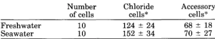
TABLE 1. Mean surface areas (in Fm2 f S.D.) of mitochondria-rich cells in sections of the primary gill eDithelium from freshwater- or seawater-adaDted trout
~~ ~ ~
Number Chloride Accessory of cells cells* cells* Freshwater 10 124 f 24 68 2 18
Seawater 10 152 f 34 70 k 27
*Difference between freshwater and seawater not significantly differ- ent ( P > 0.05).
Mitochondria-Rich Cells in Freshwater Trout
In fresh water, chloride cells were relatively volumi- nous (Table 1) and ovoid or cylindrical in shape (Figs. 1, 2). Their wide apical pole, which was flattened or slightly convex, displayed short and regularly aligned microvilli protruding into the surrounding medium (Fig. 1). Their basal region containing the nucleus was usually in contact with the basal lamina underlying the epithelium. The membranous system, in continuity with the basolateral surface, was poorly developed; in contrast to the general rule in gill chloride cells, it was not exclusively tubular and displayed portions of un- fenestrated cisternae (Fig. 7). Owing to their strong osmiophilia, these cisternae were easily distinguished from the cisternae of the endoplasmic reticulum, which were not osmiophilic (Fig. 3); they were continuous with loosely and irregularly anastomosed membranous tubules to form a n osmiophilic network located mainly in the supranuclear region (Fig. 2). Tubular extensions of this membranous system were preferentially en- countered in the apical portion of the cell, where they were intermingled with numerous independent spher- oid or ovoid membrane-bound bodies (Figs. 2). The chloride cells were attached to pavement cells by deep apical junctions from 450 to 650 nm in length (Fig. 11).
The presumptive accessory cells (Figs. 4 , 5 ) were sig- nificantly smaller, narrower, and less numerous (Tables 1, 2) than the chloride cells. They were invariably ad- jacent to the apical portion of the latter and never reached the basal lamina of the epithelium (Fig. 1). Their narrow apex, which was in contact with the ex- ternal medium, contained sparse vesicular and tubular structures (Fig. 6). They were endowed with a n exten- sive tubular system uniformly distributed throughout the cell (Figs. 1,5). At higher magnification (Fig. 4) and in stereopairs (Figs. 6,8), this tubular system consisted
A am B bl C ER G IS lbm m MB n P SL T A bbrerviations
small mitochondria-rich cell or accessory cell apical membrane
basal cells basal lamina
large mitochondria-rich cell or chloride cell endoplasmic reticulum
Golgi apparatus intercellular space
laterobasal plasma membrane mitochondria membrane-bound body nucleus pavement cell seondary lamella tubular system
GILL ACCESSORY CELLS IN FRESHWATER TROUT 315 Mitochondria-rich cell numbers (mean &S.D.) in interlamellar region of primary gill epithelium from TABLE 2.
freshwater- and seawater-adapted trout
Interdigitations between
chloride cells and Number' Chloride cells* Accessory cells** accessory cells
Freshwater 50 2.65 ? 0.90 0.94
*
0.50 0Seawater 50 2.06 2 0.90 0.92 t 0.46 1.05
*
1.6 'Number of interlamellar regions.*Difference between freshwater and seawater significantly different (P < 0.001).
**Difference not significant (P > 0.05).
exclusively of tightly anastomosed membranous tu- bules forming a continuous meshwork with numerous small regular meshes. The diameter of the tubules, how ever, was smaller (25-30 nm) in the accessory cells than in the chloride cells (50-80 nm) (Figs. 6, 9, 13). The accessory cells were bound to pavement cells by deep (450-650 nm in length) apical junctions and to adjacent chloride cells by shallower (100-200 nm) junctions (Fig. 12).
Mitochondria-Rich Cells in Seawater-Adapted Trout
When trout were progressively adapted to seawater, the size of both types of mitochondria-rich cells re- mained unaltered (Table 1); the number of accessory cells was not modified, whereas the number of chloride cells was slightly diminished (Table 2). In the accessory cells, the ultrastructural characteristics of the three main intracytoplasmic membranous systems remained unaltered. In the chloride cells, in contrast, the tubular system, which was poorly developed in freshwater (Fig. 9), became conspicuous in seawater; it consisted of tightly anastomosed membranous tubules uniformly distributed throughout the cell (Fig. 10). In both types of cells, transfer into seawater did not modify the di- ameter of the membranous tubules making up the tu- bular system.
In seawater-adapted fishes, the accessory cells sent numerous interdigitating processes into the apical por- tions of the adjacent chloride cells (Fig. 14; Table 2). As a result, the intercellular space separating the two cell types, relatively straight in freshwater, became sin- uous in seawater (Figs. 13,14). Furthermore, the apical junctions closing these expanded intercellular spaces were significantly shallower (25 to 50 nm in length) in seawater (Fig. 14) than in fresh water (Fig. 12).
DISCUSSION
Several electron microscope studies have demon- strated the existence of at least two types of chloride
Figs. 2, 3. Chloride cells i n freshwater trout gill epithelium.
Fig. 2. This ovoid cell h a s a tubular system t h a t is more abundant in the apical region (ar) than in the basal region (br). The apex of the cell is filled with large membrane-bound bodies intermingled with elements of the tubular system. X 14,000.
Fig. 3. In the supranuclear region, the membranous system orig- inating from the laterobasal cell surface shows cisternae (curved ar- rows) that are densely impregnated with reduced osmium. These cis- ternae are readily distinguished from the ER cisternae, which remain
cells in the gill epithelium of some freshwater-adapted euryhaline teleosts (Doyle and Gorecki, 1961; Straus, 1963; Bierther, 1970; Pisam et al., 1987). In the gill epithelium of the freshwater rainbow trout, in con- trast, only one type of chloride cell has been observed in conventionally stained thin sections or in specimens prepared for scanning electron microscopy (Olson and Fromm, 1973; Kendall and Dale, 1979; Laurent and Dunel, 1980). This was confirmed in the present study, as only the large, pale mitochondria-rich cells re- sembled the chloride cells of fresh-water-adapted fishes.
Upon closer examination after reduced osmium staining, the basolateral membrane invaginations of these chloride cells were found to consist partly of unfenestrated cisternae and partly of loosely anasto- mosed membranous tubules. As a result, the tubular system is poorly developed (Fig. 15) as is the case in gill chloride cells of most freshwater-adapted eury- haline fishes (Doyle and Gorecki, 1961; Pisam, 1981). In most chloride cells, the membranous system derived from infoldings of the basolateral plasma membrane is thought to contain molecules of Na+ -K + -adenosine
triphosphatase (ATPase) (Karnaky et al., 1976; Hootman and Philpott, 1979; Philpott, 1980). The scarcity of membranous tubules in chloride cells would thus be the ultrastructural counterpart of the low Na'-K+-ATPase values detected in the gills of the freshwater rainbow trout and might explain the poor adaptability of this species to seawater (Boeuf and Harache, 1984). When, however, the freshwater trout is progressively adapted t o artificial seawater, the unfenestrated cisternae are no longer visible in the basolateral membrane invaginations of the gill chlo- ride cells; they are replaced by a system of tightly anastomosed membranous tubules (Fig. 15) similar to the tight tubular network depicted in chloride cells of seawater-adapted euryhaline fishes. Thus, in this respect, chloride cells of the rainbow trout do not differ from those of other euryhaline fishes and might have a
unstained and are lined with ribosomes. x 34,000.
Figs. 4, 5.
Fig. 4.
Accessory cells i n freshwater trout gill epithelium. In the supranuclear region, the membranous system orig- inating from the laterobasal surface consists exclusivelv of anasto- mosed membranous tubules (curved arrows). X 34,000.
Flg. 5. The cell is pear-shaped, with a tubular system uniformly distributed throughout the apical (ar) and basal (br) regions. Vesicu- lar structures (V) are scarce in the narrow apex. x 12,000.
GILL ACCESSORY C E L L S IN FRESHWATER TROUT 317
Figs. 9, 10.
lium. x 36,000.
Portions of accessory and chloride cells in gill epithe-
Fig. 9. Freshwater trout. The accessory cell at left shows a tubular system made up of tightly anastomosed tubules. At right, in the chlo- ride cell, the tubular system consists of sheets and tubules loosely anastomosed.
Fig. 10. Seawater-adapted trout. The tubular system of the acces- sory cell resembles that of the freshwater fish: the density and the diameter of the tubules are not modified. In contrast, in the chloride cell, the tubular system appears as a dense network of anastomosed tubules around numerous, small, and polygonal meshes; as in the freshwater trout, the diameter of these tubules is larger than that of the membranous tubules observed in the accessory cell.
Figs. 6-8. Freshwater trout. Stereopairs of the stained membra- nous system derived from t h e laterobasal plasma membrane in mito- chondria-rich cells. x 22,000.
Fig. 6. Apex of both types of mitochondria-rich cells. In the chlo- ride cell (at left), the membranous system (curved arrows) consists of
tubules larger and more loosely anastomosed t h a n those of the acces- sory cell (at right). Numerous voluminous, apical, membrane-bound bodies are observed exclusively in the chloride cells. Note rare, small vesicular structures (V) in the apex of the accessory cell.
Fig. 7. In the supranuclear region of the chloride cell, the mem- branous system consists of unfenestrated cisternae (curved arrows) interconnected by rare tubular elements (t).
Fig. 8 . In the supranuclear region of the accessory cell, the mem- branous tubules are anastomosed t o form a n extensive network dis- playing numerous, small, regular, and polygonal meshes (arrows).
Figs, 11, 12. Apical portions of intercellular spaces in freshwater trout gill epithelium. The staining of the intercellular space disap- pears a t the level of the apical junction (‘s” located between two par- allel arrows) binding the apical portion of the chloride cell to a pave- ment cell (Fig. 11). A significantly shallower junction may be observed between a n accessory cell and two chloride cells (Fig. 12).
x 34,000.
Fig. 13. In freshwater trout, the accessory cell does not send inter- digitations into the apices of adjacent chloride cells. X 14,000.
Fig. 14. In seawater trout, the accessory cell sends numerous in- terdigitations (curved black arrows) into the apex of the chloride cell. Note the density of the tubular system in the chloride cell. X 14,000. Inset: The short apical junctions (arrows) located between chloride and accessory cells; x 34,000.
Figs. 13, 14. Apex of chloride and accessory cells in gillepithelium of trout.
GILL ACCESSORY CELLS IN FRESHWATER TROUT 319
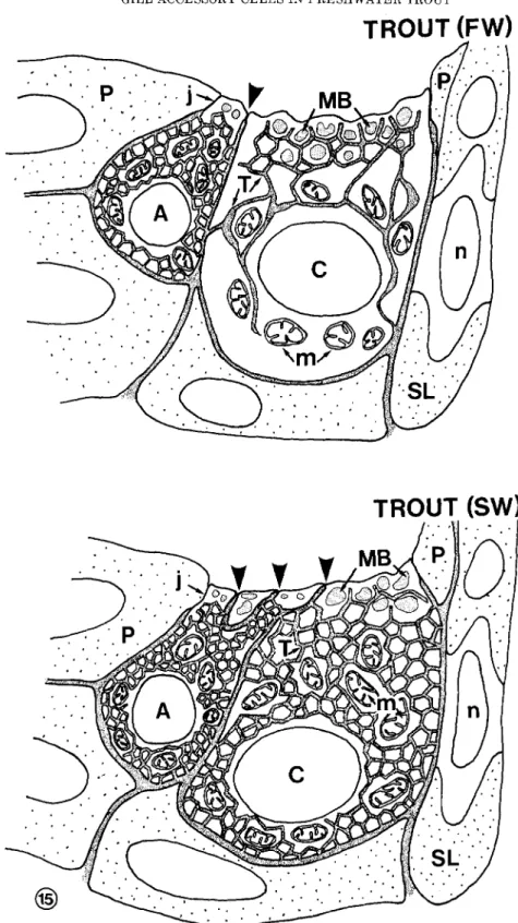
Fig. 15. Schematic representation of the ultrastructural differ- ences between “mitochondria-rich cells” from freshwater and seawa- ter trouts.
In freshwater (FW) chloride cells, the membranous system con- nected with the basolateral plasma membrane is poorly developed. In the apical region, anastomosed tubules are intermingled with large membrane-bound bodies; in the perinuclear region, the membranous system consists mainly of anastomosed cisternae. In contrast, the ac- cessory cell displays a n abundant tubular system in which tubules are anastomosed to form a dense network showing small, regular, and polygonal meshes. The apical junction binding accessory and chloride
cells (arrowhead) is shorter than the junction (i) located between the pavement cell and the accessory or chloride cells.
In seawater (SW), the size and shape of both cell types are not significantly modified. As compared with freshwater fish, the main ultrastructural alterations are: 1) Development of the tubular system in the chloride cell, which consists of a dense network of anastomosed tubules with numerous, small, polygonal meshes; and 2) formation of numerous apical interdigitations between the accessory cell and the apex of the adjacent chloride cell, sealed off by shallow junctions (ar- rowheads).
320 M. PISAM ET AL. role in the adaptation of this sedentary freshwater fish
(Boeuf and Harache, 1984) to seawater life.
Impregnation with reduced osmium of the gill epi- thelium of the freshwater trout also demonstrates the presence of smaller, denser mitochondria-rich cells dis- playing a well-developed tubular system. These small cells are characterized by the following ultrastructural features (Fig. 15): they never reach the basal lamina of the gill epithelium; they are always adjacent to the apical portion of the chloride cells and are bound to them by junctions significantly shallower than those binding the apex of chloride cells to adjacent pavement cells. Furthermore, transfer of the trout into seawater induces the formation of numerous cytoplasmic inter- digitations between these small cells and the apical portion of the chloride cells. All these ultrastructural features have been reported as specific for so-called ac- cessory cells (Laurent and Dunel, 1980; Laurent, 1984) and thus support the conclusion that the small mito- chondria-rich cells might correspond to accessory cells. Accessory cells are usually encountered in the gill ep- ithelium of seawater-adapted fishes (Laurent and Dunel, 1980); however, presumptive accessory cells in which the tubular system remained rudimentary have been found occasionally in the gill epithelium of some freshwater-adapted euryhaline fishes (Chretien and Pisam, 1986); and recently Hwang (1988) reported the presence, in the gill of some freshwater teleosts, of mul- ticellular complexes in which some chloride cells re- semble accessory cells.
Accessory cells are usually thought to have a role in the osmoregulation of seawater-adapted fishes (Sardet et al., 1979). When present in the gill epithelium of a freshwater fish such as the freshwater smolt of Salmo salar, accessory cells are presumed to participate in a
preadaptation of the fish to seawater (Pisam et al., 1988). The observations of accessory cells with a highly developed tubular system is thus rather surprising in a fresh-water sedentary fish which
“.
. .
in spite of a cer- tain degree of euryhalinity.
. . suffers significant losses in seawater. .
.”
(Boeuf and Harache, 1984). In contrast to what occurs in chloride cells, the tubular system of these accessory cells remains unaltered when the trout is adapted to seawater (Fig. 15).Furthermore, Na+ -K + -ATPase, which is an integral
part of the tubular system in chloride cells, has so far not been detected in accessory cells (Hootman and Phil- pott, 1980), so that their role in the transfer of both ions might be questioned. In the freshwater trout as in freshwater smolt, accessory cells are always associated with chloride cells. Transfer to seawater induces in both species the formation of interdigitations between the two cells and a modification of the apical junction sealing off the extended intercellular space. The chlo- ride cells and their associated accessory cells should thus be considered as a functional unit, as suggested by Sardet et al. (1979). The role of the accessory cell in this functional unit, however, remains to be established.
ACKNOWLEDGMENTS
The authors express gratitude to Professor H. Bern for his critical reading of the manuscript. The drawing (Fig. 15) was prepared by Michele Lucarain.
LITERATURE CITED
Bierther, M. 1970 Die chloridzellen des Stichlings. Z. Zellforsch.,
107:421-446.
Boeuf, G., and Y. Harache 1984 Adaptation osmotique a l’eau de mer de differentes especes (Salmo trutta, Salmo gairdneri, Salvelinus fontinalis) et hybride (Salmo trutta, Salvelinus fontinalis) de
Salmonides. Aquaculture, 40:343-358.
Chretien, M., and M. Pisam 1986 Cell renewal and differentiation in the gill epithelium of fresh- or salt-water-adapted euryhaline fish as revealed by 3H-thymidine radioautography. Biol. Cell, 56:137-
150.
Doyle, W.L., and D. Gorecki 1961 The so-called chloride cell ofthe fish gill. Physiol. Zool., 3431-85.
Hootman, S.R., and C.W. Philpott 1979 Ultrachemical localization of Na
+
, K+
activated ATPase i n chloride cells from the gills of I a euryhaline telost. Anat. Rec., 193:99-130.Hootman, S.R., and C.W. Philpott 1980 Accessory cells in teleost bran- chial epithelium. Am. J . Physiol., 238:R199-R206.
Hwang, P.P. 1988 Multicellular complex of chloride cells in the gills of freshwater teleosts. J. Morphol., 196t15-22.
Jacobs, D., E.F. Esmond, E.L. Melisky, and C.H. Hocutt 1981 Mor- phological changes in gill epithelia of heat-stressed rainbow trout, Salmo gairdneri: Evidence in support of a temperature-
induced surface area change hypothesis. Can. J . Fish. Aquat. Sci.,
38:16-22.
Karnaky, K.J., J r . 1980 Ion secreting epithelia: chloride cells in the head region of Fundulus heteroclitus. Am. J . Physiol., 238:R185-
R198.
Karnaky, K.J. Jr., L.B. Kinter, W.B. Kinter, and C.E. Stirling 1976 Teleost chloride cell. 11. Autoradiographic localization of gill Na, K-ATPase in killifish, Fundulus heteroclitus, adapted to low and high salinity environments. J . Cell Biol., 70:157-177.
Karnovsky, M.J. 1971 Use of ferrocyanide reduced osmium tetroxide in electron miscroscopy. Proc. Am. SOC. Cell Biol., 284:146 (abstract).
Kendall, M., and J . Dale 1979 Scanning and transmission electron microscopic observations of rainbow trout (Salmo gairdneri) gill.
J. Fish. Res. Board Can., 36:1072-1079.
Kikuchi, S. 1977 Mitochondria rich (chloride cells) in the gill epithelia from four species of stenohaline freshwater teleosts. Cell Tiss. Res., 180t87-98.
Laurent, P. 1984 Gill interrenal morphology, In: Fish Physiology. W.S. Hoar and D.J. Randall, eds. Academic Press, New York, pp.
73-183.
Laurent, P., and S. Dunel 1978 Relations anatomiques des ionocytes (cellules a chlorure) avec le compartiment veineux branchial: d6f- inition de deux types d’epith6lium de la branchie des poissons. C.R. Acad. Sci. Paris, 286:1447-1450.
Laurent, P., and S. Dunel 1980 Morphology of gill epithelia in fish. Am. J . Physiol., 238:R147-R159.
Morgan, M., and P. Tovell 1973 The structure of the gill of the trout,
Salmo gairdneri (Richardson). Z. Zellforsch., 142:147-162.
Olson, K., and P. Fromm 1973 A scanning electron microscopy study of secondary lamellae and chloride cells of rainbow trout (Salmo
gairdneri). Z. Zellforsch., 143:439-449.
Philpott, C.W. 1966 The use of horseradish peroxidase to demonstrate functional continuity between the plasmalemma and the unique tubular system of the chloride cell. J . Cell Biol., 31:86A (abstract).
Philpott, C.W. 1980 Tubular system membranes of teleost chloride cells: osmotic response and transport sites. Am. J . Physiol., 238:
R171-Rl84.
Pisam, M. 1981 Membranous systems in the “chloride cell” of teleo- stean fish gill, their modifications in response to the salinity of the environment. Anat. Rec., 200:401-414.
Pisam, M., A. Caroff, and A. Rambourg 1987 Two types of chloride cells in the gill epithelium of a freshwater adapted euryhaline fish: Lebistes reticulatus. Their modifications during adaptation
to salt water. Am. J. Anat., 179:40-50.
Pisam, M., P. Prunet, G. Boeuf, and A. Rambourg 1988 Ultrastruc- tural features of chloride cells in the gill epithelium of the At- lantic salmon, Salmo salar, and their modifications during smol-
tification. Am. J . Anat., 183:235-244.
Sardet, C., M. Pisam, and J . Maetz 1979 The surface epithelium of teleostean fish gills: cellular and functional adaptations of the chloride cell in relation to salt adaptation. J . Cell Biol., 80:96-
117.
Straus, L.P. 1963 A study of the fine structure of the so-called chloride cell in the gill of the guppy Lebistes reticulatus P. Physiol. Zool.,