HAL Id: tel-01575292
https://hal.inria.fr/tel-01575292v2
Submitted on 4 Oct 2017HAL is a multi-disciplinary open access archive for the deposit and dissemination of sci-entific research documents, whether they are pub-lished or not. The documents may come from teaching and research institutions in France or abroad, or from public or private research centers.
L’archive ouverte pluridisciplinaire HAL, est destinée au dépôt et à la diffusion de documents scientifiques de niveau recherche, publiés ou non, émanant des établissements d’enseignement et de recherche français ou étrangers, des laboratoires publics ou privés.
cardiac images for group-wise longitudinal analysis
Marc-Michel Rohe
To cite this version:
Marc-Michel Rohe. Reduced representation of segmentation and tracking in cardiac images for group-wise longitudinal analysis. Human health and pathology. Université Côte d’Azur, 2017. English. �NNT : 2017AZUR4051�. �tel-01575292v2�
UNIVERSITY OF NICE - SOPHIA ANTIPOLIS
DOCTORAL SCHOOL STIC
SCIENCES ET TECHNOLOGIES DE L’INFORMATION ET DE LA COMMUNICATION
P H D T H E S I S
to obtain the title of
PhD of Science
of the University of Nice - Sophia Antipolis
Specialty : Computer Science
Defended by
Marc-Michel Rohé
Reduced Representation of
Segmentation and Tracking in Cardiac
Images for Group-wise Longitudinal
Analysis
Thesis Advisor: Xavier Pennec
Thesis Co-Advisor: Maxime Sermesant
prepared at INRIA Sophia Antipolis, Asclepios Team
to be defended on July 3rd, 2017Jury :
President : Patrick Clarysse - CREATIS, CNRS, Inserm, Lyon Reviewers : Julia Schnabel - King’s College London
Alistair Young - University of Auckland Advisor : Xavier Pennec - Inria Sophia-Antipolis Co-Advisor : Maxime Sermesant - Inria Sophia-Antipolis Examinator : Andrew Taylor - University College London
Reduced Representation of Segmentation and Tracking in
Cardiac Images for Group-Wise Longitudinal Analysis
Abstract This thesis presents image-based methods for the analysis of cardiac motion to enable group-wise statistics, automatic diagnosis and longitudinal study. This is achieved by combining advanced medical image processing with machine learning methods and statistical modelling.
The first axis of this work is to define an automatic method for the segmentation of the myocardium. We develop a very-fast registration method based on convolu-tional neural networks that is trained to learn inter-subject heart registration. Then, we embed this registration method into a multi-atlas segmentation pipeline.
The second axis of this work is focused on the improvement of cardiac motion tracking methods in order to define relevant low-dimensional representations. Two different methods are developed, one relying on Barycentric Subspaces built on ref-erences frames of the sequence, and another based on a reduced order representation of the motion from polyaffine transformations.
Finally, in the last axis, we apply the previously defined representation to the problem of diagnosis and longitudinal analysis. We show that these representations encode relevant features allowing the diagnosis of infarcted patients and Tetralogy of Fallot versus controls and the analysis of the evolution through time of the cardiac motion of patients with either cardiomyopathies or obesity.
These three axes form an end to end framework for the study of cardiac motion starting from the acquisition of the medical images to their automatic analysis. Such a framework could be used for diagonis and therapy planning in order to improve the clinical decision making with a more personalised computer-aided medicine. Keywords: Medical image analysis, Non-rigid registration, Deep learning, Statis-tical model reduction, Longitudinal analysis
iii
Représentation réduite de la segmentation et du suivi des
images cardiaques pour l’analyse longitudinale de groupe
Résumé Cette thèse présente des méthodes d’imagerie pour l’analyse du mou-vement cardiaque afin de permettre des statistiques groupées, un diagnostic au-tomatique et une étude longitudinale. Ceci est réalisé en combinant des méthodes d’apprentissage et de modélisation statistique.
En premier lieu, une méthode automatique de segmentation du myocarde est définie. Pour ce faire, nous développons une méthode de recalage très rapide basée sur des réseaux neuronaux convolutifs qui sont entrainés à apprendre le recalage cardiaque inter-sujet. Ensuite, nous intégrons cette méthode de recalage dans une pipeline de segmentation multi-atlas.
Ensuite, nous améliorons des méthodes de suivi du mouvement cardiaque afin de définir des représentations à faible dimension. Deux méthodes différentes sont développées, l’une s’appuyant sur des sous-espaces barycentriques construits sur des frames de référence de la séquence et une autre basée sur une représentation d’ordre réduit du mouvement avec des transformations polyaffine.
Enfin, nous appliquons la représentation précédemment définie au problème du diagnostic et de l’analyse longitudinale. Nous montrons que ces représentations en-codent des caractéristiques pertinentes permettant le diagnostic des patients atteint d’infarct et de Tétralogie de Fallot ainsi que l’analyse de l’évolution dans le temps du mouvement cardiaque des patients atteints de cardiomyopathies ou d’obésité.
Ces trois axes forment un cadre pour l’étude du mouvement cardiaque de bout en bout de l’acquisition des images médicales jusqu’à leur analyse automatique afin d’améliorer la prise de décision clinique grâce à un traitement personnalisé assisté par ordinateur.
Mots Clefs: Analyse d’images médicales, Recalage non-rigide, Apprentissage pro-fond, Réduction statistique de modèle, Analyse longitudinale
v
Acknowledgments
First of all, I would like to warmly thank my two supervisors, Xavier Pennec and Maxime Sermesant for their guidance and support all along the three years of my Ph.D. You were always here to answer my questions and to help me during my research work and I have learned a lot from your deep and complementary scientific knowledge. Xavier, your passion for theoretical mathematics was highly communica-tive and you never refused to share your time in order to help me when I needed. I could come with one simple question and get drag into hours of interesting discus-sions about completely different topics and ideas of research. Maxime, you helped me to turn innovative mathematical concepts into practical sound applications and you always managed to came out with ideas to close the gap between theory and applications. It was a pleasure to work under both of your guidance, not only on a professional level but also on a personal level with the multiple trips abroad we had the chance to do together! I would also like to thank Nicholas Ayache for accepting me in the Asclepios team where I enjoyed outstanding working conditions and a friendly research environment for the last three years.
I am extremely grateful to Prof. Julia Schnabel and Prof. Alistair Young for accepting to be my reviewers and for spending their precious time to read and correct this manuscript. Their encouraging compliments and constructive comments on my work are invaluable. I am also indebted to Andrew Taylor and Patrick Clarysse who accepted to be member of my jury and to come at my defense. Dear committee, thank you, it has been a great honor for me to have such an outstanding jury.
During this PhD, I had the chance to have such a great time so that I always enjoyed working even when the pressure was high. Special thanks to the people who worked in my office: my ginger friend Nina who introduced me to the team and made me feel welcomed when I arrived, Krissy, Loïc D., Sofia and Yann! To Thomas with whom I probably drank thousands of coffee and had thousands of laughs. To Sophie who managed to cancel the journal club just a month after arriving in the lab. To Roch with whom I had the chance to travel to multiple places for meetings of the MD-Paedigree project. To Rocio for inviting us at her incredible wedding in Mexico. Thanks to all the other students for the good time we have spent together in and also outside the lab. Those who are not in the lab anymore but are not forgotten nonetheless: Cloclo, Nicolas C., Vikash, Milkymat, Bisheh, Loïc... And those who still need to work a little bit to reach our level and get the almighty PhD: Rafifou, Shuman, Pawel, Julian, Qiao, Wen, Luigi, Nicoco and probably many more by the time your read this!
I finally would like to thank my whole family for their love, support, encourage-ment and kindness: my mother of course, my father, my sister, my grand-mother and my cousins. You always believed in me and supported me during my long years of education and I would never have succeeded without that. Thank you for always pushing me to do what makes me happy, no matter the obstacles or challenges. This thesis is dedicated to you.
Contents
1 Clinical and Technical Context 1
1.1 Introduction . . . 1
1.2 Healthy Cardiac Structure and Function . . . 2
1.2.1 Cardiac Structure. . . 3
1.2.2 Cardiac Cycle . . . 4
1.3 From Healthy to Pathological Heart . . . 6
1.3.1 MD-Paedigree Project . . . 7
1.3.2 Cardiomyopathies . . . 9
1.3.3 Obesity . . . 11
1.4 Cardiac imaging . . . 12
1.4.1 Echocardiography . . . 13
1.4.2 Magnetic Resonance Imaging . . . 14
1.4.3 Computed Tomography . . . 17
1.5 Manuscript Organization and Objectives . . . 17
1.6 Publications . . . 23
1.6.1 Journal Articles. . . 23
1.6.2 Selective Peer-Reviewed Conference Paper . . . 24
1.6.3 Awards . . . 24
I FROM MEDICAL IMAGES TO 3D SHAPE 25 2 Registration in Medical Imaging 27 2.1 Introduction . . . 27
2.2 Registration Algorithms . . . 28
2.2.1 Similarity Measures . . . 30
2.2.2 Transformation Spaces and Regularization . . . 31
2.2.3 Optimization Methods . . . 32
2.3 LCC Log-Domain Diffeomorphic Demons . . . 33
2.3.1 Optimization Method: Alternate Optimization . . . 33
2.3.2 Deformation Parametrization: Stationary Velocity Field . . . 34
2.3.3 Similarity Metric: Local Correlation Coefficient . . . 35
2.4 Framework of Currents for Shape Registration. . . 36
2.4.1 The Formalism of Currents for Surface Representation . . . . 38
2.4.2 Surface Registration Using Currents and LDDMM . . . 40
3 SVF-Net: Learning Deformable Image Registration Using Shape Matching 43 3.1 Motivations . . . 43
3.2.1 Currents for Shape matching . . . 47
3.2.2 Elastic Body Spline Interpolation . . . 47
3.2.3 Stationary Velocity Fields for Diffeomorphism Parametrization 48 3.3 SVF-Net: Fully Convolutional Neural Network Architecture . . . 49
3.4 Experiments . . . 50
3.4.1 Training . . . 51
3.4.2 Evaluation. . . 51
3.5 Conclusions . . . 53
4 An Automatic Multi-Atlas Myocardium Segmentation 55 4.1 Chapter Overview . . . 56
4.2 Motivations . . . 57
4.3 Segmentation in Clinical Practice . . . 57
4.4 Segmentation Methods In Medical Imaging . . . 59
4.5 Multi-Atlas Segmentation: Overview of the Method. . . 60
4.6 Rigid Alignment by Landmarks Detection . . . 61
4.6.1 Method for Rigid Alignment. . . 62
4.6.2 CNNs and Heatmap for Landmarks Detection . . . 63
4.6.3 Training and Results . . . 63
4.6.4 Alignment of the Images and Cropping. . . 65
4.7 Registration of Images and Fusion of Segmentation . . . 66
4.7.1 Points Propagation . . . 66
4.7.2 Points Combination . . . 66
4.7.3 Supervised Learning of Local Distance . . . 67
4.7.4 Computation of Weights Based on Local Distance. . . 68
4.8 Conclusions . . . 70
II LOW-DIMENSIONAL REPRESENTATION OF CAR-DIAC MOTION 73 5 Barycentric Subspace for Cardiac Motion Tracking 75 5.1 Chapter Overview . . . 75
5.2 Background: Cardiac Motion Tracking . . . 76
5.2.1 Cardiac Motion Tracking Algorithms . . . 77
5.2.2 Evaluation of Tracking Methods . . . 79
5.3 Motivations: Low-Dimensional Representation of the Cardiac Motion 79 5.4 Methodology: Barycentric Subspaces . . . 82
5.4.1 Definition of the Subspace . . . 82
5.4.2 Projection of an Image to the Subspace . . . 83
5.4.3 Computation of the optimal References of a sequence of Images 84 5.4.4 Computation of an Image within the Subspace . . . 84
5.5 Using Barycentric Subspaces as a prior on the Registration . . . 85
Contents ix
5.5.2 Evaluation using a Synthetic Sequence . . . 88
5.5.3 Towards Symmetric Transitive Registration . . . 89
5.6 Conclusion. . . 90
5.A Projection of an image to the Barycentric Subspace . . . 91
5.B Frame-To-Frame Barycentric Registration Formulation . . . 92
6 Highly Reduced Model of the Cardiac Function for Fast Simulation and Personalization 93 6.1 Chapter Overview . . . 93
6.2 Motivations . . . 94
6.3 Reduced-Polyaffine Projection for Compact Cardiac Motion Repre-sentation. . . 95
6.4 Biophysical Model of the Heart Simulation Database . . . 96
6.5 Parameters Mapping through PLS Regression . . . 97
6.6 Applications . . . 99
6.6.1 Direct Highly Reduced Cardiac Function Model . . . 99
6.6.2 Personalization of Model Parameters . . . 100
6.7 Conclusion. . . 100
III APPLICATION TO DIAGNOSIS AND LONGITUDINAL ANALYSIS 101 7 Polyaffine Transformations for the Automatic Diagnosic of LV In-farct 103 7.1 Motivations . . . 104
7.2 Database of Asymptomatic Subjects and Patients with Myocardial Infarction . . . 105
7.3 Extraction of features of interest through shape and motion dimen-sionality reduction . . . 106
7.3.1 Polyaffine projection . . . 106
7.3.2 Thickness parameters . . . 108
7.4 Dimensionality reduction of the parameters and classification . . . . 110
7.4.1 Learnt Dimensionality Reduction . . . 110
7.4.2 Cross-Validation on Training Set: Classifier Selection . . . 112
7.5 Results and Validation on Testing Set . . . 113
7.6 Conclusion. . . 114
8 Cardiac Motion Signature Using Barycentric Subspaces 117 8.1 Chapter Overview . . . 117
8.2 Cardiac Motion Signature from Low-Dimensional Representation . . 118
8.2.1 Data . . . 118
8.2.2 Methods . . . 118
8.2.3 Optimal References Frames and Barycentric Curves . . . 119
8.3 Reconstruction of Cardiac Sequences . . . 121
8.3.1 Qualitative results . . . 122
8.3.2 Quantitative results . . . 123
8.4 Conclusion. . . 124
9 Longitudinal Analysis of the Cardiac Motion 125 9.1 Chapter Overview . . . 125
9.2 Motivations . . . 126
9.3 Cardiac Motion Features Extraction . . . 127
9.4 Cardiomyopathies. . . 129
9.4.1 Data . . . 129
9.4.2 Mean Motion Model . . . 130
9.4.3 Longitudinal Motion Analysis of CMP patients . . . 131
9.5 Obesity . . . 132
9.5.1 Data: Obesity Patients (CVD) . . . 132
9.5.2 Mean Motion Model Parametrized by BMI . . . 133
9.5.3 Longitudinal Motion Analysis of CVD patients . . . 134
9.6 Conclusion. . . 136
10 Conclusions and Perspectives 139 10.1 Summary of the Main Contributions . . . 139
10.2 Perspectives and Future Applications . . . 141
10.3 Virtual Patient in the Age of Artificial Intelligence: the Future of Medicine . . . 144
Chapter 1
Clinical and Technical Context
Contents
1.1 Introduction . . . 1
1.2 Healthy Cardiac Structure and Function . . . 2
1.2.1 Cardiac Structure . . . 3
1.2.2 Cardiac Cycle. . . 4
1.3 From Healthy to Pathological Heart . . . 6
1.3.1 MD-Paedigree Project . . . 7
1.3.2 Cardiomyopathies . . . 9
1.3.3 Obesity . . . 11
1.4 Cardiac imaging . . . 12
1.4.1 Echocardiography . . . 13
1.4.2 Magnetic Resonance Imaging . . . 14
1.4.3 Computed Tomography . . . 17
1.5 Manuscript Organization and Objectives . . . 17
1.6 Publications. . . 23
1.6.1 Journal Articles. . . 23
1.6.2 Selective Peer-Reviewed Conference Paper . . . 24
1.6.3 Awards . . . 24
The purpose of this chapter is to give the clinical and technical context, building the basis for the following parts of this manuscript. We start from the highest level -the patient - to give -the clinical motivation of our work. We describe what a healthy cardiac motion is and the differences seen in two medical conditions: cardiomyopathy and obesity. Then, we introduce some background on imaging techniques used in cardiology. This will bring us from a patient to a representation of its cardiac motion using medical images. Finally, we present the objectives: Using sequences of images of cardiac motion, how can we automatically analyze and derive relevant information to help diagnosis, prognosis and therapy planning?.
1.1
Introduction
Clinical practice is usually divided in three steps. First, the diagnosis, where a potential disease is identified by the clinician using the information available about
the patient and his symptoms. Then the prognosis, where a set of likely outcomes of the evolution of the patient’s disease is predicted: it usually includes the expected duration, the impact on main functions of the body, and potential symptoms of the disease. Finally, therapy planning, where all previous information are put together in order to plan one or several possible treatments, which have the best chances of curing the patient or reducing the symptoms of the disease.
In many cases, medical images are a very important source of information that can help clinicians in the process of these three steps and they are getting increas-ingly used in clinical practice. In the context of cardiac imaging, it gives clinicians knowledge about the shape of the heart (when acquiring a single image) and its function (when acquiring a series of images or a video of the beating heart with multiple frames). To compare those acquisitions over several time periods (or lon-gitudinal study) is essential to keep track of the progression of a disease, and will help with decision making and therapy planning.
Automatic analysis of medical images, using computational methods, can give clinicians additional insight on what cannot be seen by the human eye or calculated directly. A simple example of how these tools can help clinical workflow is the computation of important quantitative parameters, such as ejection fraction (derived from the size of the ventricles) or strain values. Algorithms can also help process big amount of data, which a single clinician could not. This can, for example, allow the comparison of data of one patient with a large database of subjects with known evolution and therapy in order to find similar cases. They can also replace the clinician by carrying out tasks that could be done manually, but are too time-consuming within a very time-constrained clinical workflow.
The last decades have seen spectacular advances and progress in computa-tional methods for the automatic analysis of medical images using computer vi-sion techniques, especially in cardiology. Among the most important developments led by medical image analysis, there is the automatic extraction of the geome-try of the the heart (both left and right ventricles), which is called Segmentation
[Heller 2002, Zheng 2008, Ecabert 2008], and the automatic evaluation of cardiac
motion or Cardiac Motion Tracking [Zerhouni 1988,Tobon-Gomez 2013], using Reg-istration techniques. These methods have already been applied to many clinical problems [Ferre 1999, Zhang 2004, Goshtasby 2005,Norouzi 2014]. However, they have yet to be disseminated widely in clinical routines, due to their lack of robustness and automation. The purpose of this thesis is therefore to improve those methods, in the context of cardiac motion analysis. To do so, we propose contributions to both the technical methodologies and their applications to multiple different diseases with a focus on cardiomyopathies and obese children.
1.2
Healthy Cardiac Structure and Function
The main function of the heart is to pump blood through the body. Even though the concept is straightforward, the biological machinery involved in this process is
1.2. Healthy Cardiac Structure and Function 3
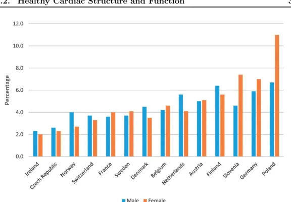
Figure 1.1: Percentage of the population (both male and female) reporting heart problems in major european countries. Taken from [Townsend 2016].
very complicated, especially considering the amount of blood and the frequency at which it has to function. A normal heart rate at rest ranges from 60 to 100 beats per minute in adults with a stroke volume - the amount of blood pumped at each heartbeat - of around 70 mL for each ventricle. This gives an idea of the imporant constraints that the heart has to endure during a human life. The consequences of a heart failure are often dramatic: shortness of breath, excessive tiredness, leg swelling and possibly death. It affects between 2% and 10% of the whole population (see Fig. 1.1) and it is the number one cause of death in many countries, accounting for approximately 25% of deaths. Therefore, it is of crucial importance to monitor heart function and to understand what separates an efficient heart from a deficient one. We present here the main characteristics of a typical heart structure and cycle. Then, we show how it differs in a pathological condition.
1.2.1 Cardiac Structure
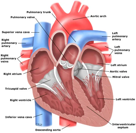
In this section, we describe the heart and its anatomy. A detailed review of the cardiac structure can be found in [Anderson 2004]. The heart is located slightly to the left of the chest between the lungs. It is cone-shaped (see Fig. 1.2) with the base (the top of the heart) positioned upwards and tapering down towards the apex (the bottom of the heart). An average adult heart has a mass of 250 to 350 grams
[Robb 1942], it is typically of the size of a fist: 12 cm long, 8 cm wide, and 6 cm
thick. Its size can vary between individuals, especially for those doing sports and exercises who tend to have larger hearts with a more efficient pumping mechanism.
The heart has two sides (left and right), both divided into two parts: atrium and ventricle making up four chambers in total. The atria are smaller than the ventricles and have thinner, less muscular walls than the ventricles. They are connected to veins, which role is to transport blood from the heart to the rest of the body, and act as chambers where blood transits. The ventricles are larger and stronger pumping chambers that send blood out of the heart, and are connected to arteries that carry blood away from the heart (Fig. 1.2).
The wall of the heart is made of four layers: the epicardium/pericardium, the myocardium and the endocardium. The epicardium is the outermost layer and is a thin layer of serous membrane. It helps lubricate and protect the outside of the heart. An additional layer, the pericardium (a thin fibrous sac that does not contract), further protects the heart and isolates it from other organs. The second layer, the myocardium, is the muscular layer of the heart: the largest one and the one that makes up for the majority of its mass. It is the most important layer, it consists of cardiac muscle tissue responsible for the contraction of the heart and therefore the pumping of the blood through the organism. Finally, the last innermost layer, the endocardium keeps the blood from sticking to the inside of the heart. It is a simple squamous endothelium layer that lies at the border with the blood pool.
Right and left ventricles have different functions. The left side of the heart sends blood all the way to the extremities of the body in the systemic circulatory loop while the right side is responsible for the pulmonary circulation to the nearby lungs. This difference in function results in a difference in muscle thickness, with the left ventricle being 15-mm thick - in order to have enough strength to push blood to the whole body - and the right ventricle being only 5-mm thick, thus making it more difficult to identify and delimitate in medical images.
1.2.2 Cardiac Cycle
The heart is a non-stopping pumping machine, repeating over and over again the same cardiac cycle: the period that starts the beginning of a heartneat and ends at to the beginning of the next. Its frequency is described by the heart rate, which is typically expressed in beats per minute. A single cycle of cardiac activity (or heartbeat) can be divided into two basic phases: diastole (roughly one third of the whole cycle) and systole. These two phases describe an alternating sequence of contraction and relaxation of the myocardium, in which blood gets delivered into both the systemic and pulmonary circulations [Boron 2012].
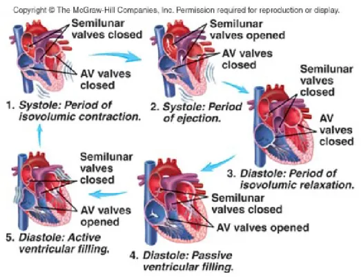
These two phases are further divided into sub-phases (see Fig. 1.4):
• Isovolumic Contraction: ventricular depolarization causes the ventricles to contract and pressure inside the ventricles to increase rapidly. Immediately after the start of the contraction, pressure in the ventricles exceeds that of in the atria, causing the atrioventricular valves to close. Pressure in the ventricles is lower than in the aorta and the pulmonary artery. Therefore, all the valves are closed and no blood can be ejected. During this phase, the ventricular
1.2. Healthy Cardiac Structure and Function 5
Figure 1.2: Anatomy of the heart. Taken from https://www.utdlab.com/.
volume remains unchanged. In the electrocardiogram (ECG), it is identified by the so-called QRS complex, which is the most visible part of the ECG signal and corresponds to the depolarization of the left and right ventricles. • Ejection: pressure in the left ventricle exceeds that of the aorta. Similarly,
pressure in the right ventricle goes beyond that of the pulmonary trunk, caus-ing the pulmonary valves to open. Both the left and right ventricles eject blood to the aorta and pulmonary arteries while the atrioventricular valves are still closed. Ejection starts rapidly and slows down as systole progresses. It corresponds to the ST segment of the ECG: first the ventricles are com-pletely depolarized, then the T wave appears with the repolarization in the second half of this phase. This is the end of the systole and the beginning of the relaxation with the diastole.
• Isovolumic Relaxation: the ventricles relax, causing a rapid drop in ventricular pressure. Shortly after, pressure in the ventricles goes below that of the
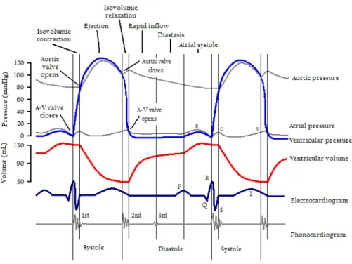
pul-Figure 1.3: Two complete cardiac cycles, together with their main events, as well as their corresponding pressure and volume curves. Taken from https://www.fastbleep.com/biology-notes/1/573.
monary artery and the aorta, and blood returns toward the ventricles, causing the semilunar valves to close while the atrioventricular valves are still closed. It corresponds to the end of the T wave of the ECG.
• Passive ventricular filling: the atrioventricular valves open and the ventricles are filled with blood coming from the atria. This passive phase accounts for most of ventricular filling. Pressure in ventricles does not change significantly, and volume increase is compensated by ventricular relaxation. No electrical activity can be found on the ECG.
• Active ventricular filling: the atria contract and complete ventricular filling. Only a small amount of blood entered the ventricles during this phase. Pres-sure in both ventricles is close to zero. Electrical activity is non-existent at the beginning of the phase, then depolarization of the atrial can be detected with the T wave, resulting in atrial contraction.
1.3
From Healthy to Pathological Heart
The work of this thesis was conducted within the MD-Paedigree project which is presented in this section. Then, we describe the clinical context centered on two
1.3. From Healthy to Pathological Heart 7
Figure 1.4: Schema representing a cardiac cycle with the active and passive systolic and diastolic phases. Taken from http://highered.mheducation.com/.
medical conditions affecting children and young adolescents: cardiomyopathies and obesity, and we show how these conditions affect the cardiac structure and function.
1.3.1 MD-Paedigree Project
MD-Paedigree aims at providing decision support to clinicians when treating their young patients in four areas: cardiomyopathies, obesity-related cardiovas-cular disease, juvenile idiopathic arthritis and neurological neuromuscardiovas-cular dis-eases. It is strongly embedded in The Virtual Physiological Human (VPH)
[Hunter 2010,Hunter 2003,Ayache 2006] framework, an European initiative which
focuses on building methodology and technology that enable collaborative investi-gation of the human body as a single complex system. This clinically-driven project regroups 7 world-renowned clinical centers of excellence: Ospedale Pediatrico Bam-bino Gesù (Italy), University College of London (United Kingdom), Istituto Gianna Gaslini (Italy), Deutsche Herzzentrum Berlin (Germany), Katholieke Universiteit Leuven & University Hospital Leuven (Belgium), Stichting Vu-VUmc (Netherlands) and Universtair Medisch Centrum Utruchet (Netherlands). This European project brings together these clinical centers with multiple technical centers (among which Inria and Siemens). This collaborative work between clinicians and computer sci-entists aims at improving interpretability of pediatric bio-medical information, data and knowledge by developing together a set of reusable and adaptable multi-scale
models for individualized, more predictive, effective and safer pediatric healthcare.
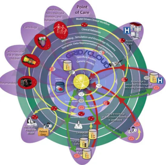
Figure 1.5: Diagram of the SOKU-Vision connecting patients and clinical centers to the models and the technical centers. On top-left the four medical conditions studied in the project (this thesis focuses on two: Cardiomyopathies and Obesity). The yellow spiral representing the models goes from the center - the simplest models - and turns around to become more and more specific and efficient. As they improve, models are validated in the different centers and integrated in the clinical workflow. Taken from http://www.md-paedigree.eu/.
MD-Paedigree validates and brings to maturity patient-specific computer-based predictive models of various paediatric diseases. It aims at increasing their potential acceptance in the clinical and biomedical research environment by making them readily available not only in the form of sustainable models and simulations, but also as newly-defined workflows for personalised predictive medicine at the point of care. These tools can be accessed and used through an innovative model-driven
1.3. From Healthy to Pathological Heart 9
Figure 1.6: (Left): Summary of ESC 2008 classification [Elliott 2007]. HCM: hy-pertrophic cardiomyopathy; DCM: dilated cardiomyopathy; ARVC: arrhythmogenic right ventricular cardiomyopathy; RCM: restrictive cardiomyopathy. Taken from
[Elliott 2012]. (Right) : Schematic representation of the different types of
car-diomyopathies as proposed in [Marcus 1982] and their effect on anatomy. Taken from Wikipedia.
infostructure powered by an established digital repository solution able to integrate multimodal health data.
MD-Paedigree implements the so-called SOKU vision which is illustrated in Fig.
1.5. The aim of this framework is to make the design and development of innovative predictive models simpler to reuse and to integrate into a clinical context. It all starts with the basic models (in the center of the diagram) which are incubated in the system with progressive semantic enrichment and model modifications in a circular way represented by the yellow spiral. During this improvement, there is an increasing intervention of both automated database-guided learning and knowledge experts validation. Then, as these models reach the status when they can be used in clinical practice, they are applied and validated by the different clinical centers and clinical researchers. In this project, four different areas of clinical pathologies were studied by the partners: cardiomyopathies, cardiovascular risk in obese children and adolescents, juvenile idiopathic arthiritis and neurological and neuromuscular disease. In this thesis, we work with the first two medical conditions and we present them in the following section.
1.3.2 Cardiomyopathies
Cardiomyopathies are a group of diseases impacting the heart leading to negative effects on the heart muscle size, shape, and function. It results in a deficiency of the heart to provide enough oxygenated blood to the rest of the body and remove carbon dioxide and other waste products. As the disease worsens and the heart weakens, classical signs and symptoms of heart failure usually occur which include: shortness of breath or trouble breathing, increased fatigue (tiredness), swelling in the ankles, feet, legs, abdomen, and veins in the neck.
There has been different ways to classify cardiomyopathies. One of the first proposed classification can be found in [Marcus 1982], in which cardiomyopathies are defined as "a heart muscle disease of unknown cause". They are classified ac-cording to their pathophysiological phenotype into dilated cardiomyopathy, hyper-trophic cardiomyopathy, or restrictive cardiomyopathy (Fig. 1.6 right). Since this first classification, more advanced classifications have been proposed to incorporate new types of diseases, such as arrhythmogenic right ventricular dysplasia (ARVD)
[Elliott 2007, Elliott 2012], and to incorporate genetic mutation testing within the
framework of classification (see Fig. 1.6left).
The differences between the four main types of cardiomyopathies according to the European Society of Cardialogy (ESC) [Elliott 2007] (see Fig. 1.6) can be sum-marized as follows [Lung 2016]:
• Hypertrophic cardiomyopathy: it is very common and it affects men and women equally of any age (1 out of every 500 people). It is caused by an enlargement and thickening of the heart muscle without any obvious cause. The ventricles, the lower chambers of the hearts and the septum thicken cre-ating narrowing or blockages in the ventricles. The efficiency of the heart to pump blood to the body is decreased. It can also cause stiffness of the ven-tricles, changes in the mitral valve, and cellular changes in the heart tissue. While the cause is not always known, hypertrophic cardiomyopathy is often inherited genetically.
• Dilated cardiomyopathy: it affects 1 out of 2,500 persons. It is associated with left ventricular remodeling, which manifests as increases in left ventricular end-diastolic and end-systolic volumes, wall thinning, and a change in chamber shape to something more spherical and less elongated. Weakened chambers of the heart no longer pump efficiently, causing the heart muscle to work harder. Possible consequences are heart failure, heart valve disease, irregular heart rate, and blood clots in the heart. The causes may be alcohol, heavy metals, coronary heart disease, cocaine use, and viral infections and it can also be inherited from a person’s parents.
• Restrictive cardiomyopathy: in opposition to other types, walls of the heart do not thicken but ventricles become stiff and rigid. It causes ventricles not to relax and not to fill with the normal blood volume. With the progression of the disease, the ventricles do not pump as well and the heart muscle weakens. It can lead to heart failure and problems with the heart valves. Possible causes of this disease are amyloidosis, hemochromatosis, and some cancer treatments. • Arrhythmogenic right ventricular dysplasia: it is a rare type of cardiomyopathy and it occurs when the muscle tissue in the right ventricle is replaced with fatty or fibrous tissue, possibly leading to disruptions in the heart’s electrical signals and arrhythmias. It usually affects teenagers and can cause sudden cardiac arrest in young athletes.
1.3. From Healthy to Pathological Heart 11
4 Obesity Update © OECD 2014
Figure 4. Measured overweight (including obesity) among children aged 5-17, 2010 or nearest year
Source: International Association for the Study of Obesity, 2013; Bös et al. (2004), Universität Karlsruhe and Ministères de l’Education nationale
et de la Santé for Luxembourg; and KNHANES 2011 for Korea.
Obesity and the economic crisis
In 2008, the world economy entered one of the most severe crises ever. Many families, especially in the hardest hit countries, have been forced to cut their food expenditures, and tighter food budgets have provided incentives for consumers to switch to lower-priced and less healthy foods.
During the 2008-09 economic slowdown, households in the United Kingdom decreased their food expenditure by 8.5% in real terms, with some evidence of an increase in calorie intake (the average calorie density of purchased foods increased by 4.8%). This change resulted in additional 0.08 g of saturated fat, 0.27 g of sugar and 0.11 g of protein per 100 g of purchased food (Institute for Fiscal Studies, Briefing Note No. 143). A similar trend was observed in Asian countries experiencing a recession in the late 1990s, with consumers switching to foods with a lower price per calorie (Block et al., 2005, Economics and Human Biology; World Bank, 2013,
Working Paper No. 6538).
Between 2008 and 2013, households in Greece, Ireland, Italy, Portugal, Spain and Slovenia decreased slightly their expenditure on fruits and vegetables, while households in other European OECD countries increased it at an average of 0.55% per year (OECD/ Imperial College analyses of passport data, Euromonitor International). Fruit and vegetable consumption was inversely related with unemployment in the United States, in the period 2007-09, and the effect was three times stronger in disadvantaged social groups at higher risk of unemployment (corresponding to a 5.6% decrease in fruit and vegetable consumption for each 1% increase in state-level unemployment). Given the size of job losses at the peak of the crisis, the most vulnerable groups may have reduced their consumption by as much as 20% (Dave and Kelly, 2012, Social Science and Medicine). Evidence from Germany, Finland and the United Kingdom shows a link between financial distress and obesity. Regardless of their income or wealth, people who experience periods of financial hardship are at increased risk of obesity, and the increase is greater for more severe and recurrent hardship (Munster et al., 2009, BMC Public Health; Conklin et al., 2013, BMC Public Health; Laaksonen et al., 2004, Obesity Research). An Australian study found that people who experienced financial distress in 2008-09 had a 20% higher risk of becoming obese than those who did not (Siahpush et al., 2014, Obesity). Financial hardship affects all household members. American children in families experiencing food insecurity are 22% more likely to become obese than children growing in other families (Metallinos-Katsaras et al., 2012, Journal of the Academy of Nutrition and Dietetics).
While some evidence suggests that shorter working hours and lack of employment are associated with more recreational physical activity (Tekin et al., 2013, NBER Working Paper No. 19234), at times of increasing unemployment any gains are likely to be offset by reduced work-related physical activity. In the United States, in the aftermath of the economic crisis, leisure-time physical activity increased by three METs (metabolic equivalents – a measure capturing both duration and intensity of physical activity) but work-related physical activity decreased by 19 METs (Colman and Dave, 2013, NBER Working Paper No. 17406). In summary, the evidence of a possible impact of the economic crisis on obesity points rather consistently to a likely increase in body weight and obesity.
Figure 1.7: (Left-Top): The International Classification of adult underweight, over-weight and obesity according to BMI as defined by the World Health Organization (WHO). Taken from [Organization 1987]. (Left-Bottom): list of medical complica-tions due to obesity. (Right): Prevalence rates of obesity in child of major developed countries for boys and girls. OECD data.
1.3.3 Obesity
Obesity is a medical condition in which abnormal or excessive fat has been accu-mulated to the point where it can have a negative impact on health. Assessment of the obesity of a person is done using the Body Mass Index (BMI). The BMI is defined as the body mass divided by the square of the height and it is universally expressed in units of kg/m2. The value of the BMI defines a classification of a per-son from underweight to obese using a benchmark proposed by the World Health Organization (WHO) (see Fig. 1.7left).
Obesity is a growing public health concerns in major developed countries. In 2008, approximately 35% of adults aged 20+ were overweight (BMI superior to 25 kg/m2) and 10% of men and 14% of women in the world were obese (BMI superior to 30 kg/m2). The worldwide prevalence of obesity worldwide has nearly doubled between 1980 and 2008 with almost 1 billion persons according to data from the Organisation de coopération et de développement économiques (OECD). Relating to children, 43 millions were estimated to be overweight and obese in 2010 (WHO data) while 92 millions were at risk. The worldwide prevalence of childhood overweight and obesity has been increasing from 4.2% in 1990 to 6.7% in 2010 and is expected to reach 9.1% in 2020 (Fig. 1.7).
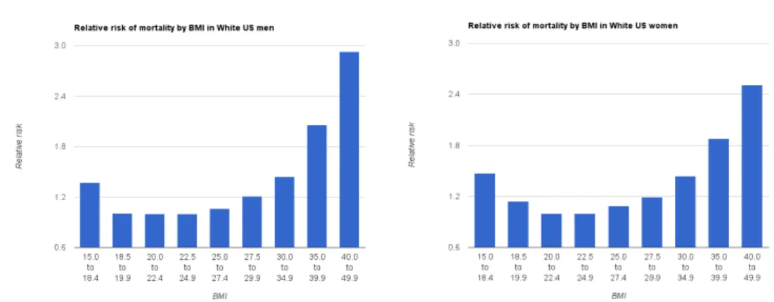
Figure 1.8: Relative risk of death over 10 years by BMI for white men (left) and women (right) who have never smoked in the United States. Taken from
[Berrington de Gonzalez 2010]
Obesity is one of the main preventable causes of death worldwide. More than 2.8 million people die each year as a result of being overweight or obese. It is associated with various diseases, particularly cardiovascular diseases, diabetes mel-litus type 2, obstructive sleep apnea, certain types of cancer, osteoarthritis and asthma and cause multiple medical complications (Fig. 1.7 left-bottom). Studies
[Berrington de Gonzalez 2010] have found a strong dependency between mortality
risk and BMI (see Fig. 1.8). It is lowest at a BMI of 18-25 kg/m2, with risk increas-ing with lower and higher BMI. A high BMI has a strong negative impact on the life expectancy and it increases the mortality risk significantly: pre-obesity (BMI of 25-30 kg/m2) reduces life expectancy by six to seven years, obesity class I (BMI of 30-35 kg/m2) by two to four years, while severe obesity (BMI ≥ 40 kg/m2) by ten years.
1.4
Cardiac imaging
To classify an individual’s condition into separate and distinct categories that allow medical decisions about treatment and prognosis to be made, clinicians have data concerning the patient’s medical condition. These data can take multiple forms: the symptoms reported by the patients, his medical history, his etiological data and the physical examination of the patient. When a cardiac problem is suspected, it is of crucial importance to analyze the myocardium motion in order to detect functional abnormalities and lesions. To do so, multiple non-invasive imaging techniques have been developed giving clinicians a vision of the heart anatomy and motion. These medical images give clinicians additional data to improve their decisions. We present the three main imaging techniques used in cardiology: Magnetic Resonance Imaging (MRI), Echocardiography (Echo), and Computed Tomography (CT) with a partic-ular focus on MRI which will be the main modality used in this manuscript.
1.4. Cardiac imaging 13
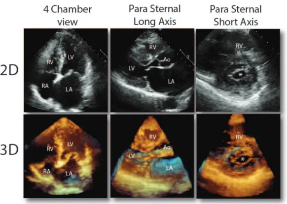
Figure 1.9: 2D and 3D echo images of the heart with the 3 different views.
1.4.1 Echocardiography
An echocardiogram (see Fig. 1.9) is an imaging technique based on the applica-tion of ultrasound (standard two-dimensional, three-dimensional, and Doppler ul-trasound). Ultrasounds are sound waves whose frequences are way higher than those audible to humans (>20,000 Hz) and ultrasonic images (also known as sonograms) are constructued by sending ultrasond pulse with a probe. The sound echoes off with varying degrees depending on the tissue. Then, these echoes are recorded and displayed as an image to the operator. Because of the difference in the reflection of the sound, the ultrasound image is able to differentiate between internal body structures such as tendons, muscles, joints, vessels and internal organs.
Echocardiography is one of the most widely used diagnostic tool in cardiology because of its ease of use, low-cost and rapidity [Belohlavek 1993]. It is used as a very efficient and fast tool to asses the cardiac anatomy and function of a patient
[Schiller 1989]. The Echo acquisition is done with the patient shirtless, lying down
on the left side. The clinician places the ultrasound probe using a gel in order to favor the transmission of the waves through the skin. Additionally, ECG electrodes can be used in order to gate the acquisition with the cardiac rhythm. Then, multiple 2D temporal acquisitions are done on different locations. An acquisition takes from 10 to 30 minutes and is completely painless and safe which is a major quality of Echocardiography.
Figure 1.10: 12 short-axis slices of a cardiac MRI from apex (top-left) to base (bottom-right).
A recent progress in echocardiography is the development of 3D temporal echocardiography. Traditional approach relies on multiple 2D acquisitions synchro-nized together using the ECG gating in order to reconstruct a 3D volume. However, a more advanced technique permits real-time three-dimensional echocardiography where the 3D acquisition is done in just one heartbeat. This technique has been applied successfully to identify structural abnormalities related to monomorphic ventricular tachycardia [Goland 2008] or to asses aortic valve area in aortic stenosis by continuity equation [Poh 2008].
The main advantages of the Echo is that it is easy to use, low-cost and completely non-invasive making it the principal imaging modality in cardiology. These qualities come at the cost of a relatively high noise to signal ratio, the presence of artifacts, and a low tissue contrast which might impair the diagnosis and decrease the quality of the assessment of the cardiac function. Therefore, other imaging techniques are often used as a complement to the Echo acquisition.
1.4.2 Magnetic Resonance Imaging
Magnetic resonance imaging (MRI) (see Fig. 1.10) is a relatively recent medical imaging technique. It was developed in the 70’s notably by Paul C. Lauterbur in 1971 [Lauterbur 1973] and Sir Peter Mansfield [Mansfield 1977]. In 2003, they both received the Nobel Prize in Medicine for their "discoveries concerning magnetic resonance imaging", although differences in tissue relaxation time values where al-ready known from the 50’s [Odeblad 1955]. MRI allows the acquisition of images of the anatomy of the body in a non-invasive manner. Its use in cardiology has been increasing over the last decades for disease detection, diagnosis, and treatment monitoring, for example to assess congenital heart disease [Razavi 2003].
The technology relies on strong magnetic fields, radio waves, and field gradients to compute images of the inside of the body. More precisely, a strong and uniform magnetic field (most of the scanners used in clinics operate at 1.5 or 3 Teslas but systems with power ranging from 0.2 to 7 T are available, mostly for research pur-pose) aligns the spin of the hydrogen atoms. Then, an additional magnetic field,
1.4. Cardiac imaging 15
Figure 1.11: (Left): A 1.5T MRI scanner: entering such a machine can be unpleasant for those who are claustrophobic or otherwise uncomfortable with the imaging device surrounding them. Taken from https://www.med-ed.virginia.edu. (Right): Principle of ECG-gated acquisition. A R-R interval on ECG, representing 1 cardiac cycle, is divided into N (here 8) frames of equal duration (A). Image data from each frame are acquired over multiple cardiac cycles and stored separately (B). When all data are added together, each frame represents a specific time of the cardiac cycle. These images are processed, and clinical indices such as the volume curve can be extracted. (C). Taken from [Paul 2004].
the pulse, is overlaid to re-orient the aligned spins. This field is disabled and the time it takes for the spins to realign with the magnetic field is detected. This time depends on the environment and the chemical nature of the molecules which allows the scanner to reconstruct an image showing different contrasts on the biological structures. Further details can be found in [Liang 1999].
Traditional MRI acquisition is modified for the acquisitons of cardiac images in several ways. The adaptation of MRI to cardiac images is often referred to as Cardiovascular magnetic resonance imaging (CMR) [Pennell 2004, Bogaert 2005]. The main difference of cardiac MRI over classical MRI applied to other organs is the necessity to acquire a whole temporal sequence of images spanning a cardiac cycle. Such an acquisition faces multiple challenges. There is a necessity to have a common starting point, for the sake of comparison between different patients. It is also needed when one wants to stack multiple 2D slice acquisition in one 3D sequence (see Fig. 1.10). Also, because such an acquisition of multiple 2D slices can take time, the motion of the lungs during the respiratory cycle can produce motion artifacts in the image, thereby making the alignment of the 2D slices challenging.
To overcome these problems, CMR uses Electrocardiography (ECG) to synchro-nize the image acquisition [Nacif 2012]. ECG records the electrical activity of the
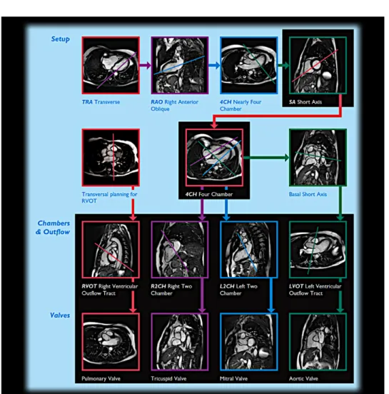
Figure 1.12: Different views of the heart acquired using Cardiac MRI and their relations with either a focus on the ventricles or the valves. Usual procedure to reconstruct a 3D cardiac sequence of the beating heart is to stack multiple multiple Short-Axis (SAX) 2D slices together.
heart using electrodes placed on the skin. The very small electrical changes on the skin coming from the depolarization during each heartbeat is detected, and similar electrical patterns correspond to similar points in the cardiac cycle. The R wave of the ECG (the most prominent wave of the QRS complex and the most easily de-tected) is used as a reference point corresponding to the end of the diastole and the start of the systole. Data acquisition is initiated after a given delay following the R wave and images are created from data collected over a series of cardiac cycles (R to R intervals) [Paul 2004]. ECG gating allows for stop motion imaging by acquiring data only during a specified portion of the cardiac cycle, typically during diastole when the heart is not moving. The patient is usually asked to hold his breath during imaging in order to alleviate respiratory motion. This can be difficult, especially for children. Therefore many artifacts such as slices misalignment are present after the
1.5. Manuscript Organization and Objectives 17
acquisition and they have to be corrected as a post-processing step.
MRI is widely used in hospitals and clinics for medical diagnosis, staging of disease and follow-up without exposing the body to ionizing radiation. MRI does not involve X-rays, as opposed to computed tomography (CT scan). While the hazards of X-rays are now well-controlled in most medical contexts, MRI can still be seen as superior to CT in this regard. Compared with CT, the defaults of MRI scans are: the acquisition takes more time, the machines are louder, and they require the subject to go into a narrow tube which can be a source of discomfort. In addition, people with medical implants or other non-removable metal inside the body may be unable to safely undergo an MRI examination.
1.4.3 Computed Tomography
Cardiac Computed Tomography [Schoenhagen 2005, Topol 2007] (also known as CT scan) is a non-invasive imaging modality that uses X-rays to take many detailed 2D pictures of the heart. Multiple X-rays images are combined to produce cross-sectional views of the body. Cardiac CT can be used to visualize the heart anatomy, coronary circulation, and great vessels (including the aorta, pulmonary veins, and arteries). In some cases, the patient is injected with an intravenous dye (iodine) before the acquisition. This contrast medium is a chemical substance that reveals what is happening inside the hollow parts of the body [Ropers 2003, Morin 2003] (such as the blood vessels, the stomach, bowel or even the fluid around the spinal cord) on the images.
The scan takes between 15 minutes and 1 hour to complete, including prepara-tion time. Similarly to MRI sequence of cardiac moprepara-tion, multiple temporal frames of CT images are acquired and ECG gated to reconstruct the whole sequence. CT images benefit from better spatial resolution (see Fig. 1.13) than MRI and echo images with isotropic voxel size in each direction. Therefore, there is no slice gap as with MRI images and no slice misalignment. With multi-slice CT scans
[Kachelrieß 2000,McCollough 1999,Pan 2004], up to 256 slices can be acquired at
the same time and the heart can be imaged during an entire cardiac cycle. But this higher quality comes at the cost of large amount of X-rays irradiation, which can be dangerous and carcinogenic [Donnelly 2001,Pearce 2012,Schulze 2014]. This is why CT acquisitions are rarely done with children and were not acquired for our project.
1.5
Manuscript Organization and Objectives
In this thesis we present a whole technical pipeline of cardiac motion analysis from the instant a patient enters the hospital to an image based group-wise statistical study of its cardiac motion. In the introduction, we have described the different dis-eases that will be the focus of this work. We presented the different cardiac imaging modalities which will be the source of our imaging data. The rest of the manuscript
Figure 1.13: 3D CT image with the 3 different views. Voxel resolution is better compared to MRI images and is isotropic in each direction.
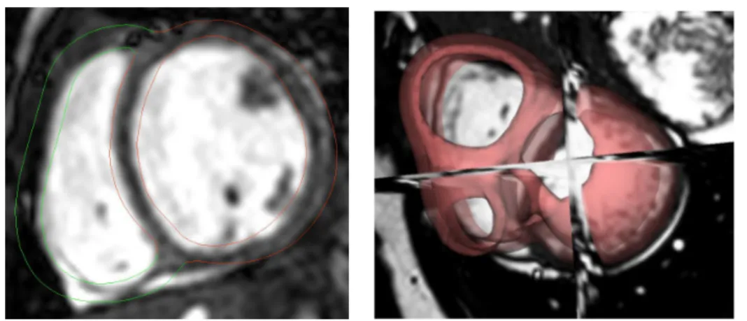
presents a whole process starting from the images to a statistical study of the car-diac motion with a focus on applications to clinical problems. The organization of the manuscript is schematically presented in Fig. 1.14. We now describe the 3 main parts of this manuscript, each of which will be driven by a key question.
Part - I
From Medical Images to 3D Shape
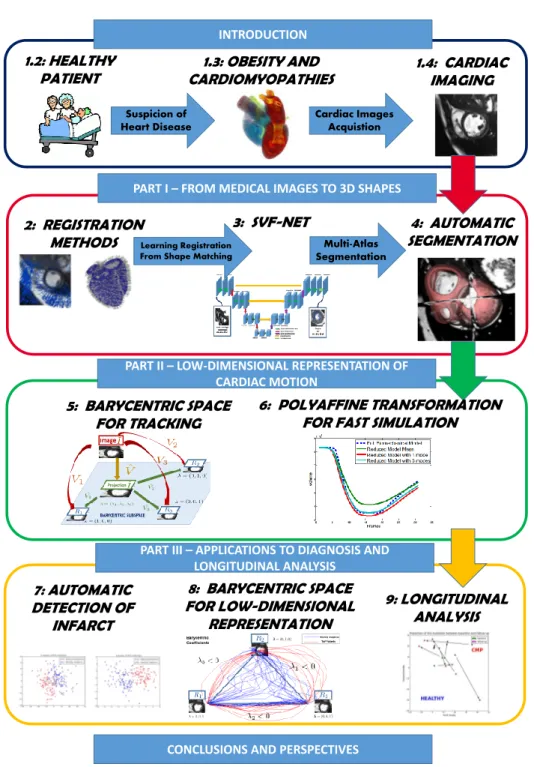
The accurate segmentation of the myocardium is of great importance in clin-ical practice. This is usually the first step of medclin-ical image analysis. The most commonly used clinical indices (e.g. the ejection fraction) to evaluate a cardiac dis-ease are derived from the information given by the segmentation of the myocardium during a cardiac motion. Therefore, clinicians often perform manual segmentation of the contours of the myocardium as the first step in the analysis of cardiac im-ages. However, considering the typical work-flow that clinicians are facing, manual segmentation is often too time-consuming and not a viable option. Furthermore, experts can have different opinions as to how to perform the segmentation (inclu-sion/exclusion of papillary muscles and trabeculations for example) leading to high inter-rater variability of manual segmentations. This makes the comparison between different patients not segmented by the same expert often difficult. This expert vari-ability can be decreased by developing automatic methods which require reduced or no user input. For all these reasons, there is a need to develop a completely automated, fast and robust segmentation method.
Going a little bit outside the direct clinical practice, most of the methods of cardiac function analysis require a segmentation of the myocardium. Cardiac mo-tion tracking algorithms, that will be the topic of Part II, usually require an initial segmentation of the myocardium at the first frame. This segmentation can be used to track the motion and monitor the main clinical indices during the cardiac cycle. It can also be used to give a local parametrization and estimation of the motion (with the AHA regions for example, as will be done in Chapter 7) for group-wise local comparison between different patients. Finally, bio-mechanical and electro-physiological simulations also rely on tetrahedral mesh segmentation of the whole
1.5. Manuscript Organization and Objectives 19
INTRODUCTION
1.2: HEALTHY
PATIENT CARDIOMYOPATHIES1.3: OBESITY AND
Suspicion of Heart Disease Cardiac Images Acquistion 1.4: CARDIAC IMAGING
PART I – FROM MEDICAL IMAGES TO 3D SHAPES
4: AUTOMATIC SEGMENTATION 2: REGISTRATION
METHODS
3: SVF-NET
PART II – LOW-DIMENSIONAL REPRESENTATION OF CARDIAC MOTION
Multi-Atlas Segmentation
Learning Registration From Shape Matching
5: BARYCENTRIC SPACE FOR TRACKING
6: POLYAFFINE TRANSFORMATION FOR FAST SIMULATION
PART III – APPLICATIONS TO DIAGNOSIS AND LONGITUDINAL ANALYSIS
CONCLUSIONS AND PERSPECTIVES
8: BARYCENTRIC SPACE FOR LOW-DIMENSIONAL REPRESENTATION 9: LONGITUDINAL ANALYSIS 7: AUTOMATIC DETECTION OF INFARCT
Figure 1.14: The global organization of the manuscript with a pipeline for cardiac motion analysis. First, from the images, a tetrahedral segmentation is extracted in Part I. Then, cardiac motion tracking is performed on the sequence of images in Part II. Finally, we show applications of this representation in diagnosis and longitudinal analysis in Part III.
myocardium. These simulations can be used for therapy planning and for quan-titative understanding of the functioning of the heart in healthy and pathological subjects.
This non-exhaustive list of applications of segmentation leads to our first objec-tive with the question driving part I of this manuscript:
• Can we develop a fast, automatic and accurate segmentation of the myocardium represented by tetrahedral mesh for shape analysis, cardiac motion tracking, and bio-mechanical simulations?
The main objective is to compute the segmentation of the myocardium. The method we use to get the segmentation rely heavily on registration methods. We actually show that the two main topic of medical imaging: registration and segmen-tation are intrinsically related and can be used together to perform one task or the other. In Chapter 2, we present the background of registration in medical imaging with the focus on two different algorithms that are used extensively in the remaining of the manuscript: Log-Demons for image registration and LDDM on Currents for shape registration. In Chapter 3, we use shape segmentation matching to train a predictive registration algorithm and we develop a new fast and robust algorithm for image registration. Finally, in Chapter4, we go the other way around and use image registration to compute shape segmentation by developing an automatic, robust and fast multi-atlas segmentation pipeline. Results show that our method has good ac-curacy and robustness while being faster than traditional multi-atlas segmentation algorithms.
The main contributions developed in this part are:
• A method for computing reference transformations between pair of images, using mesh segmentations which are registered in the space of currents. These transformations can be used efficiently to train a learning-based registration algorithm.
• A fully convolutional neural network for 3D registration prediction. Our ar-chitecture is able to detect global features and deformations that could not be detected with a sliding-window approach (for ex. [Yang 2016]). It also proves to be faster at testing time as only one pass of the whole image is required. • A fast and robust end-to-end framework for myocardium segmentation
lever-aging the speed of the SVF-net registration with a large number of atlases.
Part - II
Low-Dimensional Representation of Cardiac Motion
Part I was focused on the segmentation of the myocardium using the image of the first frame of the sequence (end-diastole). In this part, we develop methods to efficiently compute the motion of the heart during a cardiac cycle. The main clinical
1.5. Manuscript Organization and Objectives 21
application is to analyze the cardiac deformations at a local or global level. This can provide an estimate of the cardiac function efficiency and possibly indicators of heart disease. Tracking the tissue with physical markers is not directly possible in a clinical setting because it is invasive. Therefore, noninvasive methods relying on medical images have been developed in recent years to track automatically the myocardium.
The aim of this part can be summed up with the general question:
• Can we improve cardiac motion tracking algorithms in order to have a more accurate estimation of the main clinical indices and an efficient representa-tion?
In Chapter 5, we first give an overview of the traditional methods for cardiac motion tracking. Then, we propose an innovative approach to study the cardiac motion. Standard methods rely on the registration of each frame with respect to the first frame taken as a single reference. We challenge this methodology by consid-ering multiple references in the cardiac sequence. We build a Barycentric Subspace using these references and use it in the registration process as an additional a-priori to improve the tracking along the sequence. Results show that this innovative ap-proach leads to substantial improvement of the tracking accuracy at end-systole and the estimation of the ejection fraction. Then, in Chapter6, we present the polyaffine algorithm, a reduced-order model for tracking cardiac motion. This algorithm has the advantage not only to track the motion but also to give a representation with a low-dimensional number of parameters that have physiological meaning. We in-troduce local basis and reduced-order affine parameters and show that they give an efficient representation of the cardiac motion with better interpretability. We also use this representation to build a reduced-order model of the cardiac motion for fast simulation and personalization.
The main contributions developed in this part are:
• The introduction of a new method for dimension reduction and low-dimensional subspace analysis: Barycentric Subspace Analysis [Pennec 2015] in the context of medical images.
• The methods for computing the coordinates of an image within a Barycentric Subspace, for choosing the reference frames building the optimal subspace, and for reconstructing an image given the coordinates and the references.
• An extension of the polyaffine parameters framework with an additional pa-rameters reduction by keeping only the 6 most relevant papa-rameters and the use of this parametrization of the motion to build a very fast reduced-order model of the cardiac function.
Part - III
Previous parts were focused on the improvement of the computational methods of the heart in order to compute a robust, accurate and automatic low-dimensional representation of the cardiac motion. In this last part of the manuscript, we show how this representation can be used in several clinical problems to improve diag-nosis, prognosis and therapy planning. We have seen how cardiac motion tracking can provide crucial information such as the ejection fraction and strain values to the cardiologist. But these are simple numbers which do not encompass all the relevant information of the full deformation of the myocardium during the cycle. A representation of the motion that goes beyond the classical clinical parameters, while still represented by a low number of parameters, could be used efficiently to improve the diagnosis of a disease.
Once a patient is diagnosed and its present cardiac condition evaluated, a clin-ician would like to have information about the possible evolution of the disease in order to chose the most adapted therapeutic option. One possible way to provide this information is to perform a longitudinal analysis of the motion. This kind of study is particularly challenging since two time-dimensions have to be coupled: the time of the cardiac motion (heartbeat) and the evolution of this motion through different time points. Furthermore, to build such a model, one needs to gather multiple cardiac motions of the same patient over a large period of time (to detect measurable differences). Waiting for years to acquire the data makes it difficult to build large enough database, especially when working with children who tend to have a large opt-out rate of clinical studies. For these reasons, there has been very few studies on longitudinal analysis of cardiac motion, even though this has been a trending topic in brain analysis for many years.
This leads to the key question:
• Can we perform group-wise statistics on a low-dimensional representation of the cardiac motion to improve diagnosis, prognosis and therapy planning? This part is divided in 3 chapters focusing each on a different clinical question and applied each to a different population. In Chapter7, we classify two populations of healthy subjects and infarcted patients based on a prior reduction of dimension-ality of the motion using a reduced number of polyaffine parameters. We show that our low-dimensional representation allows for very good classification results using classical machine learning algorithms. Furthermore, we are able to quantify the importance of each of the parameters in the classification. These parameters represent clinical indices that can be understood by clinicians and provide insights into what is the main impact of an infarct on the motion. In Chapter 8, we use the Barycentric representation of the motion presented in Chapter 5as an efficient cardiac motion signature. We apply this representation to two populations, one of healthy subjects and one of patients with Tetralogy of Fallot (ToF). We show that the signature of each population presents significant differences, corresponding in particular to a longer systolic duration for the ToF population. Finally in Chapter
9, we perform two longitudinal analyses. The first analysis concerns a population of adolescents with cardiomyopathies. The evolution of their cardiac motion has
1.6. Publications 23
been studied for two time points spanning one year. The second analysis was an experimental study of the evolution of the cardiac motion in response of a clinical test with several acquisitions spanning 2 hours. Healthy subjects and obese patients followed this experimental protocol and we compare their respective responses.
The main contributions of this part are:
• The use of a low-dimensional representation with polyaffine transformations to classify a population of hearts with myocardial infarction. The method favor-ably compares with state-of-the-art in term of classification accuracy (AUC, sensitivity and accuracy) inline with the best competitive methods.
• The group-wise analysis of the features extracted from the projection and its application in the context of the study of Tetralogy of Fallot.
• The longitudinal analysis of the cardiac motion for two medical conditions: cardiomyopathies and obesity using a low-dimensional representation based on polyaffine transformations.
Perspectives
In the last chapter of this thesis, we conclude by summarizing the key con-tributions of this work. We also recap the key objectives and goals of the work. Finally, we propose some perspectives for future works in order to extend the meth-ods which are described in this thesis. Some of these suggestions are already under development in collaboration with other PhD students.
1.6
Publications
The contributions described led to 3 journal papers (1 accepted, 1 accepted subject to minor revisions and 1 in preparation) and 6 conference papers.
1.6.1 Journal Articles
• M.-M. Rohé, M. Sermesant, and X. Pennec. Low-Dimensional Represen-tation of Cardiac Motion Using Baryncetric Subspaces: a New Group-Wise Paradigm for Estimation, Analysis, and Reconstruction. Accepted subject to minor revisions at Medical Image Analysis Chapter 5and Chapter 8.
• M.-M. Rohé, M. Sermesant, and X. Pennec. SVF-Net: Learning Deformable Image Registration Using Intensity and Geometric Features for the Automatic Segmentation of the Myocardium. In preparation for submission to a journal. Chapter3 and Chapter 4.
• A. Suinesiaputra, M.-M. Rohé, et al. Statistical shape modeling of the left ventricle: myocardial infarct classification challenge. IEEE Journal of Biomedical and Health Informatics, 2017.
1.6.2 Selective Peer-Reviewed Conference Paper
• M.-M. Rohé, M. Sermesant, and X. Pennec. Barycentric Subspace Analysis: a new Symmetric Group-wise Paradigm for Cardiac Motion Tracking. In MIC-CAI 2016 - the 19th International Conference on Medical Image Computing and Computer Assisted Intervention (oral presentation).
• M.-M. Rohé, N. Duchateau, M. Sermesant, and X. Pennec. Combination of Polyaffine Transformations and Supervised Learning for the Automatic Diag-nosis of LV Infarct. In Statistical Atlases and Computational Modeling of the Heart (STACOM 2015), Munich, Germany, 2015. Chapter 7.
• M.-M. Rohé, R. Molléro, M. Sermesant, and X. Pennec. Highly Reduced Model of the Cardiac Function for Fast Simulation. In IEEE - IVMSP Work-shop 2016, Image, Video, and Multidimensional Signal Processing WorkWork-shop (IVMSP), 2016 IEEE 12th, Bordeaux, France. Chapter 6.
• M.-M. Rohé, M. Datar, T. Heimann, M. Sermesant and X. Pennec. SVF-Net: Learning Deformable Image Registration Using Shape Matching. In MICCAI 2017 - the 20th International Conference on Medical Image Comput-ing and Computer Assisted Intervention. Chapter3.
• R. Molléro, D. Neumann, M.-M. Rohé, M. Datar, H. Lombaert, N. Ayache, D. Comaniciu, O. Ecabert, M. Chinali, G. Rinelli, X. Pennec, M. Sermesant, and T. Mansi. Propagation of Myocardial Fibre Architecture Uncertainty on Electromechanical Model Parameter Estimation: A Case Study. In Functional Imaging and Modeling of the Heart, FIMH 2015, Maastricht.
• S. Jia, C. Camaioni, M.-M. Rohé, P. Jaïs, X. Pennec, and M. Sermesant. Prediction of Post-Ablation Outcome in Atrial Fibrillation Using Shape Pa-rameterization and Partial Least Squares Regression. In Functional Imaging and Modeling of the Heart, FIMH 2017, Toronto.
1.6.3 Awards
• A Student Travel Award was awarded at the conference MICCAI 2016 for the paper: Barycentric Subspace Analysis: a new Symmetric Group-wise Paradigm for Cardiac Motion Tracking.
Part I
FROM MEDICAL IMAGES TO
3D SHAPE
Chapter 2
Registration in Medical Imaging
Contents
2.1 Introduction . . . 27
2.2 Registration Algorithms . . . 28
2.2.1 Similarity Measures . . . 30
2.2.2 Transformation Spaces and Regularization. . . 31
2.2.3 Optimization Methods . . . 32
2.3 LCC Log-Domain Diffeomorphic Demons . . . 33
2.3.1 Optimization Method: Alternate Optimization . . . 33
2.3.2 Deformation Parametrization: Stationary Velocity Field . . . 34
2.3.3 Similarity Metric: Local Correlation Coefficient . . . 35
2.4 Framework of Currents for Shape Registration . . . 36
2.4.1 The Formalism of Currents for Surface Representation . . . . 38
2.4.2 Surface Registration Using Currents and LDDMM . . . 40
This chapter introduces the concept of registration which will be extensively used in the rest of this manuscript in different applications related to cardiac images analysis. The process of registration consists in finding correspondences between a pair (or a group) of images or meshes. Along with the segmentation of organs, it has been one of the principal challenges in medical imaging and a very important tool for analysis. We first introduce the concept, the applications in medical imaging and the mathematical formalism. Then we go deeper and present two specific different registration algorithms which we use in the rest of this work. This is not meant to be a full review of registration methods (a comprehensive review can be found for instance in [Sotiras 2013] and more recent advances in the field are discussed
in [Schnabel 2016]) but an overview of the principal possible choices in order to
position the methods that we use with respect to the state-of-the-art.
2.1
Introduction
Registration is a key instrument in computational anatomy and has gained an increasing importance in the past years. Many applications of registration have been developed and we give here some of the main ones. The registrations of images between different subjects (inter-patient registration) can be used to find