HAL Id: cea-02164621
https://hal-cea.archives-ouvertes.fr/cea-02164621
Submitted on 17 Dec 2020
HAL is a multi-disciplinary open access
archive for the deposit and dissemination of
sci-entific research documents, whether they are
pub-lished or not. The documents may come from
teaching and research institutions in France or
abroad, or from public or private research centers.
L’archive ouverte pluridisciplinaire HAL, est
destinée au dépôt et à la diffusion de documents
scientifiques de niveau recherche, publiés ou non,
émanant des établissements d’enseignement et de
recherche français ou étrangers, des laboratoires
publics ou privés.
Deciphering the uranium target proteins in human
dopaminergic SH-SY5Y cells
Claude Vidaud, Mélanie Robert, Eduardo Paredes, Richard Ortega, Emilie
Avazeri, Lun Jing, Jean-Marie Guigonis, Carole Bresson, Veronique Malard
To cite this version:
Claude Vidaud, Mélanie Robert, Eduardo Paredes, Richard Ortega, Emilie Avazeri, et al.. Deciphering
the uranium target proteins in human dopaminergic SH-SY5Y cells. Archives of Toxicology, Springer
Verlag, 2019, 93 (8), pp.2141-2154. �10.1007/s00204-019-02497-4�. �cea-02164621�
HAL Id: hal-03066498
https://hal.archives-ouvertes.fr/hal-03066498
Submitted on 15 Dec 2020
HAL is a multi-disciplinary open access
archive for the deposit and dissemination of
sci-entific research documents, whether they are
pub-lished or not. The documents may come from
teaching and research institutions in France or
abroad, or from public or private research centers.
L’archive ouverte pluridisciplinaire HAL, est
destinée au dépôt et à la diffusion de documents
scientifiques de niveau recherche, publiés ou non,
émanant des établissements d’enseignement et de
recherche français ou étrangers, des laboratoires
publics ou privés.
Deciphering the uranium target proteins in human
dopaminergic SH-SY5Y cells
Claude Vidaud, Mélanie Robert, Eduardo Paredes, Richard Ortega, Emilie
Avazeri, Lun Jing, Jean-Marie Guigonis, Carole Bresson, Veronique Malard
To cite this version:
Claude Vidaud, Mélanie Robert, Eduardo Paredes, Richard Ortega, Emilie Avazeri, et al.. Deciphering
the uranium target proteins in human dopaminergic SH-SY5Y cells. Archives of Toxicology, Springer
Verlag, 2019, 93 (8), pp.2141-2154. �10.1007/s00204-019-02497-4�. �hal-03066498�
DECIPHERING THE URANIUM TARGET PROTEINS IN HUMAN
DOPAMINERGIC SH-SY5Y CELLS
Claude Vidaud1*, Mélanie Robert1, Eduardo Paredes2, Richard Ortega3,4, Emilie Avazeri1, Lun Jing5, Jean-Marie
Guigonis5, Carole Bresson2 and Véronique Malard6*.
1 CEA, DRF, Institute of Biosciences and Biotechnologies of Aix Marseille (BIAM), Bagnols-sur-Cèze, France. 2 Den – Service d’Etudes Analytiques et de Réactivité des Surfaces (SEARS), CEA, Université Paris-Saclay,
F-91191, Gif sur Yvette, France.
3 Chemical Imaging and Speciation, CENBG, University of Bordeaux, UMR 5797, 33175 Gradignan, France 4 CNRS, IN2P3, CENBG, UMR 5797, 33175 Gradignan, France
5 Laboratory Transporter in Imaging and Radiotherapy in Oncology (TIRO), UMR E4320 CEA - Université Nice
Sophia Antipolis, 06107, Nice, France
6 Laboratory of Protein-Metal Interactions (LIPM), Institute of Biosciences and Biotechnologies of Aix Marseille
(BIAM), UMR7265 CEA – CNRS - Aix Marseille Univ, CEA Cadarache, F-13108
*e-mail:
cl_vidaud@hotmail.fr; ORCID ID: 0000-0003-0165-2732, +33-6-88-18-76-61
veronique.malard@cea.fr; ORCID ID: 0000-0002-0020-0518, +33-4-42-25-42-77 The authors declare that they have no conflict of interest.
Acknowledgements
We acknowledge the Transversal Toxicology Program run by the Direction de la Recherche Fondamentale (DRF) of the Commissariat à l’énergie atomique et aux énergies alternatives (CEA) and the financial support from this program. E.P. also thanks the CEA Enhanced Eurotalents program, co-funded by the European Commission through the Marie Sklodowska-Curie COFUND program under the 7th Framework Program for Research and Technological Development.
This work was also partially funded by the CNRS Interdisciplinary Mission through the PEPS (Projet Exploratoire Premier Soutien) Faidora program (Faibles Doses, Risques, Alertes).
Abstract
Uranium (U) is the heaviest naturally occurring element ubiquitously present in the Earth's crust. Human exposure to low levels of U is therefore unavoidable. Recently, several studies have clearly pointed out that the brain is a sensitive target for U, but the mechanisms leading to the observed neurological alterations are not fully known.
To deepen our knowledge of the biochemical disturbances resulting from U(VI) toxicity in neuronal cells, two complementary strategies were set up to identify the proteins that selectively bind U(VI) in human dopaminergic SH-SY5Y cells. The first strategy relies on the selective capture of proteins capable of binding U(VI), using immobilized metal affinity chromatography, and starting from lysates of cells grown in a U(VI)-free medium. The second strategy is based on the separation of U-enriched protein fractions by size-exclusion chromatography, starting from lysates of U(VI)-exposed cells.
High-resolution mass spectrometry helped us to highlight 269 common proteins identified as the urano-proteome. They were further analyzed to characterize their cellular localization and biological functions. Four canonical pathways, related to the protein ubiquitination system, gluconeogenesis, glycolysis, and the actin cytoskeleton proteins, were particularly emphasized due to their high content of U(VI)-bound proteins. A semi-quantification was performed to concentrate on the ten most abundant proteins, whose physico-chemical characteristics were studied in particular depth. The selective interaction of U(VI) with these proteins is an initial element of proof of the possible metabolic effects of U(VI) on neuronal cells at the molecular level.
Key words
1. Introduction
The heaviest naturally occurring element, uranium (U), is inherently present in the environment or results from anthropic activities. Natural U is poorly radioactive, and its chemical toxicity prevails over its radiotoxicity. Under atmospheric conditions and in most environmental systems, the most stable form of U is the di-oxo cation (U(VI)O2)2+ (uranyl), with U in the +VI oxidation state. It is simply referred to as “U(VI)” in the remainder of
this article.
In the event of exposure, U reaches first the blood and then its main target organs, namely the kidneys and the bones, where it accumulates for years while exerting its adverse effects. Less severe effects are, however, observed in the liver, the lungs, and the reproductive and nervous systems (ATSDR 2013; UNSCEAR 2017). Some epidemiological studies have shown behavioral disorders in humans, as well as a correlation between urinary U levels and impaired cognitive test results in veterans of the Gulf War (McDiarmid et al. 2000). Over the last few years, several studies have clearly pointed out that the brain is a sensitive target for U (Dinocourt et al. 2015; UNSCEAR 2017). Exposure to U(VI) induces neurobehavioral changes such as increased locomotor activity, sleep-wake cycle disturbances, decreased memory, and increased anxiety in animal models. The mechanisms by which these neurological alterations are induced are not fully known at the molecular level. Changes in the levels of neurotransmitters and their metabolites involved in acetylcholine and dopaminergic pathways have been observed in animal brains after exposure to U(VI) (Barber et al. 2007; Bensoussan et al. 2009; Bussy et al. 2006). We have recently quantified the expression of dopamine-related genes in the SH-SY5Y cell line, a model of dopaminergic neuronal cells, after their exposure to concentrations of U(VI) ranging from non-toxic to sub-toxic (Carmona et al. 2018). The expression of the monoamine oxidase B (MAO-B) gene was found to be statistically significantly decreased, while other dopamine-related gene expressions were not modified. These original results suggest that the inhibition of dopamine catabolism, but also that of other MAO-B substrates, could constitute selective effects of uranium neurotoxicity. MAO-By exposing this cellular model to a non-toxic U(VI) concentration, we have also revealed the isotopic fractionation of U during its uptake by the cells, thereby suggesting the existence of a high-affinity transporter protein responsible for U(VI) uptake (Paredes et al. 2018; Paredes et al. 2016).
In order to improve our general understanding of the metabolic disturbances resulting from U(VI) toxicity in neuronal cells, additional information, such as the identification of the proteins that selectively bind U(VI) in cellulo (i.e., the “urano-proteome”), is needed. This knowledge is also of key importance for describing the intracellular chemical species of U(VI), as well as its speciation, because the mechanisms of uptake, metabolism, accumulation, toxicity, and excretion in which an element is involved are highly dependent on the corresponding chemical species and their localization (Michalke et al. 2018; Templeton and Fujishiro 2017). Multidimensional approaches combining chromatographic methods and associated with proteomic studies and metal quantification are powerful methods for deciphering the metalloproteome (Bishop et al. 2018; Szpunar and Lobinski 2002). That said, given the low concentration and complex speciation of metals in biological systems, the screening of their target proteins still remains a challenge, in particular for the actinides, such as U(VI), because of their non-covalent interactions with proteins. Furthermore, identifying the target proteins within complex protein mixtures from small bio-fluid samples or cell lysates requires selective, low-consumption separation strategies.
The most relevant way to identify the target proteins of U(VI) is to selectively capture the proteins capable of binding this cation. Chromatographic processes using molecular recognition provide a rational basis for this screening. Such processes have the advantage of providing a relative affinity scale, thereby making it possible to discriminate potential targets from among numerous proteins. Immobilized metal affinity chromatography (IMAC) is a widely accepted technique based on this molecular recognition that is used for enriching natural and recombinant metal targets (Aryal et al. 2012; Porath et al. 1975). A proteomic strategy based on IMAC has previously been implemented to capture and identify proteins with a high affinity for U(VI), taking into account the physico-chemical properties of U(VI) and the small amounts of crude human protein extracts generally available for such experiments. To do this, a dedicated U(VI)-loaded support was first specifically designed, and then an IMAC strategy was developed and validated (Basset et al. 2008). This method was successfully applied
to kidney cell extracts, leading to the identification of U(VI)-affine proteins (Dedieu et al. 2009). In this study, this IMAC method was used to isolate proteins capable of selectively binding U(VI) from SH-SY5Y cell lysates. Another way to identify the target proteins is to isolate the U(VI)-protein complexes formed in cellulo in response to U(VI) exposure. To achieve this goal, size-exclusion chromatography (SEC), known for its mild mobile phase conditions and separation process, was selected to fractionate the mixture of free proteins and U(VI)-protein complexes from the extracts of SH-SY5Y cells exposed to U(VI). Size-exclusion chromatography was coupled to quadrupole inductively coupled plasma mass spectrometry (q-ICP-MS) to identify the U-enriched protein fractions and to determine the relative proportion of U in these fractions.
The originality of the present approach lies in the combination of these two chromatographic strategies to isolate and identify the urano-proteome. The SH-SY5Y cells were first differentiated (Presgraves et al. 2004) to acquire a neuronal dopaminergic phenotype. Next, SEC and IMAC were applied to the protein extracts from these cells grown respectively with and without U(VI). We were particularly interested in the molecular effects induced by continuous rather than acute U(VI) exposure. So, to prevent any metabolic disturbance due to toxicity, the cells were submitted to a seven-day period of continuous exposure to a non-toxic U(VI) concentration (10 µM) (Carmona et al. 2018). The proteins from the U-enriched SEC fractions and those selectively captured by IMAC were identified thanks to high-resolution mass spectrometry. The proteins belonging to both populations constitute a part of the urano-proteome, giving more accurate information on the pathways affected in response to a non-toxic U(VI) exposure. The ten most abundant U(VI) proteins were characterized with particular attention.
2. Materials and methods
All experiments were conducted at room temperature unless otherwise stated. Ultra-pure water (18 MΩ cm at 25°C) was provided by a Milli-Q (Millipore, Billerica, MA, USA) installation. All of the buffers were carefully filtered (0.22 µm) and degassed before being used for the chromatographic processes.
2.1. Materials
The main reagents and salts were supplied by Sigma–Aldrich Fluka (Saint-Quentin-Fallavier, France).
Protein absorbance was measured on a Varian Cary® 50 UV-Vis spectrophotometer (Agilent, Les Ulis, France). Spectra/Por® (Sigma–Aldrich Fluka) membranes (6000–8000) were used for the dialysis. The microcon® centrifugal filters were from Millipore (Molsheim, France).
The HiTrap® DEAE Fast Flow phase (1 mL) was from GE Healthcare (Vélizy, France). The Duolite™ C467 resin was provided by Rohm & Haas (Philadelphia, PA, USA) and was initially used in its sodium salt form. Eagle’s minimum essential medium (EMEM, ATCC, 30-2003), F12 medium (Life Technologies, 21765-029), fetal bovine serum (FBS, ATCC, 30-2020), and penicillin/streptomycin (Gibco-Thermo Fisher Scientific, 15070-063) solutions were used to prepare the culture medium for cell growth and exposure experiments. The culture medium consisted of an equal mix of EMEM and F12 media supplemented with 10% FBS and 1% penicillin/streptomycin. TrypLE Express 1X/EDTA (Gibco-Thermo Fisher Scientific, 12605-010) or trypsin 0.25% EDTA-free (Life Technologies, 25050-014) was used for the trypsinization of the cells. Phosphate-buffered saline (PBS, pH 7.4) formulated without CaCl2 or MgCl2 (Gibco 10010-015) was used to wash the cells
after trypsinization. Retinoic acid (RA) and 12-O-tetradecanoylphorbol-13-acetate (TPA), used for cell differentiation, were purchased from Sigma-Aldrich (R2625 and P8139, respectively). A retinoic acid solution (3 mg mL-1) was prepared in sterile dimethyl sulfoxide (DMSO, Sigma-Aldrich) under nitrogen atmosphere in
opaque tubes and stored at -80°C. Also, TPA was re-suspended at 3 mg mL-1 in sterile DMSO, and the resulting
solution was stored at -20°C. Plasma Pure Plus 67-70% HNO3 (SCP Science, Baie-d'Urfé, Canada) was used to
prepare the U(VI) stock solution. NaHCO3 analytical reagent (Normapur grade), anhydrous Na2CO3 (99.95%
(ultrapure grade ≥99.9%, Sigma-Aldrich) were used to prepare the buffer solution. Ammonium acetate (Normapur grade, VWR Prolabo) was used to prepare the mobile phase for size-exclusion chromatography (SEC) experiments.
2.2 U(VI) solution preparation
Natural uranium powder (U3O8) was produced in-house, and then dissolved in 0.5 M ultrapure HNO3 to prepare
a U(VI) stock solution at 151 mM.
For the SEC experiments, an intermediate U(VI) solution (30 mM, pH = 8-8.5) was extemporaneously prepared (1:5 dilution ratio) by dropwise addition of the U(VI) stock solution into a buffer made of 0.1 M NaHCO3, 0.1 M
Na2CO3, 0.15 M NaCl, and 0.05 M TRIS, in order to prevent U(VI) precipitation. The 10 µM exposure solution
was then obtained by extemporaneous dilution of the intermediate solution in the culture medium.
For the IMAC experiments, the U(VI)-loaded beads were prepared according to operating conditions published elsewhere (Basset et al. 2008). Briefly, the 151 mM U(VI) stock solution was diluted in 50 mM ammonium acetate buffer (pH 4.0) to ensure 0.7 mM U(VI) per mL of beads. After the beads were preconditioned in 50 mM ammonium acetate buffer at pH 4.0, they were left in contact with this U(VI) solution overnight at 4°C under gentle stirring, and then stored at 4°C until use.
2.3. SH-SY5Y cell culture, differentiation, and exposure to U(VI)
Commercial human SH-SY5Y (ATCC, CRL-2266, batch 59740436) cells were differentiated into neuron-like cells prior to U(VI) exposure experiments. Cells were grown in 175 cm² flasks at 37°C in 5% CO2 for ten days.
The culture medium was replaced with fresh medium every three or four days. Afterward, the cells were passaged by trypsinization using TrypLE Express 1X/EDTA and then seeded at 25,000 cells cm-2. They were
differentiated into neuron-like cells according to the method described by Presgraves et al. (Presgraves et al. 2004). Briefly, the culture medium was replaced with fresh medium containing 10 µM RA, and the cells were incubated for 3.5 days. The culture medium was then replaced with fresh medium containing 80 nM TPA and left for an additional 3.5 days to obtain differentiated cells. The dopaminergic phenotype in SH-SY5Y human cells was verified by phase-contrast microscopy to check for neural differentiation and by western blot analysis for tyrosine hydroxylase expression (Carmona et al. 2018).
For the IMAC strategy, the cells were cultured for seven days in U(VI)-free medium.
For the SEC strategy, the cells were exposed for seven days to a freshly prepared 10 µM U(VI) solution as described above. This U(VI) solution was replaced once three days after exposure began. The cells were then trypsinized using an EDTA-free trypsin, collected, and washed twice with PBS. The cell pellets were stored at −80 °C until use.
2.4. Soluble protein extraction and sample preparation for chromatography
The cells were suspended at 30x106 cells per mL in 0.02 M TRIS pH 7.5 containing anti-proteases (Roche
cocktail, EDTA-free) and 0.002 M spermine base (Sigma). The cells were disrupted by one shot at 1000 bar using a cell disruption system (Constant Systems, Northants, UK), then incubated for 30 min at 4°C. The extracts were then ultra-centrifuged (1 h at 100 000 g) and the supernatants collected. Aliquots were stored at -20 °C until use.
2.5. Ion exchange and IMAC
2.5.1 Ion exchange
A first ion exchange chromatography purification step (Dedieu et al. 2009) was performed with an Åkta Purifier 10 System (GE Healthcare, Uppsala, Sweden) equipped with on-line UV detection. Protein samples from cell extracts were dialyzed (overnight at 4°C) against 0.02 M HEPES buffer. Because of their tendency to precipitate in this buffer regardless of pH (8.0 or 7.4), the samples were ultra-filtered (Microcon, 3000 Da, Millipore) for 10 min at 10 000 g before use. The aliquots from these protein samples (2 mL at ~660 µg mL-1) were injected onto a
1 mL HiTrap DEAE Fast Flow column (0.2 mL min-1 flow rate). Reproducible processes led to similar
chromatograms regardless of pH (8.0 or 7.4), and pH 7.4 was selected. The flow-through fraction (A1) was collected. Then, the ionic strength was linearly increased up to 0.5 M NaCl, leading to the collection of two other
populations (A2 and A3). No protein was then eluted with buffer above 0.5 M and up to 1 M NaCl. The UV absorbance ( = 260 and 280 nm) and the protein content (BCA assay) were checked for A1, A2 and A3.
2.5.2. Protein capture by IMAC
U(VI)-free and U(VI)-loaded Duolite C467 beads were prepared as previously described (Basset et al. 2008). The batch conditions were chosen because of the small bead volume used. Before each experiment, 50 µL of U(VI)-free and U(VI)-loaded beads were conditioned by rinsing six times with 0.05 M HEPES, 0.5 M NaCl pH 7.4 buffer plus 0.05% Brij as surfactant (binding buffer). A high NaCl concentration prevents ion exchange with U(VI)-loaded beads, and the Brij surfactant enables or strongly limits hydrophobic interactions with the support (Basset et al. 2008; Dedieu et al. 2009).
In parallel, the three fractions collected (A1, A2, A3) from the ion exchange chromatography were first extensively dialyzed overnight against the binding buffer. Aliquots of these fractions were then adjusted to a protein concentration around 60 µg.mL-1, and then 0.2 M NaHCO
3 (5 µL per mL) was added. A sample of each
fraction (1 mL) was placed in contact with 50 µL of the conditioned U(VI)-loaded and U(VI)-free beads (room temperature, gentle stirring for 60 min). The supernatants were discarded. The U(VI)-loaded and U(VI)-free beads were washed seven times with 1 mL of the binding buffer and dialyzed overnight at 4°C against the same buffer to ensure the elimination of all proteins non-specifically bound to the beads. Then, the proteins specifically bound to the U(VI)-loaded and U(VI)-free beads (control fractions) were recovered separately by bringing the beads into contact with 500 µL of a 0.2 M sodium carbonate solution for 90 min. The beads were discarded, and the protein solutions were desalted in ammonium carbonate by eight successive concentration-dilution steps in micro-concentrator systems at 8°C and 13 000 g (Microcon, 3000 Da, Millipore) to reach volumes of ~90-120 µL. The proteins potentially found in the control fractions reflect non-specific interactions with the support and were not considered further for the proteomic study.
2.6. Size-exclusion chromatography (SEC) coupled to inductively coupled plasma mass spectrometry (ICP-MS)
A Smartline chromatographic system (KNAUER, Berlin, Germany) was used. The column was a 4.6 x 150 mm BEH 200 (Waters, Milford, USA) with 1.7 µm particle size and 200 Å pore size. The mobile phase was made of 100 mM ammonium acetate in ultrapure water, and the flow rate was 0.3 mL.min-1 (dual Pump 1000). To
estimate the molecular weight range of the eluted populations of proteins, the column was calibrated before use using 100 ppm gel filtration standard proteins (Bio-Rad, Marnes-la-Coquette, France) : thyroglobulin (≈ 670 kDa), ferritin (≈ 440 kDa), anti-IgE (≈ 150 kDa), ovotransferrin (≈ 75 kDa), ovalbumin (≈ 43 kDa), and aprotinin (≈ 6.5 kDa).
Cell lysates (20 µL) were manually injected into the column using a Rheodyne injector valve (model 9725), with monitoring of the UV signal at = 280 nm (Smartline PDA detector 2800). For the detection of the protein fractions containing uranium, the UV detector was connected in series to a quadrupole ICP-MS (q-ICP-MS, X7, Thermo Fisher Scientific) using PEEK tubing (i.d. 125 µm), with the 238U+ signal being monitored. The
integration time per chromatographic point with q-ICP-MS was 10 ms, and approximately 5800 points were acquired for a run time of 10 min.
The three main protein fractions enriched in U(VI) (denoted F1, F2, F3) were identified and collected separately.
2.7. High-resolution electrospray ionization mass spectrometry (HR ESI-MS)
Proteins were first extracted using trichloroacetic acid (TCA, final concentration 10%). Pellets were mixed with 30 µL of Laemmli LDS buffer (Invitrogen). Samples were heated at 99°C for 5 min and loaded (30 µL per well) onto a NuPAGE 4-12% Bis-Tris gel (Invitrogen). They were subjected to a short denaturing electrophoresis migration (5 min) at 200 V in MES SDS 1× running buffer. The polyacrylamide band containing the entire LDS-soluble proteome was processed for an in-gel trypsin digestion using sequencing-grade trypsin (Promega, Charbonnières-les-Bains, France) and ProteaseMAX Surfactant (Promega) at 0.01%.
The resulting peptides (10 μL) were analyzed using either an ESI-Q Exactive HF mass spectrometer (Thermo Fisher Scientific) incorporating an ultra-high-field Orbitrap analyzer and coupled to an Ultimate 3000 RSL Nano LC System (Dionex-LC Packings) for IMAC fractions, or an Ultimate 3000 RSL Capillary LC System (Thermo Fisher Scientific) for SEC fractions.
For nano-liquid chromatography, the samples were loaded and desalted on-line on a reversed-phase Acclaim PepMap100 C18 micro precolumn (5 μm, 100 Å, 300 μm internal diameter x 5 mm, Thermo Fisher Scientific) and separated on an Acclaim PepMap100 C18 nano column (3 μm, 100 Å, 75 μm internal diameter x 50 cm, Thermo Fisher Scientific) at a 0.3 μL/min flow rate for 60 min with a 2.5-40% gradient of solvent B (80% acetonitrile, 20% water, 0.1% formic acid) against solvent A (0.1% formic acid, 99.9% water). A top20 data-dependent method was used for MS/MS spectrum acquisition. Full-scan mass spectra were recorded from 350 to 1500 m/z with an automatic gain control target set at 3x10⁶ ions and a resolution of 60 000. Each MS/MS scan
was initiated at a resolution of 15 000 for ions with potential charge of 2+ and 3+ and with a dynamic exclusion of 10 s. The MS/MS spectra were recorded with an automatic gain control target set at 1x10⁵ ions.
For capillary liquid chromatography, the system was set up for pre-concentration mode using a 300 μm x 5 mm trap column in back-flush configuration at 40°C (P/N 6720.0315). An EASY-Spray 15 cm x 150 μm column (P/N ES806) was connected to the system and coupled to the system with an EASY-Spray source (P/N ES081) operating at 40°C. The flow rate was 1.2 μL/min with a 5-45% gradient of solvent B (80% acetonitrile, 20% water, 0.1% formic acid) against solvent A (0.1% formic acid, 99.9% water) for 180 min. A top15 data-dependent method was used for MS/MS spectrum acquisition. Full-scan mass spectra were registered from 350 to 1500 m/z with an automatic gain control target set at 3x10⁶ ions and a resolution of 70 000. Each MS/MS scan
was initiated at a resolution of 17 500 for ions with potential charge of 2+, 3+, and 4+ and with a dynamic exclusion of 20 s. The MS/MS spectra were recorded with an automatic gain control target set at 5x10⁴ ions.
2.8. Proteomic data analysis
All MS raw data files were analyzed by Proteome Discoverer software 2.1.1.21 (Thermo Fisher Scientific, France) using the Sequest HT search engine against a database of protein sequences (UniProt database release 2015_2). The XCorr confidence threshold was maintained above 0.7 for all parameters. Precursor mass tolerance was set to 10 ppm, and fragment ion tolerance was 0.02 Da, with permission for two missed cleavages in the trypsin digests. A decoy database search strategy was also used to estimate the false discovery rate (FDR) for ensuring the reliability of the proteins identified: a 1% target FDR was the strict criterion and a 5% target FDR was the relaxed criterion using Percolator; also, at least two peptides were required to match a protein entry in view of its identification. The strict maximum parsimony principle was applied, and only peptide spectra with at least medium confidence were considered for protein grouping. Carbamidomethylation on cysteine was set as a static modification and oxidation of methionine as a dynamic modification. For relative quantitation, both unique and razor peptides were considered a highly confident identification and used for quantification. Reporter ion abundances were corrected for isotopic impurities based on the manufacturer’s data sheets. Signal-to-noise ratio (S/N) values were used to represent the reporter ion abundance and an average reporter S/N threshold of 10 and above were required for quantitation spectra. The S/N values of the peptides, which were summed from the S/N values of the identified peptide sequences (PSMs), represent the abundance of the proteins. The number of PSM's is the total number of identified peptide spectra matched for the protein.
The number of peptides per protein was determined for each sample. The relative quantitative proteomic analysis was calculated according to the method of normalized spectral abundance factors (NSAF) (Zybailov et al. 2006). The normalized spectral abundance factor is a ratio calculated by normalizing the peptide numbers obtained for a protein (i) by the respective molecular weight (MW) as follows:
2.9 Integrative biological analysis
The lists of proteins of interest obtained from the two analytical strategies were concatenated and the duplicates removed. A Venn diagram was built in order to determine the population of proteins common to both pools (urano-proteome).
The list of the common proteins obtained after Venn analysis was uploaded into Ingenuity Pathway Analysis (IPA) software (QIAGEN) for biological analysis. This data set was processed to investigate gene functional annotation and distribution into known cell functions and canonical pathways, according to the IPA knowledge database. Associations with known canonical pathways were expressed by ratios (in percentage), meaning the number of proteins that belong to a canonicalpathway divided by the total number of known proteins mapping this pathway. The significance of highlighted functions and pathways was calculated using a Fisher's exact test with a p-value set at 0.05.
3. Results
3.1 Study design
We chose two complementary analytical strategies to collect and identify the proteins capable of binding U(VI) in cellulo. Human dopaminergic SH-SY5Y cells were cultured in a U(VI)-free medium or exposed to a non-toxic concentration (10 µM) of U(VI) for seven days (Carmona et al. 2018). The IMAC approach relies on selective protein capture from lysates of cells grown in a free medium, leading to the direct identification of U(VI)-affine proteins. The SEC strategy consists in a fractionation of lysates of cells exposed to U(VI) to collect protein fractions enriched in U. The protein fractions obtained from SEC experiments should contain both free proteins and U(VI)-protein complexes. The proteins common to both strategies, identified using nano-liquid chromatography coupled to high-resolution electrospray ionization tandem mass spectrometry (nano-LC–HR ESI-MS/MS), are referred to herein as the urano-proteome. The experimental design is depicted in Figure 1.
3.2 Proteins captured by IMAC on U(VI)-loaded and U(VI)-free beads
An initial ion exchange chromatography step was performed to fractionate the protein samples and eliminate some residual DNA (Dedieu et al. 2009). When the protein sample (2 mL, 660 µg mL-1) was injected onto the
column, a flow-through fraction (denoted A1) was first eluted, followed next by a large A2 fraction in the first third of the NaCl gradient and finally by a narrow A3 fraction in the last third of the NaCl gradient. The protein contents in these A1, A2 and A3 fractions were in the range of around 180, 718, and 110 µg, respectively, and the protein recovery was around 76%. Half of each of these three fractions was then placed in contact with the U(VI)-loaded beads, while in parallel the other half was placed in contact with the U(VI)-free beads to serve as the control. All of the specifically captured proteins obtained from these six IMAC experiments were submitted to nano-LC–HR ESI-MS/MS analysis for identification. The few proteins eluted from U(VI)-free beads but also present in the populations eluted from U(VI)-loaded beads were not considered as U(VI)-affine proteins.
3.3 U(VI)-containing protein fractions obtained by SEC
There is very little knowledge concerning cytosolic U(VI)-bound proteins in any type of cell (Dedieu et al. 2009). The bonds between proteins and U(VI)are often weak and labile because they are ionic and generally solvent-exposed (Pible et al. 2006). Therefore, the SEC method was selected for the fractionation of the mixture of proteins from the extracts of SH-SY5Y cells exposed to U(VI). For the fractionation to be as efficient as possible while maintaining the best possible integrity of the U(VI)-protein complexes, we selected a last-generation stationary phase packed with bridged-ethylene hybrid inorganic-organic (BEH) support material of less than 2 µm in particle diameter. Figure 2 shows the chromatograms monitored by UV detection as well as by q-ICP-MS following the 238U+ signal. Under our experimental conditions, the proteins were eluted in less than
eight minutes. The UV elution profile shows the distribution of proteins with apparent molecular weights ranging from 600 kDa to 3 kDa according to the calibration curve, reflecting the complexity of this protein sample.
The ICP-MS elution profile shows the presence of U among the entire protein population, but distributed in three main peaks that were collected and denoted as fractions F1, F2 and F3. Around 50% of the U(VI) was quantified in the F2 fraction, whereas the F1 and F3 fractions accounted for 40% and 10%, respectively. Given the SEC separation mode and the fractionation range of the selected phase, the complexes of U(VI) with small ligands are eluted last. So we can assume that the U(VI) found in these three fractions is present in the form of U(VI)-bound proteins. The final protein peak, eluted after seven minutes, did not contain a significant amount of U(VI) and was not considered in our study. Therefore, so as not to discard any of the U(VI)-bound proteins, these three fractions - F1, F2 and F3 - were subjected to proteomic analysis.
3.4 Proteomic analysis
The proteins from all nine samples obtained with both analytical techniques (A1, A2, A3 eluted from U(VI)-loaded beads; A1, A2, A3 eluted from U(VI)-free beads for IMAC; and F1, F2, F3 for SEC) were analyzed separately using nano-liquid chromatography coupled to high-resolution electrospray ionization tandem mass spectrometry (nano-LC–HR ESI-MS/MS).
Using the IMAC strategy, 433 different proteins were selected using the criteria indicated in the Materials and methods section. First of all, only 14 proteins were isolated from the U(VI)-free beads used for the IMAC control: we located six human keratins in the three A1, A2 and A3 control fractions, endoplasmin and cytoplasmic actin G1 in the A1 control fraction, 10-kDa heat shock protein in the A2 control fraction, and peptidylprolyltransisomerase FKBP2 in the A3 control fraction. Apart from human keratins, which are often found as an experimental contaminant in such analyses, the four other proteins can be non-specifically bound to the matrix of the beads. They represent around 1% of the proteins captured on the U(VI)-loaded beads. They were eliminated from the protein list of the IMAC strategy and not considered in our proteomic analysis. The detailed results for the identification of the 433 proteins are given in Table S1.
The F1, F2 and F3 fractions from the SEC strategy led to the identification of 1257 proteins (Table S2). This difference between the 433 proteins captured by IMAC and the 1257 proteins from the SEC experiment is not surprising, since in the SEC process, free and U(VI)-bound proteins can be eluted in the same fraction, according to their molecular size.
3.4.1 Identification of the common IMAC and SEC protein population
The protein lists obtained from the two chromatographic strategies were concatenated (Table S3). A Venn diagram analysis was performed, leading to 269 proteins common to the two methods (Fig. 3). Because these 269 proteins are both affine for U(VI) and also present in U(VI)-rich protein fractions, we can reasonably assume that these are the U(VI)-bound proteins present in the SEC fractions. So, 62% of the proteins captured by IMAC (269/433) and 21% of the proteins isolated by SEC (269/1257) belong to the urano-proteome pool.
In a previous study using the same IMAC methodology, 64 U(VI)-affine proteins were identified from human kidney-2 cell extracts (Dedieu et al. 2009). The proteomic tools and techniques have progressed considerably over the past ten years, making it possible to identify increased numbers of proteins with a high confidence level. These 64 proteins, however, were compared to the 269 common proteins. Forty-two out of the 64 proteins (65%) are present among the 269 currently selected proteins. Interestingly, and despite the fact that both the cell lines and the proteomic tools used are different, the population of proteins overlapping these two lists is considerable, thereby validating these targets. Concerning SEC, 162 proteins out of 269 (60%) and 96 proteins out of 269 (36%) belonged to the F1 and F2 fractions, respectively, and accounted for 90% of the uranium. The last F3 fraction contained only 11 of these 269 proteins (4%). These results are consistent with both the absorbance and U(VI)-content profiles registered during the SEC process.
3.4.2 Cellular localization and biological function of the urano-proteome
The urano-proteome was further analyzed to identify the cellular localization, biological function and metabolic pathways that contain a high proportion of U(VI)-bound proteins. The localization and the biological functions
of the 269 common proteins were predicted with the IPA software (Table S4). The distribution of these proteins, shown in Figure 4, is as follows: 70% are found in the cytoplasm, 18% in the nucleus, 4% in the extracellular space and 6% in the plasma membrane. A very gentle method of mechanical cell lysis was selected in order to preserve the integrity of the U(VI) complexes as much as possible, and a detergent-free buffer was used. Using in situ cellular chemical mapping, we have observed that U distribution is mainly cytoplasmic for the same cellular model and exposure conditions (Carmona, 2018). We have also previously shown that 2.5 times fewer proteins are recovered by mechanical lysis compared to chemical lysis; the main proteins affected are membrane proteins and large complexes that are solubilized only in the presence of a detergent (Malard et al. 2005). It is also interesting to note that 49 nuclear proteins could be obtained by mechanical lysis of the nuclei (Table S4). In particular, the urano-proteome contains six ribonucleoproteins (hnRNPs). The hnRNPs are multifunctional RNA-binding proteins involved in processing heterogeneous nuclear RNAs into mature mRNAs and acting as trans factors in regulating gene expression (Chaudhury et al. 2010). The hnRNPs undergo nucleocytoplasmic shuttling, transporting mRNA out of the nucleus. They are also essential for the numerous cytoplasmic functions (Han et al. 2010). Twenty-one transcription or translation regulator proteins were also found. Although most of the urano-proteome consists of cytoplasmic proteins, these results also suggest that U(VI) interacts with proteins involved in gene expression regulation.
The analysis of the biological function of the 269 common proteins also shows that the urano-proteome contains mainly enzymes (42%) (Fig. 4). Although these data are not direct evidence of the possible metabolic effects of U(VI) on cells, a selective binding of U(VI) to these proteins indicates possible modes of action of U(VI) on cellular metabolism.
3.4.3 Pathway analysis
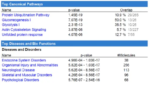
To go further in this analysis, the main canonical pathways along with the main diseases and functions were determined using IPA (Table 1 and Table S4). The most highly altered canonical pathways were related to the protein ubiquitination system, gluconeogenesis, glycolysis, and actin cytoskeleton protein in terms of the number of proteins found in the urano-proteome in comparison with the total number of proteins in each pathway, expressed in percentage form.
Table 1: Top five canonical pathways and top five diseases associated with the urano-proteome, as analyzed by IPA. Names are ordered according to p-value (p<0.05). The ratio value is also shown, as is the number of proteins implicated.
Gluconeogenesis and glycolysis
The highest percentage is observed for gluconeogenesis with 13 proteins (50%), followed by glycolysis with ten proteins (38.5%, including isoforms). The role of glucose in physiological and pathological brain function was reviewed a few years ago (Mergenthaler et al. 2013). The mammalian brain is the main consumer of glucose,
because glucose is its main source of energy. Its metabolism provides the fuel for physiological brain function through the generation of ATP, the foundation for neuronal and non-neuronal cellular maintenance, as well as the generation of neurotransmitters. Therefore, tight regulation of glucose metabolism is critical for brain physiology, and disturbed glucose metabolism in the brain underlies several diseases affecting the brain (Mergenthaler et al. 2013). In this context, it is very interesting to see that seven of the nine glycolysis enzymes belong to the urano-proteome (see Fig. 5). We can also notice that these proteins are found in SEC fraction F2, which contains the highest amount of U(VI).
In the gluconeogenesis pathway, many steps are the opposite of those found in glycolysis. This is why both pathways are found in the main canonical pathways. Of the 13 enzymes of gluconeogenesis found in the urano-proteome (IPA analysis), nine are common to those of glycolysis. The four others are specific to gluconeogenesis and concern the transport of oxaloacetate and malate from the mitochondria to the cytosol (MDH1 and MDH2); the oxidative decarboxylation of malate to pyruvate (ME2: NAD-dependent malic enzyme); and ME1 (malic enzyme 1), which links the glycolytic and citric acid cycles through the reversible oxidative decarboxylation of malate. The effect of uranyl nitrate on enzymes of carbohydrate metabolism has been studied in kidneys of rodents exposed in vivo to natural U(VI). The activity of lactate dehydrogenase increased, whereas the activities
of isocitrate, succinate and malate dehydrogenases, glucose-6-phosphatase, and fructose-1,6-bisphosphatase significantly decreased following natural U(VI) exposure (Banday et al. 2008). In another study, the effect of uranyl nitrate has been investigated in isolated human and mouse kidney cortex tubules metabolizing the physiological substrate lactate. Uranyl nitrate in the millimolar range reduced lactate removal and gluconeogenesis in a concentration-dependent fashion: lactate dehydrogenase, pyruvate carboxylase, phosphoenolpyruvate carboxykinase, and glucose-6-phosphatase fluxes were lowered by 20 to 45% (Renault et al. 2010). Our present results show that U(VI) could have an impact on both pathways through its binding with enzymes belonging to both pathways. They are in agreement with studies demonstrating that uranium affects the activity of these enzymes. It is nevertheless necessary to mention that in brain metabolism, gluconeogenesis is a very minor pathway, with most of the glucose supplied to the brain being provided by the blood. Interestingly, disturbed glucose metabolism can be the foundation for the development of a wide variety of disorders in the brain, particularly neurodegenerative diseases (Mergenthaler et al. 2013).
Proteasome
Twenty-nine proteins out of the 265 in the protein ubiquitination pathway are found in the urano-proteome (Table 1). Using tandem mass spectrometry, Tai et al. (Tai et al. 2010) identified 30 proteins of the brain 26S proteasome. Strikingly, five out of the seven proteins from proteasome subunits alpha described in this paper and six out of seven proteins from proteasome subunits beta belong to the urano-proteome. These findings highlight our previous results with regard to evaluating the response of a human lung cell line (A549) to U(VI) by a proteomic approach. Uranyl treatment triggered differential expression of 14 proteins identified as ubiquitinylated fragments of cytokeratin 8 and cytokeratin18. We hypothesized that U(VI) could cause an abnormality in the ubiquitin degradation system that may lead to an accumulation of cytokeratin degradation products in U(VI)-exposed cells (Malard et al. 2005). The binding of U(VI) to many of the proteins in the 26S subunit of the proteasome could explain these results obtained previously.
Actin cytoskeleton pathway
Thirteen proteins from the actin cytoskeleton pathway were found in the urano-proteome, among them cytoplasmic beta-actin itself. Again, in a previous study, we demonstrated that the expression of genes and proteins coding for actin and tubulin was down-regulated in human renal cells upon exposure to uranium (Prat et al. 2005). Actin was also recently demonstrated to bind U(VI), identifying this protein as a potential uranyl target
in a zebrafish model (Eb-Levadoux et al. 2017).
Actin is a highly conserved cytoskeleton protein that polymerizes to produce filaments (F-actin) that form cross-linked networks in the cytoplasm of cells. The controlled polymerization of monomeric actin (G-actin) into F-actin and the subsequent depolymerization of F-F-actin play key roles in cell division, motility, and contraction (Fletcher and Mullins 2010). In neurons, actin polymerization is involved in fundamental neurobiological
processes, the dynamic plasticity of dendritic spines, formation and motility of growth cones, and polarized cargo sorting at the axon initial segment (Konietzny et al. 2017).
Other proteins related to actin cytoskeleton formation were identified in the urano-proteome and are detailed below. One example is cellular non-muscle myosin-9, which appears to play a role in cytokinesis, cell shape, and specialized functions such as secretion and capping. During cell spreading, myosin-9 fulfills an important function in cytoskeleton reorganization and has different roles in the regulation of the actin network (Betapudi 2010). Profilin-1 binds to actin and affects the structure of the cytoskeleton. It prevents the polymerization of actin at high concentrations but enhances it at low concentrations (Alkam et al. 2017). In neurons, profilin-1 maintains G/F-actin ratios together with capping proteins (Konietzny et al. 2017). Cofilin-1 binds to F-actin and exhibits pH-sensitive F-actin depolymerizing activity. It regulates actin cytoskeleton dynamics and plays a role in the regulation of cell morphology and cytoskeletal organization. It is thus an important protein for normal progress through mitosis and normal cytokinesis (Bernstein and Bamburg 2010). In neurons, cofilin-1 modulates dendritic spine morphology and is involved in long-term potentiation through the enhancement of actin depolymerization in spines (Konietzny et al. 2017). Filamin-A promotes orthogonal branching of actin filaments, links actin filaments to membrane glycoproteins, anchors various transmembrane proteins to the actin cytoskeleton and serves as a scaffold for a wide range of cytoplasmic signaling proteins (Razinia et al. 2012). Vinculin, an F-actin-binding protein involved in cell-matrix adhesion and cell-cell adhesion (Le Clainche et al. 2010), as well as alpha-actinin-1 and -4, F-actin cross-linking proteins that are involved in neurons in dendrite elaboration and branching. Moesin and ezrin are probably involved in connections of major cytoskeletal structures to the plasma membrane. These proteinsare mainly actin-binding proteins. These results suggest that the binding of U(VI) with several actin partners could have an impact on actin network regulation.
Based on the urano-proteome data, the top five diseases and biological disorders given by IPA analysis are endocrine system disorders, organismal injury and abnormalities, neurological disease, skeletal and muscular disorders, and psychological disorders (Table 1). Our proteomics results from a cellular model of U(VI) exposure are consistent with the reported effects of uranium on animal models. The United Nations Scientific Committee on the Effects of Atomic Radiations (UNSCEAR 2017) recently reviewed radiobiological and toxicological effects of exposure to U(VI). In terms of effects on the rat brain, brain toxicity leading to neurobehavioral impairments, associated with increased locomotor activity, as well as perturbation of the sleep-wake cycle, decreased memory, and increased anxiety were reported. The Committee mentioned that the mechanisms underlying these neurobehavioral disturbances are not clearly understood (UNSCEAR 2017).
3.4.5 Most abundant common proteins
To analyze the most abundant common proteins, the normalized spectral abundance factor (NSAF) was used (Zybailov et al. 2006). The NSAF represents the relative proportion of each protein in a sample and is used for quantitative proteomics. This value made it possible to select the most abundant U(VI)-affine proteins for each analytical strategy.
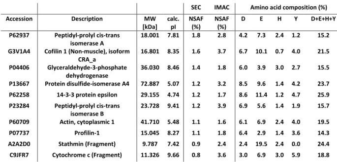
The NSAFs were first calculated for each of the 269 common proteins to enable their ranking in two independent lists corresponding to the two chromatographic strategies. Then, the ten most abundant proteins common to the two populations were extracted from the NSAF ranking established from the SEC experiments because they correspond to U(VI)-bound proteins in cellulo. Table 2 presents these proteins with some of their physico-chemical characteristics, i.e., molecular weights, calculated isoelectric points, and their composition in some amino acids known to coordinate with U(VI) (both the oxygen of aspartate, glutamate, and tyrosinate and the nitrogen of histidine are hard Lewis bases).
Table 2: The ten most abundant proteins from among the 269 common proteins were extracted from the NSAF ranking established from the SEC experiments. Their molecular weights and their calculated isoelectric points are shown, as are their calculated compositions in some amino acids. D: aspartate; E: glutamate; H: histidine; Y: tyrosine.
SEC IMAC Amino acid composition (%)
Accession Description MW [kDa] calc. pI NSAF (%) NSAF (%) D E H Y D+E+H+Y P62937 Peptidyl-prolyl cis-trans isomerase A 18.001 7.81 1.8 2.8 4.2 7.3 2.4 1.2 15.2
G3V1A4 Cofilin 1 (Non-muscle), isoform CRA_a 16.801 8.35 1.6 3.7 6.7 10.1 0.7 4.0 21.5 P04406 Glyceraldehyde-3-phosphate dehydrogenase 36.030 8.46 1.4 1.8 6.0 3.9 3.0 2.7 15.5 P13667 Protein disulfide-isomerase A4 72.887 5.07 1.2 3.2 8.5 9.6 1.4 4.2 23.7 P62258 14-3-3 protein epsilon 29.155 4.74 1.2 1.7 8.6 11.4 1.2 4.7 25.9 P23284 Peptidyl-prolyl cis-trans isomerase B 23.728 9.41 1.2 3.9 6.9 5.6 1.4 1.9 15.7 P60709 Actin, cytoplasmic 1 41.710 5.48 1.1 1.6 6.1 6.9 2.4 4.0 19.5 P07737 Profilin-1 15.045 8.27 1.1 1.8 6.4 2.9 1.4 3.6 14.3
A2A2D0 Stathmin (Fragment) 9.787 7.42 0.9 2.4 2.4 19.5 2.4 0.0 24.4
C9JFR7 Cytochrome c (Fragment) 11.326 9.66 0.8 3.6 3.0 6.9 3.0 5.9 18.8
First of all, the NSAF values calculated for the proteins from the SEC strategy are smaller than those from the IMAC strategy, in keeping with IMAC selectivity. The molecular weights range from 9 kDa (stathmin fragment) to 73 kDa (protein disulfide-isomerase A4), i.e., rather small proteins. All of them were isolated both in fractions F1 and F3 of the SEC experiments, meaning that they are probably associated with other proteins in the F1 fraction. None of these ten proteins were eluted in the F2 fraction, indicating that the U(VI) proteins in the F2 fraction are less abundant taking into account their NSAFs (data not shown). The pI values indicate that 70% of these ten proteins were globally positively charged. As expected, they were isolated in the A1 fraction corresponding to the flow-through of the ion exchange step performed before the IMAC, the remaining 30% being isolated in the A2 and A3 fractions (data not shown).
Aspartate, glutamate, and tyrosine are hard Lewis bases that contain oxygen from carboxylates or phenolates and that are able to bind U(VI). The theoretical aspartate + glutamate + tyrosine + histidine frequency is around 17.4 in vertebrates (Bairoch). Interestingly, out of these ten selected proteins, six present higher frequencies up to 25.9. Furthermore, PDIA4 (P13667) presents two clusters of amino acids prone to U(VI) binding, one long cluster (ED EEEEEEEDDD EEEDDLE) in its N-terminal part and another small cluster(EP EEFDSD) in its C-terminal part. Such sequences are not evidenced in the nine remaining proteins. Nonetheless, profilin-1 (P07737) presents six phosphorylation sites, five of them on serine and one on tyrosine residues. Phosphorylations have been shown to reinforce the binding of U(VI) to glutamate and aspartate. Even if it remains rather difficult to predict the U(VI) binding sites in proteins, our results are consistent with what is known about uranium coordination chemistry.
Apart from these preliminary considerations, this list of the ten most common proteins in the urano-proteome reveals some interesting information about the effects of U(VI). Peptidyl-prolyl cis-trans isomerase A (P62937) is the most abundant U(VI)-bound protein when SH-SY5Y cells are exposed to U(VI), and peptidyl-prolyl cis-trans isomerase B (P23284) is also one of the ten proteins on the present list. Both of them accelerate the correct folding of proteins (Freedman et al. 2017; Schmidpeter and Schmid 2015). Furthermore, peptidyl-prolyl cis-trans isomerase B interacts with protein disulfide-isomerase A4, which is also on this list (Schonbrunner and Schmid 1992). Glyceraldehyde-3-phosphate dehydrogenase (P04406) is one of the enzymes of the main pathways presented above and is involved in glycogenesis. In addition, four of these ten proteins are actin and actin-binding proteins, all of them playing a crucial role in the regulation of the cytoskeleton (Pollard 2016; Schmitz and Bereiter-Hahn 2002). Two other proteins complete this selection: 14-3-3 protein epsilon and cytochrome c. Once bound to 14-3-3 protein epsilon, U(VI) may affect the ability of the former to regulate several signaling
pathways. The presence of cytochrome c in the ten most abundant proteins of the urano-proteome reinforces previously published results on the potential role of cytochrome c in oxidative stress induced by uranium (Shaki et al. 2013). All of these results highlight how uranyl could affect this cell line at different levels of cell functions.
4. Conclusions
Advances in the description of metalloproteomes provide a better understanding of the mechanism and precise role of metalloproteins in the brain (Lothian et al. 2013). Because the brain is a sensitive target of U(VI), the aim of the present study was to identify U(VI) target proteins in neuronal cells exposed to non-toxic U(VI) concentrations. We crossed two complementary approaches, which is more relevant than using a single identification method, in order to isolate, identify, and characterize proteins capable of selectively binding U(VI) in neuron-like cell lysates. This strategy enabled the identification of 269 common U(VI)-affine proteins (the urano-proteome). Functional annotation and distribution into known cell functions and canonical pathways were performed thanks to Ingenuity Pathway Analysis software. Among these pathways, ubiquitination, glycolysis/gluconeogenesis, actin cytoskeleton signaling, and unfolded protein response were particularly highlighted by the presence of several U(VI)-affine proteins in each of them. Furthermore, a semi-quantitative analysis of these 269 proteins was performed to focus on the ten most abundant U(VI)-affine proteins. Nine of them belong to these five canonical pathways, confirming that when neuronal cells are exposed to U(VI), these pathways could be disrupted, not only by the number of U(VI)-affine proteins potentially altered in each pathway, but also by the particular abundance of some of them.
Although uranium has already been suggested to have an impact on the cytoskeleton, carbohydrate metabolism, protein ubiquitination, and oxidative stress, our study is the first to identify a large number of proteins from these metabolic pathways that are linked to an exposure to uranium in cellulo. These results pave the way for a better understanding of the mechanisms involved in the toxic effects of uranium.
FIGURE CAPTIONS
Fig. 1: Experimental design of the analytical strategy.
Fig. 2: UV and q-ICP-MS chromatograms obtained for the lysates of SH-SY5Y cells exposed to 10 µM U(VI). The dashed rectangles indicate the three protein fractions collected.
Fig. 3: Venn diagram of the 269 common proteins.
Fig. 4: Cellular localization and protein function of the urano-proteome. Fig. 5: Glycolysis pathway and U(VI)-bound enzymes.
REFERENCES