RESEARCH OUTPUTS / RÉSULTATS DE RECHERCHE
Author(s) - Auteur(s) :
Publication date - Date de publication :
Permanent link - Permalien :
Rights / License - Licence de droit d’auteur :
Bibliothèque Universitaire Moretus Plantin
Dépôt Institutionnel - Portail de la Recherche
researchportal.unamur.be
University of Namur
Age-related morphometric changes of the tidemark in the ovine stifle
Hontoir, Fanny; Pirson, Romain; Simon, Vincent; Clegg, Peter D.; Nisolle, Jean-François; Kirschvink, Nathalie; Vandeweerd, Jean-Michel
Published in:
Anatomia, Histologia, Embryologia DOI:
10.1111/ahe.12449
Publication date: 2019
Document Version
Early version, also known as pre-print
Link to publication
Citation for pulished version (HARVARD):
Hontoir, F, Pirson, R, Simon, V, Clegg, PD, Nisolle, J-F, Kirschvink, N & Vandeweerd, J-M 2019, 'Age-related morphometric changes of the tidemark in the ovine stifle', Anatomia, Histologia, Embryologia, vol. 48, no. 4, pp. 366-374. https://doi.org/10.1111/ahe.12449
General rights
Copyright and moral rights for the publications made accessible in the public portal are retained by the authors and/or other copyright owners and it is a condition of accessing publications that users recognise and abide by the legal requirements associated with these rights. • Users may download and print one copy of any publication from the public portal for the purpose of private study or research. • You may not further distribute the material or use it for any profit-making activity or commercial gain
• You may freely distribute the URL identifying the publication in the public portal ?
Take down policy
If you believe that this document breaches copyright please contact us providing details, and we will remove access to the work immediately and investigate your claim.
RESEARCH OUTPUTS / RÉSULTATS DE RECHERCHE
Author(s) - Auteur(s) :
Publication date - Date de publication :
Permanent link - Permalien :
Rights / License - Licence de droit d’auteur :
Bibliothèque Universitaire Moretus Plantin
Dépôt Institutionnel - Portail de la Recherche
researchportal.unamur.be
Age-related morphometric changes of the tidemark in the ovine stifle
Hontoir, Fanny; Pirson, Romain; Simon, Vincent; Clegg, Peter D.; Nisolle, Jean-François; Kirschvink, Nathalie; Vandeweerd, Jean-Michel
Published in:
Anatomia, Histologia, Embryologia DOI:
DOI:10.1111/ahe.12449
Publication date: 2019
Document Version
Early version, also known as pre-print
Link to publication
Citation for pulished version (HARVARD):
Hontoir, F, Pirson, R, Simon, V, Clegg, PD, Nisolle, J-F, Kirschvink, N & Vandeweerd, J-M 2019, 'Age-related morphometric changes of the tidemark in the ovine stifle' Anatomia, Histologia, Embryologia.
https://doi.org/DOI:10.1111/ahe.12449
General rights
Copyright and moral rights for the publications made accessible in the public portal are retained by the authors and/or other copyright owners and it is a condition of accessing publications that users recognise and abide by the legal requirements associated with these rights. • Users may download and print one copy of any publication from the public portal for the purpose of private study or research. • You may not further distribute the material or use it for any profit-making activity or commercial gain
• You may freely distribute the URL identifying the publication in the public portal ?
Take down policy
If you believe that this document breaches copyright please contact us providing details, and we will remove access to the work immediately and investigate your claim.
For Review Only
Age-related morphometric changes of the tidemark in the ovine stifle
Journal: Anatomia, Histologia, Embryologia Manuscript ID Draft
Wiley - Manuscript type: Original Article Date Submitted by the
Author: n/a
Complete List of Authors: Hontoir, Fanny; Université de Namur, URVI - Department of Veterinary Medicine
Pirson, Romain; Université de Namur, URVI - Department of Veterinary Medicine
Simon, Vincent; Université de Namur, URVI - Department of Veterinary Medicine
Clegg, Peter; University of Liverpool Faculty of Health and Life Sciences, Department of Musculoskeletal Biology, Institute of Ageing and Chronic Disease (IACD)
Nisolle, Jean-François; CHU UCL Namur - Site Godinne
Kirschvink, Nathalie ; Université de Namur, URVI - Department of Veterinary Medicine
Vandeweerd, J.M.; Université de Namur, URVI - Department of Veterinary Medicine
Keywords: sheep, cartilage, knee, osteoarthritis, ageing
Abstract:
Though the ovine stifle is commonly used to study osteoarthritis, there is limited information about the age-related morphometric changes of the tidemark. The objective of this study was to document the number of tidemarks in the stifle of research sheep without clinical signs of osteoarthritis and of various ages (n = 80). Articular cartilage of the medial and lateral tibial condyles and of the medial and lateral femoral condyles was assessed by histology: (1) to count the number of
tidemark; and (2) to assess the OARSI (OsteoArthritis Research Society International) score for structural changes of cartilage.
The number of tidemarks varied between anatomical regions respectively from 4.2 in the medial femoral condyle to 5.0 in the lateral tibial
condyle. The axial part showed a significant higher number of tidemarks than the abaxial part, for all regions except the medial tibial condyle. While the tidemark count strongly correlated to age (Spearman Correlation coefficient=0.70; 95% confidence interval 0.67 to 0.73; P<0.0001), the OARSI score was weakly correlated to age in our cohort of sheep (Spearman Correlation coefficient=0.25; 95% confidence interval 0.19 to 0.30; P<0.0001). Interestingly, no tidemark was seen in the three animals aged 6 months.
Our data indicate that the number of tidemarks increases with age and vary with anatomical region. The regional variation also revealed a higher number of tidemarks in the tibia than in the femur. This could be attributed to the local variation in cartilage response to strain and to the
For Review Only
difference in chondrocyte biology and density.
3 4 5 6 7 8 9 10 11 12 13 14 15 16 17 18 19 20 21 22 23 24 25 26 27 28 29 30 31 32 33 34 35 36 37 38 39 40 41 42 43 44 45 46 47 48 49 50 51 52 53 54 55 56 57 58 59
For Review Only
Age-related morphometric changes of the tidemark in the ovine stifle.
1 2
Running title: Tidemark in the ovine stifle 3
Fanny Hontoir1, Romain Pirson1, Vincent Simon1, Peter Clegg2, Jean-François Nisolle,
4
Nathalie Kirschvink1, Jean-Michel E. Vandeweerd1
5 6
1 Department of Veterinary Medicine, Integrated Veterinary Research Unit (IVRU) – Namur
7
Research Institute for Life Sciences (NARILIS), Faculty of Sciences, University of Namur, rue 8
de Bruxelles, 61, 5000 Namur, Belgium 9
2 Department of Musculoskeletal Biology, Institute of Ageing and Chronic disease, University
10
of Liverpool, Liverpool L69 3BX, UK 11
3 Centre Hospitalier Universitaire (CHU) UCL Namur‐Mont Godinne, Université Catholique
12
de Louvain, 5530 Yvoir, Belgium 13 14 15 16 17 18 19 Corresponding author: 20 Fanny Hontoir 21 61, rue de Bruxelles 22 5000 Namur 23 Tel: 0032 496 53 51 45 24 [email protected] 25 26 3 4 5 6 7 8 9 10 11 12 13 14 15 16 17 18 19 20 21 22 23 24 25 26 27 28 29 30 31 32 33 34 35 36 37 38 39 40 41 42 43 44 45 46 47 48 49 50 51 52 53 54 55 56 57 58 59 60
For Review Only
Summary 27
Though the ovine stifle is commonly used to study osteoarthritis, there is limited information 28
about the age-related morphometric changes of the tidemark. The objective of this study was to 29
document the number of tidemarks in the stifle of research sheep without clinical signs of 30
osteoarthritis and of various ages (n = 80). Articular cartilage of the medial and lateral tibial 31
condyles and of the medial and lateral femoral condyles was assessed by histology: (1) to count 32
the number of tidemark; and (2) to assess the OARSI (OsteoArthritis Research Society 33
International) score for structural changes of cartilage. 34
The number of tidemarks varied between anatomical regions respectively from 4.2 in the medial 35
femoral condyle to 5.0 in the lateral tibial condyle. The axial part showed a significant higher 36
number of tidemarks than the abaxial part, for all regions except the medial tibial condyle. 37
While the tidemark count strongly correlated to age (Spearman Correlation coefficient=0.70; 38
95% confidence interval 0.67 to 0.73; P<0.0001), the OARSI score was weakly correlated to 39
age in our cohort of sheep (Spearman Correlation coefficient=0.25; 95% confidence interval 40
0.19 to 0.30; P<0.0001). Interestingly, no tidemark was seen in the three animals aged 6 months. 41
Our data indicate that the number of tidemarks increases with age and vary with anatomical 42
region. The regional variation also revealed a higher number of tidemarks in the tibia than in 43
the femur. This could be attributed to the local variation in cartilage response to strain and to 44
the difference in chondrocyte biology and density. 45
46
Key words: sheep – cartilage – stifle – osteoarthritis - ageing 47
48
Number of figures in this manuscript: 4 49
Number of tables in this manuscript: 1 50 51 52 3 4 5 6 7 8 9 10 11 12 13 14 15 16 17 18 19 20 21 22 23 24 25 26 27 28 29 30 31 32 33 34 35 36 37 38 39 40 41 42 43 44 45 46 47 48 49 50 51 52 53 54 55 56 57 58 59 60
For Review Only
Introduction 53
Osteoarthritis is a degenerative process of the diarthrodial (synovial) joint characterized by 54
progressive degeneration of the articular cartilage, combined with subchondral bone sclerosis 55
and osteophyte formation, leading to reduced joint function (Grynpas, Albert, Katz, Lieberman, 56
Pritzker, 1991; McIlwraith, 1996, p.34). Histology is considered as a gold standard technique 57
to assess normality of cartilage, disease development (Lahm, Kreuz, Oberst, Haeberstroh, Uhl 58
et al., 2006; Wucherer, Ober, Cozemius, 2012; Zamli, Adams, Tarlton, Sharif, 2013), and 59
efficacy of treatments (Huang, Simonian, Norman, Clark, 2004; Hoeman, Hurtig, Rossomacha, 60
Sun, Chevrier et al., 2005; Zscharnak, Hepp, Richter, Aigner, Schultz et al., 2010) in research 61
studies on osteoarthritis. 62
Different scoring scales have been described for microscopic assessment of cartilage, based on 63
several histological criteria such as the Mankin score, the “modified Mankin score” (Thomas, 64
Fuller, Whittles, Sharif, 2007; Piskin, Gulbahar, Tomak, Gukman, Hokelek et al., 2007; Daubs, 65
Markel, Manley, 2006), and the ICRS (International Cartilage Repair Society) -II scoring scale 66
(Mainil-Varlet, Van Damme, Nesic, Knutsen, Kandel, Roberts et al., 2010). Species-specific 67
scoring scales have been proposed by the Osteoarthritis Research Society International 68
(OARSI) histopathology initiative to ensure comparison between studies using animal models 69
of osteoarthritis, in mice (Glasson, Chambers, Van Den Berg, Little, 2010), rats (Gerwin, 70
Bendele, Glasson, Carlson, 2010), guinea pigs (Kraus, Huebner, DeGroot, Bendele, 2010), 71
rabbits (Laverty, Girard, Williams, Hunziker, Pritzker, 2010), dogs (Cook, Kuroki, Visco, 72
Pelletier, Schulz et al., 2010), horses (McIlwraith, Frisbie, Kawcak, Fuller, Hurtig et al., 2010), 73
goats and sheep (Little, Smith, Cake, Read, Murphy et al., 2010). For example in sheep, the 74
histopathological assessment includes the following parameters: cartilage structure, percentage 75
of the surface area affected by structural damage, chondrocyte density, cell cloning, 76
interterritorial Toluidine blue staining, and tidemark variations. 77 3 4 5 6 7 8 9 10 11 12 13 14 15 16 17 18 19 20 21 22 23 24 25 26 27 28 29 30 31 32 33 34 35 36 37 38 39 40 41 42 43 44 45 46 47 48 49 50 51 52 53 54 55 56 57 58 59
For Review Only
78
The tidemark is the limit between the hyaline cartilage and the calcified cartilage (Meachim & 79
Allibone, 1984; Oegema, Carpenter, Hofmeister, Thompson, 1997; Burr, 2004). At 80
microscopy, the tidemark appears as a non-cellular line of about 10 µm strongly stained with 81
hematoxylin-eosin, or toluidine blue (Lyons, Stoddart, McClure, McClure, 2005). A trilaminar 82
organization has been demonstrated by combining different histochemical staining 83
(hematoxylin and eosin, picrosirius red, toluidine blue and safranin O), with a distal lamina (to 84
the side of the non-calcified cartilage), a proximal lamina (to the side of the calcified-cartilage) 85
and a central lamina. The proximal and distal laminae differ in their chemistry and, hence, in 86
their tinctorial properties. It is therefore suggested that the central lamina is actually an 87
artefactual zone due to the interpenetration of colorations of the proximal and the distal laminae 88
(Lyons et al., 2005). 89
The general consensus is that the tidemark is the result of accumulation of non-specific 90
molecules at the interface of calcified and hyaline cartilage caused by discontinuous 91
mineralization (Oegema et al., 1997). The tidemark seems to be derived from apoptotic 92
chondrocytes, and to include several molecules such as phospholipides, alkaline phosphatase, 93
type X collagen, adenosine triphosphatase, deoxyribonucleic acid, lectins, and High Mobility 94
Group Box chromosomal protein 1 (HMGB1) (Lyons et al. 2005; Simkin 2012). Chondrocytes 95
are not present in the tidemark but a few can be partially embedded in its mineralizing side 96
(Lyons et al., 2005). 97
98
Tidemark alterations, i.e. duplication, advancement and vascular invasion have been associated 99
to disease such as rheumatoid arthritis (Fassbender, Seibel, Hebert, 1992; Suber & Rosen, 2009) 100
or osteoarthritis (Oettmeier, Abendroth, Oettmeier, 1989; Bonde et al., 2005; Hulth, 1993; Suri, 101
Gill, Massena de Camin, Wilson, McWilliams et al., 2007; Bullough & Jagannath, 1983; 102 3 4 5 6 7 8 9 10 11 12 13 14 15 16 17 18 19 20 21 22 23 24 25 26 27 28 29 30 31 32 33 34 35 36 37 38 39 40 41 42 43 44 45 46 47 48 49 50 51 52 53 54 55 56 57 58 59
For Review Only
Oegema et al., 1997). In the OARSI score, it is observed whether the tidemark is duplicated 103
(score 1) and whether blood vessels from the subchondral bone cross the tidemark to the 104
calcified cartilage (score 2) or to the hyaline cartilage (score 3). 105
106
However, multiple tidemarks can be observed in normal joints (Oegema et al., 1997; Oettmeier 107
et al., 1989). The number of tidemarks has been reported to change with ageing in humans, with 108
an average increase from 1.5 to 2.5 in femur and humerus after the age of 60 (Lane & Bullough, 109
1980). Duplicated tidemarks were visible in mature normal canine femoral articular cartilage 110
(Oegema et al., 1997). In a study on 28 cynomolgus monkeys, as many as ten tidemarks were 111
observed in normal primates over 20 years old while at least two tidemarks were present in all 112
animals (Miller, Novatt, Hamerman, Carlson, 2004). In horses, the number of tidemarks was 113
higher in non-athletic than in racehorses with articular pathology (Muir, Peterson, Sample, 114
Scollay, Markell, 2008). In non-working and working German shepherd dogs, the tidemark 115
duplication in the femur and the tibia has been suggested to be related to ageing (Francuski, 116
Radovanović, Andrić, Krstić, Bogdanović et al., 2014). 117
Since tidemark duplication and advancement could be observed in diseased but also in healthy 118
animals, it is important to document how tidemark varies with age in a population of research 119
animals. The sheep, in particular, is commonly used as a large animal model for osteoarthritis 120
(Little et al., 2010). In sheep, there is limited information about the variation of the number of 121
tidemarks (Appleyard, Burkhardt, Ghosh, Read, Cake et al., 2003; Frisbie, Cross, McIlwraith, 122
2006). Most of the sheep used in research are skeletally mature sheep (Huang et al., 2004; 123
Burger, Mueller, Wlodarczyk, Goost, Tolba et al., 2007; Dattena, Pilichi, Rocca, Mara, Casu et 124
al., 2009) aged between 3 and 6 years old (Hoeman et al., 2005). 125
The objectives of this study were to document the variation of the number of tidemarks of the 126
stifle in a large cohort of sheep without clinical signs of osteoarthritis and of various ages. 127 3 4 5 6 7 8 9 10 11 12 13 14 15 16 17 18 19 20 21 22 23 24 25 26 27 28 29 30 31 32 33 34 35 36 37 38 39 40 41 42 43 44 45 46 47 48 49 50 51 52 53 54 55 56 57 58 59
For Review Only
128
Materials and methods 129
Population 130
Eighty pairs of hindlimbs were collected, between 2012 and 2018, from crossed Texel ewes, 131
euthanatized for reasons other than hind limb lameness (mastitis, metritis), within six hours of 132
euthanasia. Animals were aged between 6 months and 3 years old (N=28), 4 to 6 years old 133
(N=31) and 7 to 11 year old (N=21). Animals had no clinical signs of osteoarthritis (lameness, 134
articular swelling, and pain at manipulation). They had been used for teaching anatomy and 135
were not euthanized for the purpose of the current study. The experimental protocol (KI 10/148) 136
was approved by the local ethical committee for animal welfare. 137
138
Gross anatomy 139
After soft tissue dissection and joint opening, synovium and articular surfaces were assessed by 140
one investigator in a blinded manner following OARSI recommendations (Little et al., 2010). 141
Synovium was evaluated for macroscopic alterations (normal, slight, mild, moderate, marked 142
and severe): discoloration, vascularity, thickening and synovial proliferation. Macroscopic 143
scores for cartilage damages were: score 0 for intact cartilage surface; score 1 for surface 144
roughening; score 2 for deeper defects (fibrillation, fissures) not involving the subchondral 145
bone; score 3 for erosions down to the subchondral bone (less than 5 mm diameter); score 4 for 146
large erosions down to the subchondral bone (more than 5 mm diameter). Scoring was 147
performed in four areas of interest: the middle part of the medial tibial condyle (or plateau) 148
(MTC), of the medial femoral condyle (MFC), of the lateral tibial condyle (LTC) and of the 149
lateral femoral condyle (LFC) (Figure 1). Joint margins were observed for the presence of 150
osteophytes. Joint surfaces were digitally photographed (Sony Alpha DSLR-A230 digital 151
camera) with standardized lighting conditions for records (two Sony Illustar SM-300 lighting). 152 3 4 5 6 7 8 9 10 11 12 13 14 15 16 17 18 19 20 21 22 23 24 25 26 27 28 29 30 31 32 33 34 35 36 37 38 39 40 41 42 43 44 45 46 47 48 49 50 51 52 53 54 55 56 57 58 59
For Review Only
153
Histology 154
Four mm-thick osteochondral slabs were collected from the middle part of the medial tibial 155
condyle (or plateau), medial femoral condyle, lateral tibial condyle and lateral femoral condyle 156
(Figure 1). A total of 640 samples (80 sheep x 2 limbs x 4 regions) were collected. After 48-h 157
fixation in 10% (v/v) neutral buffered formalin, samples were transferred to 70% (v/v) ethanol 158
for further processing (Little et al., 2010). They were decalcified in DC3 (non-ionic surfactants, 159
hydrochloric acid, EDTA, VWR International, Leuven, Belgium) for 2 days and embedded in 160
paraffin, and then 4-𝜇m sections were cut. Sections were deparaffinised with xylene and graded 161
ethanol, and then stained with Toluidine blue. 162
Each slice was examined for cartilage structure and tidemark count. Scoring of cartilage 163
structure followed the OARSI recommendations for histological evaluation of structural 164
changes in ovine articular cartilage (Little et al., 2010). Each region being divided into two 165
subregions (abaxial (Ab) and axial (Ax)), 1280 subregions were assessed (640 regions x 2). 166
Assessments were performed in duplicates by two observers to obtain a mean score. Tidemark 167
counts were obtained by one blinded observer in six equidistant locations per anatomical region. 168
Mean number was calculated and recorded. Sheep, age and limb identities were blinded to 169 histological scorers. 170 171 Statistical analysis 172
Statistics were performed with GraphPad Prism 7.03 (GraphPad Software, La Jolla). Statistical 173
significance was set at 0.05. Firstly, the dataset was assessed for normality, skewness and 174
kurtosis. Due to the moderate positive skewness, to kurtosis, and to non-normal distribution of 175
the data, nonparametric statistics were conducted (Pearce & Frisbie, 2010). Wilcoxon matched-176 3 4 5 6 7 8 9 10 11 12 13 14 15 16 17 18 19 20 21 22 23 24 25 26 27 28 29 30 31 32 33 34 35 36 37 38 39 40 41 42 43 44 45 46 47 48 49 50 51 52 53 54 55 56 57 58 59
For Review Only
pairs signed rank test and Friedman test were used to compare data from left and right limbs, 177
and to compare data from the different (sub-)regions of each limb. 178
Kruskal-Wallis test followed by a Dunn’s multiple comparison test enabled to test difference 179
between age groups for tidemark count and OARSI scoring. Mean tidemark count and mean 180
OARSI scores of both limbs was considered for each sheep. Correlation between age and 181
tidemark number or OARSI scoring of the sheep was assessed using the Spearman’s rank order 182
test. Correlation was considered very weak (0.00-0.19), weak (0.20-0.39), moderate (0.40-183
0.59), strong (0.60-0.79) and very strong (0.80-1.00) depending on the absolute value of the 184 coefficient. 185 186 Results 187 Gross anatomy 188
Macroscopic assessment of cartilage for the 1280 anatomic areas revealed 911 zones of intact 189
cartilage (71.2%), 315 score-1 lesions (24.6%), 50 score-2 lesions (3.9%) and 4 score-3 lesions 190
(0.3%). Score-2 and -3 erosions were found in 11 of the 80 sheep (13.75%). No score-4 lesion 191
was found. No signs of joint inflammation (effusion, synovitis) and no osteophyte was detected 192 at gross anatomy. 193 194 Histology 195
Thirty slides presented artifacts (folding, shredding, splitting) preventing tidemark count. Thus, 196
1250 of the 1280 sub-regions were appropriately assessed. 197
There was no significant difference between left and right limbs for tidemark count (P= 0.5898), 198
and for OARSI scores (P = 0.2761).The tidemark count (P<0.0001) showed difference upon 199
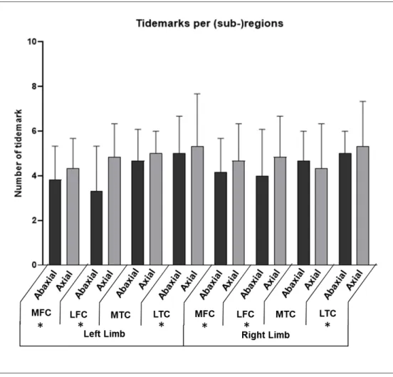
(sub-)regions. The axial sub-region had a significant higher number of tidemarks than the 200
abaxial sub-region, for all regions except in the medial tibial condyle (Figure 3). The number 201 3 4 5 6 7 8 9 10 11 12 13 14 15 16 17 18 19 20 21 22 23 24 25 26 27 28 29 30 31 32 33 34 35 36 37 38 39 40 41 42 43 44 45 46 47 48 49 50 51 52 53 54 55 56 57 58 59
For Review Only
of tidemarks in the four regions was ranked as MFC < LFC < MTC < LTC, with an average 202
number of 4.2, 4.5, 4.8 and 5.0, respectively; those differences were statistically significant, 203
except between MFC and LFC. 204
The OARSI scores significantly differed with ()regions (Figure 4), with the axial sub-205
regions showing higher scores than abaxial sub-regions (P<0.0001). OARSI scores in the four 206
regions were ranked as LFC < LTC < MFC < MTC, with an average score of 2.0, 2.6, 5.0 and 207
5.3, respectively. The differences were not significant between regions of the same bone. 208
209
The three age groups had significant different tidemark count (P<0.0001) and OARSI scores 210
(P=0.0197) (Table 1), with a strong positive correlation between age and the number of 211
tidemarks (Spearman Correlation coefficient = 0.70, 95% confidence interval 0.67 to 0.73; P < 212
0.0001). However, the OARSI score was weakly correlated to age in our cohort of sheep 213
(Spearman Correlation coefficient = 0.25, 95% confidence interval 0.19 to 0.30; P < 0.0001). 214
The correlation between OARSI scores and tidemark count was weak as well (Spearman 215
Correlation coefficient = 0.19, 95% confidence interval 0.13 to 0.24; P < 0.0001). In the three 216
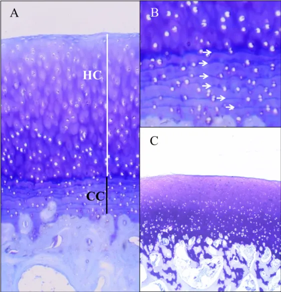
young animals aged 6 months, no tidemark was visible (Figure 2). 217
218
Discussion 219
In this study, the number of tidemarks increased significantly with age. Interestingly, no 220
tidemark was identified in the three sheep aged 6 months. This is in agreement with reports that 221
calcified cartilage layer does not begin to develop until well into the first year postpartum 222
(Martinelli, Eurell, Les, Fyhrie, Bennett, 2002). In horses, functional adaptation of articular 223
cartilage occurs during maturation (Brama, TeKoppele, Bank, Barneveld, van Weeren, 2002). 224
Cartilage-bone interface is a dynamic area where duplication of the tidemark and thickening of 225 3 4 5 6 7 8 9 10 11 12 13 14 15 16 17 18 19 20 21 22 23 24 25 26 27 28 29 30 31 32 33 34 35 36 37 38 39 40 41 42 43 44 45 46 47 48 49 50 51 52 53 54 55 56 57 58 59
For Review Only
calcified cartilage are due to micro-trauma at the bone cartilage-interface and quick repair 226
process in response to mechanical stresses over time (Burr & Schaffler, 1997). 227
The effect of constraints on tidemark duplication is also illustrated by the variation of number 228
of tidemarks between anatomical regions. Constraints are higher in the medial compartment 229
due to the asymmetry of load bearing and contact area in the stifle (Thomas, Resnick, Alazraki, 230
Daniel, Greenfield, 1975; Baliunas Hurwitz, Ryals, Karrar, Case et al., 2002; Lee-Shee, Dickey, 231
Hurtig, 2007; Taylor, Poepplau, Konig, Ehrig, Zachow, 2011). This is associated with a higher 232
deterioration of cartilage and higher OARSI scores in those anatomical regions, as 233
demonstrated by studies in sheep (Vandeweerd, Hontoir, Kirschvink, Clegg, Nisolle et al., 234
2013; Hontoir, Clegg, Simon, Kirschvink, Nisolle et al., 2017), and man (Arøen, Løken, Heir, 235
Alvik, Ekeland et al., 2004; Neogi, Felson, Niu, Lynch, Nevitt et al., 2009; Flanigan, Harris, 236
Trinh, Siston, Brophy, 2010). In the current study, OARSI scores were also higher in the medial 237
tibial and femoral condyles than in the lateral tibial and femoral condyles, with the axial side 238
being more affected. 239
In the current study, the number of tidemarks was higher in the tibia than in the femur. A 240
difference in number of tidemarks has also been described in dogs (Francuski et al., 2014). In 241
femoral cartilage, tidemark multiplication was more frequently observed in working dogs than 242
in working dogs, whilst in the tibial cartilage it was more frequently observed in non-243
working dogs. This particularity has not been described elsewhere. However, regional 244
differences of cartilage mechanobiology and cell biology could account for change in tidemark 245
number. Mechanically, the cartilage strain is not homogeneous through the joint after exercise: 246
for example, in human, the cartilage strain (percentage of thickness change) is higher in the 247
tibia (30%) compared to the femur (20%) after a 30-minutes jogging (Moscher, Smith, Collins, 248
Liu, Hancy et al., 2005; Sanchez-Adams, Leddy, McNulty, O’Conor, Guilak, 2014). Moreover, 249
the cartilage response to loading is different for tibial and femoral cartilage. In vivo assessment 250 3 4 5 6 7 8 9 10 11 12 13 14 15 16 17 18 19 20 21 22 23 24 25 26 27 28 29 30 31 32 33 34 35 36 37 38 39 40 41 42 43 44 45 46 47 48 49 50 51 52 53 54 55 56 57 58 59
For Review Only
of cartilage response to load has been performed in human using compositional imaging, this 251
technique revealed that tibial cartilage showed an homogeneous response for deep and 252
superficial layers, whilst the femur showed an opposite response for both layers, suggesting a 253
transport of water to the deep zone of cartilage in the femur, in opposition to the general squeeze 254
of water of both tibial layers (Souza, Kumar, Calixto, Singh, Schooler et al., 2014). 255
Biologically, tibial and femoral cartilage shows different pattern, with higher 256
glycosaminoglycans and collagen content, higher chondrocyte density and proliferation rate in 257
the femur than in the tibia (Stenhamre, Slynarski, Petrén, Tallheden, Lindahl, 2008). It should 258
be reminded here that chondrocyte reaction to mechanical load varies from enhanced matrix 259
synthesis (anabolism) to catabolism, apoptosis and necrosis depending on the frequency, the 260
amplitude, or the strain-scheme for example (Sanchez-Adams et al., 2014; Bleuel, Zacke, 261
Brüggemann, Niehoff, 2015; Iijima, Ito, Nagai, Tajino,Yamaguchi et al., 2017). As the 262
tidemark originates from the chondrocytes activity (Havelka, Horn, Spohrová, Valouch, 1984) 263
and apoptosis (Simkin, 2012), the higher number of tidemarks in the tibia could be explained 264
by the combination of higher strain and lower cell yield in the tibia compared to the femur. 265
266
The correlation between the number of tidemarks and the OARSI score was weak in our sheep 267
population. In a recent research study in man, the tidemark count poorly and non-significantly 268
correlated to the human OARSI scores in the middle part of 42 lateral tibial condyles, with 269
OARSI scores ranging from 0 (normal) to 4 (superficial delamination to mid-zone erosion). 270
(Deng, Wang, Yin, Chen, Guo et al., 2016). These results support the idea, also proposed by 271
other authors (Lane & Bullough, 1980; Bonde et al., 2005; Oegema et al., 1997; Muir et al., 272
2008; Francuski et al., 2014), that tidemark multiplication is not a unique feature of 273
osteoarthritis and can be found in normal animals. OARSI scores in the current study were low. 274 3 4 5 6 7 8 9 10 11 12 13 14 15 16 17 18 19 20 21 22 23 24 25 26 27 28 29 30 31 32 33 34 35 36 37 38 39 40 41 42 43 44 45 46 47 48 49 50 51 52 53 54 55 56 57 58 59
For Review Only
In addition, we found no osteophytes, a feature of osteoarthritis (Little et al., 2010; Cake, Read, 275
Corfield, Daniel, Burkhardt et al., 2013). 276
277
Since there was no osteoarthritic sheep in the current research population, it is not possible to 278
infer on the association between OA and the number of tidemarks. The use of the sheep as an 279
animal model for osteoarthritis requires the surgical induction of the disease to ensure the 280
development of moderate to severe cartilage damages (Little et al., 2010). For example, in a 281
lateral meniscectomy model, average OARSI scores can reach up to 16 +/-3 for cartilage (with 282
erosion of cartilage and loss of proteoglycans to the mid/deep zone), associated to moderate 283
synovitis and osteophytes in the lateral femoral and tibial condyles (Gelse, Körber, Schöne, 284
Raum, Koch, 2017). Obviously such cases were not identified in the current population. 285
One could argue that the decalcification process is a limitation of the current study and would 286
impair assessment of the tidemark. The tidemark is basically seen as the limit between the 287
calcified cartilage and the hyaline cartilage (Meachim & Allibone, 1984; Oegema et al., 1997; 288
Burr, 2004; Lyons et al., 2005). However, the tidemark is not only featured by presence of 289
calcium deposition; it contains multiple molecules (phospholipids, alkaline phosphatase, 290
adenosine triphosphatase, DNA, lectins) revealed by a wide range of histologic stains 291
(Dmitrovsky, Lane and Bullough, 1978; Havelka et al., 1984; Oettmeir et al., 1989; Lyons et 292
al., 2005). Furthermore, we have purposely conducted the study according to the OARSI 293
recommendation for assessment of cartilage and osteochondral junction in ovine, i.e. with a 294
decalcification step during the histological processing of osteochondral samples (Little et al., 295
2010). Another limitation is the lack of one-year old sheep to determine the apparition of the 296
first tidemark. Those animals are not frequently available for research since they are young 297
skeletally mature animal at the beginning of their reproductive career, and therefore not likely 298 to be reformed. 299 3 4 5 6 7 8 9 10 11 12 13 14 15 16 17 18 19 20 21 22 23 24 25 26 27 28 29 30 31 32 33 34 35 36 37 38 39 40 41 42 43 44 45 46 47 48 49 50 51 52 53 54 55 56 57 58 59
For Review Only
300
Conclusion 301
Documentation of animal models is a concern in research and should be pursued to ensure 302
accurate evaluation of the model and of the tested hypothesis. In the current study, we 303
demonstrated that the multiplication of the tidemark is associated to ageing in the stifles of our 304
sheep population aged between 6 months and 11 years old, without clinical signs of 305
osteoarthritis. The tidemark count was weakly correlated to OARSI scores, confirming that 306
tidemark count is not a feature of osteoarthritis. This might have implications in the 307
interpretation of the OARSI histological score in sheep. Indeed, ageing seems to be more 308
relevant to tidemark count than osteoarthritis progression in the sheep, as well as in man and 309 dogs. 310 311 3 4 5 6 7 8 9 10 11 12 13 14 15 16 17 18 19 20 21 22 23 24 25 26 27 28 29 30 31 32 33 34 35 36 37 38 39 40 41 42 43 44 45 46 47 48 49 50 51 52 53 54 55 56 57 58 59
For Review Only
312
Ackowledgements 313
We acknowledge Nadine Antoine and Joelle Piret for their help in histology. 314
315
Conflict of interest statement 316
None of the authors of this paper has a financial or personal relationship with people or 317
organizations that could inappropriately influence or bias the content of the paper. 318
319
Funding Information 320
This study was supported by the University of Namur (UNamur), NARILIS (Namur Research 321
Institute for Life Science). 322
323
References 324
Appleyard, R.C., Burkhardt, D., Ghosh, P., Read, R., Cake, M., Swain, M.V., & Murrell, G.A. 325
(2003). Topographical analysis of the structural, biochemical and dynamic biomechanical 326
properties of cartilage in an ovine model of osteoarthritis. Osteoarthritis and Cartilage, 11, 65-327
77. https://doi.org/10.1053/joca.2002.0867. 328
Arøen, A., Løken, S., Heir, S., Alvik, E., Ekeland, A., Granlund, O.G., & Engebretsen, L. 329
(2004). Articular cartilage lesions in 993 consecutive knee arthroscopies. American Journal of 330
Sports Medicine, 32, 211–215. https://doi.org/10.1177/0363546503259345. 331
Baliunas, A.J., Hurwitz, D.E., Ryals, A.B., Karrar, A., Case, J.P., Block, J.A., & Andriacchi, 332
T.P. (2002). Increased knee joint loads during walking are present in subjects with knee 333
osteoarthritis. Osteoarthritis and Cartilage, 10, 573-579. doi:10.1053/joca.2002.0797. 334 3 4 5 6 7 8 9 10 11 12 13 14 15 16 17 18 19 20 21 22 23 24 25 26 27 28 29 30 31 32 33 34 35 36 37 38 39 40 41 42 43 44 45 46 47 48 49 50 51 52 53 54 55 56 57 58 59
For Review Only
Bleuel, J., Zaucke, F., Brüggemann, GP., & Niehoff, A. (2015). Effects of cyclic tensile strain 335
on chondrocyte metabolism: a systematic review. PLoS One, 10, e0119816. doi: 336
10.1371/journal.pone.0119816. 337
Bonde, H.V., Talman, M.L.M., & Kofoed, H. (2005). The area of the tidemark in osteoarthritis: 338
a three-dimensional stereological study in 21 patients. Acta pathologica, microbiologica et 339
immunologica Scandinavia, 113, 349-352. https://doi.org/10.1111/j.1600-340
0463.2005.apm_113506.x 341
Brama, P.A., TeKoppele, J.M., Bank, R.A., Barneveld, A., & van Weeren, P.R. (2002). 342
Development of biochemical heterogeneity of articular cartilage: influences of age and exercise. 343
Equine Veterinary Journal, 34, 265-269. https://doi.org/10.2746/042516402776186146. 344
Bullough, P.G., & Jagannath, A. (1983). The morphology of the calcification front in articular 345
cartilage. Journal of Bone and Joint Surgery, 65B, 72–78. doi: 10.1302/0301-346
620X.65B1.6337169. 347
Burger, C., Mueller, M., Wlodarczyk, P., Goost, H., Tolba, R.H., Rangger, C., Kabir, K., & 348
Weber, O. (2007). The sheep as a knee osteoarthritis model: early cartilage changes after 349
meniscus injury and repair. Laboratory animals, 41, 420-431. doi: 350
10.1258/002367707782314265. 351
Burr, D.B., 2004. Anatomy and physiology of the mineralized tissues: role in the pathogenesis 352
of osteoarthrosis. Osteoarthritis and Cartilage, 12, S20-S30. 353
https://doi.org/10.1016/j.joca.2003.09.016. 354
Burr, D.B., & Schaffler, M.B. (1997). The involvement of subchondral mineralized tissues in 355
osteoarthrosis: quantitative microscopic evidence. Miscroscopic research techniques, 37, 343-356
357. https://doi.org/10.1002/(SICI)1097-0029(19970515)37:4<343::AID-JEMT9>3.0.CO;2-L 357
Cake, M.A., Read, R.A., Corfield, G., Daniel, A., Burkhardt, D., Smith, M.M., & Little, C.B. 358
(2013). Comparison of gait and pathology outcomes of three meniscal procedures for induction 359 3 4 5 6 7 8 9 10 11 12 13 14 15 16 17 18 19 20 21 22 23 24 25 26 27 28 29 30 31 32 33 34 35 36 37 38 39 40 41 42 43 44 45 46 47 48 49 50 51 52 53 54 55 56 57 58 59
For Review Only
of knee osteoarthritis in sheep. Osteoarthritis and Cartilage, 21, 226-36. doi: 360
10.1016/j.joca.2012.10.001. 361
Clark, J.M., & Huber, J.D. (1990). The structure of the human subchondral plate. Journal of 362
Bone and Joint Surgery Britain, 72, 866-873. doi: 10.1302/0301-620X.72B5.2211774. 363
Cook, J.L., Kuroki, K., Visco, D., Pelletier, J.-P., Schulz, L., & Lafeber, FP.J.G. (2010). The 364
OARSI histopathology initiative - recommendations for histological assessments of 365
osteoarthritis in the dog. Osteoarthritis and Cartilage, 18: S66-S79. doi: 366
10.1016/j.joca.2010.04.017. 367
Dattena, M., Pilichi, S., Rocca, S., Mara, L., Casu, S., Masala, G., Manunta, L., Manunta, A., 368
Passino, E.S., Pool, R.R., & Cappai, P. (2009). Sheep embryonic stem-like cells transplanted 369
in full-thickness cartilage defects. Journal of tissue engineering and regenerative medicine, 3, 370
175-187. doi: 10.1002/term.151. 371
Daubs, B.M., Markel, M.D., & Manley, P.A. (2006). Histomorphometric analysis of articular 372
cartilage, zone of calcified cartilage, and subchondral bone plate in femoral heads from 373
clinically normal dogs and dogs with moderate or severe osteoarthritis. American Journal of 374
Veterinary Research, 67, 1719-1724. https://doi.org/10.2460/ajvr.67.10.1719. 375
Deng, B., Wang, F., Yin, L., Chen, C., Guo, L., Chen, H., Gong, X., Li, Y., & Yang, L. (2016). 376
Quantitative study on morphology of calcified cartilage zone in OARSI 0-4 cartilage from 377
osteoarthritic knees. Current Research in Translational Medicine, 64, 149–154. doi: 378
10.1016/j.retram.2016.01.009. 379
Dmitrovsky, E., Lane, LB., & Bullough, P.G. (1978). The characterization of the tidemark in 380
human articular cartilage. Metabolic Bone Disease and Related Research, 1, 115-118. 381 https://doi.org/10.1016/0221-8747(78)90047-4. 382 3 4 5 6 7 8 9 10 11 12 13 14 15 16 17 18 19 20 21 22 23 24 25 26 27 28 29 30 31 32 33 34 35 36 37 38 39 40 41 42 43 44 45 46 47 48 49 50 51 52 53 54 55 56 57 58 59
For Review Only
Fassbender, H.G., Seibel, M., & Hebert, T. (1992). Pathways of destruction in metacarpal and 383
metatarsal joints of patients with rheumatoid arthritis. Scandinavian Journal of Rheumatology, 384
21, 10-16. https://doi.org/10.3109/03009749209095055. 385
Flanigan, D.C., Harris, J.D., Trinh, T.Q., Siston, R.A., & Brophy, R.H. (2010). Prevalence of 386
chondral defects in athletes' knees: a systematic review. Medicine and science in sports and 387
exercise, 42, 1795-801. doi: 10.1249/MSS.0b013e3181d9eea0. 388
Francuski, J.V., Radovanović, A., Andrić, N., Krstić, V., Bogdanović, D., Hadzić, V., 389
Todorović, V., Lazarević Macanović, M., Sourice Petit, S., Beck-Cormier, S., Guicheux, J., 390
Gauthier, O., & Kovacević Filipović, M. (2014). Age-related changes in the articular cartilage 391
of the stifle joint in non-working and working German shepherd dogs. Journal of comparative 392
pathology, 151, 363-374. doi: 10.1016/j.jcpa.2014.09.002. 393
Frisbie, D.D., Cross, M.W., & McIlwraith, C.W. (2006). A comparative study of articular 394
cartilage thickness in the stifle of animal species used in human pre-clinical studies compared 395
to articular cartilage thickness in the human knee. Veterinary and Comparative Orthopaedics 396
and Traumatology, 19, 142-146. doi:10.1055/s-0038-1632990. 397
Gelse, K., Körber, L., Schöne, M., Raum, K., Koch, P., Pachowsky, M., Welsch, G., & Breiter, 398
R. (2017). Transplantation of Chemically Processed Decellularized Meniscal Allografts. 399
Cartilage, 8, 180-190. doi: 10.1177/1947603516646161. 400
Gerwin, N., Bendele, A.M., Glasson, S., & Carlson, C.S. (2010). The OARSI histopathology 401
initiative - recommendations for histological assessments of osteoarthritis in the rat. 402
Osteoarthritis and Cartilage, 18: S24-S34. doi: 10.1016/j.joca.2010.05.030. 403
Glasson, S.S., Chambers, M.G., Van Den Berg, W.B., & Little, C.B. (2010). The OARSI 404
histopathology initiative - recommendations for histological assessments of osteoarthritis in the 405
mouse. Osteoarthritis and Cartilage, 18: S17-S23. doi: 10.1016/j.joca.2010.05.025. 406 3 4 5 6 7 8 9 10 11 12 13 14 15 16 17 18 19 20 21 22 23 24 25 26 27 28 29 30 31 32 33 34 35 36 37 38 39 40 41 42 43 44 45 46 47 48 49 50 51 52 53 54 55 56 57 58 59
For Review Only
Grynpas, M., Albert, B., Katz, I., Lieberman, I., & Pritzker, K.P.H. (1991). Subchondral bone 407
in osteoarthritis. Calcified Tissue International, 49, 20–26. Doi: 10.1007/BF02555898. 408
Havelka, S., Horn, V., Spohrová, D., & Valouch, P. (1984). The calcified-non calcified cartilage 409
interface: the tidemark. Acta Biologica Hungary, 35, 271-279. 410
Hoeman, C.D., Hurtig, M., Rossomacha, E., Sun, J., Chevrier, A., Shive, M.S., & Buschmann, 411
M.D. (2005). Chitosan-Glycerol Phosphate/Blood Implants improve Hyaline Cartilage Repair 412
in Ovine Microfracture Defects. The Journal of Bone And Joint Surgery, 87, 2671-2686. 413
doi:10.2106/JBJS.D.02536. 414
Hoemann, C., Kandel, R., Roberts, S., Saris, D.B.F., Creemers, L., Mainil-Varlet, P., Méthot, 415
S., Hollander, A.P., & Buschmann, M.D. (2011). International Cartilage Repair Society (ICRS) 416
Recommended Guidelines for Histological Endpoints for Cartilage Repair Studies in Animal 417
Models and Clinical Trials. Cartilage, 2, 153– 172. doi: 10.1177/1947603510397535. 418
Hontoir, F., Clegg, P., Simon, V., Kirschvink, N., Nisolle, J.-F., & Vandeweerd, J.-M. (2017). 419
Accuracy of computed tomographic arthrography for assessment of articular cartilage defects 420
in the ovine stifle. Veterinary Radiology and Ultrasound, 58, 512-523. doi: 10.1111/vru.12504. 421
Huang, F.S., Simonian, P.T., Norman, A.G., & Clark, J.M. (2004). Effects of small 422
incongruities in a sheep model of osteochondral autografting. The American Journal of sports 423
medicine, 32, 1842-1848. https://doi.org/10.1177/0363546504264895. 424
Hulth, A. (1993). Does osteoarthrosis depend on growth of the mineralized layer of cartilage? 425
Clinic Orthopaedics Related Research, 287, 19–24. doi: 10.1097/00003086-199302000-00004. 426
Iijima, H., Ito, A., Nagai, M., Tajino, J., Yamaguchi, S., Kiyan, W., Nakahata, A., Zhang, J., 427
Wang, T., Aoyama, T., Nishitani, K., & Kuroki, H. (2017). Physiological exercise loading 428
suppresses post-traumatic osteoarthritis progression via an increase in bone morphogenetic 429
proteins expression in an experimental rat knee model. Osteoarthritis and Cartilage, 25, 964-430 975. doi: 10.1016/j.joca.2016.12.008. 431 3 4 5 6 7 8 9 10 11 12 13 14 15 16 17 18 19 20 21 22 23 24 25 26 27 28 29 30 31 32 33 34 35 36 37 38 39 40 41 42 43 44 45 46 47 48 49 50 51 52 53 54 55 56 57 58 59
For Review Only
Jeffery, A.K., Blunn, G.W., Archer, C.W., & Bentley, G. (1991). Three-dimensional collagen 432
architecture in bovine articular cartilage. Journal of Bone and Joint Surgery, 73, 795-801. 433
https://doi.org/10.1016/j.joca.2017.02.673. 434
Kraus, V.B., Huebner, J.L., DeGroot, J., & Bendele, A. (2010). The OARSI histopathology 435
initiative - recommendations for histological assessments of osteoarthritis in the guinea pig. 436
Osteoarthritis and Cartilage, 18, S35-S52. https://doi.org/10.1016/j.joca.2010.04.015. 437
Lahm, A., Kreuz, P., Oberst, M., Haeberstroh, J., Uhl, M., & Maier, D. (2006). Subchondral 438
and trabecular bone remodelling in canine experimental model of osteoarthritis. Archives of 439
Otrthopaedic and Trauma Surgery, 126, 582-587. doi: 10.1007/s00402-005-0077-2. 440
Lane, L.B., & Bullough, P.G., (1980). Age-related changes in the thickness of the calcified 441
cartilage and the number of tidemarks in adult human articular cartilage. The journal of bone 442
and joint surgery, 62, 372–375. doi: 10.1302/0301-620X.62B3.7410471. 443
Laverty, S., Girard, C.A., Williams, J.M., Hunziker, E.B., & Pritzker, K.P.H. (2010). The 444
OARSI histopathology initiative - recommendations for histological assessments of 445
osteoarthritis in the rabbit. Osteoarthritis and Cartilage, 18, S53-S65. doi: 446
10.1016/j.joca.2010.05.029. 447
Lee-Shee, N.K., Dickey, J.P., & Hurtig, M.B. (2007). Contact mechanics of the ovine stifle 448
during simulated early stance in gait. An in vitro study using robotics. Veterinary and 449
comparative orthopaedics and traumatology, 20, 70-72. doi: 10.1055/s-0037-1616591. 450
Little, C.B., Smith, M.M., Cake, M.A., Read, R.A., Murphy, M.J., & Barry, F.P. (2010). The 451
OARSI histopathology initiative - recommendations for histological assessments of 452
osteoarthritis in sheep and goats. Osteoarthritis and Cartilage, 18, 80-92. 453
http://dx.doi.org/10.1016/j.joca.2010.04.016. 454
Lyons, T.J., Stoddart, R.W., McClure, S.F., & McClure, J. (2005).The tidemark of the chondro-455
osseous junction of the normal human knee joint. Journal of molecular histology, 36, 207–215. 456 https://doi.org/10.1007/s10735-005-3283-x. 457 3 4 5 6 7 8 9 10 11 12 13 14 15 16 17 18 19 20 21 22 23 24 25 26 27 28 29 30 31 32 33 34 35 36 37 38 39 40 41 42 43 44 45 46 47 48 49 50 51 52 53 54 55 56 57 58 59
For Review Only
Mainil-Varlet, P., Van Damme, B., Nesic, D., Knutsen, G., Kandel, R., & Roberts, S. (2010). 458
A new histology scoring system for the assessment of the quality of human cartilage repair: 459
ICRS II. American Journal of Sports Medicine, 38, 880-890. doi: 10.1177/0363546509359068. 460
Martinelli, M.J., Eurell, J., Les, C.M., Fyhrie, D., & Bennett, D. (2002). Age-related 461
morphometry of equine calcified cartilage. Equine Veterinary Journal, 34, 274-278. 462
https://doi.org/10.2746/042516402776186100. 463
McIlwraith, C.W. (1996). Joint Disease in the Horse. Philadelphia, PA: Saunders. 464
McIlwraith, C.W., Frisbie, D.D., Kawcak, C.E., Fuller, C.J., Hurtig, M., & Cruz, A. (2010). 465
The OARSI histopathology initiative - recommendations for histological assessments of 466
osteoarthritis in the horse. Osteoarthritis and Cartilage, 18, S93-S105. 467
https://doi.org/10.1016/j.joca.2010.05.031. 468
Meachim, G., & Allibone, R. (1984). Topographical variation in the calcified zone of upper 469
femoral articular cartilage. Journal of Anatomy, 139, 341-352. 470
Miller, L.M., Novatt, J.T., Hamerman, D., & Carlson, C.S. (2004). Alterations in mineral 471
composition observed in osteoarthritic joints cynomolgus monkeys. Bone, 35, 498-506. 472
https://doi.org/10.1016/j.bone.2004.03.034. 473
Mosher, TJ., Smith, H.E., Collins, C., Liu, Y., Hancy, J., Dardzinski, B.J., & Smith, M.B. 474
(2005). Change in knee cartilage T2 at MR imaging after running: a feasibility study. 475
Radiology, 234, 245-249. https://doi.org/10.1148/radiol.2341040041. 476
Muir, P., Peterson, A.L., Sample, S.J., Scollay, S.C., Markell, M.D., & Kalscheur, V.L. (2008). 477
Exercise-induced metacarpophalangeal joint adaptation in the Thoroughbred racehorse. 478
Journal of anatomy, 213, 706–717. doi: 10.1111/j.1469-7580.2008.00996.x. 479
Neogi, T., Felson, D., Niu, J., Lynch, J., Nevitt, M., Guermazi, A., Roemer, F., Lewis, C.E., 480
Wallace, B., & Zhang, Y. (2009). Cartilage loss occurs in the same subregions as subchondral 481 3 4 5 6 7 8 9 10 11 12 13 14 15 16 17 18 19 20 21 22 23 24 25 26 27 28 29 30 31 32 33 34 35 36 37 38 39 40 41 42 43 44 45 46 47 48 49 50 51 52 53 54 55 56 57 58 59
For Review Only
bone attrition: a within-knee subregion-matched approach from the multicentre osteoarthritis 482
study. Arthritis and rheumatism, 61, 1539-1544. doi: 10.1002/art.24824. 483
Oegema, T.R., Carpenter, R.J., Hofmeister, F., & Thompson, R.C. (1997). The interaction of 484
the zone of calcified cartilage and subchondral bone in osteoarthritis. Microscopy research and 485
technique, 37, 324–332. https://doi.org/10.1002/(SICI)1097-0029(19970515)37:4<324::AID-486
JEMT7>3.0.CO;2-K 487
Oettmeier, R., Abendroth, K., & Oettmeier, S. (1989). Analyses of the tidemark on human 488
femoral heads. II. Tidemark changes in osteoarthrosis: a histological and histomorphometric 489
study in non-decalcified preparations. Acta morphologica Hungarica, 37, 169-180. 490
Pearce, G.L., & Frisbie, D.D. (2010). Statistical evaluation of biomedical studies. Osteoarthritis 491
and Cartilage 18, S117-122. doi: 10.1016/j.joca.2010.04.014. 492
Piskin, A., Gulbahar, M.Y., Tomak, Y., Gulman, B., Hokelek, M., Kerimoglu, S. Koksal, B., 493
Alic, T., & Kabak, Y.B. (2007). Osteoarthritis models after anterior cruciate ligament resection 494
and medial meniscectomy in rats. A histological and immunohistochemical study. Saudi 495
Medical Journal, 28, 1796–1802. 496
Sanchez-Adams, J., Leddy, H.A., McNulty, A.L., O'Conor, C.J., & Guilak, F. (2014). The 497
mechanobiology of articular cartilage: bearing the burden of osteoarthritis. Current 498
Rheumatology Reports, 16, 451. doi: 0451-6. doi: 10.1007/s11926-014-499
0451-6. 500
Simkin, P.A. (2012). Consider the tidemark. The Journal of Rheumatology, 39, 890-892. doi: 501
10.3899/jrheum.110942. 502
Souza, R.B., Kumar, D., Calixto, N., Singh, J., Schooler, J., Subburaj, K., Li, X., Link, T.M., 503
& Majumdar, S. (2014). Response of knee cartilage T1rho and T2 relaxation times to in vivo 504
mechanical loading in individuals with and without knee osteoarthritis. Osteoarthritis and 505
Cartilage, 22, 1367-1376. doi: 10.1016/j.joca.2014.04.017. 506 3 4 5 6 7 8 9 10 11 12 13 14 15 16 17 18 19 20 21 22 23 24 25 26 27 28 29 30 31 32 33 34 35 36 37 38 39 40 41 42 43 44 45 46 47 48 49 50 51 52 53 54 55 56 57 58 59
For Review Only
Stenhamre, H., Slynarski, K., Petrén, C., Tallheden, T., & Lindahl, A. (2008). Topographic 507
variation in redifferentiation capacity of chondrocytes in the adult human knee joint. 508
Osteoarthritis and Cartilage, 16, 1356-1362. doi: 10.1016/j.joca.2008.03.025. 509
Suber, T., & Rosen, A. (2009). Apoptotic cell blebs: repositories of autoantigens and 510
contributors to immune context. Arthritis and Rheumatism, 60, 2216-2219. doi: 511
10.1002/art.24715. 512
Suri, S., Gill, S.E., Massena de Camin, S., Wilson, D., McWilliams, D.F., & Walsh, D.A. 513
(2007). Neurovascular invasion at the osteochondral junction and in osteophytes in 514
osteoarthritis. Annals of Rheumatic Diseases, 66, 1423–1428. doi: 10.1136/ard.2006.063354 515
Taylor, W.R., Poepplau, B.M., Konig, C., Ehrig, R.M., Zachow, S., Duda, G.N., & Heller, M.O. 516
(2011). The medial-lateral force distribution in the ovine stifle joint during walking. Journal of 517
Orthopaedic Research, 29, 567-571. doi: 10.1002/jor.21254. 518
Thomas, C.M., Fuller, C.J., Whittles, C.E., & Sharif, M. (2007). Chondrocyte death by 519
apoptosis is associated with cartilage matrix degradation. Osteoarthritis and Cartilage, 15, 27– 520
34. https://doi.org/10.1016/j.joca.2006.06.012. 521
Thomas, R.H., Resnick, D., Alazraki, N.P., Daniel, D., & Greenfield, R. (1975). Compartmental 522
evaluation of osteoarthritis of the knee: a comparative study of available diagnostic modalities. 523
Radiology, 116, 585-94. https://doi.org/10.1148/116.3.585. 524
Vandeweerd, J.M., Hontoir, F., Kirschvink, N., Clegg, P., Nisolle, J.F., Antoine, N., & Gustin, 525
P. (2013). Prevalence of Naturally Occurring Cartilage Defects in the Ovine Knee. 526
Osteoarthritis and Cartilage, 21,1125-1131. doi: 10.1016/j.joca.2013.05.006. 527
Wucherer, K.L., Ober, C.P., & Conzemius, M.G. (2012). The use of delayed gadolinium 528
enhanced magnetic resonance imaging of cartilage and T2 mapping to evaluate articular 529
cartilage in the normal canine elbow. Veterinary Radiology and Ultrasound, 53, 57-63. doi: 530 10.1111/j.1740-8261.2011.01867.x. 531 3 4 5 6 7 8 9 10 11 12 13 14 15 16 17 18 19 20 21 22 23 24 25 26 27 28 29 30 31 32 33 34 35 36 37 38 39 40 41 42 43 44 45 46 47 48 49 50 51 52 53 54 55 56 57 58 59
For Review Only
Zamli, Z., Adams, M.A., Tarlton, J.F., & Sharif, M. (2013). Increased Chondrocyte Apoptosis 532
Is Associated with Progression of Osteoarthritis in Spontaneous Guinea Pig Models of the 533
Disease. Internatinal Journal of Molecular Sciences, 14, 17729-17743. doi: 534
10.3390/ijms140917729. 535
Zscharnak, M., Hepp, P., Richter, R., Aigner, T., Schultz, R., Somerson, J., Josten, C., Bader, 536
A., & Marquass, B. (2010). Repair of chronic osteochondral defects using predifferentiated 537
mesenchymal stem cells in an ovine model. American Journal of Sports Medicine, 38, 1857-538 1869. doi: 10.1177/0363546510365296. 539 3 4 5 6 7 8 9 10 11 12 13 14 15 16 17 18 19 20 21 22 23 24 25 26 27 28 29 30 31 32 33 34 35 36 37 38 39 40 41 42 43 44 45 46 47 48 49 50 51 52 53 54 55 56 57 58 59
For Review Only
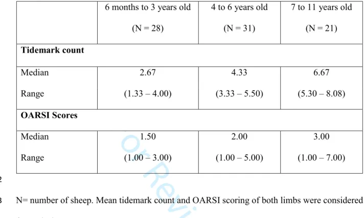
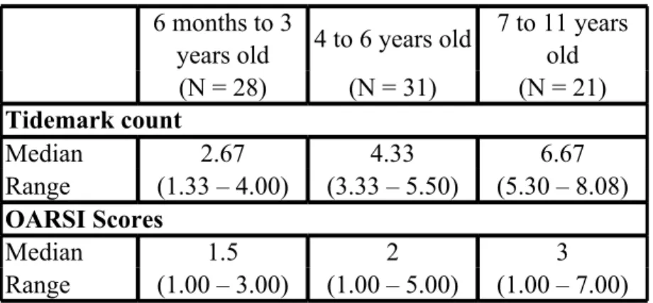
Table 1: Tidemark count and OARSI score values (median and interquartile range) for the three 540
age groups. 541
6 months to 3 years old (N = 28) 4 to 6 years old (N = 31) 7 to 11 years old (N = 21) Tidemark count Median 2.67 4.33 6.67 Range (1.33 – 4.00) (3.33 – 5.50) (5.30 – 8.08) OARSI Scores Median 1.50 2.00 3.00 Range (1.00 – 3.00) (1.00 – 5.00) (1.00 – 7.00) 542
N= number of sheep. Mean tidemark count and OARSI scoring of both limbs were considered 543
for each sheep. 544
The tidemark count (P<0.0001) and the OARSI scores (P=0.0197) differed significantly 545 between groups. 546 547 3 4 5 6 7 8 9 10 11 12 13 14 15 16 17 18 19 20 21 22 23 24 25 26 27 28 29 30 31 32 33 34 35 36 37 38 39 40 41 42 43 44 45 46 47 48 49 50 51 52 53 54 55 56 57 58 59
For Review Only
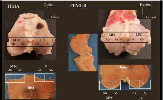
Figure legends 548
Figure 1. Sampling sites in the middle third of the medial tibial condyle (MTC), medial femoral 549
condyle (MFC), lateral tibial condyle (LTC) and lateral femoral condyle (LFC). Tibial slabs 550
were centered on the intercondylar eminence (black lines). Femoral slabs were obtained in the 551
centre of the middle third of the circumference of the condyle (black lines and dotted black 552
box). White rectangles illustrate the histological slices that were obtained, each abaxial (Ab) 553
and axial (Ax) part being assessed separately at microscopy. White arrows highlight cartilage 554
555
Figure 2. The osteochondral junction at histology. 556
A. The white line indicates non-calcified hyaline cartilage (HC); the black line is the calcified 557
cartilage (CC). 558
B. White arrows indicate tidemarks. 559
C. Histological slide showing the absence of tidemark in a sample of cartilage of the medial 560
femoral condyle in a 6 months old sheep. 561
562
Figure 3: Number of tidemarks in the different sub-regions for right and left limbs, expressed 563
as median and interquartile range (bar). Asterisks means that statistical significance (P<0.05) is 564
reached for the difference between the axial and the abaxial part of the region. 565
MFC, LFC: medial and lateral femoral condyle, respectively; MTC, LTC: medial and lateral 566
femoral condyle, respectively. 567
568
Figure 4: OARSI scores in the different sub-regions for right and left limbs, expressed as 569
median and interquartile range (bar). Asterisks means that statistical significance (P<0.05) is 570
reached for the difference between the axial and the abaxial part of the region. 571 3 4 5 6 7 8 9 10 11 12 13 14 15 16 17 18 19 20 21 22 23 24 25 26 27 28 29 30 31 32 33 34 35 36 37 38 39 40 41 42 43 44 45 46 47 48 49 50 51 52 53 54 55 56 57 58 59
For Review Only
MFC, LFC: medial and lateral femoral condyle, respectively; MTC, LTC: medial and lateral 572
femoral condyle, respectively. 573 3 4 5 6 7 8 9 10 11 12 13 14 15 16 17 18 19 20 21 22 23 24 25 26 27 28 29 30 31 32 33 34 35 36 37 38 39 40 41 42 43 44 45 46 47 48 49 50 51 52 53 54 55 56 57 58 59
For Review Only
Figure 1. Sampling sites in the middle third of the medial tibial condyle (MTC), medial femoral condyle (MFC), lateral tibial condyle (LTC) and lateral femoral condyle (LFC). Tibial slabs were centered on the intercondylar eminence (black lines). Femoral slabs were obtained in the centre of the middle third of the circumference of the condyle (black lines and dotted black box). White rectangles illustrate the histological
slices that were obtained, each abaxial (Ab) and axial (Ax) part being assessed separately at microscopy. White arrows highlight cartilage.
155x96mm (300 x 300 DPI) 3 4 5 6 7 8 9 10 11 12 13 14 15 16 17 18 19 20 21 22 23 24 25 26 27 28 29 30 31 32 33 34 35 36 37 38 39 40 41 42 43 44 45 46 47 48 49 50 51 52 53 54 55 56 57 58 59
For Review Only
Figure 2. The osteochondral junction at histology.
A. The white line indicates non-calcified hyaline cartilage (HC); the black line is the calcified cartilage (CC). B. White arrows indicate tidemarks.
C. Histological slide showing the absence of tidemark in a sample of cartilage of the medial femoral condyle in a 6 months old sheep.
92x95mm (300 x 300 DPI) 3 4 5 6 7 8 9 10 11 12 13 14 15 16 17 18 19 20 21 22 23 24 25 26 27 28 29 30 31 32 33 34 35 36 37 38 39 40 41 42 43 44 45 46 47 48 49 50 51 52 53 54 55 56 57 58 59
For Review Only
Figure 3: Number of tidemarks in the different sub-regions for right and left limbs, expressed as median and interquartile range (bar). Asterisks means that statistical significance (P<0.05) is reached for the difference
between the axial and the abaxial part of the region.
MFC, LFC: medial and lateral femoral condyle, respectively; MTC, LTC: medial and lateral femoral condyle, respectively. 90x85mm (300 x 300 DPI) 3 4 5 6 7 8 9 10 11 12 13 14 15 16 17 18 19 20 21 22 23 24 25 26 27 28 29 30 31 32 33 34 35 36 37 38 39 40 41 42 43 44 45 46 47 48 49 50 51 52 53 54 55 56 57 58 59
For Review Only
Figure 4: OARSI (OsteoArthritis Research Society International) scores in the different sub-regions for right and left limbs, expressed as median and interquartile range (bar). Asterisks means that statistical significance (P<0.05) is reached for the difference between the axial and the abaxial part of the region. MFC, LFC: medial and lateral femoral condyle, respectively; MTC, LTC: medial and lateral femoral condyle,
respectively. 92x92mm (300 x 300 DPI) 3 4 5 6 7 8 9 10 11 12 13 14 15 16 17 18 19 20 21 22 23 24 25 26 27 28 29 30 31 32 33 34 35 36 37 38 39 40 41 42 43 44 45 46 47 48 49 50 51 52 53 54 55 56 57 58 59
For Review Only
Table 1: Tidemark count and OARSI score values (median
and interquartile range) for the three age groups. 6 months to 3
years old 4 to 6 years old
7 to 11 years old (N = 28) (N = 31) (N = 21) Tidemark count Median 2.67 4.33 6.67 Range (1.33 – 4.00) (3.33 – 5.50) (5.30 – 8.08) OARSI Scores Median 1.5 2 3 Range (1.00 – 3.00) (1.00 – 5.00) (1.00 – 7.00) N= number of sheep. Mean tidemark count and OARSI scoring of both limbs were considered for each sheep. The tidemark count (P<0.0001) and the OARSI scores (P=0.0197) differed significantly between groups.
3 4 5 6 7 8 9 10 11 12 13 14 15 16 17 18 19 20 21 22 23 24 25 26 27 28 29 30 31 32 33 34 35 36 37 38 39 40 41 42 43 44 45 46 47 48 49 50 51 52 53 54 55 56 57 58 59