RESEARCH OUTPUTS / RÉSULTATS DE RECHERCHE
Author(s) - Auteur(s) :
Publication date - Date de publication :
Permanent link - Permalien :
Rights / License - Licence de droit d’auteur :
Bibliothèque Universitaire Moretus Plantin
Institutional Repository - Research Portal
Dépôt Institutionnel - Portail de la Recherche
researchportal.unamur.be
University of Namur
Auxin activity and molecular structure of 2-alkylindole-3-acetic acids
Antolic, Snjezana; Dolusic, Eduard; Kozic, Erika K.; Kojic-Prodic, Biserka; Magnus, Volker;
Ramek, Michael; Tomic, Sanja
Published in:
Plant Growth Regulation DOI:
10.1023/A:1022894914226
Publication date: 2003
Document Version
Publisher's PDF, also known as Version of record
Link to publication
Citation for pulished version (HARVARD):
Antolic, S, Dolusic, E, Kozic, EK, Kojic-Prodic, B, Magnus, V, Ramek, M & Tomic, S 2003, 'Auxin activity and molecular structure of 2-alkylindole-3-acetic acids', Plant Growth Regulation, vol. 39, no. 3, pp. 235-252. https://doi.org/10.1023/A:1022894914226
General rights
Copyright and moral rights for the publications made accessible in the public portal are retained by the authors and/or other copyright owners and it is a condition of accessing publications that users recognise and abide by the legal requirements associated with these rights. • Users may download and print one copy of any publication from the public portal for the purpose of private study or research. • You may not further distribute the material or use it for any profit-making activity or commercial gain
• You may freely distribute the URL identifying the publication in the public portal ? Take down policy
If you believe that this document breaches copyright please contact us providing details, and we will remove access to the work immediately and investigate your claim.
Auxin activity and molecular structure of 2-alkylindole-3-acetic acids
Snježana Antolic´
1, Eduard Dolušic´
1, Erika K. Kožic´
1, Biserka Kojic´-Prodic´
1, Volker
Magnus
1,*, Michael Ramek
2and Sanja Tomic´
11Ruder Boškovic´ Institute, Bijenicˇka cesta 54, p. p. 180, HR-10002 Zagreb, Croatia;2Institut für physikalische
und theoretische Chemie, Technische Universität Graz, A-8010 Graz, Austria; *Author for correspondence (e-mail: [email protected]; phone: +385-1-4561-002; fax: +385-1-4561-177)
Received 12 March 2002; accepted in revised form 19 August 2002
Key words: 2-Ethylindole-3-acetic acid, 2-Propylindole-3-acetic acid, Auxin, Conformational analysis,
Indole-3-acetic acid, X-ray crystallography
Abstract
2-Methylindole-3-acetic acid (2-Me-IAA) is a known auxin, but its 2-ethyl homologue has been considered in-active. Here we show that the compound previously bioassayed as ‘2-ethylindole-3-acetic acid’ (2-Et-IAA) was, in fact, 3-(3-methylindol-2-yl)propionic acid. The proper 2-Et-IAA and its 2-(n-propyl) homologue (2-Pr-IAA) are prepared, unambiguously characterized, and their auxin activity is demonstrated in the Avena coleoptile-sec-tion straight-growth test. Their half-optimal concentracoleoptile-sec-tions are approximately the same as for 2-Me-IAA (2 × 10⁻⁵mol L−1), and hence about ten times larger than for unsubstituted indole-3-acetic acid (IAA) and its
deriva-tives alkylated in positions 4, 5, 6 or 7. The optimal response elicited by 2-Et-IAA and 2-Pr-IAA is about half that observed for 2-Me-IAA. These characteristics place the three 2-alkyl-IAAs along the borderline between the classes of strong and weak auxins, thus corroborating the results of interaction similarity analysis, a mathemati-cal approach based on the capability of auxin molecules to participate in non-bonding interactions with a gen-eralized receptor protein. X-ray diffraction analysis shows no explicit structural features to be blamed for the decrease in auxin activity caused by attaching a 2-alkyl substituent to the IAA molecule; sterical interference of the 3-CH2COOH group and the 2-alkyl moiety is barely recognizable in the crystalline state. Quantum-chemical
calculations and molecular dynamics simulations suggest that 2-alkyl-IAAs, in the absence of crystal-packing restraints, prefer conformations with the CH2-COOH bond tilted to the heterocyclic ring system. Substantially
higher conformational energy (and hence lower abundance) is predicted for planar conformers which were pre-viously shown to prevail for IAA and many of its derivatives substituted in the benzene moiety of the indole nucleus. This shift in the rotational preferences of the -CH2COOH moiety may be one of the reasons for the
reduced plant-growth promoting activity of 2-alkyl-IAAs.
Abbreviations: 2-Et-IAA – 2-ethylindole-3-acetic acid, 2-Pr-IAA – 2-(n-propyl)indole-3-acetic acid, IAA –
in-dole-3-acetic acid, TLC – thin-layer chromatography
Introduction
A large number of indole-3-acetic acids (IAAs) sub-stituted in the side chain and in ring-positions 4–7 have been screened for their plant-growth regulating properties (Antolic´ et al. 1996, 1999; Hatano et al. 1987; Jönsson 1961; Katayama et al. 1998; Katekar 1979; Kojic´-Prodic´ et al. 1991; Nigovic´ et al. 2000;
Porter and Thimann 1965), but derivatives with sub-stituents in the (pyrrole) 2-position have received lit-tle attention. Yet, these IAA derivatives are of theo-retical interest because large 2-substituents may block access to the carboxyl group, a structural element which appears to be critical for recognition by pro-teins which participate in auxin physiology.
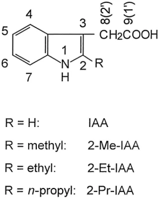
2-Methylindole-3-acetic acid (2-Me-IAA; Fig-ure 1) is somewhat less active than IAA in a variety of bioassays (Hoffmann et al. 1952; Kögl and Kos-termans 1935; Muir et al. 1949; Muir and Hansch 1953; Nigovic´ et al. 2000; Porter and Thimann 1965; Rescher et al. 1996; Sell et al. 1952). Adding a 2-me-thyl group to 5-chloro-IAA, 7-chloro-IAA, 4,7-dichloro-IAA and 5,7-4,7-dichloro-IAA also reduces auxin activity (Hoffmann et al. 1952; Muir et al. 1949). On the other hand, there have only been few attempts to introduce substituents other than methyl into the IAA 2-position. Kögl and Kostermans (1935) allegedly prepared 2-ethylindole-3-acetic acid (2-Et-IAA) and found no growth-promoting activity, but could not, in 1935, establish the chemical identity of their product beyond reasonable doubt. In fact, Wal-ton et al. (1968) attributed the same structure to a compound with a melting point about 100° above that reported by Kögl and Kostermans (1935). Porter and Thimann (1965) synthesized 2-chloroindole-3-acetic and 2-bromoindole-3-acetic acids, both of which were claimed to be more active than IAA in the split pea-stem curvature bioassay. Unfortunately, the C2-halo-gen bond is subject to acid-catalyzed hydrolysis (Por-ter and Thimann 1965; Lawson et al. 1960) and the hydrochloric and hydrobromic acids thus formed not only accelerate the decomposition, but may also cause an acid-growth response (Cosgrove 1998).
Here we demonstrate that the compound Kögl and Kostermans (1935) described as ‘2-Et-IAA’ was, in fact, its isomer, 3-(3-methylindol-2-yl)propionic acid which is not expected to be an auxin. Authentic 2-Et-IAA and its homologue, 2-(n-propyl)indole-3-acetic acid (2-Pr-IAA) are prepared by an unequivocal route, characterized by spectroscopic and crystallo-graphic methods, and shown to be auxins. Their plant-growth promoting properties are rationalized considering their molecular structures in the solid state and in solution. Interaction similarity analysis also suggests that the two 2-alkyl-IAAs fit well into the auxin family.
Materials and methods
Bioassays
Auxin activity was monitored by the Avena coleop-tile-section straight-growth test (Mitchell and Living-ston 1968; Larsen 1961). In brief, seeds of Avena
sativa L. cv. Pula and cv. Valiant were soaked in
run-ning tap water for 6 h, sown onto moist vermiculite, and germinated at 27 °C, for ca. 80 h, in complete darkness, except for a short daily exposure to red light. Coleoptiles, 25–30 mm in length, were selected under green light and 10 mm sections (1 per coleop-tile) were excised starting 2 mm below the tip. They were temporarily (ca. 2 h) stored floating on distilled water before being distributed, in aliquots of 10–15, to disposable polystyrene Petri dishes (diameter: 3 cm) containing 2.7 mL of auxin solution in glass-dis-tilled water. A water control and a complete series of IAA dilutions were included in each set of bioassays. After 20 h of growth in darkness at 27 °C, shadow graphs (original size) of the sections on photographic paper were prepared and measured to the nearest 0.1 mm. To the dose-response data were fitted fourth- and fifth-degree polynomials and the function
y⫽ d ⫹ 共a/s兲关1/共x ⫹ c兲兴exp兵⫺ 关ln共x ⫹ c兲 ⫺ b兴2/2s2其
wherein y is the length of the coleoptile sections in mm, x is the negative logarithm of the auxin concen-tration in mol L−1, and a, b, c, d and s are shape
pa-rameters optimized by the fitting process. The curve Figure 1. Structural formulae of the auxins studied indicating the
numbering of selected positions. For the 3-side chain, the crystal-lographic numbering conventions used in this report differ from IUPAC recommendations (shown in parentheses).
optimally representing the data points was chosen to estimate the maximal elongation and the optimal and half-optimal concentrations.
Preparative methods General
Melting points were determined in open capillaries and were not corrected. UV spectra were measured using a Varian Cary 5 digital spectrophotometer. NMR spectra were recorded at 20 °C on a Varian Gemini 300 spectrometer operating at 300 MHz for
1H and at 75 MHz for 13C. Chemical shifts are
re-ported in parts per million downfield from tetrameth-ylsilane. Column chromatography was on silica gel 60 (Merck), particle diameter: 0.063–0.2 mm. Thin-layer chromatography (TLC) was on silica gel GF254
(Merck) developing with dichloromethane/methanol/ acetic acid (90:10:1, solvent A1; 100:50:2, solvent A2;
90:45:5, solvent A3; 100:20:5, solvent A4; 90:5:1,
sol-vent A5), 2-propanol/ethyl acetate/25% aq. ammonia
(35:45:20; solvent B), and 2-propanol/ethyl acetate/ water (24:65:11, solvent C). In addition to detection by UV absorption, chromatograms were sprayed with the Ehrlich reagent (1% p-dimethylaminobenzalde-hyde in a 1/1 mixture of ethanol and 35% HCl). The solvents employed were of analytical purity; the use of phosgene-free chloroform (stabilized with 1% eth-anol) and peroxide-free diethyl ether and dioxane is essential. 4-ketohexanoic (homolevulinic) acid was obtained as follows: methyl 4-nitrohexanoate was prepared by triethylamine-catalyzed condensation of 1-nitropropane and methyl acrylate (Kloetzel 1948), purified by column chromatography (eluent dichloro-methane containing decreasing proportions of n-hep-tane), and its sodium salt (prepared in 20% v/v aq. EtOH) was subjected to acid decomposition (Nef re-action) (Kloetzel 1948). 2-Ethylindole and 2-(n-pro-pyl)indole were synthesized by sodium amide-medi-ated cyclization of N-propyl and N-butyl-o-toluidine (Verley and Beduwé 1925). All other chemicals used, including 2-Me-IAA, were obtained through commer-cial suppliers. The essential steps in the syntheses performed herein are summarized in Figure 2.
2-Ethylgramine
[N,N-dimethyl-(2-ethylindole-3-)methanamine]
2-Ethylindole was treated with formaldehyde/dimeth-ylamine in dioxane/acetic acid, essentially as de-scribed by Le Goffic et al. (1973), except that the product was recrystallized from acetone/cyclohexane
(1:9). M. p. 115–117 °C [lit. (Le Goffic et al. 1973) 119–121 °C; from acetone/water]. Rf = 0.7 (solvent
A2), 0.6 (solvent A3), 0.7 (solvent B).
1
H-NMR (CD3OD). Gramine moiety:␦7.60 (dd, 1H, J4,5= 6.8
Hz, J4,6= 1.4 Hz, H-4), 7.06 (ddd, 1H, J5,6= 7.2 Hz,
J5,7 = 1.2 Hz, H-5), 7.11 (ddd, 1H, H-6), 7.35 (dd,
1H, J6,7 = 7.1 Hz, H-7), 3.68 (s, 2H, CH2), 2.34 (s,
6H, 2CH3); ethyl moiety:␦2.90 (q, 2H, Jvic= 7.6 Hz,
CH2), 1.40 (t, 3H, CH3). 13C-NMR (CD 3OD). Gramine moiety: ␦141.7 (C-2), 107.0 (C-3), 130.6 (C-3a), 120.1, 119.4 (C-4, C-6), 121.9 (C-5), 111.7 (C-7), 137.4 (C-7a), 53.8 (CH2), 45.5 (2CH3); ethyl moiety:␦20.5 (␣-C), 14.7 (-C). 2-(n-Propyl)gramine [N,N-dimethyl-(2-n-propylindole-3-)methanamine]
2-(n-Propyl)indole, when processed as described for the preparation of 2-ethylgramine, afforded 74% of the title compound, m. p. 98 – 100 °C, Rf= 0.8
(sol-vent A2), 0.4 (solvent A4), 0.8 (solvent B).
1H-NMR
(CD3OD). Gramine moiety: ␦7.51 (ddd, 1H, J4,5 =
7.3 Hz, J4,7= 1.0 Hz, H-4), 6.97 (ddd, 1H, J5,6= 7.0 Hz, J5,7= 1.2 Hz, H-5), 7.02 (ddd, 1H, J4,6= 1.5 Hz, H-6), 7.25 (dd, 1H, J6,7= 7.1 Hz, H-7), 3.59 (s, 2H, CH2), 2.25 (s, 6H, 2CH3); n-propyl moiety:␦2.76 (t, 2H, Jvic= 7.6 Hz,␣-CH2), 1.74 (sextet, 2H,-CH2), 0.97 (t, 3H, Jvic= 7.4 Hz, CH3). 13 C-NMR (CD3OD). Gramine moiety: ␦140.2 (C-2), 107.8 (C-3), 130.5 (C-3a), 120.0, 119.4 (C-4, C-6), 121.9 (C-5), 111.7 (C-7), 137.4 (C-7a), 53.9 (CH2), 45.6 (2CH3); n-pro-pyl moiety: 29.4 (␣-C), 24.3 (-C), 14.6 (CH3).
2-Ethylindole-3-acetic acid (2-Et-IAA)
The reaction and work-up were performed in an effi-cient fume hood, as highly toxic HCN gas is formed as a by-product. Adapting a method for the synthesis of indole-3-acetic acid (Snyder and Pilgrim 1948), a solution of 2-ethylgramine (1.71 g, 8.46 mmol) and KCN (2.74 g, 42.1 mmol) in 95% ethanol (17.0 mL) and water (3.7 mL) was boiled until TLC (solvents A5and B) showed complete consumption of the
start-ing gramine (70 h). After coolstart-ing to room tempera-ture, water (3.4 mL) and solid KOH (3.7 g, 65.5 mmol) were added and boiling was resumed for 10 h. The cooled solution was diluted with water (43 mL) and neutral by-products were extracted with diethyl ether. The organic phase was re-partitioned against 5% aq. Na2CO3 which was added to the aqueous
phase. The latter was then acidified to pH 2.5 with concd HCl and extracted with diethyl ether. Evapora-tion of the organic phase gave the crude title
com-pound (1316 mg, 77%). Repeated recrystallization from 1. ethyl acetate/cyclohexane (20:1) and 2. chlo-roform/ethyl acetate (2:1) afforded off-white crystals, m. p. 190–197 °C (decomposition) [lit. (Walton et al. 1968): decomposition at 193–203 °C; crystallization from acetone/petrol]. Rf= 0.5 (solvent A5), 0.4
(sol-vent B). 1
H-NMR (CD3OD). Indole-3-acetic acid
moiety:␦7.53 (d, 1H, J4,5= 7.6 Hz, H-4), 7.05 (ddd,
1H, J5,6= 7.0 Hz, J5,7= 1.4 Hz, H-5), 7.11 (ddd, 1H,
J4,6= 1.4 Hz, H-6), 7.34 (dd, 1H, J6,7= 7.6 Hz, H-7),
3.75 (s, 2H, CH2); ethyl moiety:␦2.87 (q, 2H, Jvic=
7.6 Hz, CH2), 1.38 (t, 3H, CH3).
13C-NMR (CD 3OD).
Indole-3-acetic acid moiety:␦140.3 2), 104.3 3), 130.1 3a), 119.9, 119.0 4, C-6), 121.8 (C-5), 111.7 (C-7), 137.4 (C-7a), 31.1 (CH2), 176.9
(COOH); ethyl moiety: ␦20.5 (␣-C), 14.9 (-C). UV
(95% EtOH). max(log⑀) 226.5 (4.47), 276.2 (3.81,
shoulder), 283.1 (3.85), 290.4 (3.80); reference val-ues for IAA: 222.7 (4.50), 276.3 (3.73, shoulder), 282.0 (3.76), 290.0 (3.69); 2-Me-IAA: 225.8 (4.44),
276.1 (3.77, shoulder), 282.3 (3.80), 289.8 (3.73).
X-ray structural analysis. See Figure 3a. 2-(n-Propyl)indole-3-acetic acid (2-Pr-IAA)
The title compound was prepared from 2-(n-propyl)-gramine in essentially the same way as 2-Et-IAA (crude yield: 82%) and repeatedly recrystallized from chloroform to yield off-white crystals, m. p. 150–153 °C [lit. (Rokach 1973): 154–158 °C]. Rf = 0.5
(sol-vent B), 0.7 (sol(sol-vent C). 1
H-NMR (CD3OD).
Indole-3-acetic acid moiety: ␦7.53 (d, 1H, J4,5 = 7.6 Hz,
H-4), 7.05 (ddd, 1H, J5,6 = 6.6 Hz, J5,7 = 1.4 Hz, H-5), 7.11 (dd, 1H, H-6), 7.34 (dd, 1H, J6,7= 7.4 Hz, H-7), 3.75 (s, 2H, CH2); n-propyl moiety:␦2.82 (t, 2H, Jvic = 7.5 Hz, ␣-CH2), 1.82 (sextet, 2H, -C), 1.06 (t, 3H, Jvic= 7.4 Hz, CH3). 13C-NMR (CD 3OD).
Indole-3-acetic acid moiety:␦138.8 2), 105.1 3), 130.1 3a), 119.9, 119.1 4, C-6), 121.8 (C-5), 111.7 (C-7), 137.4 (C-7a), 31.2 (CH2), 176.9
(COOH); n-propyl moiety: ␦29.3 (␣-C), 24.4 (-C), 14.5 (␥-C). UV (95% EtOH). max (log ⑀) 226.4
Figure 2. Chemical reactions discussed in the text. The names of the compounds shown are as follows. 1 = 4-phenylhydrazonohexanoic
acid, 2 = 2-ethylindole-3-acetic acid (2-Et-IAA), 3 = 3-(3-methylindol-2-yl)propionic acid, 4 = 2-alkylindole, 5 = 2-alkylgramine or N,N-dimethyl-(2-alkylindole-3-)methanamine, 6 = 2-alkylindole-3-acetonitrile, 7 = 2-alkylindole-3-acetic acid; ‘alkyl’ in compounds 4 – 7 is ethyl or n-propyl.
(4.49), 276.5 (3.84), 283.1 (3.88), 290.4 (3.82). X-ray
structural analysis. See Figure 3b. 3-(3-Methylindol-2-yl)propionic acid
4-Phenylhydrazonohexanoic acid was subjected to Fischer cyclization in abs. ethanol/sulfuric acid, as detailed by Kögl and Kostermans (1935) in their at-tempted synthesis of 2-ethylindole-3-acetic acid. Yield 44% (after one recrystallization from water), m. p. 97–99 °C [The product obtained by Kögl and Ko-stermans (1935) melted at 100–101 °C]. Rf= 0.5
(sol-vent B). 1 H-NMR (CDCl3).␦8.05 (broadened s, 1H, H-1), 7.49 (d, 1H, J4,5= 7.8 Hz, H-4), 7.08 (ddd, 1H, J5,6= 6.4 Hz, J5,7= 1.1 Hz, H-5), 7.13 (ddd, 1H, J4,6 = 1.4 Hz, H-6), 7.26 (dd, 1H, J6,7= 8.1 Hz, H-7), 2.24 (s, 3H, CH3), 3.03 (t, 2H, Jvic= 6.8 Hz, Ar-CH2), 2.71 (t, 2H, CH2COOH). 13C-NMR (CDCl 3). ␦133.0 (C-2), 107.4 (C-3), 129.0 (C-3a), 119.0, 118.2 (C-4, C-6), 121.4 (C-5), 110.4 (C-7), 135.2 (C-7a), 8.2 (CH3),
20.4 (Ar-CH2), 33.8 (CH2COOH), 179.4 (COOH).
X-ray structural analysis. See Figure 4.
Figure 3. Molecular structures [ORTEP II (Johnson 1976)] of (a) 2-ethylindole-3-acetic acid (2-Et-IAA) and (b) 2-(n-propyl)indole-3-acetic
acid (2-Pr-IAA) showing the crystallographic numbering conventions referred to in the text. The thermal ellipsoids are scaled at the 30% probability level.
X-ray crystallography
Crystals suitable for X-ray structure analysis were prepared by slow evaporation of 2 mL solutions con-taining 5–10 mg mL−1of compound in CHCl
3. The
crystals were grown at room temperature overnight. The compounds studied have no chiral centers and accordingly crystallize in the centrosymmetric space groups: P1¯ [3-(3-methylindol-2-yl)propionic acid] and P21/a (2-Et-IAA and 2-Pr-IAA). Data were
col-lected on an Enraf-Nonius CAD-4 diffractometer (Ta-ble 1) with graphite-monochromated CuK␣ (2-Et-IAA) and MoK␣ (2-Pr-IAA) radiation and rescaled for decay on the basis of intensity reduction of stan-dard reflections. Data for 3-(3-Me-indol-2-yl)propi-onic acid were exceptionally collected at 133K on a Huber/Stoe diffractometer with attached Siemens Kappa Charge-Coupled Device electronic area detec-tor and using a molybdenum sealed tube. Lorentz and polarization corrections were applied using the
HEL-ENA (Spek 1993) program. Empirical absorption
cor-rection [⌿-scan, PLATON (Spek 1997)] was applied for 2-Et-IAA, and semiempirical from equivalents [SADABS (Sheldrick 1996)] for 3-(3-methylindol-2-yl)propionic acid. The structures were solved by the
SHELXS (Sheldrick 1997a) program and refined
us-ing SHELXL (Sheldrick 1997b). Hydrogen atoms were generated on stereochemical grounds. The non-H atoms were refined anisotropically; details of the refinement procedures are listed in Table 1. Scat-tering factors are those included in SHELXL (Sheld-rick 1997b). Molecular geometry was calculated by the program PLATON (Spek 1997). Drawings were prepared using ORTEPII (Johnson 1976) incorpo-rated in PLATON (Spek 1997). The final atomic co-ordinates and equivalent isotropic thermal parameters are listed in Tables 2a, 2b, 2c*. Calculations were * Lists of atomic coordinates, anisotropic displace-ment parameters and structure factors have been de-posited with the IUCr [Reference: CCDC-139864 for 2-Et-IAA and CCDC-139865 for 3-(3-methylindol-2-yl)propionic acid). Copies may be obtained through Figure 4. Molecular structure [ORTEP II (Johnson 1976)] of 3-(3-methylindol-2-yl)propionic acid. The elementary cell contains two
non-equivalent molecules, A and B, of the indolic acid and a molecule of chloroform, the solvent from which the crystals subjected to X-ray analysis were obtained. Generally, the number ‘1’ was appended to the atom numbers in molecule B. Only the two carbons at the junction of the benzene and pyrrole rings required different labeling to avoid confusion: C31⬘ and C71⬘ in molecule A and C311 and C711 in molecule
performed on a Silicon Graphics OCTANE worksta-tion in the Laboratory for Chemical Crystallography and Biocrystallography, RuWer Boškovic´ Institute, Zagreb, Croatia.
Computational methods Molecular modeling
Conformational analysis for undissociated 2-alkyl-IAA molecules and the corresponding carboxylate an-ions was performed by the semiempirical methods PM3 and AM1 from the MOPAC package of pro-grams (Stewart 1990). The conformations (i.e. the corresponding potential-energy surfaces) of 2-Me-IAA and 2-Et-2-Me-IAA were also, to some extent, ana-lyzed by ab initio self-consistent field calculations using the Restricted Hartree-Fock (RHF) formalism and the 6–31G* basis set which was previously found to be adequate for IAA and its ring-substituted deriv-atives (Ramek et al. 1995, 1996; Ramek and Tomic´ 1998a, 1998b, 1999). The latter calculations were per-formed with the program GAMESS (Schmidt et al. 1993). The resulting conformers are grouped into 1.
T (tilted) conformers with the CH2-COOH bond
per-pendicular, or near-perper-pendicular, to the ring plane and 2. P (planar) conformers with that bond in the ring plane (or nearly so).
To investigate possible conformational transitions of 2-alkyl-IAAs in aqueous solution, molecular dy-namics simulations were performed by the program DISCOVER (DISCOVER, release 1997) using the CVFF (Dauber-Osguthorpe et al. 1988) force field and periodic boundary conditions. The optimized conformations were ‘soaked’ in a cubic box of water with the starting dimensions of 17 × 17 × 17 Å3.
Electrostatic interactions were considered up to a dis-tance of 15 Å. The use of two cut-off values utilized together with a smoothing function, for the range of 13.5–15 Å, enabled proper treatment of long distance electrostatic interactions. After equilibrating the sys-tem for 10 ps at 300 K, ca. 2 ns of productive simu-lation was carried out at a temperature ranging from 273–393 K, using 1 fs time steps.
Similarity analysis
The 2-alkyl-IAAs studied herein were compared with three classes of auxin-related compounds: 1) strong auxins, 2) weak auxins with weak antiauxin proper-ties, and 3) inactive compounds with structural rela-tionships to the auxin family. Details of the classifi-cation and the mathematical background were described previously (Tomic´ et al. 1998a, 1998b). In brief, the interaction energies between the 2-alkyl-IAAs and selected ‘probes’ were calculated for pre-defined arrays of sampling points, at the molecular surface and at a grid enclosing the molecule. Two of the probes employed reflect general properties of the amino acid residues surrounding the active site of the postulated auxin receptor(s): H2O for hydrophilic
(and thus hydrated under physiological conditions), and DRY for hydrophobic residues. Three further probes conventionally termed NH2
+, CH
3 and O
re-flect the interactions with specific functional groups present in many protein amino acids.
P and T conformers of auxin molecules required
separate treatment. The resulting ‘molecular interac-tion fields’ were compared to those obtained for typi-cal representatives of classes 1 to 3. This was accom-plished by calculating the Carbó similarity index (Carbó and Calabuig 1992), averaging, if required, over multiple P or T conformers and over multiple class representatives: the larger the similarity index, the closer the connection to the respective class. The class representatives chosen here were: 4-chloro-2-methylphenoxyacetic acid, IAA and 4-chloro-IAA for class 1, indole-3-propionic acid and 4,7-dichloroin-dole-3-acetic acid for class 2, and benzoic acid for class 3.
Results and discussion
Growth-promoting properties of 2-Et-IAA and 2-Pr-IAA
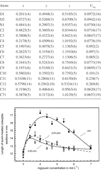
2-Et-IAA and 2-Pr-IAA prepared as outlined in the Materials and Methods section were active auxins. Typical dose-response curves are presented in Fig-ure 5. Because of the relatively weak response, bio-assays were performed with two oat cultivars and re-peated altogether 10 times for 2-Et-IAA and 9 times for 2-Pr-IAA. Dose-dependent growth-promotion was observed in every single case. The results of the ex-periments in which both alkyl homologues were as-The Managing editor, International Union for
Crys-tallography, 5 Abbey Square, Chester CH1 2HU, En-gland. Due to the high anisotropy of part of the 2-Pr-IAA molecule these data are not deposited.
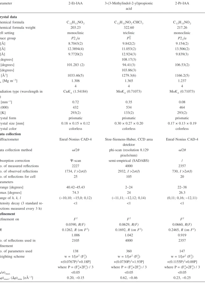
Table 1. Experimental details for X-ray structure analysis.
Parameter 2-Et-IAA 3-(3-Methylindol-2-yl)propionic acid
2-Pr-IAA
Crystal data
Chemical formula C12H13NO2 C12H13NO2·CHCl3 C13H15NO2
Chemical formula weight 203.23 322.60 217.26
Cell setting monoclinic triclinic monoclinic
Space group P21/a P1 P21/a a [Å] 8.7043(2) 9.842(2) 9.154(2) b [Å] 12.3894(4) 11.053(2) 13.506(2) c [Å] 9.7720(2) 12.924(3) 9.839(3) ␣[degrees] 108.17(3) [degrees] 101.283 (2) 94.41(3) 106.53(2) ␥[degrees] 103.86(3) V [Å3] 1033.46(5) 1279.3(6) 1166.2(5) Dx[Mg m−3] 1.306 1.365 1.237 Z 4 4 4
Radiation type (wavelength in Å)
CuK␣(1.54184) MoK␣(0.71073) MoK␣(0.71073)
[mm−1] 0.72 0.35 0.08
F (000) 432 534 464
T [K] 293(2) 133(2) 293(2)
Crystal form prismatic prismatic prismatic
Crystal size [mm] 0.18 × 0.15 × 0.12 0.30 × 0.27 × 0.20 0.17 × 0.13 × 0.19
Crystal color colorless colorless colorless
Data collection
Diffractometer Enraf-Nonius CAD-4 Stoe-Siemens-Huber, CCD area detektor
Enraf-Nonius CAD-4
Data collection method /2 phi-scan (resolution 8.129 pixels/mm)
/2 Absorption correction ⌿-scan semi-empirical (SADABS) /
No. of measured reflections 2227 4000 2357
No. of observed reflections 1734, I >2(I) 2932, I >2(I) 730, I >2(I) No. of reflections for cell
parameters
25 105 20
range [degrees] 40.42–45.43 2–24 22–38
max [degrees] 74.3 24 26.3
Range of h, k, l (−10,10; −15,0; 0,12) (−11,11; −12,12; 0,14) (0,11; 0,16; −12,11) Intensity decay (3 standard
re-flections measured every 3 h)
<1 <1 <1
Refinement
Refinement on F2 F2 F2
R 0.0390, R(F) 0.0629, R(F) 0.0660, R(F)
wR 0.1262, R (on F2) 0.1692, R (on F2) 0.2465, R (on F2)
S 1.006 1.042 0.919
No. of reflections used in refinement
2105 4000 2357
No. of parameters used 138 360 147
Weighting scheme w = 1/[2(F o 2) w = 1/[2(F o 2) w = 1/[2(F o 2) +(0.0767P)2+0.18P] +(0.0730P)2+1.93P] +(0.1155P)2+0.00P] where P = (Fo2+2F c 2) / 3 where P = (F o 2+2F c 2) / 3 where P = (F o 2+2F c 2) / 3 (⌬/)max <0.05 <0.05 <0.05
sayed in parallel with IAA are summarized in Table 3. The values found for the optimal and half-optimal concentrations of IAA are obviously close to the re-spective data from a previous, larger, set of experi-ments (Nigovic´ et al. 2000). The corresponding val-ues for 2-Et-IAA and 2-Pr-IAA were consistently larger (i.e. the negative logarithms shown in Table 3 were smaller) by ca. 0.5 orders of magnitude for the optimal and by ca. 1 order of magnitude for the half-optimal concentrations and, thus, about the same as previously determined for 2-Me-IAA (Nigovic´ et al. 2000). However, the maximal response for 2-Et-IAA and 2-Pr-IAA was only 20–50% of the IAA control, while 2-Me-IAA reached 86% (Nigovic´ et al. 2000). In a similar fashion, increasing chain length of n-alkyl substituents at ring position 4, 5, and 6 had little ef-fect on the optimal and half-optimal concentrations, but substantially reduced the maximal response (Nigovic´ et al. 2000).
Chemical syntheses
The synthetic procedures employed are summarized in Figure 2. As shown in its upper part, 4-phenylhy-drazonohexanoic acid (1) may undergo Fischer cy-clization in two ways: either C-3 or C-5 may establish a bond to one of the phenyl ortho-position to yield, after elimination of ammonia, 2-Et-IAA (2) or 3-(3-methylindol-2-yl)propionic acid (3). 2-Et-IAA was al-legedly formed using the protocol adopted by Kögl
and Kostermans (1935). If this was correct, then the aliphatic region of the 1
H-NMR spectrum should show a 2H singlet (CH2COOH), a 2H quartet (CH2
-CH3), and a 3H triplet (CH2-CH3). The product we
obtained repeating the protocol of Kögl and Koster-mans (1935) showed, however, a 3H singlet (CH3)
and two 2H triplets (CH2-CH2-COOH), the signals
expected for 3-(3-methylindole-2-yl)propionic acid. Table 2a. Fractional atomic coordinates and equivalent isotropic
displacement parameters (Ueq= (1/3)⌺i⌺jU ijaiaja i.aj; in Å2) for 2-Et-IAA. Atom x y z Ueq O1 0.29744(13) 0.01798(11) 1.00399(10) 0.0586(4) O2 0.41839(13) −0.03697(11) 0.83763(10) 0.0599(4) N1 0.00614(14) 0.22382(11) 0.58349(10) 0.0458(4) C2 0.00693(15) 0.15531(12) 0.69484(10) 0.0426(4) C3 0.10795(15) 0.07187(11) 0.68679(10) 0.0385(4) C4 0.27358(16) 0.03014(12) 0.49737(10) 0.0450(4) C5 0.30446(18) 0.06772(14) 0.37279(10) 0.0518(5) C6 0.23640(18) 0.16254(15) 0.31237(10) 0.0520(5) C7 0.13522(17) 0.22231(13) 0.37473(10) 0.0474(4) C8 0.13982(17) −0.02324(13) 0.78365(10) 0.0462(5) C9 0.29675(16) −0.01422(11) 0.87949(10) 0.0403(4) C10 −0.09259(19) 0.17843(16) 0.79996(10) 0.0566(5) C11 −0.0076(3) 0.2359(2) 0.9275(2) 0.0804(8) C31 0.17047(14) 0.08811(11) 0.56308(10) 0.0372(4) C71 0.10327(14) 0.18415(12) 0.50015(10) 0.0397(4)
Table 2b. Fractional atomic coordinates and equivalent isotropic
displacement parameters (Ueq= (1/3)⌺i⌺jU ijaiaja
i.aj; in Å2) for
3-(3-methylindol-2-yl)propionic acid, molecules A and B
Atom x y z Ueq Molecule A O1 −0.0183(4) −0.3328(3) 0.0635(2) 0.0523(11) O2 −0.0676(3) −0.5174(3) 0.1057(2) 0.0427(10) N1 −0.0691(3) −0.2040(3) 0.4934(2) 0.0345(11) C2 −0.1669(4) −0.3120(3) 0.4159(3) 0.0305(11) C3 −0.2964(4) −0.3242(3) 0.4496(3) 0.0319(11) C4 −0.3703(4) −0.1798(4) 0.6264(3) 0.0388(14) C5 −0.3159(5) −0.0734(4) 0.7222(3) 0.0419(16) C6 −0.1723(5) −0.0042(4) 0.7470(3) 0.0413(14) C7 −0.0791(4) −0.0391(4) 0.6755(3) 0.0383(14) C8 −0.1267(4) −0.3931(4) 0.3137(3) 0.0335(12) C9 −0.0986(5) −0.3203(4) 0.2317(3) 0.0434(16) C10 −0.0611(4) −0.4004(4) 0.1283(3) 0.0380(12) C11 −0.4325(4) −0.4280(4) 0.3887(3) 0.0467(16) C31 −0.2785(4) −0.2192(4) 0.5525(3) 0.0321(12) C71 −0.1350(4) −0.1469(3) 0.5784(3) 0.0319(12) Molecule B O11 0.0949(3) −0.0870(3) −0.0970(2) 0.0369(9) O21 −0.1272(3) −0.1397(2) −0.0632(2) 0.0314(8) N11 −0.1944(3) −0.5237(3) −0.4255(2) 0.0274(10) C21 −0.2178(3) −0.5191(3) −0.3198(2) 0.0270(11) C31 −0.2567(3) −0.6442(3) −0.3165(2) 0.0265(11) C41 −0.2961(4) −0.8722(3) −0.4747(3) 0.0309(11) C51 −0.2925(4) −0.9261(4) −0.5849(3) 0.0345(11) C61 −0.2540(4) −0.8461(4) −0.6495(3) 0.0341(12) C71 −0.2190(4) −0.7102(4) −0.6041(3) 0.0310(11) C81 −0.2019(4) −0.3883(3) −0.2317(3) 0.0301(11) C91 −0.0546(4) −0.2928(3) −0.2109(3) 0.0284(11) C101 −0.0344(4) −0.1667(3) −0.1164(3) 0.0276(11) C111 −0.3006(4) −0.6860(4) −0.2213(3) 0.0374(12) C311 −0.2613(3) −0.7328(3) −0.4257(2) 0.0264(11) C711 −0.2230(3) −0.6545(3) −0.4921(2) 0.0265(11) Cl1a 0.3921(3) 0.1523(3) −0.1035(2) 0.0683(7) Cl2a 0.5076(2) 0.3981(2) 0.0722(2) 0.0884(8) Cl3a 0.6199(2) 0.1799(2) 0.0641(2) 0.1078(9) C12a 0.4663(5) 0.2247(4) 0.0360(3) 0.0561(17) a atoms of chloroform (solvent from which the compound was
The results of X-ray crystallography shown in Fig-ure 4 confirmed that this was indeed the correct struc-ture.
Chandra et al. (1980) allegedly synthesized the proper 2-Et-IAA, as well as 2-Pr-IAA, using a slight modification of the approach of Kögl and Koster-mans. Their procedure was, however, not described in sufficient detail and the identity of their products was not documented by error-proof data. Here we prepare 2-Et-IAA (2) from the preformed indole ring system (4, R = C2H5), via 2-ethylgramine (5, R = C2H5) and
2-ethylindole-3-acetonitrile (6, R = C2H5) (lower part
of Figure 2), as reported by Walton et al. (1968) with-out experimental details. An analogous approach was used to synthesize 2-Pr-IAA, which was previously obtained by catalytic reduction of 2-ethylindolopy-rone (Rokach 1973). The lots of 2-Et-IAA and 2-Pr-IAA used in bioassays had melting points consistent with the values given by Walton et al. (1968) and Rokach (1973), while TLC suggested a purity well above 98%. The identity of the 2-alkyl-IAAs was fur-ther confirmed by NMR-spectroscopy and X-ray crystallography (Figure 3a, b).
X-ray crystallography
The molecular structures of 2-Et-IAA, 2-Pr-IAA and 3-(3-methylindole-2-yl)propionic acid in the crystal-line state were determined by X-ray structure analy-sis. The results are shown in Figures 3 and 4. Details of the molecular geometry are presented in Tables 4, 5 and 6. Deviations of the molecular geometry of the benzene moiety of the indole ring system, including shortening of the C4-C5 bond (arithmetic mean: 1.375(5) Å; Table 4) and closing of the bond angle C6-C7-C71 (arithmetic mean: 117.4(4)°; Table 5), have already been observed for a large number of other indolic compounds (Nigovic´ et al. (2000) and references cited therein). In the pyrrole ring, alkyl substitution at C2 slightly decreases the endocyclic bond angle at that position, as revealed by comparing the data in Table 5 to the corresponding values for IAA [110.1(2)°] and 2-Me-IAA [108.2(2)°] (Nigovic´ et al. 2000). Comparable changes at the site of sub-stitution were also observed for IAAs alkylated in the benzene moiety (Nigovic´ et al. 2000) indicating ad-justments of orbital hybridization due to electronic substituent effects.
The overall conformations of the molecules stud-ied is described by torsion angles (Table 6) C2-C3-C8-C9 (T1) which defines the orientation of the CH2
-COOH group relative to the indole ring plane, and N1-C2-C10-C11 (T3) which reflects the respective orientation of the 2-alkyl substituent. The distal ends of the 2- and 3-substituents are turned to the same side of the ring plane. Both the C8-C9 (CH2-COOH)
and the C10-C11 (CH2-CH3 in 2-Et-IAA; CH2-CH2
in 2-Pr-IAA) bonds are near-perpendicular to the aro-matic system. Moreover, while the carboxyl group in IAA and 2-Me-IAA is coplanar with the C3-C8 bond and thus turned away from C2 (Nigovic´ et al. 2000), the COOH-moiety in 2-Et-IAA and 2-Pr-IAA is ori-Table 2c. Fractional atomic coordinates and equivalent isotropic
displacement parameters (Ueq= (1/3)⌺i⌺jU ijaiaja i.aj; in Å2) for 2-Pr-IAA. Atom x y z Ueq O1 0.2011(4) 0.4948(3) 0.5185(3) 0.0972(16) O2 0.0727(4) 0.5269(3) 0.6709(3) 0.0942(14) N1 0.4841(4) 0.2907(3) 0.9357(4) 0.0750(14) C2 0.4825(5) 0.3605(4) 0.8344(4) 0.0716(17) C3 0.3808(5) 0.4322(4) 0.8421(4) 0.0647(17) C4 0.2178(5) 0.4509(4) 1.0192(5) 0.0776(19) C5 0.1907(6) 0.4078(5) 1.1365(6) 0.092(2) C6 0.2625(7) 0.3194(5) 1.1910(6) 0.097(3) C7 0.3623(6) 0.2727(4) 1.1306(5) 0.085(2) C8 0.3441(5) 0.5243(4) 0.7549(4) 0.0773(19) C9 0.1931(6) 0.5169(3) 0.6431(5) 0.0695(17) C10 0.5802(6) 0.3502(5) 0.7392(5) 0.101(3) C11 0.5108(11) 0.2804(11) 0.6150(8) 0.238(7) C12 0.5790(14) 0.2562(10) 0.5324(11) 0.269(8) C31 0.3196(5) 0.4064(4) 0.9563(4) 0.0620(17) C71 0.3878(5) 0.3172(4) 1.0129(5) 0.0657(19)
Figure 5. Elongation growth induced by IAA, 2-Et-IAA and
2-Pr-IAA in the Avena coleoptile section straight-growth bioassay. The error bars are standard errors of the mean. The data shown are from a single experiment and are intended to illustrate the general shape of the dose-response curves. For quantitative comparisons, the re-sults of a number of experiments were averaged to yield the values shown in Table 3.
ented in a plane perpendicular to that bond and thus pushes its OH group towards the alkyl chain at the 2-position. These features of the crystal structures in-dicate that there is no major sterical crowding for the distal ends of the 2- and 3-substituents. For
3-(3-me-thylindol-2-yl)propionic acid, the positions of C8 (CH2) and C11 (CH3) above or below the ring plane
are described by torsion angles C31-C3-C2-C8 and C4-C31-C3-C11 (Table 6). The C8-C9 (CH2-CH2)
Table 3. Auxin activity of 2-alkylindole-3-acetic acids in the Avena coleoptile-section straight-growth test performed with two different oat
cultivars.
Compounda Number of assays Negative logarithm of
optimal concentrationbin
mol L−1
Relative optimal elonga-tionb, c Negative logarithm of half-optimal concentra-tionbin mol L−1 cv. Valiant IAA 4 4.2±0.1 1.00 5.8±0.2 2-Et-IAA 4 3.6±0.1 0.21±0.04 4.6±0.2 2-Pr-IAA 4 3.8±0.1 0.35±0.06 4.9±0.3 cv. Pula IAA 4 4.0±0.1 1.00 5.3±0.4 2-Me-IAAd 6 3.7±0.1 0.86±0.07 4.7±0.1 2-Et-IAA 4 3.5±0.1 0.40±0.05 4.2±0.2 2-Pr-IAA 4 3.6±0.0 0.49±0.05 4.5±0.2
aSee Figure 1 for abbreviations.bArithmetic mean ± standard error of the mean.cElongation at optimal concentration divided by the
elon-gation at the optimal IAA concentration for the same lot of coleoptiles; elonelon-gation = length of auxin-treated coleoptile section minus length of coleoptile sections in the water control for the respective lot of coleoptiles.dQuoted from Nigovic´ et al. (2000) for comparison. For IAA,
in that set of experiments (performed with cv. Pula), the negative logarithm of the optimal concentration was 4.1±0.0 and the negative loga-rithm of the half-optimal concentration was 5.5±0.1 (n = 15).
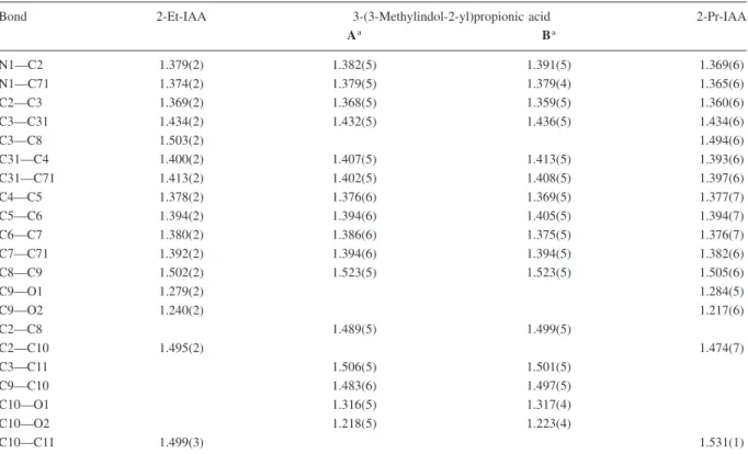
Table 4. Bond lengths (Å) for 2-Et-IAA, 3-(3-Me-indol-2-yl)propionic acid and 2-Pr-IAA.
Bond 2-Et-IAA 3-(3-Methylindol-2-yl)propionic acid 2-Pr-IAA
Aa Ba N1—C2 1.379(2) 1.382(5) 1.391(5) 1.369(6) N1—C71 1.374(2) 1.379(5) 1.379(4) 1.365(6) C2—C3 1.369(2) 1.368(5) 1.359(5) 1.360(6) C3—C31 1.434(2) 1.432(5) 1.436(5) 1.434(6) C3—C8 1.503(2) 1.494(6) C31—C4 1.400(2) 1.407(5) 1.413(5) 1.393(6) C31—C71 1.413(2) 1.402(5) 1.408(5) 1.397(6) C4—C5 1.378(2) 1.376(6) 1.369(5) 1.377(7) C5—C6 1.394(2) 1.394(6) 1.405(5) 1.394(7) C6—C7 1.380(2) 1.386(6) 1.375(5) 1.376(7) C7—C71 1.392(2) 1.394(6) 1.394(5) 1.382(6) C8—C9 1.502(2) 1.523(5) 1.523(5) 1.505(6) C9—O1 1.279(2) 1.284(5) C9—O2 1.240(2) 1.217(6) C2—C8 1.489(5) 1.499(5) C2—C10 1.495(2) 1.474(7) C3—C11 1.506(5) 1.501(5) C9—C10 1.483(6) 1.497(5) C10—O1 1.316(5) 1.317(4) C10—O2 1.218(5) 1.223(4) C10—C11 1.499(3) 1.531(1)
bond is near-perpendicular to the ring-plane and ap-proximately coplanar with the COOH group.
Conformational analysis
It has been argued that the conformations adopted by bioactive compounds, in the absence of restraints im-posed by the crystal lattice, are more directly related to their biological properties. Under these conditions, a number of conformers coexist; the smaller the con-formational strain (energy), the larger the respective subpopulation of conformers, a correlation expressed in quantitative terms by the Boltzmann-Maxwell
dis-tribution. As complete conformational analysis by ex-perimental methods is so far not feasible for 2-alkyl-IAAs, we resorted to computational chemistry. Table 7 summarizes the results of conformational analysis performed for isolated molecules of 2-alkyl-IAAs using semiempirical PM3 and ab initio Re-stricted Hartree-Fock/6–31G* calculations. The con-formers are described by torsion angles T1 to T4 (consult Table 6 for definitions). For all three com-pounds, the conformations with the C8-C9 and C10-C11 bonds near-perpendicular to the ring plane have the lowest energies. The global minima for 2-Et-IAA (C, C*) and 2-Pr-IAA (B) correspond to conformers Table 5. Bond angles (degrees) for 2-Et-IAA, 3-(3-methylindol-2-yl)propionic acid and 2-Pr-IAA.
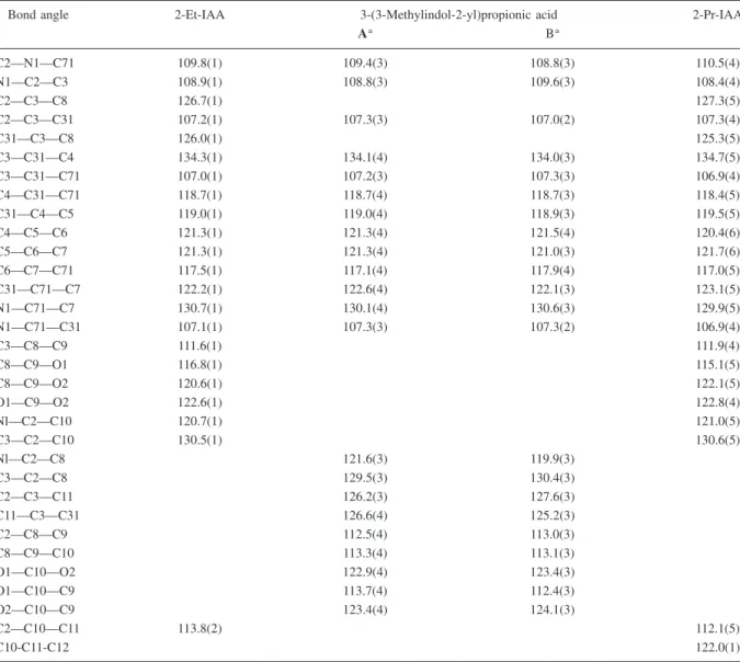
Bond angle 2-Et-IAA 3-(3-Methylindol-2-yl)propionic acid 2-Pr-IAA
Aa Ba C2—N1—C71 109.8(1) 109.4(3) 108.8(3) 110.5(4) N1—C2—C3 108.9(1) 108.8(3) 109.6(3) 108.4(4) C2—C3—C8 126.7(1) 127.3(5) C2—C3—C31 107.2(1) 107.3(3) 107.0(2) 107.3(4) C31—C3—C8 126.0(1) 125.3(5) C3—C31—C4 134.3(1) 134.1(4) 134.0(3) 134.7(5) C3—C31—C71 107.0(1) 107.2(3) 107.3(3) 106.9(4) C4—C31—C71 118.7(1) 118.7(4) 118.7(3) 118.4(5) C31—C4—C5 119.0(1) 119.0(4) 118.9(3) 119.5(5) C4—C5—C6 121.3(1) 121.3(4) 121.5(4) 120.4(6) C5—C6—C7 121.3(1) 121.3(4) 121.0(3) 121.7(6) C6—C7—C71 117.5(1) 117.1(4) 117.9(4) 117.0(5) C31—C71—C7 122.2(1) 122.6(4) 122.1(3) 123.1(5) N1—C71—C7 130.7(1) 130.1(4) 130.6(3) 129.9(5) N1—C71—C31 107.1(1) 107.3(3) 107.3(2) 106.9(4) C3—C8—C9 111.6(1) 111.9(4) C8—C9—O1 116.8(1) 115.1(5) C8—C9—O2 120.6(1) 122.1(5) O1—C9—O2 122.6(1) 122.8(4) Nl—C2—C10 120.7(1) 121.0(5) C3—C2—C10 130.5(1) 130.6(5) Nl—C2—C8 121.6(3) 119.9(3) C3—C2—C8 129.5(3) 130.4(3) C2—C3—C11 126.2(3) 127.6(3) C11—C3—C31 126.6(4) 125.2(3) C2—C8—C9 112.5(4) 113.0(3) C8—C9—C10 113.3(4) 113.1(3) O1—C10—O2 122.9(4) 123.4(3) O1—C10—C9 113.7(4) 112.4(3) O2—C10—C9 123.4(4) 124.1(3) C2—C10—C11 113.8(2) 112.1(5) C10-C11-C12 122.0(1)
aA and B are the two conformers of 3-(3-methylindol-2-yl)propionic acid which coexist in the crystalline state. Consult Figure 4 for the
with the side chains in the 2- and 3-positions tilted to the same side of the ring-plane. Essentially the same conformation exists in the crystal structures (Table 6), except for the opposite orientation of the carboxyl oxygens (i.e. O1 and O2 interchanged). The latter is due to intermolecular hydrogen bonds in which the carboxyl group participates (data not shown), and re-flects the influence of the crystal environment on the molecular conformation. Conformers with the C8-C9 bond in the ring plane have markedly higher energies. To investigate the behavior of 2-alkyl-IAA mole-cules in aqueous solution, molecular dynamics simu-lations were performed. As shown in Figure 6 for the example of 2-Pr-IAA, the population of conformers with both side chains tilted to the indole ring plane was much larger than that of molecules with one of the side chains approximately in that plane. The tran-sitions of the 2-alkyl chain between opposite orienta-tions relative to the ring plane are much more fre-quent than the corresponding transitions of the 3-CH2COOH moiety.
IAA and its 2-alkyl derivatives are carboxylic ac-ids with pK values around 4.8, and may thus ionize at physiological pH. However, the pH-equivalent
in-side the active sites of the proteins engaged in auxin
perception will remain unknown until these proteins are completely characterized. With respect to this un-clear situation, conformational analysis was also per-formed for the carboxylate ions of 2-alkyl-IAAs using the semiempirical PM3 and AM1 methods. The re-sults obtained by the latter approach (which afforded more consistent results for dissociated molecules) are
presented in Table 8, indicating that tilted conforma-tions are lower in energy than their planar rotamers, as they are in the undissociated acids. However, ion-ization decreases the energy difference between the planar and the tilted conformations, and hence the difference in their abundance under physiological conditions.
Interaction similarity analysis
We previously used interaction similarity analysis to define four classes of auxrelated compounds, in-cluding growth inhibitors in addition to the classes defined in the Materials and Methods section (Tomic´ et al. 1998a, 1998b). As 2-alkyl-IAAs are obviously not inhibitors (at physiological concentrations), here we only investigate how they fit into classes 1–3. For this purpose the representative conformations of the carboxylate ions listed in Table 8 were considered. For each compound, an equal number of P and T conformations was included. The enhanced flexibility of the 2-alkyl residues, proceeding from 2-Me-IAA to 2-Pr-IAA, was taken into account by considering an increasing number of conformers. For each set of conformers, molecular interaction fields (MIFs) were calculated according to the protocols specified by Tomic´ et al. (1998a, 1998b), using the probes dis-cussed in the Materials and Methods section.
The interaction similarity indices calculated from the above molecular interaction fields identified the three 2-alkyl-IAAs either as strong (class 1) or as weak (class 2) auxins, depending to some extent on Table 6. Selected torsion angles (o) for 2-Et-IAA, 3-(3-Me-indol-2-yl)propionic acid and 2-Pr-IAA.
Torsion angle 2-Et-IAA 3-(3-Methylindol-2-yl)propionic acid 2-Pr-IAA
Aa Ba C2—C3—C8—C9 (T1b) −105.9(2) 104.6(6) C31—C3—C8—C9 78.5(2) −79.2(6) C3—C8—C9—O1 100.7(1) −99.0(5) C3—C8—C9—O2 (T2b) −79.0(2) 77.9(6) C3—C2—C8—C9 106.8(5) 123.4(4) C31—C3—C2—C8 −178.5(4) 177.1(3) C2—C8—C9—C10 −178.9(4) −175.0(3) C8—C9—C10—O1 −171.4(4) 177.9(3) C8—C9—C10—O2 7.8(6) −2.5(5) C4—C31—C3—C11 0.7(8) −3.9(6) N1—C2—C10—C11 (T3b) −96.1(2) 81.3(7) C2—C10—C11—C12 (T4b) −173.8(1)
aA and B are the two conformers which coexist in the crystalline state. Consult Figure 4 for the numbering of corresponding atoms.bLabels
the specific protocol employed. In each case was 2-Me-IAA somewhat better (about 10% larger differ-ences for similarity indices) distinguished from class 3 (inactive compounds) than 2-Et-IAA and 2-Pr-IAA, as demonstrated by the examples presented in Ta-ble 9. This is in general accord with the results of the bioassays: 2-Me-IAA is at the borderline between strong and weak auxins; 2-Et-IAA and 2-Pr-IAA are about as ‘active’ as 2-Me-IAA if half-optimal concen-trations are considered, but the maximal response is only about one-half that for 2-Me-IAA (Table 3, cv. Pula).
Concluding remarks
Kögl and Kostermans (1935) attempted the synthesis of 2-Et-IAA by an ambiguous method and could not,
at that time, easily verify the structure of their prod-uct. Repeating their protocol, we obtained a com-pound of about the same melting point, but NMR spectra and X-ray data revealed a different identity: 3-(3-methylindol-2-yl)propionic acid. It is not sur-prising that this compound is inactive in the Avena curvature test (Kögl and Kostermans 1935), because the auxin activity of indole-2-acetic acid in the same bioassay is also ‘a lot smaller’ (Schindler 1958) than that of its positional isomer, IAA. The authentic 2-Et-IAA and its homologue, 2-Pr-2-Et-IAA show definite auxin activity.
Like any hormone, auxins must interact with spe-cific cell proteins to trigger a biological response. In spite of much effort, and some initial success (Mc-Donald 1997; Woo et al. 2002), the complete set of these proteins has not yet been characterized. Accord-ing to Katekar’s (1979) model of the idealized auxin-Table 7. Representative conformations of 2-alkylindole-3-acetic acids characterized by torsion angles T1 (C2-C3-C8-C9), T2
(C3-C8-C9-O2), T3 (N1-C2-C10-C11), T4 (C2-C10-C11-C12) and relative energies (⌬E). The values in the shaded areas were computed by an ab initio approach (6-31G*), the other ones by the semiempirical PM3 method. Near-planar (P) conformations have T1 ⬃ 0°, tilted (T) conforma-tions T1 ⬃ 90°, with a margin of ±35°.
binding site, the acidic head group and the planar body of an auxin are recognized by separate sections of a polypeptide chain which engulfs the target mol-ecule. According to our X-ray data for 2-Me-IAA (Nigovic´ et al. 2000), 2-Et-IAA and 2-Pr-IAA (this paper), neither the indole nucleus nor the CH2COOH
group, considered separately, show structural features which are not encountered in other, more active, in-dole auxins. Also, the molecular structures in the solid state provide no indications for major sterical crowd-ing involvcrowd-ing the 3-CH2COOH and 2-alkyl groups.
However, when not exposed to crystal packing forces, Figure 6. Molecular dynamics simulations for 2-(n-propyl)indole-3-acetic acid (2-Pr-IAA) in aqueous solution. Presented are the
tempera-ture regime and the changes of torsion angles T1, T3 and T4 within a time interval of 2 nanoseconds.
Table 8. Representative, AM1-optimized conformations of the carboxylate anions of 2-alkylindole-3-acetic acids characterized by torsion
angles T1 (C2—C3—C8—C9), T3 (N1—C2—C10—C11), T4 (C2—C10—C11—C12) and relative energies (⌬E). The torsion angle T2 cannot be unambiguously defined because the two oxygen atoms in the resonance-stabilized carboxylate ion are equivalent. Near-planar (P) conformations have T1 ⬃ 0°, tilted (T) conformations have T1 ⬃ 90°, with a margin of ±25°.
Compound Conformation Conformational type T1 degrees T3 degrees T4 degrees ⌬Eakcal mol−1
2-Me-IAA A P 6 2.1 B T 70 0 2-Et-IAA A P 20 −7 0 B P 4 78 1.2 C T 99 81 0.6 D T 100 −82 1.2 2-Pr-IAA A P 2 −6 −179 0.5 B P 2 −11 83 1.6 C P 4 94 −67 1.7 D P 1 86 −175 0.9 E T 102 106 −179 0 F T 101 105 −75 0.7 G T 88 −82 180 1.5 H T 78 −90 75 1
aThe origin of the scale (⌬E = 0) corresponds to the conformation with the lowest energy (global minimum) and is defined separately for
2-alkyl-IAAs appear to have distinct conformational preferences. Thus, conformational analysis for iso-lated molecules and molecular dynamics simulations in aqueous solution strongly suggest that alkyl substi-tution at the 2-position ‘pushes’ the -CH2COOH
group into conformations with T1 ⬃ 90° (‘T-confor-mations’). Planar (P) conformations (T1 ⬃ 0°), on the other hand, which are preferred by most other in-dole auxins which have so far been subjected to de-tailed conformational analysis (Ramek et al. 1996; Ramek and Tomic´ 1998a, 1998b, 1999), are signifi-cantly less populated for undissociated 2-alkyl-IAAs, becoming somewhat more abundant when the car-boxyl group ionizes. These conformational prefer-ences in solution (i.e. before entering the active site of an auxin-binding protein) would affect the energy input needed to give the 3-side chain the right twist to make both the carboxyl group and the indole ring snap into their optimal positions in their respective compartments of the auxin-binding site. Half-optimal concentrations in auxin bioassays have been inter-preted as the dissociation constants (Kd) of the
auxin-receptor complexes involved in the biological re-sponse (Libbenga et al. 1986). In accord with this view, it follows from the data presented in Table 3 that the binding energy (-⌬G) of IAA (calculated as ⌬G = RTlnKd; T = 298 K) is 0.8 to 1.5 kcal mol
−1
more negative than for its 2-alkyl derivatives (note that more negative binding energy means stronger binding). This is indeed within the energy range re-quired for interconversion of the more abundant con-formations of 2-alkyl-IAAs (Tables 8 and 9), and the energy spent on such adjustments would reduce the net binding energy. An energy of 0.8 to 1.5 kcal/mol would also be sufficient for slight displacement of the polypeptide chain around the indole-binding
compart-ment to create space for the 2-alkyl group. However, if this should be the necessary, it would be difficult to understand why 2-Et-IAA and 2-Pr-IAA have about the same half-optimal concentrations, and hence about the same net binding energies, as 2-Me-IAA, even though the size of the 2-alkyl groups varies within wide limits.
On the other hand, IAA and its 2-alkyl derivatives differ considerably with respect to the growth re-sponse they elicit at their optimal concentrations, a phenomenon for which Katekar (1979) introduced the term ‘efficacy’. This effect could be rationalized by assuming that bulky substituents in the IAA 2-posi-tion prevent the auxin-loaded receptor from assuming the conformation which is optimal for initiating a growth response. However, the following kinetic ex-planation appears at least as likely. To avoid a never-ending growth-stimulating effect of an auxin-loaded ‘receptor’ protein, there must be ways of disposing of it (by metabolism, forced unloading in the next step of the signal transduction sequence etc.). The rate at which this occurs, relative to the rate of formation of the auxin-protein complex, would determine its sta-tionary concentration, which would translate into the growth rate. The ‘induced fit’ of an auxin molecule to an auxin-binding site, in the above sense, is accom-plished by random conformational changes (through thermal movement) and selection for optimal (largest free-energy gain) protein-ligand interactions, a pro-cess which takes time. As 2-alkyl-IAAs have differ-ent conformational preferences than IAA, it is plau-sible to expect that they accommodate themselves more slowly in a binding site nature designed for IAA, and the stationary concentration of the auxin-receptor complex should decrease accordingly. In-deed, the ‘maximal response’ for 2-Me-IAA is only Table 9. Typical examples for the differences between mean similarity indices relating 2-alkylindole-3-acetic acids to auxin classes 1 (highly
active) and 3 (inactive) using the ‘probes’ listed.
Set of molecular interaction fields Compound Probec
H2O NH2+ CH 3 O DRY Aa 2-Me-IAA 0.150 0.064 0.142 0.055 0.341 2-Et-IAA 0.139 0.063 0.124 0.048 0.341 2-Pr-IAA 0.138 0.062 0.128 0.048 0.369 Bb 2-Me-IAA 0.136 0.118 0.118 0.123 0.295 2-Et-IAA 0.132 0.118 0.119 0.117 0.252 2-Pr-IAA 0.123 0.116 0.110 0.108 0.189
aP conformers aligned by the program SEAL (.)bT conformers aligned by optimizing molecular interaction fields at the molecular surface. crepresent relevant functional groups (NH
2 +, CH
3, O), as well as hydrophilic (H2O) and hydrophobic (DRY) elements in the polypeptide
about 85% of the value found for IAA in the same lot of Avena coleoptiles (Nigovic´ et al. 2000). For 2-Et-IAA and 2-Pr-2-Et-IAA, additional time should be required for the tail of the 2-substituent (mobility confirmed by molecular dynamics simulations) to assume a confor-mation which does not interfere with simultaneous binding of the indole nucleus and the carboxyl group. As expected, the maximal response for the latter two auxins reaches only 20–50% of the value obtained for IAA. In a similar fashion, alkylation of IAA at the benzene ring does not markedly affect their half-op-timal concentrations, but efficacy drops as the length of the alkyl chain increases (Nigovic´ et al. 2000).
It has been argued that differences in auxin activ-ity can also be due to differences in metabolic stabil-ity. This possibility must always be borne in mind, even though there is little, if any, unequivocal sup-porting evidence. IAA is preferentially metabolized by oxidation at the 2-position which occurs either di-rectly (Ernstsen et al. 1987; Klämbt 1959; Reinecke and Bandurski 1983) or following conjugation with aspartic or glutamic acids (Catalá et al. 1992; Plüss et al. 1989; Östin et al. 1992, 1995, 1998; Riov and Bangerth 1992; Tsurumi and Wada 1986a, 1986b; Tuominen et al. 1994). The compounds studied here cannot be oxidized at the 2-position which is blocked by an alkyl substituent. It is thus unlikely that 2-alkyl-IAAs are metabolized faster than the unsubstituted parent compound.
Acknowledgements
Supported by grants no. 00980608 and 00981010 by the Ministry of Science and Technology of the Re-public of Croatia and by US-Croatian Research Agreement JF202. We are grateful to Vladislav Tomišic´ for recording the UV spectra.
References
Antolic´ S., Kojic´-Prodic´ B., Tomic´ S., Nigovic´ B., Magnus V. and Cohen J.D. 1996. Structural studies on monofluorinated deriva-tives of the phytohormone indole-3-acetic acid (auxin). Acta Crystallogr., Sect. B: Struct. Sci. 52: 651–661.
Antolic´ S., Salopek B., Kojic´-Prodic´ B., Magnus V. and Cohen J.D. 1999. Structural characterization and auxin properties of dichlorinated indole-3-acetic acids. Plant Growth Regul. 27: 21–31.
Carbó R. and Calabuig B. 1992. Molecular quantum similarity measures and n-dimensional representation of quantum objects. I. Theoretical foundations. Int. J. Quant. Chem. 42: 1681–1693. Catalá C., Östin A., Chamarro J., Sandberg G. and Crozier A. 1992. Metabolism of indole-3-acetic acid by pericarp discs from im-mature and im-mature tomato (Lycopersicon esculentum Mill.). Plant Physiol. 100: 1457–1463.
Chandra U., Gupta A.A. and Sengupta A.K. 1980. Studies on po-tential juvenile hormone analogs: part I – Synthesis & physical studies of some heterocyclic geranyl & citronellyl ethers, their epoxides, hydrochlorides & bromides. Indian J. Chem., Sect B: Org. Chem. Incl. Med. Chem. 19: 528–531.
Cosgrove D.J. 1998. Cell wall loosening by expansins. Plant Phys-iol. 118: 333–339.
Dauber-Osguthorpe P., Roberts V.A., Osguthorpe D.J., Wolff J., Genest M. and Hagler A.T. 1988. Structure and energetics of ligand binding to proteins: Escherichia coli difolate reductase-trimethoprim, a drug-receptor system. Proteins: Struct., Funct., Genet. 4: 31–47.
DISCOVER, release 1997. Molecular Simulations, Inc., San Di-ego, CA, USA.
Ernstsen A., Sandberg G. and Lundström K. 1987. Identification of oxacetic acid, and metabolic conversion of indole-3-acetic acid to oxindole-3-indole-3-acetic acid in seeds of Pinus
sylves-tris. Planta 172: 47–52.
Hatano T., Katayama M. and Marumo S. 1987. 5,6-Dichloroindole-3-acetic acid as a potent auxin: its synthesis and biological ac-tivity. Experientia 43: 1237–1239.
Hoffmann O.L., Fox S.W. and Bullock M.W. 1952. Auxin-like ac-tivity of systematically substituted indoleacetic acids. J. Biol. Chem. 196: 437–441.
Johnson C.K. 1976. ORTEPII. Report ORNL – 5138. Oak Ridge National Laboratory, Tennessee, USA.
Jönsson L. 1961. Chemical structure and growth activity of auxins and antiauxins. In: Ruhland W. (ed.), Encyclopedia of Plant Physiology. Vol. 14. Springer-Verlag, Berlin, pp. 958–1006. Katayama M., Kato Y., Hatano T., Hatori M. and Marumo S. 1998.
Synthesis and biological activities of 5,6-difluoroindole-3-ace-tic acid; a new fluoroindole auxin. J. Pes5,6-difluoroindole-3-ace-ticide Sci. 23: 289– 295.
Katekar G.F. 1979. Auxins: on the nature of the receptor site and molecular requirements for auxin activity. Phytochemistry 18: 223–233.
Kearsley S.K. and Smith G.M. 1990. SEAL. Tetrahedron Computer Methodol. 3: 615.
Klämbt H.D. 1959. Die 2-Hydroxy-indol-3-essigsäure, ein pflan-zliches Indolderivat. Naturwissenschaften 46: 649.
Kloetzel M.C. 1948. Reactions of nitroparaffins. II. Addition of ni-troparaffins to unsaturated esters. J. Am. Chem. Soc. 70: 3571– 3576.
Kögl F. and Kostermans D.G.F.R. 1935. Über die Konstitutions-Spezifizität des Hetero-auxins. Hoppe Seyler’s Z. physiol. Chem. 235: 201–216.
Kojic´-Prodic´ B., Nigovic´ B., Tomic´ S., Ilic´ N., Magnus V., Giba Z. et al. 1991. Structural studies on 5-(n-alkyl)-substituted deriva-tives of the plant hormone indole-3-acetic acid. Acta Crystal-logr., Sect. B: Struct. Sci. 47: 1010–1019.
Larsen P. 1961. Biological determination of natural auxin. In: Ru-hland W. (ed.), Encyclopedia of Plant Physiology. Vol. 14. Springer-Verlag, Berlin, pp. 521–582.
Lawson W.B., Patchornik A. and Witkop B. 1960. Substitution, oxidation and group participation in the bromination of indoles. J. Am. Chem. Soc. 82: 5918–5923.
Le Goffic F., Gouyette A. and Ahoud A. 1973. Une nouvelle syn-thèse de l’ellipticine et ses analogues structuraux. Tetrahedron 29: 3357–3362.
Libbenga K.R., Maan A.C., van der Linde P.C.G. and Mennes A.M. 1986. Auxin receptors. In: Chadwick C.M. and Garrod D.R. (eds), Hormones, Receptors and Cellular Interactions in Plants. Cambridge University Press, Cambridge, UK, pp. 1–68. McDonald H. 1997. Auxin perception and signal transduction.
Physiol. Plant. 100: 423–430.
Mitchell J.W. and Livingston G.A. 1968. Methods of studying plant hormones and growth-regulating substances. Agriculture Hand-book No. 336. Agricultural Research Service, United States Department of Agriculture, Washington, DC, USA.
Muir R.M. and Hansch C. 1953. On the mechanism of action of growth regulators. Plant Physiol. 28: 218–232.
Muir R.M., Hansch C.H. and Gallup A.H. 1949. Growth regulation by organic compounds. Plant Physiol. 24: 359–366.
Nigovic´ B., Antolic´ S., Kojic´-Prodic´ B., Kiralj R., Magnus V. and Salopek-Sondi B. 2000. Correlation of structural and physico-chemical parameters with the bioactivity of alkylated deriva-tives of indole-3-acetic acid, a phytohormone (auxin). Acta Crystallogr., Sect. B: Struct. Sci. 56: 94–111.
Östin A., Catalá C., Chamarro J. and Sandberg G. 1995. Identifi-cation of glucopyranosyl--4,1-glucopyranosyl- -1-N-oxin-dole-3-acetyl-N-aspartic acid, a new IAA-catabolite, with liquid chromatography-tandem mass spectroscopy. J. Mass Spectrom. 30: 1007–1017.
Östin A., Kowalczyk M., Bhalerao R.P. and Sandberg G. 1998. Metabolism of indole-3-acetic acid in Arabidopsis. Plant Phys-iol. 118: 285–296.
Östin A., Monteiro A.M., Crozier A., Jensen E. and Sandberg G. 1992. Analysis of indole-3-acetic acid metabolites from
Dal-bergia dolichopetala by high performance liquid
chromatogra-phy-mass spectrometry. Plant Physiol.: 63–68.
Plüss R., Jenny T. and Meier H. 1989. IAA-induced adventitious root formation in greenwood cuttings of Populus tremula and formation of 2-indolone-3-acetylaspartic acid, a new metabo-lite of exogenously supplied indole-3-acetic acid. Physiol. Plant. 75: 89–96.
Porter W.L. and Thimann K.V. 1965. Molecular requirements for auxin action – I. Halogenated indoles and indoleacetic acids. Phytochemistry 4: 229–243.
Ramek M., Tomic´ S. and Kojic´-Prodic´ B. 1995. Systematic ab ini-tio SCF conformaini-tional analysis of indol-3-ylacetic acid phy-tohormone (auxin): comparison with experiment and molecular mechanics calculations. Int. J. Quantum Chem.: Quantum Biol. Symp. 22: 75–81.
Ramek M.L., Tomic´ S. and Kojic´-Prodic´ B. 1996. Comparative ab initio SCF conformational study of 4-chloro-indole-3-acetic acid and indole-3-acetic acid phytohormones (auxins). Int. J. Quantum Chem. 60: Quantum Biol. Symp. 23: 3–9. Ramek M. and Tomic´ S. 1998a. RHF conformational analysis of
the auxin phytohormones n-ethyl-indole-3-acetic acid (n = 4, 5, 6). Int. J. Quantum Chem. 70: 1169–1175.
Ramek M. and Tomic´ S. 1998b. Ab initio RHF investigation of mono- and dichlorinated indole-3-acetic acid (IAA) phytohor-mones. J. Mol. Struct. (Theochem.) 454: 167–173.
Reinecke D.M. and Bandurski R.S. 1983. Oxindole-3-acetic acid, an indole-3-acetic acid catabolite in Zea. Plant Physiol. 71: 211–213.
Rescher U., Walther A., Schiebl C. and Klämbt D. 1996. In vitro binding affinities of 4-chloro-, 2-methyl-, 4-methyl, and 4-eth-ylindoleacetic acid to auxin-binding protein 1 (ABP1) correlate with their growth-stimulating activities. J. Plant Growth Regul. 15: 1–3.
Riov J. and Bangerth F. 1992. Metabolism of auxin in tomato fruit tissue. Formation of high molecular weight conjugates of ox-indole-3-acetic acid via the oxidation of indole-3-acetylaspartic acid. Plant Physiol. 100: 1396–1402.
Rokach J. 1973. Indole derivatives. Canadian Patent 926410. Schindler W. 1958. Indol-2-essigsäure. Helv. Chim. Acta 41: 1441–
1443.
Schmidt M.W., Baldridge K.K., Boatz J.A., Elbert S.T., Gordon M.S., Jensen J.H. et al. 1993. General atomic and molecular electronic structure systems. J. Comput. Chem. 14: 1347–1363. Sell H.M., Wittwer S.H., Rebstock T.L. and Redemann C.T. 1952. Comparative stimulation of parthenocarpy in the tomato by var-ious indole compounds. Plant Physiol. 28: 481–487. Sheldrick G.M. 1996. SADABS program for absorption correction.
University of Göttingen, Göttingen, Germany.
Sheldrick G.M. 1997a. SHELXS-97. Program for crystal structure solution. University of Göttingen, Göttingen, Germany. Sheldrick G.M. 1997b. SHELXL97. Program for crystal structure
refinement. University of Göttingen, Göttingen, Germany. Snyder H.R. and Pilgrim F.J. 1948. The preparation of
3-indoleace-tic acid; a new synthesis of tryptophol. J. Am. Chem. Soc.: 3770–3771.
Spek A.L. 1993. HELENA program for data reduction. University of Utrecht, Utrecht, The Netherlands.
Spek A.L. 1997. PLATON. Molecular geometry program. Version of 1997. University of Utrecht, Utrecht, The Netherlands. Stewart J.J.P. 1990. MOPAC 6.0, available from Quantum
Chem-istry Program Exchange. Indiana University, Bloomington, In-diana, USA.
Tomic´ S., Gabdoulline R.R., Kojic´-Prodic´ B. and Wade R.C. 1998a. Classification of auxin plant hormones by interaction property similarity indices. J. Computer-Aided Mol. Design 12: 63–79.
Tomic´ S., Gabdoulline R.R., Kojic´-Prodic´ B. and Wade R. 1998b. Classification of auxin related compounds based on similarity of their interaction fields: Extension to a new set of compounds. Internet J. Chem. 1: 26.
Tsurumi S. and Wada S. 1986a. Identification of 3-hydroxy-2-in-dolinone-3-acetylaspartic acid as a new indole-3-acetic acid metabolite in Vicia roots. Plant Cell Physiol. 27: 559–562. Tsurumi S. and Wada S. 1986b. Dioxindole-3-acetic acid
conju-gates formation from indole-3-acetylaspartic acid in Vicia seed-lings. Plant Cell Physiol. 27: 1513–1522.
Tuominen H., Östin A., Sandberg G. and Sundberg B. 1994. A novel metabolic pathway for indole-3-acetic acid in apical shoots of Populus tremula (L.) × Populus tremuloides (Michx.). Plant Physiol. 106: 1511–1520.
Verley M.A. and Beduwé J. 1925. Méthode générale de prépara-tion des dérivés substitués de l’indole. Bull. Soc. Chim. France 37: 189–191.
Ramek M. and Tomic´ S. 1999. Quantum chemical conformational analysis of the auxin phytohormone 4-methyl-3-indoleacetic acid. Int. J. Quantum Chem. 75: 1003–1008.
Walton E., Jenkins S.R., Nutt R.F. and Holly F.W. 1968. Some analogs of 1-p-chlorobenzyl-5-methylindole-3-acetic acid. J. Med. Chem. 11: 1252–1255.
Woo E.-J., Marshall J., Bauly J., Chen J.-G., Venis M., Napier R.M. et al. 2002. Crystal structure of auxin-binding protein 1 in com-plex with auxin. EMBO J. 21: 2877–2885.

![Figure 3. Molecular structures [ORTEP II (Johnson 1976)] of (a) 2-ethylindole-3-acetic acid (2-Et-IAA) and (b) 2-(n-propyl)indole-3-acetic acid (2-Pr-IAA) showing the crystallographic numbering conventions referred to in the text](https://thumb-eu.123doks.com/thumbv2/123doknet/14566743.726934/6.892.241.660.97.719/molecular-structures-johnson-ethylindole-crystallographic-numbering-conventions-referred.webp)
![Figure 4. Molecular structure [ORTEP II (Johnson 1976)] of 3-(3-methylindol-2-yl)propionic acid](https://thumb-eu.123doks.com/thumbv2/123doknet/14566743.726934/7.892.172.720.100.515/figure-molecular-structure-ortep-johnson-methylindol-propionic-acid.webp)