Antiinflammatory property of a local medicinal plant
(Ziziphus lotus .L)
DEMOCRATIC AND POPULAR REPUBLIC OF ALGERIA
MINISTRY OF HIGHER EDUCATION AND SCIENTIFIC RESEARCH
UNIVERSITY OF JIJIEL
لــجـيج ىــــــــيــحي نب قيدصلا دمحم ةــــعماـــج
Master's thesis
Branch: Biological Sciences
Specialization: Biochemistry
Theme
Examiner’s committee: Presented by:
President : Mrs. HIRECHE Saliha
KAHLAT Fatima Zahra
Examiner : Dr. LAHOUEL Asma
Supervisor: Dr. CHERBAL Asma
Academic Year 2019-2020
Order number (library):……….…..…
ةايحلاو ةيعيبطلا مولعلا ةيلك
ةيئيزجلا ايجولويبلا مسق
ةيولخلاو
Faculty of Nature and Life
Sciences
Department of Molecular
and Cellular Biology
Antiinflammatory property of a local medicinal plant
(Ziziphus lotus .L)
DEMOCRATIC AND POPULAR REPUBLIC OF ALGERIA
MINISTRY OF HIGHER EDUCATION AND SCIENTIFIC RESEARCH
UNIVERSITY OF JIJIEL
لــجـيج ىــــــــيــحي نب قيدصلا دمحم ةــــعماـــج
Master's thesis
Branch: Biological Sciences
Specialization: Biochemistry
Theme
Examiner’s committee: Presented by:
President : Mrs. HIRECHE Saliha
KAHLAT Fatima Zahra
Examiner: Dr. LAHOUEL Asma
Supervisor: Dr. CHERBAL Asma
Academic Year 2019-2020
Order number (library):…….…..…
ةايحلاو ةيعيبطلا مولعلا ةيلك
ةيولخلاو ةيئيزجلا ايجولويبلا مسق
Faculty of Nature and Life Sciences
Department of Molecular and Cellular Biology
Acknowledgements
All my thanks and gratitude to God, who gave me strength, patience and a
lot of courage to move forward firmly in this work and complete it until the end.
I do not hesitate to thank my teacher and supervisor for my graduation
project, Dr. Asma Charbel thank you very much for your support and advices.
I would also like to thank Dr. Asma Lahouel and Mrs. Saliha Hireche for
their evaluation of this academic work.
My family and friends, I offer you a lot of love and gratitude for your moral
support, especially in the Corona pandemic
.To you mom, who represents for me the symbol of kindness, the source of
tenderness and the example of the dedication that has never ceased to encourage and
pray for me.
To my very dear PAPA who I love very much and who has always believed in
me and put at my disposal all the necessary means for me to succeed I thank you for
your support and especially for the confidence and the freedom you have granted me
may God protect you and keep you for me.
With enormous pleasure and immense joy, I dedicate this work to all my
family, my brother Zakaria, my sisters Saffia, Soumia and Hiba. I also dedicate this
thesis to all my friends (Meriam, Souad, Nesrine, Imane and Sara)
I dedicate this work to the spirit of two people who are very dear to my heart,
my friend B. Waffa, and my teacher B. Hussein the role model to me, who has always
been eagerly awaiting my achievements but who died in 2011. I have successfully
completed my graduation note. May God bless you, my sincere prayers to you always.
List of abbreviations ... iii
List of figures……….v
List of tables ...………vii
Introduction………...1 Chapter 1: Inflammation I. Generalities on inflammation………..2 I.1. Definition………..………...2 I.2. Etiology………..……….….2 I.3. Signs ………...………...2
I.4. Pathophysiology of inflammation ……….……….………...…..3
I.4.1. Types of inflammation ……….………..……….………...…..3
I.4.1.1. Acute Inflammation……….……….……….……3
I.4.1.2. Chronic Inflammation………..……….………...…3
I.4.3. Pathologies related to inflammation ………..……….………….…4
I.5. The inflammatory cells ………..….……….…..….……4
I.6. Mediators of inflammation……….………..…...6
I.6.1. General mediator of inflammation ……….………...…6
I.6.2. Acute-phase proteins……….……….…………..…7
I.7. Stages of inflammatory response and their mechanism of regulation….……….……….…...7
I.7.1 Initiation of inflammatory responses……….……….……….……….……….……….….….7
I.7.2. Vascular and cellular changes phase……….……….……….……….……….………..…....8
I.7.3. The repair phase (resolution) ……….……….……….….…..9
I.8. Inflammatory response mechanism……….……….……….…...10
I.8.1. TLR signaling……….……….……….……….……….……….……….……….…...…...…10
I.8.2. NF-κB pathway ………..……….……….……….……….………...11
I.8.3. MAPK pathway……….……….……….……….……….……….……….……….…….…..12
I.8.4. JAK-STAT pathway……….……….……….……….……….……….……….………..…...13
I.10.1. Non-steroidal antiinflammatory drugs………...……….……..….…...15
I.10.2. Steroidal antiinflammatory drugs………..………..…...…16
I.10.3. Medicinal plants with antiinflammatory activity……….……….………...18
Chapter 2: « Ziziphus Lotus. L » the medicinal plant I.1. Generalities……….…………..….19
I.2. Botanic description……….………..….…19
I.3. Taxonomy……….……….……….….…..20
I.4. Biochemical composition of Ziziphus lotus………...……….……….………...…...20
I.4.1. Primary metabolites……….……….……….……….……….……….……….………....….21
I.4.2. Secondary metabolites ……….……….………...…..21
I.5. Pharmacological and biological activities of Ziziphus lotus compounds …………..……...…23
Chapter 3: The anti-inflammatory effect of ziziphus lotus I. Antiinflammatory effect of Ziziphus lotus against skin inflammation induced by 12-O-tetrade canoylphorbol-13-acetate (TPA)……….. ……….……..….…………24
II. Anti-inflammatory effect of Ziziphus lotus against edema induced by carrageenan and experimental trauma. ……….……….……….…..…………27
III. Antiinflammatory activities of a polyphenol rich extract from Ziziphus lotus. L fruit pulp against obesity……….……….……….………...…….….30
Conclusion
List of abbreviations
AA: Ascorbic acid AP-1: Activator protein-1
BHA: Butylated hydroxyanisole. ButP: Flavonoid‐butanol phase CLRs: C-type lectin receptors COX: Cyclooxygenases CRP: C-reactive protein, CSF: Colony stimulating factor
DAMPs: Danger associated molecular patterns EAP: Flavonoid‐ethyl acetate phase of the pulp ER: Endoplasmic reticulum
ERK: Extracellular signal-regulated kinase ESR: Erythrocyte sedimentation rate FRAP: Ferric‐reducing antioxidant powers HC: Hydrocortisone
HETE: Hydroxy eicosatetraenoic acid HFAD: High fat diet
HFADP: High fat diet supplemented with the Z. lotus pulp HOMA‐IR: Homeostatic model assessment of insulin resistance HPETE: Hydroperoxy eicosatetraenoic acid
IKK: IκB kinase
iNOS: Induced nitric oxide synthetase IRF3: Interferon regulatory factor 3 JNK: C-Jun N-terminal kinases LOX: Lipoxygenases
MBP: Mannose-binding protein
MCP‐1 : Monocyte chemotactic protein 1 MyD88: Myeloid differentiation factor-88 NF-Κb: Nuclear factor kappa B
Nk cell: Naturel killer cells NLRs: NOD-like receptors
NSAIDs: Nonsteroidal anti-inflammatory drugs PAF: Platelet Activator Factor
PAMPs: Pathogen associated molecular patterns PCK : Protéine Kinase C
PCT : Procalcitonin
PHA : Phyto-hemagglutinin PLA2 : Phospholipase A2 PMNs: Polynuclear neutrophils
PolyP: Phenolic extract of Z. lotus pulp PRRs: Pattern recognition receptors RIG.I: retinoic acid-inducible gene I RLRs: I-like receptors
ROS: reactive oxygen species
SAA protein: Serum amyloid protein A SAIDs: Steroidal anti-inflammatory drugs S-Lx: Sialyl-Lewis X molecules
TCHO: Total cholesterol TG: Thapsigargin
TGF: transforming growth factor TLRs: Toll-like receptors
TNFα: Tumor necrosis factor alpha TP: Tannins pulp
TPA: 12-O-tetrade canoylphorbol-13-acetate TXB2: Thromboxane B2
ZJE: Ziziphus jujube extract ZLP: Ziziphus lotus polyphenols
List of figures
Figure 1. The mast cell in acute inflammation………...…..8
Figure 2. Cellular change phase of inflammation. ………....9
Figure 3. Inflammatory pathway components. ………...10
Figure 4. TLR signaling pathway. ………...….11
Figure 5. NF-κB signaling pathway. ………...….12
Figure 6. MAPK signaling pathway. ………...…13
Figure 7. JAK-STAT signaling pathway. ………14
Figure 8. The antiinflammatory mechanism of Non-steroidal and steroidal anti- inflammatory drugs. ……….……….17
Figure 9. Location of Ziziphus lotus plant in Algeria. ……….………19
Figure 10. Ziziphus lotus. L plant. ………...20
Figure 11. Common structure of jujubogenins (a), lotogenins (b), and lotusines (c) found in Z. lotus. ………...………...………….22
Figure 12. Effects of Z. lotus essential oil on TPA-induced ear thickness and water content in BLAB/c mice……….………..…...24
Figure 13. Histological sections of mouse ear skin biopsies (magnification x l00) ……….25
Figure 14. Ear dermis area measurements to estimate thickening of epidermis ………..…26
Figure 15. Area measurements to estimate thickening of dermis. ………...……….…26
Figure 16. Influence of Ziziphus lotus oil (200 and 300 mg / kg orally) assessed by carrageenan-induced edema. ……….…….………29
Figure 17. Percentage inhibition of edema induced by carrageenan at the level of the rat's leg pretreated with Ziziphus lotus oil (200 and 300 mg / kg orally) ………..…29
Figure 18. Influence of Ziziphus lotus oil (200 and 300 mg / kg orally) assessed by edema induced by experimental trauma………..…...…29
Figure 19. Percentage inhibition of edema induced by experimental trauma in the paw of
the rat pretreated with Ziziphus lotus oil (200 and 300 mg / kg orally). ……….….…...29
Figure 20. In vitro antioxidant properties of the Ziziphus lotus pulp………...…31 Figure 21. Effect of the Ziziphus lotus pulp on different in vivo parameters in rats. …...32 Figure 22. Z. lotus polyphenol effect on MTT cell viability and proliferation rate of RAW
264.7 cells. ………..…….34
Figure 23. mRNA expression of different inflammatory cytokines in response to
different treatments in RAW 264.7 cells. ………..……….……34
Figure24. (a) mRNA expression of nitric oxide synthase (iNOS); (b) quantity of nitric oxide (NO)
produced in RAW 264.7 cells……….….36
List of tables
Table 1. The differences between acute and chronic inflammation………...…...3
Table 2. Characteristics of acute and chronic inflammation. ……...………...……...3
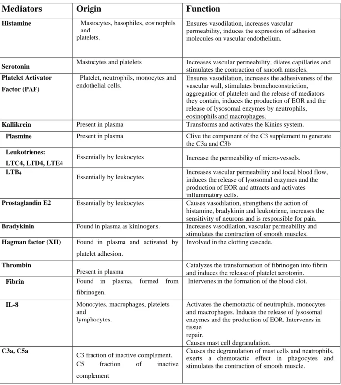
Table 3. Inflammatory mediator’s function. ………..…...…….6
Table 4. Biomarkers of inflammation. ……….………..15
Table 5. Examples of nonsteroidal antiinflammatory drugs. ………...…..…....16
Table 6. Mechanisms of antiinflammatory action of the medicinal plants………...18
Table 7. Percentage of primary compositions of Ziziphus lotus. ………..………….21
Table 8. Composition in secondary metabolites of the different organs of Ziziphus lotus……….22
Table 9. Antiinflammatory effect of the Z lotus leaf’s aqueous extract on carrageenan-induced rats………..……….…30
Introduction
Elimination of pathogens such as infection, viruses, trauma, and damaged tissues requires an immune response called inflammation in order to preserve the survival of the organisms. To eliminate a pathogen, inflammation depends on a combination of cellular, local, and peripheral reactions. The inflammatory reaction includes the production of cytokines, which exert local and systemic actions; its control depends on a subtle balance between pro and antiinflammatory cytokines. (Kumar et al., 2017).
The complex events and mediators involved in the inflammatory reaction can induce, maintain or aggravate many diseases. Therefore, the use of antiinflammatory agents is helpful in the therapeutic treatment of these pathologies, but sometimes causes dose dependent pathologies and diseases (Vane et al., 1998). In allopathic medicine, steroidal anti-inflammatory drugs (cortisone and derivatives) or non-steroidal drugs (NSAIDs) are prescribed, depending on the case, the best-known being aspirin, their main drawbacks are poor digestive tolerance and the many contraindications (Lydyard et al.,
2011). NSAIDs can be responsible for headaches or dizziness, more or less serious
digestive side effects (nausea, pain or heartburn, ulcer or bleeding in the digestive tract), allergic reactions (rash, asthma) and renal failure in certain rare circumstances. In order to limit the onset of side effects, especially digestive effects, NSAIDs should be used at the minimum effective dose and for the shortest possible time, especially in the elderly. Indeed, in people over 65, the side effects of NSAIDs are more common and often more serious (Lydyard et al., 2011).
The incorporation and use of medicinal plants in the treatment of several inflammatory reactions, particularly rheumatism, are common practices in traditional medicine. Today it is a remarkable fact that antiinflammatory substances of plant origin are of growing interest because they offer advantages over conventional anti-inflammatory, such as for example the absence of side effects; it must be said that these products may have a smaller potential, but studies of structure-activity relationships may lead to obtaining more effective preparations. (Kulkarni et al., 2010).
Among the substances of natural origin that manifest this activity, we will notice the Ziziphus
lotus. L, to this aim this thesis studies: the antiinflammatory effect of Z. lotus against skin
inflammation induced by 12-O-tetradecanoylphorbol-13 acetate (TPA), edema induced by Carrageenan and experimental trauma and obesity induced by high fat diet.
Chapter 1:
Inflammation
I. Generalities on Inflammation
I.1. Definition
Inflammation is one of the immune system's defense mechanisms against exogenous or endogenous pathogens in order to eliminate them and repair damaged tissues. Acute inflammation lasts for a few days, while chronic inflammation lasts for months or several years (Kantari et al., 2008).
I.2. Etiology
The causes of the inflammatory reaction are multiple and divided in non-infectious factors and non-infectious factors.
- Non-infectious factors
Physical: burn, frostbite, physical injury, trauma, lionizing radiation.
Chemical: glucose, fatty acids, toxins, alcohol, chemical irritants (including
fluoride, nickel and other trace elements).
Biological: damaged cells. Psychological: excitement.
- Infectious factors: Bacteria, Viruses, Other microorganisms (Chen et al.,
2018).
I.3. Signs
There are five cardinal signs of inflammation listed by Celsius Cornelius, a Roman encyclopedic of ancient times, and Rudolf Virchow:
- Redness: vasodilatation, increased blood flow. - Swelling: extravascular accumulation of fluid. - Heat: vasodilatation, increased blood flow. - Pain
I.4. Pathophysiology of inflammation I.4.1. Types of inflammation
I.4.1.1. Acute inflammation
This type of inflammation is one of the immediate immune mechanisms to eliminate pathogens and it is characterized by a rapid immune response in addition to platelet adhesion, mast cells and neutrophils to the endothelium. It is the typical response of the innate immune system, resident in complete healing of the initial lesion (table 1)
(Medzhitov, 2008).
I.4.1.2. Chronic Inflammation
When the body cannot eliminate the pathogen (aggression persists or tissues undergoing) the inflammatory response continues for months or years (table 2)
(Medzhitov, 2008).
Table 1. The differences between acute and chronic inflammation (Feldman, 2017).
Table 2. Characteristics of acute and chronic inflammation (Chandrasoma et al., 1991).
I.4.3. Pathologies related to inflammation
There are a lot of major organs and tissues affected because of the secondary effect of inflammation.
- Disorders in which the main pathogenic role returns to inflammation:
Asthma, rheumatoid arthritis, arteriosclerosis, drop, osteoarthritis, Hashimoto’s thyroiditis, Chronic obstructive pulmonary diseases, Alzheimer’s diseases, Scattered lupus erythematous, eczema, Crohn’s diseases, ankylosing spondylitis, hemorrhagic colitis (Nathan, 2002).
- Infectious diseases in which inflammation contributes to pathology
Hepatitis C, tuberculosis, gastritis induced by Helicobacter pillory, bacterial dysentery
(Nathan, 2002).
- Diseases of various origins in which post-inflammatory fibrosis is the main cause of the pathology:
Idiopathic pulmonary fibrosis, viral or alcoholic liver cirrhosis, bilharzias (Nathan,
2002).
I.5. The inflammatory cells I.5.1. Polynuclear neutrophils
The most important cells involved in the acute inflammatory process (40 to 70% of immune defense cells), which allow the release of cytokines, chemokines and proteins to trigger inflammation, after chemo-attractants which stimulate them to repair cells
(Descamps-Latscha and Witko-Sarsat, 1996; Eming et al., 2007). I.5.2. Mast cells
One of the characteristics of these cells is that they contain granules in the cytoplasm. These granules release inflammatory mediators when stimulating the cell, such as histamine, serotonin, heparin, and cytokines that stimulate the inflammatory reaction (Williams and Galli, 2000).
These cells are found in connective tissue and they are involved in tissue repair
I.5.3. Monocytes
These cells are considered the primary drivers of inflammation as they migrate to the site of infection and then divide into macrophages to eliminate the pathogen through phagocytosis. They participate in tissue repair by releasing inflammatory mediators that stimulate other inflammatory cells (Eming et al., 2007).
I.5.4. Platelets
Platelets provide chemokines and activators for the circulation of blood cells and vascular wall cells. They produce eicosanoids and release peptides and other mediators acting on vascular permeability to induce hemostasis and healing of tissue damage. They activate complement, release proteases, and provide growth factors for fibroblasts, vascular endothelial cells and smooth muscle cells. In addition, they stimulate inflammatory phenomena via leukocytes and endothelial cells, because they interact with microorganisms (Steinhubl, 2007; Medzhitov, 2008).
I.5.5. Polynuclear basophils
These cells play an important role in inflammation despite their small number (less than 1% of inflammatory cells), as they are considered as a macrophage and involved in allergies thanks to their granules containing inflammatory mediators (Rankin,
2004).
I.5.6. Polynuclear eosinophils
Eosinophils make up 1-6% of inflammatory cells. Their main function is to attack pests through the content of their granules. They are also involved in the modulation and propagation of the adaptive immune response by directly activating T lymphocytes (Hogan et al., 2008).
I.5.7. Lymphocytes
Two main types of lymphocytes are B cells and T cells. B cells have immunoglobulins on their surface and if stimulated by antigen, differentiate into plasma cells produce antibodies specific to the antigen. T cells participate in all mediated immunity (Lydyard et al., 2011).
I.6. Mediators of inflammation
I.6.1. General mediators of inflammation
A variety of chemical mediators from circulation system, inflammatory cells, and injured tissue actively modulate the inflammatory response (table 3). They include vasoactive amines and peptides (e.g., bradykinin), eicosanoids, pro-inflammatory cytokines and acute phase proteins (Halliwell et al., 2015).
Table 3. Inflammatory mediator’s function (Rankin 2004, Kumar et al, 2017).
Mediators Origin Function
Histamine Mastocytes, basophiles, eosinophils and
platelets.
Ensures vasodilation, increases vascular permeability, induces the expression of adhesion molecules on vascular endothelium.
Serotonin Mastocytes and platelets Increases vascular permeability, dilates capillaries and stimulates the contraction of smooth muscles. Platelet Activator
Factor (PAF)
Platelet, neutrophils, monocytes and endothelial cells.
Ensures vasodilation, increases the adhesiveness of the vascular wall, stimulates bronchoconstriction, aggregation of platelets and the release of mediators they contain, induces the production of EOR and the release of lysosomal enzymes by neutrophils, eosinophils and macrophages.
Kallikrein Present in plasma Transforms and activates the Kinins system.
Plasmine Present in plasma Clive the component of the C3 supplement to generate the C3a and C3b
Leukotrienes: LTC4, LTD4, LTE4
Essentially by leukocytes Increase the permeability of micro-vessels. LTB4
Essentially by leukocytes Increases vascular permeability and local blood flow, induces the release of lysosomal enzymes and the production of EOR and attracts and activates inflammatory cells.
Prostaglandin E2 Essentially by leukocytes Causes vasodilation, strengthens the action of histamine, bradykinin and leukotriene, increases the sensitivity of neurons and is responsible for pain. Bradykinin Found in plasma as kininogens. Increases vasodilation, vascular permeability and
stimulates the contraction of smooth muscles. Hagman factor (XII) Found in plasma and activated by
platelet adhesion.
Involved in the clotting cascade.
Thrombin
Present in plasma
Catalyzes the transformation of fibrinogen into fibrin and induces the release of platelet serotonin. Fibrin Found in plasma, formed from
fibrinogen.
Intervenes in the formation of the blood clot.
IL-8 Monocytes, macrophages, platelets and
lymphocytes.
Activates the chemotactic of neutrophils, monocytes and macrophages. Induces the release of lysosomal enzymes and the production of EOR. Intervenes in tissue
repair.
Causes mast cell degranulation. C3a, C5a
C3 fraction of inactive complement. C5 fraction of inactive complement
Causes the degranulation of mast cells and neutrophils, exerts a chemotactic effect in phagocytes and stimulates the contraction of smooth muscle.
I.6.2. Acute-phase proteins
Acute-phase proteins play an important role in innate immunity, as they contribute to the elimination of bacteria and pathogens such as trauma and infections. Among these proteins:
- C- reactive protein (CRP): Binds the bacterial phosphocholine, activates complement through C1q, acts as an opsonin. The increased level of interleukin 6 stimulates the liver to synthesize and secrete C-reactive protein in obesity case. This last is the accumulation of abnormal or excessive fat that may interfere with the maintenance of an optimal state of health. The excess of macronutrients in the adipose tissues stimulates them to release inflammatory mediators such as tumor necrosis factor α and interleukin 6, and reduces production of adiponectin, predisposing to a pro-inflammatory state and oxidative stress (Ellulu et al., 2017).
- Serum amyloid A (SAA): Activates complement (through C1q), acts an opsonin.
- Mannose-binding lectin (MBL): Binds to mannose on bacteria, attaches to
phagocyte; activates complement via classical pathway.
- Complement components: Chemotaxis, opsonization, and lysis.
- Mental-binding proteins: Removal of essential metal ions required for bacteria
growth.
- Fibrinogen: Coagulation factors.
- α1-antitrypsin, α1-antichymotrypsin: Protease inhibitors (Lydyard et al., 2011).
I.7. Stages of inflammatory response and their regulation I.7.1 Initiation of inflammatory responses
This stage is considered to be the vascular phase, where blood flow increases and vascular permeability changes which allows plasma proteins to seep into the tissues (exudation) (Börzsei et al., 2008).
This step begins immediately after the activation of platelets and mast cells, in which inflammatory mediators such as serotonin, histamine and arachidonic acid derivatives are released in addition to factor XII, fibrin and bradykinin.
In some cases, neuritis occurs due to the sensitivity of sensory cells to capsaicin. These cells secrete mediators that stimulate platelets and plasma perfusion by acting on endothelial cells and smooth muscles in blood vessels (Birklein and Schmelz, 2008).
Richardson and Vasko (2002) confirm that sensory neurons contain cyclooxygenases, which play an important role in the synthesis of prostaglandins.
I.7.2. Vascular and cellular changes phase
Inflammation mediators cause rapid changes in the wall of blood vessels and stimulate leukocyte transmission by increasing the expression of cell adhesion molecules causing changes in tight connections in endothelial cells and thus allowing passage of blood cells, antibacterial proteins, coagulation factors, antibodies and PMNs (figure 1).
Figure 1. The mast cell in acute inflammation (Lydyard et al., 2011).
PMNs leave the bloodstream because of their recognition of adhesion molecules displayed on endothelial cells. The expression of these adhesion molecules is induced by pro-inflammatory cytokines released by macrophages. PMNs form weak attachments through their sialyl-Lewis X (S-Lx) molecules to E-selectin on the endothelial surface. Binding to chemokines, eg IL-8, activates PMN to express an active form of LFA-1 which then binds to ICAM-1 on the endothelial surface. This leads to arrest and flattening of the PMN, which then extravagates by squeezing between the endothelial cells. PMN then migrates into the tissues to the source of the inflammatory mediators (figure 2).
Figure 2. Cellular change phase of inflammation (Lydyard et al., 2011).
I.7.3. The repair phase (resolution)
Stopping the inflammatory process and repairing damaged tissue requires several inflammatory mediators, such as anti-inflammatory cytokines (IL10, TGF-b1)
(Lawrence et al., 2007), expression of soluble receptors like TNFα, as well as apoptosis
of inflammatory cells by negative feedback mechanisms to avoid the harmful risks of the inflammatory response and limit the damage to the tissues, ensuring a return to silence of the inflammatory response and also preparing the tissue repair phase (Serhan et al., 2009
; Serhan, 2010).
Tissue repair involves macrophages, endothelial cells and fibroblasts. As these anti-inflammatory molecules neutralize the inflammatory phase, repair of the damage begins. Various cells, including myofibroblasts and macrophages, both of which make collagen, mend tissues. Macrophage products, including epidermal growth factor, platelet-derived growth factor, fibroblast growth factor, and transforming growth factor, are important in the repair process. In addition, acute phase proteins contribute to the healing process (Lydyard et al., 2011).
I.8. Inflammatory response mechanism
The inflammatory pathway is made up of inducers, sensors, mediators and target tissues (figure 3) (Medzhitov, 2008).
Figure 3. Inflammatory pathway components (Medzhitov, 2008).
According to Chen et al. (2018) the activation of the inflammatory pathways is based on the activation of the following inflammatory receptors and pathways: TLRs, NF-κB, MAPK and JAK-STAT.
I.8.2.1. Pattern recognition receptor activation.
Classes of pattern recognition receptors (PRRs) families include the Toll-like receptors (TLRs). TLRs are a family of highly conserved, mammalian PRRs that participate in the activation of the inflammatory response. Signaling through TLRs activates intracellular signaling cascades that lead to nuclear translocation of AP-1 and NF-κB or IRF3, which regulates the inflammatory response.
Transmission of PAMPs and DAMPs is mediated by myeloid differentiation factor-88 (MyD88) along with TLRs. Signaling through TLRs activates an intracellular signaling cascade that leads to nuclear translocation of transcription factors, such as activator protein-1 (AP-1) and NF-κB or interferon regulatory factor 3 (IRF3) (figure4).
Figure 4. TLR signaling pathway (Chen et al., 2018). I.8.2.2. NF-κB pathway
The NF-κB transcription factor plays important roles in inflammatory, immune response, survival, and apoptosis processes. The NF-κB family includes five related transcription factors: P50, p52, RelA (p65), RelB, and c-Rel. NF-κB activity is induced by a range of stimuli, including pathogen-derived substances, intercellular inflammatory cytokines, and many enzymes. Under physiological conditions, IκB proteins present in the cytoplasm inhibit NF-κB. PRRs use similar signal transduction mechanisms to activate IκB kinase (IKK), which is composed of two kinase subunits, IKKα and IKKβ, and a regulatory subunit, such as IKKγ. IKK regulates NF-κB pathway activation through IκB phosphorylation. IκB phosphorylation results in its degradation by the proteasome and the subsequent release of NF-κB for nuclear translocation and gene transcription activation. This pathway regulates pro-inflammatory cytokine production and inflammatory cell recruitment, which contribute to the inflammatory response. This pathway is triggered by TLRs and inflammatory cytokines, such as TNF and IL-1, leading to activation of RelA/p50 complexes that regulate expression of inflammatory cytokines. NF-κB signaling requires IKK subunits. which regulate pathway activation through IκB phosphorylation (figure5) (Chen et al., 2018).
Figure 5. NF-κB signaling pathway (Chen et al., 2018). I.8.2.3. MAPK pathway
MAPKs are a family of serine/threonine protein kinases that direct cellular responses to a variety of stimuli, including osmotic stress, mitogens, heat shock, and inflammatory cytokines (such as IL-1, TNF-α, and IL-6), which regulate cell proliferation, differentiation, cell survival and apoptosis. The mammalian MAPKs include extracellular-signal-regulated kinase ERK1/2, p38 MAP Kinase, and c-Jun N-terminal kinases (JNK). Each MAPK signaling pathway comprises at least three components: a MAPK, a MAPK kinase (MAPKK), and a MAPK kinase kinase (MAPKKK).
MAPKKKs phosphorylate and activate MAPKKs, which in turn phosphorylate and activate MAPKs. ERKs are generally activated by mitogens and differentiation signals,while inflammatory stimuli and stress activate JNK and p38. MKK1 and MKK2 activate ERK1/2, MKK4 and MKK7 activate JNK, and MKK3 and MKK6 activate p38.
Activation of the MAPKs, including Erk1/2, JNK, leads to phosphorylation and activation of p38 transcription factors present in the cytoplasm or nucleus, which initiates the inflammatory response.
This pathway mediates intracellular signaling initiated by extracellular stimuli, such as stress and cytokines. MAPKKKs phosphorylate and activate MAPKKs, which in turn phosphorylate and activate MAPKs. The mammalian MAPK family includes
Erk1/2, JNK, and p38. In the Erk1/2 pathway, Erk1/2 is activated by MKK1/2, which is activated by Raf. In the JNK pathway, JNK is activated by MKK4/7, which is activated by MEKK1/4, ASK1, and MLK3. In the p38 pathway, p38 is activated by MKK3/6, which is activated by MLK3, TAK, and DLK. Activated MAPKs phosphorylate various proteins, including transcription factors, resulting in regulation of inflammatory responses (figure6) (Chen et al., 2018).
Figure 6. MAPK signaling pathway (Chen et al., 2018). I.8.2.4. JAK-STAT pathway
The highly conserved JAK-STAT pathway involves diverse cytokines, growth factors, interferons, and related molecules, such as leptin and growth hormone, and is a signaling mechanism through which extracellular factors can control gene expression. Receptor-associated JAKs are activated by ligands and phosphorylate one other, creating docking sites for STATs, which are latent, cytoplasmic transcription factors. Cytoplasmic STATs recruited to these sites undergo phosphorylation and subsequent dimerization before translocation to the nucleus. Tyrosine phosphorylation is essential for STAT dimerization and DNA binding. Therefore, JAK/STAT signaling allows for the direct translation of an extracellular signal into a transcriptional response. For example, binding of IL-6 family members to plasma membrane receptors activates the
JAK-STAT proteins. STAT proteins translocated into the nucleus bind target gene promoter regions to regulate transcription of inflammatory genes (Chen et al., 2018).
Following IL-6 binding, signal is transduced by a receptor to activate the JAKs, which then activate STATs. STATs are dephosphorylated in the nucleus, leading to activation of downstream cytokines. Dysregulation of NF-κB, MAPK, or JAK-STAT activity is associated with inflammatory, autoimmune, and metabolic diseases, and cancer. Signaling through transcription factors result in secretion of cytokines.
Multiple transcription factors regulate a variety of inflammatory genes, such as IL-1, TNF-α, IL-6 colony stimulating factor (CSF), interferons, transforming growth factor (TGF), and chemokines (figure 7) (Chen et al., 2018).
Figure 7. JAK-STAT signaling pathway (Chen et al., 2018). I.9. Biomarkers of inflammation
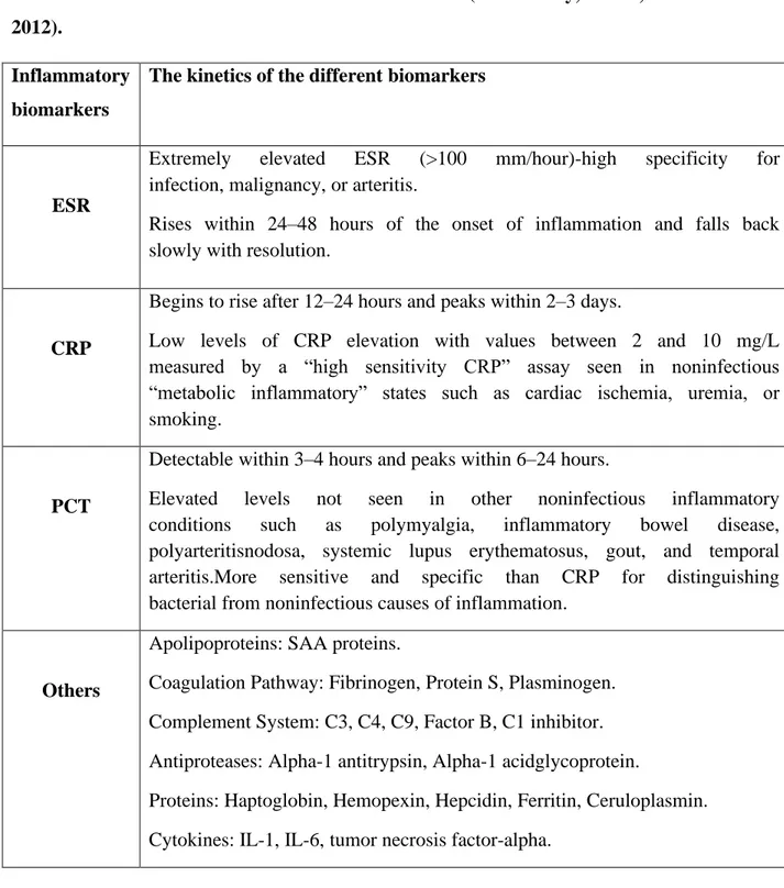
The biological marker is considered one of the reliable means for diagnosing diseases and predicting their complications. It is also one of the strategies that indicates the effectiveness of the drug in the course of the prescription, where this biological marker is measured for physiological or pathological states (table 4) (Niehues, 2018).
Table 4. Biomarkers of inflammation (Markanday, 2015; Watson et al., 2012).
Inflammatory biomarkers
The kinetics of the different biomarkers
ESR
Extremely elevated ESR (>100 mm/hour)-high specificity for infection, malignancy, or arteritis.
Rises within 24–48 hours of the onset of inflammation and falls back slowly with resolution.
CRP
Begins to rise after 12–24 hours and peaks within 2–3 days.
Low levels of CRP elevation with values between 2 and 10 mg/L measured by a “high sensitivity CRP” assay seen in noninfectious “metabolic inflammatory” states such as cardiac ischemia, uremia, or smoking.
PCT
Detectable within 3–4 hours and peaks within 6–24 hours.
Elevated levels not seen in other noninfectious inflammatory conditions such as polymyalgia, inflammatory bowel disease, polyarteritisnodosa, systemic lupus erythematosus, gout, and temporal arteritis.More sensitive and specific than CRP for distinguishing bacterial from noninfectious causes of inflammation.
Others
Apolipoproteins: SAA proteins.
Coagulation Pathway: Fibrinogen, Protein S, Plasminogen. Complement System: C3, C4, C9, Factor B, C1 inhibitor. Antiproteases: Alpha-1 antitrypsin, Alpha-1 acidglycoprotein.
Proteins: Haptoglobin, Hemopexin, Hepcidin, Ferritin, Ceruloplasmin. Cytokines: IL-1, IL-6, tumor necrosis factor-alpha.
I.10. Treatment of inflammation
I.10.1. Nonsteroidal antiinflammatory drugs
Doctors rely on non-steroidal drugs as a way to reduce pain, fever, and inflammation. Although these drugs are chemically not homogeneous (table 5), they do have some commonalities in the non-selective inhibition of cyclooxygenase
NSAIDs reduce pain and inflammation by blocking cyclooxygenases (cox), enzymes that are needed to produce prostaglandins. Most NSAIDs block two different cyclooxygenases called Cox-l and Cox-2. Cox-2, found in joint and muscle, contributes to pain and inflammation but can cause bleeding because they block the Cox-l enzyme that protects the lining of the stomach from acid (Saraf et al., 2008). So, a selective NSAID that blocks Cox-2 but not Cox-1 might reduce pain and inflammation in joints but leave the stomach lining intact (figure 8) (Vane et al., 1998).
Table 5. Examples of nonsteroidal antiinflammatory drugs (Wallace and Staats, 2005).
However, the use of NSAIDs is associated with many adverse effects with a considerable prevalence of new diseases and mortality (Bidaut-Russell, 2001). The side effects of treatment with NSAIDs are attributed to their non-selective inhibition of cyclooxygenase isoforms including COX-1 which is constitutively present in most human tissues. This has the role of regulating a number of physiological processes such as the maintenance of the integrity of the gastric mucosa, renal function, and platelet aggregation (figure 8) (Vonkeman et al., 2008).
I.10.2. Steroidal antiinflammatory drugs
Steroidal anti-inflammatory drugs (SAIDs) are used to treat asthma, rheumatoid arthritis, and autoimmune diseases, as these drugs belong to cortisol derivatives and share the mechanism of action with auto-glucocorticoids that bind to glucocorticoid receptors in the cytoplasm to stimulate the increase in transcription of genes that encode anti-inflammatory proteins (lipocortin-1 and interleukin 10) and inhibit In other hand genes that encode pro-inflammatory proteins (cytokines, enzymes, receptors and adhesion molecules).
Structural class Scientific name Trade name Salicylates Salicylic acetyl
Salsalate (sodium salicylates) Diflusinal
Aspirin Dolobid
Propionic acid derivatives
Acetic acid derivatives
Ibuprofen Fenoprofen calcium Flubiprofen Ketoprofen Diclofenac Ibuprofene Nalfon Ansaid Nalfon Voltarene Indoles Indomethacin Tolmetin Sulindac Indocine, Tolectie cliniril
While these drugs are effective in treating much chronic pathology, they also cause dangerous side effects in some disorders. These disorders can be acute such as high blood pressure, deregulation of the natural synthesis of glucocorticoids at the end of treatment, euphoria with insomnia up to acute psychosis and the development of peptic ulcers. Chronic disorders can also manifest themselves such as osteoporosis, cataracts and weight gain (Fernandes et al., 2005; Barnes., 1998; Henzen, 2003).
Figure 8. The antiinflammatory mechanism of Non-steroidal and steroidal anti-inflammatory drugs (Kumar et al., 2017).
I.10.1. Medicinal plants with antiinflammatory activity
Medicinal plants are widely used in traditional medicine around the world for the relief of inflammatory diseases such as rheumatoid arthritis, asthma, bronchitis, eczema, osteoarthritis, gout, allergic rhinitis, gastric ulcers. and duodenal (Setty and Sigal, 2005;
Wiart, 2007). Table 6 shows some traditional medicinal plants with their mechanisms of
action.
Table 6. Mechanisms of antiinflammatory action of the medicinal plants
Chapter 2:
″Ziziphus lotus ″ the
medicinal plant
I.1. Generalities
The Mediterranean climate favors the growth of numerous plant species, some of which have various ecological, nutritional, and medicinal potential properties (Naili et al.,
2010).
The genus Ziziphus belongs to the family Rhamnaceae, intrinsically widely adapted to environmental stresses such as dry and hot climates, which makes it suitable for growth in challenging environments characterized by degraded land and limited water resources (figure 9) (Hammer, 2001). The genus Ziziphus consists of about 170 species of spiny shrubs and small trees distributed in warm-temperate and subtropical regions throughout the world (Islam and Simmons, 2006).
Figure 9. Location of Ziziphus lotus plant in Algeria. The populations are indicated by bold red points (Boussaid et al., 2018).
I.2. Botanic description
Ziziphus lotus.L is a dicotyledon plant from the Rhamnacea family, locally
called "Sedra" (Borgi et al., 2007). It is a shrub in the form of a bush not exceeding 2.5m with flexed twigs, very thorny white grey growing in zigzag. The leaves are small short, and oval more at least elliptical 1 to 2cm long and 7mm wide.
The flowers of Ziziphus lotus L. are very visible yellow with open star sepals, a bisexual extra ovary and bloom in June. An ovoid-olong fruit, having the shape and size of a beautiful olive (figure10). First green and then yellow, it turns dark red when it is walled, in October. Its thick pulp can be a greenish white or yellowish brown, a little glutinous and has a sweet and tangy flavor (Amara and Benabdeli., 2017).
Figure 10. Ziziphus lotus. L plant (Boussaid et al., 2018; Amara,2016; Adelia and Samavatib, 2015).
I.3. Taxonomy (Quezel et al., 1962) Kingdom: Plantae
Division (Phylum): Tracheophyta Class: Dichotyledoneae
Family: Rhamnaceae Genus: Ziziphus
Species: Ziziphus lotus.
I.4.Biochemical composition of Ziziphus lotus
Phytochemical studies carried out on Z. lotus show the presence of primary and secondary metabolites (Catoir et al., 1999).
I.4.1. Primary metabolites
The pulp of Zizyphus lotus L. is very rich in nutrients, composed of 12.8 to 13.6% carbohydrates including: 5.6% sucrose, 1.5% glucose, 2.1% fructose and 1 % starch (Jawanda et al., 1981). The pectin extracted from the pulp contains D Galactose, 2,3,6 Tri-o-acetyl, which gives it anti-diarrheal properties and helps lower plasma cholesterol levels (Tomoda et al., 1985). The following amino acids are found in the pulp: asparagine, arginine, glutamic acid, aspartic acid, glycine, serine and threonine (Bal,
1981). It is an important source of vitamin C and vitamin A (Bal et al., 1978; Bal, 1981).
Dried fruits contain several volatile substances responsible for the specific flavor of the fruit: seventy-eight compounds are identified, among them capric acid (19.98%), succinic acid and malic acid (15.64%) (Ahmedov and Halmatov, 1969). The pericarp and seeds are characterized by the presence of phospholipids, and a predominance of palmitoleic acid in the extracted oil (Goncharova et al., 1990). The almonds of "Zizyphus lotus L." are very rich in sulfur proteins (Nour et al., 1987). They also contain saponins which have medicinal values, and cyclopeptide alkaloids which are used in Chinese medicine as sedative substances (Ghedira et al., 1995).
These metabolites are collated in table 7. The Carbohydrates represent a significant proportion followed by the lipids then the proteins.
Table 7. Percentage of primary compositions of Ziziphus lotus (Chouaibi, 2011). Components % Values Proteins 19,11% Carbohydrates 40,87% Lipides 32,92% Sugars 20%
I.4.2. Secondary metabolites
Studies on the various species of the genus Ziziphus lead to the isolation and characterization of cyclopeptide alkaloids, flavonoids, sterols, tannins and triterpene saponins (table 8) (Abdoul-Azize, 2016).
Table 8. Composition in secondary metabolites of the different organs of Ziziphus lotus.
Figure 11. Common structure of jujubogenins (a), lotogenins (b), and lotusines (c) found in Z. lotus (Renault et al., 1997; Ghedira et al., 1993; Le Crouéour et al., 2002; Maciuk et al., 2004).
I.5. Pharmacological and biological activities of Z. lotus compounds
Studies have shown that the different parts of the plant exhibit multiple biological activities:
The Ziziphus lotus plant is widely used in the treatment of certain diseases because of its antifungal, antibacterial (Ghazghazi et al., 2014; Rsaissi et al., 2013; Aziz
et al., 1998), antiulcerogenic and gastroprotective (Bakhtaoui et al., 2014), anti
-inflammatory (Abdoul-Azize et al., 2013; Benammar et al., 2010; Borgi et al., 2007), sedative, analgesic and antispasmodic (Borgi et al., 2007), sterilant, hypotensive, cardiotonic, antioxidant (Benammar., 2010), antidiabetic and hypoglycemic effects
(Abdel-Zaher et al., 2005; Benammar et al., 2014).
Ziziphus lotus seeds induce an antigen / antibody reaction and inhibition of
histamine release. Its two active ingredients: jujuboside and protojujuboside increase serum IgG, they are also remedying for coughs, diarrhea, asthma, encephalopathy, vomiting and ophthalmopathy (Eddouks et al., 2004).
The leaves are used as a diuretic, emollient, expectorant, to promote hair growth, anticancer, sedative, blood purification. They are known to be hypoglycemic and cause an increase in the level of glycogen in the liver (Preeti and Shalini, 2014). The active ingredient in the leaves (jujuboside a) has a synergistic effect with phenylalanine on the central nervous system.
The roots, leaves and bark have an antibacterial and antifungal effect. Flavonoids and saponins from leaves and root bark have antiinflammatory and analgesic activity
(Ghedira et al., 1993).
The fruit has an anticancer, anti-inflammatory, hepato-protective, antioxidant, anti-insomnia, immunostimulant and neuro-protective effect (Guo et al., 2015).
Chapter 3:
Antiinflammatory effect
of Ziziphus lotus
I-
The antiinflammatory effect of Ziziphus lotus against skin inflammation
induced by 12-O-tetrade canoylphorbol-13-acetate (TPA)
To investigate whether the seed essential oil of Z. lotus is able to attenuate the inflammation in the skin, Al-Reza, et al. (2010) working on TPA-induced skin inflammation model to assess the antiinflammatory effect of topically applied essential oil of Z. lotus in vivo. Both of figure 12 A and B represent the different results of Z. lotus essential oil effect on TPA-induced ear thickness and water contenent in BALB/c mice.
Administration of TPA on the ear of the BALB/c mice caused a marked increase in both ear thickness and skin water content. The ear thickness was measured for TPA induced ear was 0.54 mm, as compared to control (0.23 mm). In other hand, Treatment with 1% and 10% of essential oil caused significant decrease in ear thicknesses, which were measured to be 0.30 and 0.35 mm, as well as reduce the water content about 51% and 53% in the TPA-induced skin inflammation model, respectively.
In the same study, the effect of 1% and 10% of essential oil on TPA-induced ear was also comparable to that of 1% of hydrocortisone (HC) (0.50 ± 0.1 mm), which was used as a positive control. Increase in skin water content induced by TPA was also suppressed by essential oil treatment in a concentration- dependent manner (figure 12B). The results show that the inhibitory effect of 1% and 10% of essential oil on TPA-induced skin inflammation was better than that of 1% of hydrocortisone (HC). Thus, these results demonstrate that essential oil of Z. lotus exerted potential antiinflammatory effects in
vivo.
Figure 12. Effects of Z. lotus essential oil on TPA-induced ear thickness and water content in BLAB/c mice. Mice were pretreated with indicated concentrations of
essential oil (EO) and 1% hydrocortisone (HC) for 30 mm and TPA (4 mM) was applied to induce skin inflammation. After 4 h the increase in ear thickness (A) and skin water content (B) was measured. Each column shows the mean ± SD of triplicate determinations (Al-Reza, et al., 2010).
To know more about Z. lotus antiinflammatory effect on skin inflammation
Al-Reza, et al. (2010) examining the mouse ear tissue morphology. The histological analysis
clearly confirmed that Z. lotus essential oil inhibited the inflammatory responses of skin inflammation in animal model. Also, TPA application resulted in a marked increase in ear thickness with clear evidence of edema, epidermal hyperplasia, and substantial inflammatory cell infiltration in the dermis with accompanying connective tissue disruption (figure 13A and B). By histological comparison, 1% of essential oil treatment reduced ear thickness and associated pathological indicators to an extent comparable to eugenol and the positive control hydrocortisone (HC) (figure 13C–E).
Figure 13. Histological sections of mouse ear skin biopsies (magnification x l00) (Al-Reza, et al., 2010).
To further elucidate the effect of Z. lotus essential oil on TPA-induced skin inflammation, the thickness of epidermal and dermal area of mice ear tissue were measured. As shown in figure 14, the thickness of epidermal area of essential oil treated
ear tissue (15.2 ± 2.9 µm²) was found to be nearly same as control (12.3 ± 2.1 µm²), whereas, the thicknesses of epidermal area for TPA, HC and eugenol were 19.9 ± 3.7, 17.8 ± 3.6 and 16.2 ± 3.1 µm², respectively. Further, the thickness of dermal area of essential oil treated ear tissue provides the evidence that Z. lotus essential oil exhibited highest antiinflammatory activity among all the tested samples (figure 15). The thicknesses of dermal area for essential oil, TPA, HC and eugenol were 185.5 ± 20.4, 249.7 ± 40.1, 309.7 ± 28.6 and 204.8 ± 28.2 µm², respectively.
Figure 14. Area measurements to estimate thickening of epidermis (Al-Reza, et al., 2010).
Figure 15. Area measurements to estimate thickening of ear dermis ( Al-Reza, et al., 2010).
The results of this study are consistent with the studies that confirm that Treating mice with TPA leads to pathological changes at the skin level, and these changes are mainly due to cellular, biochemical, and molecular alterations (Kendler et al., 1987;
Nakamura et al., 1998; Nakamura et al., 2000). Also, the treatment with TPA
stimulates the infiltration of inflammatory cells that cause a rise in the amount of H2O2, and this last causes cellular oxidation (Wei and Frenkel, 1993; Bhasin et al., 2003).
The hydrodistillation of Z. lotus seeds confirmed the presence of phenolic compounds, mono- and sesquiterpene hydrocarbons and their oxygenated derivatives
(Al-Reza et al., 2009) also, there are some studies that have confirmed that mono-cysteine
hydrocarbons and their oxygenated derivatives are the main components of plant-based essential oils and these last have a strong antiinflammatory effect (Peana et al., 2002;
Fernandes et al., 2007; Ko et al., 2008) while some studies confirmed that
antiinflammatory effect of Z. lotus seed is due to an antigen / antibody reaction and inhibition of histamine release. Its two active ingredients: jujuboside and protojujuboside increase serum IgG, they are also remedying for cough, diarrhea, asthma, encephalopathy, vomiting and ophthalmopathy (Eddouk et al., 2004).
II. Anti-inflammatory effect of Ziziphus lotus against edema
induced by carrageenan and experimental trauma
Indomethacin and diclofenac are the most drugs using to demonstrate the antiinflammatory effect of Ziziphus lotus plant, where indomethacin exerts an anti-inflammatory effect mainly by inhibiting the enzymatic activity of COX (Vane et al.,
1971). Thus, it reduces the production of pro-inflammatory mediators such as: histamine,
serotonin, kinins and prostaglandins, produced during this immune process and, therefore, constitutes an interesting therapeutic strategy for the treatment of inflammation
(Medzhitov and Ruslan., 2008).
El Hachimi et al. (2017) use indomethacin as reference to evaluate Z. lotus
aintinflammatory effect against edema induced by carrageenan and experimental trauma that's because carrageenan is the standard experimental model of acute inflammation used to assess the antiinflammatory activity of several natural and synthetic compounds
(Vogel, 2002; Boominathan et al., 2004). In the control animals, the sub plantar
injection of carrageenan produces a local edema which gradually increases after the injection of the phlogistic agent then the volume of the rat paw was measured at 1 hour 30
minutes, three and six hours after injection of the carrageenan. Indomethacin, at a dose of 10 mg / kg, significantly reduces the volume of paw edema by 88.83 ± 0.68; 71.57 ± 1.08 and 67.00 ± 1.23% respectively at 1h 30 min, three and six hours (figure 16,17).
At a dose of 200 mg / kg, the seed oil of Z. lotus significantly inhibited the development of edema of the rat paw induced by carrageenan from the first hour of the experiment, with a maximum reduction. 64.33 ± 0.72% at 1 hour 30.
However, the antiinflammatory effect of Z. lotus oil remains weaker than that of indomethacin (10 mg / kg) during the six-hour experiment. By increasing the dose to 300 mg / kg, the oil showed strong antiinflammatory activity. These effects are greater than those obtained with indomethacin (10 mg / kg) (figure 17).
Furthermore, the evaluation of the antiinflammatory activity of the seed oil of Z.
lotus by the edema induced by experimental trauma in the rat showed a maximum
inhibition of the edema at 1 hour 30 with an inhibition of edema equal to 43.40 ± 2.85 and 86.82 ± 3.46% respectively at the dose of 200 and 300 mg / kg, relative to indomethacin (p <0.05), with 50 ,70% inhibition of edema (figure 19).
The results obtained from antiinflammatory tests show that Z. lotus oil has an effect on the three phases of inflammation and significantly reduces the edema induced by carrageenan and by experimental trauma, with a good dose – effect relationship, since better inhibition is obtained with the dose of 300 mg / kg (El Hachimi et al.,2017).
The effects of Z. lotus oil manifested with the same antiinflammatory mechanism due to indomethacin, from the first hour of the experiment. It is concluded that Z. lotus seed oil contains antiserotonin and antihistamine compounds, as well as prostaglandin inhibitors, such as the indomethacin used as a reference substance (El Hachimi et al.,
2014; Zhang et al., 2017). Also, the high efficiency of Z. lotus oil could be linked to the
chemical profile of this oil, because it is rich in biologically active molecules such as polyphenols, sterols (Chouaibi et al., 2012) and oleic acid which is an acid monounsaturated fat essential for human consumption (El Hachimi et al., 2017). These molecules can play a decisive role in this biological activity. The results of this study are consistent with the results of HANI et al. (2020) in which edema of the mouse legs was achieved using carrageenin as inflammation inducer and diclofenac as a reference (table 9).
Figure 16. Influence of Ziziphus lotus oil (200 and 300 mg / kg orally) assessed by carrageenan-induced edema (El Hachimi et al., 2017).
Figure 17. Percentage inhibition of edema induced by carrageenan at the level of the rat's leg pretreated with Ziziphus lotus oil (200 and 300 mg / kg orally) (El Hachimi et al., 2017).
Figure 19. Percentage inhibition of edema induced by experimental trauma in the paw of the rat pretreated with Ziziphus lotus oil (200 and 300 mg / kg orally) (El Hachimi et al., 2017).
Table 9. Antiinflammatory effect of the Z. lotus leaf’s aqueous extract on
carrageenan-induced rats (HANI et al., 2020).
III. Antiinflammatory activities of a polyphenol rich extract from
Ziziphus lotus. L fruit pulp against obesity
Some studies have shown that obesity is a low-grade inflammation that results in a significant increase in circulating levels of many inflammatory cytokines, including necrosis factor tumor alpha (TNF-α) and interleukin-6 (IL - 6) (Murtaza et al., 2017;
Esser et al., 2014; Aboura et al., 2016). According to Esser et al. (2014) inflammation
is a link between obesity, metabolic syndrome, and type 2 diabetes. Therefore, Ghalem et
al. (2018) investigated the biological activities of Z. lotus against obesity and related
disorders. Hence, male Wistar rats received either control diet, high fat diet (HFAD), or HFAD supplemented with the Z. lotus pulp (HFADP) for 14 weeks and RAW 264.7 cells were used for the determination of antiinflammatory activities in vitro.
The results obtained by Ghalem et al. (2018) confirm that in Z. lotus, the total polyphenol extract had the highest yield w/w (31%) compared to flavonoid (4.80%) and tannin extracts (2.32%). Also, The Z. lotus pulp contained sugar (26%) and fibers (28%) as major primary metabolites, expressed as percent of dry matter. In other hand, Ihara et
al. (1999) and Maritim et al. (2003) has been previously shown that obesity is
were carried out in vitro and in vivo to study the antiinflammatory effect of the fruit pulp of Z. lotus in the mouse model suffering from obesity and oxidative stress as an inflammatory factor.
Figure 20a represents the ferric‐reducing antioxidant powers (FRAP) of the different extracts of the Z. lotus pulp in comparison to AA and BHA as standards. The reducing power of the Z. lotus extract, AA, and BHA increased in a dose‐dependent manner (figure 20a). The reducing power of BHA was significantly more pronounced relative to the plant extracts and AA. Interestingly, PolyP and AA had similar values of ferric antioxidant power assay at different concentrations (figure 20a). FRAP values of the flavonoid extract (butanol and ethyl acetate extract) and the tannin extract of the Z.
lotus pulp were significantly lower than those of AA and BHA.
Figure 20. In vitro antioxidant properties of the Ziziphus lotus pulp. (a) Ferric‐reducing antioxidant power assay; (b) total antioxidant capacity. PolyP: phenolic extract from the pulp; EAP: flavonoid‐ethyl acetate phase of the pulp; ButP: flavonoid‐butanol phase; TP: tannins pulp; AA: ascorbic acid; BHA: butylated hydroxyanisole. Data represent means ± SEM (n = 3) (Ghalem et al., 2018).
The measurement of the total antioxidant capacity of the Z. lotus pulp extracts, expressed as equivalents of ascorbic acid, showed that PolyP had a higher antioxidant capacity (0.704 ± 0.011 mg/mg extract) than the tannin extract from the pulp (TP) (0.505 ± 0.065 mg/ mg extract) and flavonoids‐containing butanol phase (ButP) (0.440 ± 0.072 mg/mg extract) (figure 20b). Hence, reducing and antioxidant activities exhibited by the
pulp extract might be due to the presence of polyphenols in the Z. lotus pulp. In fact, the major phenolic compounds present in Z. lotus fruit are phenolic acids (gallic acid and syringic acid), flavanol(catechin), and flavonols (quercetin and kaempferol) (Hammi et
al., 2017). Interestingly, theses flavonoids and phenolic acids have exhibited antioxidant
and antiinflammatory properties (Gutierrez‐ Grijalva et al., 2017).
In the same study, a highly significant increase in weight gain was observed in rats receiving HFAD compared to the controls (Ctr) (figure 21a). Interestingly, introduction of the Z. lotus pulp in HFAD (HFADP group) significantly decreased the body weight gain induced by the HFAD diet. In the same experiment the weight gain weight gain in the HFADP group was lower than control group only in the last four weeks of experiment. Plasma concentrations of total cholesterol (TCHO), triglycerides (TG), glucose, and insulin increased significantly in HFAD‐fed rats compared to ND‐fed rats (figure 21b–e).
Figure 21. Effect of the Ziziphus lotus pulp on different in vivo parameters in rats. Weight gain in rats as a function of time undergoing different treatments (a). Total cholesterol (b), triglyceride (c), glucose (d), insulin (e), and HOMA‐IR (f), carbonyl protein (G), levels on the day of sacrifice. Protein levels of interleukin‐6 (IL‐6) (H); tumor necrosis