HAL Id: tel-02443938
https://tel.archives-ouvertes.fr/tel-02443938
Submitted on 17 Jan 2020HAL is a multi-disciplinary open access archive for the deposit and dissemination of sci-entific research documents, whether they are pub-lished or not. The documents may come from teaching and research institutions in France or abroad, or from public or private research centers.
L’archive ouverte pluridisciplinaire HAL, est destinée au dépôt et à la diffusion de documents scientifiques de niveau recherche, publiés ou non, émanant des établissements d’enseignement et de recherche français ou étrangers, des laboratoires publics ou privés.
swallowing sound
Hajer Khlaifi
To cite this version:
Hajer Khlaifi. Preliminary study for detection and classification of swallowing sound. Bioengineering. Université de Technologie de Compiègne, 2019. English. �NNT : 2019COMP2485�. �tel-02443938�
Par Hajer KHLAIFI
Thèse présentée
pour l’obtention du grade
de Docteur de l’UTC
Preliminary study for detection and classification of
swallowing sound
Soutenue le 21 mai 2019
Spécialité : Bio-ingénierie et Sciences et Technologies de
l’Information et des Systèmes : Unité de Recherche Biomécanique et
Preliminary study for detection and
classification of swallowing sound
By: Hajer KHLAIFI
jury member:
Rapporteur: Yannick KERGOSIEN, Professeur, University of Cergy Pontoise, UFR of Science and Technology
Rapporteur:Mohamed Ali MAHJOUB, MCF-HDR, National Engineering School of Sousse, LATIS Laboratory
Examiner:Sophie DABO, Professeur, University of Lille, Wooden bridge area, LEM UMR CNRS 9221 Laboratory
Examiner: Monia TURKI, University Professor, National Engineering School of Tunis, University Campus - El Manar II
Examiner: Sofiane BOUDAOUD, Maître de Conférences, University of Tech-nology of Compiègne, BMBI UMR 7338
Invited guest: Carla TARAMASCO TORO, Professeur, Escuela de Ingeniería Civil Informática Universidad de Valparaíso, Chile
Thesis Director:Jacques DEMONGEOT, Professeur Emérite, AGEIS EA 7407 UGA Grenoble
Thesis Director:Dhafer MALOUCHE, MCF-HDR, Higher School of Statistics and Information Analysis, Tunis
Thesis Director: Dan ISTRATE, ECC Rang A, University of Technology of Compiègne, BMBI UMR 7338
A thesis submitted in fulfillment of the requirements
for the degree of Bioengineering, Information Science and Technology and systems
Date of defense: 21 Mai 2019
To mum and dad...
Abstract
The diseases affecting and altering the swallowing process are multi-faceted, affect-ing the patient’s quality of life and ability to perform well in society. The exact nature and severity of the pre/post-treatment changes depend on the location of the anomaly. Effective swallowing rehabilitation, clinically depends on the inclusion of a video-fluoroscopic evaluation of the patient’s swallowing in the post-treatment evaluation. There are other available means such as endoscopic optical fibre. The drawback of these evaluation approaches is that they are very invasive. However, these methods make it possible to observe the swallowing process and identify areas of dysfunction during the process with high accuracy.
"Prevention is better than cure" is the fundamental principle of medicine in gen-eral. In this context, this thesis focuses on remote monitoring of patients and more specifically monitoring the functional evolution of the swallowing process of people at risk of dysphagia, whether at home or in medical institutions, using the minimum number of non-invasive sensors. This has motivated the monitoring of the swallow-ing process based on the capturswallow-ing only the acoustic signature of the process and modeling the process as a sequence of acoustic events occuring within a specific time frame.
The main problem of such acoustic signal processing is the automatic detection of the relevent sound signals, a crucial step in the automatic classification of sounds during food intake for automatic monitoring. The detection of relevant signal re-duces the complexity of the subsequent analysis and characterisation of a particular swallowing process. The-state-of-the-art algorithms processing the detection of the swallowing sounds as distinguished from environmental noise were not sufficiently accurate. Hence, the idea occured of using an adaptive threshold on the signal re-sulting from wavelet decomposition.
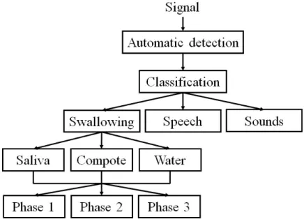
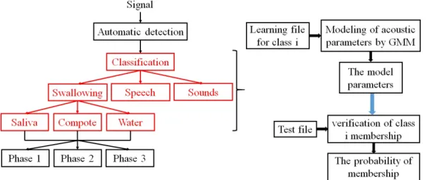
The issues related to the classification of sounds in general and swallowing sounds in particular are addressed in this work with a hierarchical analysis that aims to first identify the swallowing sound segments and then to decompose them into three
vi
characteristic sounds, consistent with the physiology of the process. The coupling between detection and classification is also addressed in this work.
The real-time implementation of the detection algorithm has been carried out. However, clinical use of the classification is discussed with a plan for its staged de-ployment subject to normal processes of clinical approval.
Keywords: Wavelet decomposition, signal processing, detection, classification, GMM, HMM.. . .
Résumé
Les maladies altérant le processus de la déglutition sont multiples, affectant la qual-ité de vie du patient et sa capacqual-ité de fonctionner en société. La nature exacte et la gravité des changements post/pré-traitement dépendent de la localisation de l’anomalie. Une réadaptation efficace de la déglutition, cliniquement parlant, dépend généralement de l’inclusion d’une évaluation vidéo-fluoroscopique de la déglutition du patient dans l’évaluation post-traitement des patients en risque de fausse route. La restriction de cette utilisation est due au fait qu’elle est très invasive, comme d’autres moyens disponibles, tels que la fibre optique endoscopique. Ces méthodes permettent d’observer le déroulement de la déglutition et d’identifier les lieux de dysfonctionnement, durant ce processus, avec une précision élevée.
"Mieux vaut prévenir que guérir" est le principe de base de la médecine en général. C’est dans ce contexte que se situe ce travail de thèse pour la télésurveil-lance des malades et plus spécifiquement pour suivre l’évolution fonctionnelle du processus de la déglutition chez des personnes à risques dysphagiques, que ce soit à domicile ou bien en institution, en utilisant le minimum de capteurs non-invasifs. C’est pourquoi le principal signal traité dans ce travail est le son.
La principale problématique du traitement du signal sonore est la détection au-tomatique du signal utile du son, étape cruciale pour la classification auau-tomatique de sons durant la prise alimentaire, en vue de la surveillance automatique. L’étape de la détection du signal utile permet de réduire la complexité du système d’analyse sonore. Les algorithmes issus de l’état de l’art traitant la détection du son de la dég-lutition dans le bruit environnemental n’ont pas montré une bonne performance. D’où l’idée d’utiliser un seuil adaptatif sur le signal, résultant de la décomposition en ondelettes.
x
la déglutition en particulier sont abordées dans ce travail avec une analyse hiérar-chique, qui vise à identifier dans un premier temps les segments de sons de la dég-lutition, puis à le décomposer en trois sons caractéristiques, ce qui correspond par-faitement à la physiologie du processus. Le couplage est également abordé dans ce travail.
L’implémentation en temps réel de l’algorithme de détection a été réalisée. Cepen-dant, celle de l’algorithme de classification reste en perspective. Son utilisation en clinique est prévue.
Mots-clés : Déglutition, sons déglutitoires, décomposition en ondelettes, traite-ment de signal, détection, classification, GMM, HMM.
Acknowledgements
Firstly, I would like to express my sincere gratitude to several persons who helped me morally and professionally to conduct this work. I owe a deep sense of gratitude to my teachers, Mr. Dan ISTRATE, Mr. Jacques DEMONGEOT, Mr. Jérôme BOUDY and Mr. Dhafer MALOUCHE, for their sincere guidance and inspiration in complet-ing this project.
I am extremely thankful to Ms. Catherine MARQUE, Ms. Carla TARAMASCO, Ms. Pascale CALABRESE, Ms. Gila BENCHETRIT, Ms. Pascale VIGNERON, Mr. Atta BADII, Mr. Sofiane BOUDAOUD, Mr. Mounim A. EL YACOUBI, Mr. Hassan ESSAFI, Mr. Antonio GLARIA, Mr Salim BOUZEBDA and all UTC members for their kind guidance and encouragement.
Besides my advisors, I would like to thank my friends and my office colleagues, Meriem YAHIAOUI, Khalida AZUDIN, Tomas RODENAS, Vincent ZALC, Teddy HAPPILLON, Jean-Baptiste TYLCZ and Halim TANNOUS, Nassreddine NAZIH, Ala Eddine YAHIAOUI, who have more or less contributed to the preparation of this work.
To everyone I met during this project.
I thank my little brother, Zied, for his presence and understanding... I would like to let him know how precious his presence was, during the months he stayed with me.
Last but not the least, I would like to thank my family: my parents, my sisters and my brothers for supporting me spiritually throughout writing this thesis and my life in general.. . .
Contents
Acknowledgements xiii
1 Introduction 1
1.1 Ageing of the population. . . 1
1.2 Dysphagia consequences . . . 3
1.2.1 Epidemiology of dysphagia . . . 3
1.2.2 Multidisciplinary care of dysphagia . . . 4
1.3 Medical desertification . . . 6
1.4 Telemedicine . . . 8
1.5 e-SwallHome project . . . 10
1.6 Objectives . . . 11
1.7 Document organisation. . . 12
2 Anatomy and Physiology 13 2.1 Swallowing and breathing: fundamental couple . . . 13
2.1.1 Anatomy . . . 13 2.1.2 Physiology. . . 16 2.2 Dysphagia . . . 18 2.2.1 Etiology of Dysphagia . . . 21 2.2.2 Semiology of dysphagia . . . 23 3 State-of-the-art 27 3.1 Screening for dysphagia . . . 27
3.2 Swallowing Monitoring Modalities . . . 32
3.3 Sound Recognition Domain . . . 42
3.4 Breathing monitoring . . . 54
xvi
4 Proposed system 59
4.1 The proposed Automatic Detection Algorithm . . . 59
4.1.1 The Gaussian mixture model . . . 64
4.1.2 Hidden Markov Model (HMM). . . 66
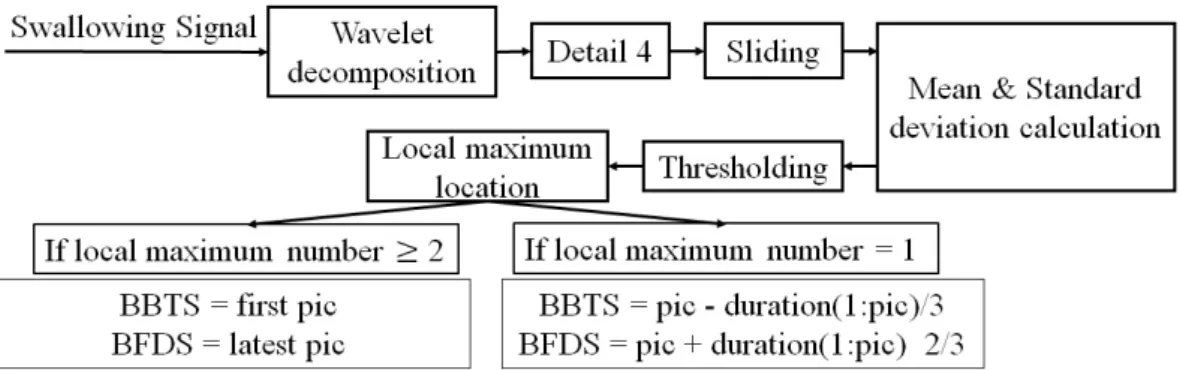
4.1.3 Local maximum detection algorithm . . . 66
4.1.4 Classification types . . . 68
4.2 Methods for breath analysis . . . 70
4.3 Conclusion . . . 71
5 Results 73 5.1 Recording protocol and swallowing monitoring database . . . 73
5.1.1 Data acquisition protocol . . . 73
5.1.2 Materials . . . 76
5.1.3 Procedure and Database . . . 77
5.1.4 Sound signals . . . 78
5.1.5 Breathing signals . . . 83
5.2 Automatic detection algorithm results . . . 84
5.2.1 Evaluation methodology. . . 85
5.2.2 Developed detection application . . . 86
5.3 Classification results . . . 88
5.3.1 Evaluation methodology. . . 88
5.3.2 Classification . . . 89
5.3.3 Assessment of Swallowing Sounds Stages. . . 97
5.4 Breathing analysis results . . . 101
5.4.1 Discussion . . . 103
6 Conclusions and perspectives 105 Bibliography 109 A List of Publications 125 B Theory and Methods for signal processing and pattern recognition 127 B.1 Fourier Decomposition . . . 127
B.1.1 Continuous Fourier transform . . . 127
B.1.3 Fast Fourier transform . . . 128
B.2 Wavelet Decomposition . . . 129
B.2.1 The Continuous Wavelet Transform . . . 130
B.2.2 Discrete wavelet transform . . . 131
B.2.3 Fast wavelet transform . . . 131
B.3 Acoustical features . . . 133
B.4 The Gaussian mixture model . . . 135
B.4.1 Expectation Maximisation (EM) algorithm . . . 137
B.5 Markovian models . . . 137
B.5.1 Markovian process . . . 137
B.5.2 Hidden Markov Model (HMM). . . 138
B.5.3 Forward-Backward Probabilities . . . 139
B.5.4 Recognition method . . . 140
The a Posteriori Marginal Method (PMM) . . . 140
Viterbi algorithm . . . 141
EM algorithm for parameter estimation . . . 142
C Identification grid for swallowing disorders and associated factors 145
D Information note-Consent 147
E Protocol 153
List of Figures
1.1 Distributions of otolaryngologists on French territory . . . 7
1.2 Distributions of physiotherapists on French territory. . . 7
2.1 Anatomy of human superior digestive system BCCampus ( https://open-textbc.ca/anatomyandphysiology/2303-anatomy-of-nose-pharynx-mouth-larynx/) 14 2.2 Respiratory and digestive tracts in Human . . . 16
2.3 Lateral view of bolus propulsion during swallowing. . . 19
3.1 Dynamic sEMG topography in a swallowing process . . . 37
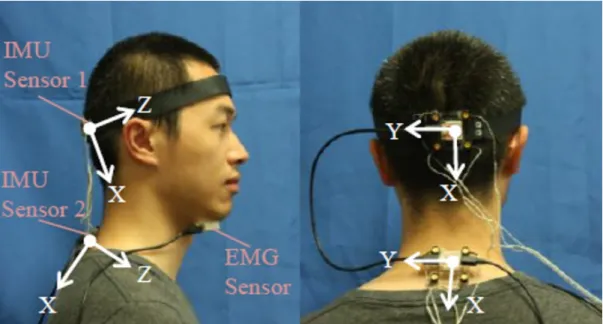
3.2 Sensor placement on the body and Frame axes . . . 42
3.3 Characteristics of the swallowing sound wave . . . 50
4.1 Global Proposed System . . . 60
4.2 Diagram of the automatic detection algorithm . . . 60
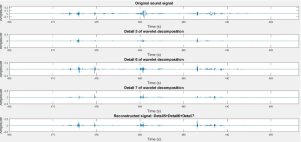
4.3 Wavelet decomposition into 12 levels. . . 61
4.4 Reconstructed Signal as linear combination of selected details . . . 62
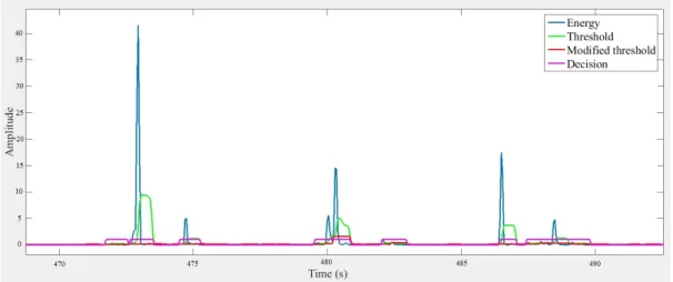
4.5 Procedure of detection algorithm with different calculated features . . 63
4.6 Recognition process. . . 65
4.7 Hidden Markov Model algorithm. . . 66
4.8 Local maximum detection algorithm . . . 67
4.9 The Visuresp interface . . . 71
5.1 Best microphone position (in yellow) . . . 77
5.2 A typical swallowing sound (a) time-domain signal (b) the correspond-ing spectrogram. (’au’ arbitrary units for normalised amplitude Aboofazeli and Moussavi (2004)) . . . 79
5.3 A typical swallowing sound of water in time-domain signal and its corresponding spectrogram . . . 82
xx
5.5 Respiratory signals and the corresponding apnea segment . . . 84
5.6 Result of the proposed automatic detection algorithm . . . 85
5.7 Some examples of automatic detection and errors that can be vali-dated by the detection algorithm where green panels refer to reference event and pink panels refer to detections . . . 85
5.8 Graphical interface of the recording and detection application in real time . . . 87
5.9 Detection results. . . 87
5.10 Classification evaluation process with leave-one-out approach . . . 89
5.11 Good recognition rate per class using manually annotated segments . 90
5.12 Good recognition rate per class using sounds obtained from micro-phone fixed on the vest . . . 92
5.13 Good recognition rate per class using Grenoble database . . . 92
5.14 Good recognition rate per class using different data . . . 93
5.15 Spectrograms of the sounds of compote swallowing . . . 94
5.16 Spectrograms of the sounds of water swallowing . . . 94
5.17 Spectrograms of the sounds of saliva swallowing . . . 94
5.18 Good recognition rate per class using manual annotated swallowing sounds according to textures. . . 95
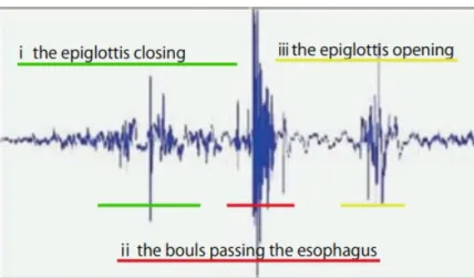
5.19 Typical decomposition of the swallowing sound into three character-istic sounds . . . 98
5.20 Respiratory signal and detected boundaries of respiratory phases (in-spiration and expiration) during the subject’s rest . . . 102
5.21 Results of automatic detection on the synchronized sound and respi-ratory signals . . . 102
B.1 Wavelet Functions Examples . . . 132
B.2 Mel filter bank (containing 10 filters and starts at 0Hz and ends at 8000Hz). . . 134
B.3 Steps of MFCC features calculation . . . 134
C.1 Identification grid for swallowing disorders and associated factors (Desport et al.,2011) . . . 146
List of Tables
3.1 State-of-the-art summary. . . 53
4.1 Roll-off point calculated according to textures (*Comp=Compote) . . . 62
4.2 Correspondence of the coefficients in mel and frequency scales for each coefficient using 24 filters . . . 65
5.1 Sound database . . . 78
5.2 Swallowing event duration . . . 81
5.3 Swallowing event duration according to textures . . . 81
5.4 Grenoble’s database (acquired as part of the e-SwallHome project by Calabrese et al. (1998)) . . . 83
5.5 Sehili’s database . . . 83
5.6 Automatic detection results . . . 88
5.7 Confusion matrix of the classification of manually annotated segments 91
5.8 Automatic Detection Algorithm results for data obtained from micro-phone attached to the vest . . . 91
5.9 Confusion matrix using sounds obtained from microphone fixed on the vest . . . 92
5.10 Confusion matrix of Grenoble database . . . 93
5.11 Confusion matrix of the classification of manually annotated swal-lowing sounds according to textures . . . 95
5.12 Confusion matrix of the classification of automatically detected sounds 97
5.13 Confusion matrix of the classification of automatically detected swal-lowing sounds according to textures . . . 97
5.14 Swallowing phases duration for Water sounds . . . 97
5.15 Swallowing phases duration for Compote sounds . . . 97
5.16 Mean ± standard deviation for delay in detection of boundaries of swallowing sound phases obtained by HMM . . . 99
xxii
5.17 Confusion matrix of the classification of water sounds of the swallow-ing phases usswallow-ing HMM. . . 99
5.18 Delays in detection boundaries of swallowing phases in water sounds (ms) . . . 99
5.19 Confusion matrix of the classification of compote sounds of the swal-lowing phases using HMM . . . 100
5.20 Delays in detection boundaries of swallowing phases in compote sounds (ms) . . . 100
5.21 Confusion matrix of the classification of water sounds of the swallow-ing phases usswallow-ing local maximum algorithm . . . 100
5.22 Confusion matrix of the classification of compote sounds of the swal-lowing phases using local maximum algorithm . . . 100
5.23 Delays in detection boundaries of swallowing phases in water sounds (ms) . . . 101
5.24 Delays in detection boundaries of swallowing phases in compote sounds (ms) . . . 101
List of Abbreviations
ANR National Research Agency NRA BTS Bolus Transit Sound
CA Cervical Auscultation
DREES Directorate of Research, Studies, Evaluation and Statistics DWT Discrete Wavelet Transform
ECPFR Echelles Cliniques Prédictives de Fausse Route EGG Electroglottography
EM Expectation Maximisation EMG Electromyography
ENMG Electroneuromyogram FAR False Alarm Rate FDS Final Discrete Sound
FEES Fiberoptic Endoscopic Evaluation Swallowing FFT Fast Fourier Transform
GMM Gaussian Mixture Model GUSS Gugging Swallowing Screen HMMs Hidden Markov Models
ICT Information an Communication Technologies IDS Initial Discrete Sound
IESEI Inspiration Expiration Swallowing Expiration Inspiration IESI Inspiration Expiration Swallowing Inspiration
ISEI Inspiration Swallowing Expiration Inspiration IMU Inertial Measurement Unit
INED National Institute for Demographic Studies
INSEE National Institute of Statistics and Economic Studies KNN K-Nearest Neighbours
LDA Linear Discriminant Analysis LES Lower Esophageal Sphincter
xxiv
LFCC Linear Frequency Cepstral Coefficients LIRA Limited Receptive Area
MEBD Modified Evan’s Blue Dye MER Missed Event Rate
MFCC Mel Frequency Cepstral Coefficients MsFS Mel-scale Fourier Spectrum
ORR Overall Recognition Rate
PASS Practical Aspiration Screening Schema PER Partial Event Rate
RIP Respiratory Inductance Plethysmography sEMG Surface Electromyography
STFT Short Time Fourier Transform SVM Support Vector Machine
TOR-BSST Toronto Bedside Swallowing Screening Test UES Upper Oesophageal Sphincter
VER Validated Event Rate
VFSS Videofluoroscopic Swallowing Study WDT Waveform Dimension Trajectory WFD Waveform Fractal Dimension WHO World Health Organization WPD Wavelet Packet Decomposition
Chapter 1
Introduction
Breathing, drinking, eating, protection from cold and heat are among the essential el-ements for survival. Some primary needs are met by nature, including breathing, but the majority of them are not and require voluntary action by the individual. Among them, swallowing is one of these needs. Swallowing is a complex and vital process in humans. It provides two vital functions at once: nutrition and protection of the respiratory tract. Swallowing, defined as a voluntary act, enables transport of sub-stance, saliva or liquid or chewed solids, from the oral cavity to the stomach passing through the pharynx, œsophagus while ensuring the airways security. Swallowing is a repetitive act that occurs 300 times per hour during a meal and is estimated to occur about 600 to 2000 times a day in healthy people Amft and Troster (2006). Eat-ing and drinkEat-ing are physiological necessities, but also a source of individual and social pleasure. Indeed, to eat is to gather, to share, to desire, to see, to smell, to salivate, to taste, and so on. If eating is a necessity for everyone, a pleasure for many, a sin for some, it is also a danger for others. Difficulties in swallowing, disorders are known as dysphagia. Dysphagia can occur at any age, but it is more common in older adults in general and their management requires a multidisciplinary approach to recognise, characterise and propose rehabilitation measures. However, in France, the number of practitioners is decreasing across the healthcare professions including general practitioners, specialised doctors or medical auxiliaries.
1.1
Ageing of the population
Dysphagia in the elderly is called presbyphagia. It can be divided into two cate-gories: (i) Primary presbyphagia which refers to the effects of normal ageing on the organs involved in swallowing and (ii) Secondary presbyphagia which refers to the
2 Chapter 1. Introduction
accumulation of normal aging and related changes to diseases which can lead to se-vere dysphagia such as stroke and Parkinson’s disease. Mccomas, Upton, and Sica (1974) found that there is a little loss of functioning motoneurons before the age of 60 years but there is a striking and progressive depletion subsequently with age, by applying electrical stimuli of low, gradually increasing intensity, to single motor units.
As a world phenomenon, the ageing populations effects both developed and emerging countries. The French population like other European peoples is also age-ing.People aged 65 and over increased from 13.9% in 1990 to 18.8% in 2016. The chal-lenge will be to adapt the capacities of the structures caring the loss of autonomy so that they accompany this important increase. By 2050, according to the population projections calculated by Eurostat, the number of people aged 65 and over would be 27.2% which is a significant and increasing demographic trend.
Swallowing disorders are common in the elderly, from 9% for those aged 65 to 74 living at home to 28% after 85 years Audrey (2013). Previous studies estimate that the prevalence of dysphagia after stroke ranges from 33% to 73% Mann, Han-key, and Cameron (2000) and Paciaroni et al. (2004). In another study, cited by var-ious authors, Greuillet and Couturier estimate a prevalence ranging from 12% to 68% among institutionalised elderly populations. For home-stay patients, a ques-tionnaire study found that 10% to 16% had symptoms of dysphagia Audrey (2013). In institutions, dysphagia reaches 30% to 60%. According to the Nestlé Institute of Health Science Science (2016), 40% of people over 75 have swallowing disorders. Up to 66% of those affected are residents of a Medico-Social Institution.
As the world’s population ages, these statistics will increase. In France, the per-centage of people living alone in their homes between 65 and 79 years is 27.1% whereas for people aged 80 or over is 48.8% according to the National Institute of Statistics and Economic studies (INSEE). The higher risks associated with dyspha-gia for those living alone, make for an alarming situation given that a significant percentage of dysphagic elderly live in institutions and at home.
Ageing, according to Stedman’s Medical Dictionary (26th edition, p 38) is "the gradual deterioration of a mature organism from time-dependent irreversible changes in structure that are intrinsic to the particular species, and that eventually lead to a decreased ability to cope with the stresses of the environment, thereby increasing
the probability of death.". The effects of ageing result in changes in the mouth, phar-ynx and œsophagus which is called presbyphagia. Studies have shown a decrease in tongue mass in elderly patients Ney et al. (2009). This phenomenon of decreased skeletal muscle is well known in elderly as sarcopenia. As a result, tongue position and movements are often altered in the elderly Baum and Bodner (1983) and lin-gual pressure decreases with age Robbins et al. (1995) causing prolonged oral phase Nicosia et al. (2000). Studies have also shown that the pharyngeal phase of swallow-ing is significantly prolonged in the elderly Ney et al. (2009) and Rofes et al. (2011). Changes in lip posture may also cause drooling of saliva which is common in older persons Cook and Kahrilas (1999). A radiological study has shown that 90% of 90-year-olds had impaired œsophageal motility with one third having complete loss of the primary peristaltic waves responsible for transport of feed Zboralske, Amberg, and Soergel (1964). Similar observations have been obtained by a nanometric study which examined the effect of the age on secondary œsophageal peristalsis Ren et al. (1995); frequency of secondary peristalsis and lower esophageal sphincter relaxation in response to air distention was significantly lower in the elderly than in the young. With advancing age, loss of teeth reduces masticatory performance and increases duration for chewing. Influence of masticatory behavior on muscle compensations during the oral phase of swallowing of smokers was proposed by Silva et al. (2019). A study on the influence of food thickeners on the size of bolus particles swallowed was also the focus of study proposed by Yamaguchi et al. (2019)
1.2
Dysphagia consequences
The consequences of dysphagia are highly variable, ranging from no discernible ef-fect to airway obstruction or severe aspiration pneumonia.
1.2.1 Epidemiology of dysphagia
Incidence of pneumonia is estimated to be around one-third of person with stroke, and increases mortality by three Hilker et al. (2003). It is associated with a quarter of deaths in the first month after stroke Worp and Kappelle (1998); 27% to 37% of dysphagic patients die within 3 to 6 months Singh and Hamdy (2006). In a study conducted by Zhou, among 70 patients with stroke recruited, 6 patients (8.6%) had pneumonia within 1.7±2.4 days (end: 0∼6 days) compared to the occurring instant
4 Chapter 1. Introduction
of stroke. In a retrospective study of 40 dysphagic patients in the geriatric category, Croghan et al. (1994) described the occurrence of pneumonia in 43% within one year compared to the first videoradioscopic examination of swallowing and at 30% of deaths related to these pneumonias.
Dysphagia is responsible for about 4000 deaths per year in France Audrey (2013). According to the National Institute for Demographic Studies (INED), the mortality rate in France in general population in relation to suffocation by swallowing disorder is 5.99 per 100000 for men, and 6.1 per 100000 for women.
The consequences of dysphagia are multiple, ranging from a deterioration in a patient’s quality of life to death. The medical management of dysphagia for reha-bilitation purposes requires multidisciplinary care to ensure rehareha-bilitation and good practice of swallowing and to avoid any incidence of dysphagia.
1.2.2 Multidisciplinary care of dysphagia
The objective of the management of swallowing disorders is to ensure a feeding modality that guarantees the safest possible nutritional state with regard to respira-tory function and respects the person’s quality of life as much as possible, whether in terms of reducing the discomfort caused by the dysphagia experienced or in terms of eating pleasure. For populations at risk of swallowing disorders, the prevention of complications of this disorder leads to the development of screening tests based on care protocols. For a patient for whom the diagnosis of dysphagia has been con-firmed, the clinician must understand the cause of the disorder and how well it is tolerated.
Following a swallowing complication, the diagnosis of dysphagia begins with a simple questioning of the patient and his or her relatives. Clinical examination is essential for the diagnosis of dysphagia, but it is not effective in detecting and pre-venting cases of food chocking, which are often silent. The treatment of dysphagia is mainly etiological. Videoradiography is the gold standard for determining the mechanism of dysphagia and the modalities of symptomatic treatment. Rehabilita-tion and adaptaRehabilita-tion of food textures Ross et al. (2019) to the mechanisms of dyspha-gia can help preserve oral nutrition. In addition to enteral nutrition, maintaining an oral diet is important to preserve the patient’s quality of life Professionnelles (2007) and Audrey (2013). This involves multidisciplinary follow-up.
The therapeutic aspect is essentially based on multidisciplinary care that requires cooperation between medical staff (doctors, the nursing staff), the patient, their fam-ily or the members of the institution where they reside. Rehabilitation and dietary management techniques are the most widespread.
The nurse ensures the good practice of medical and logopedic instructions and tracks the warning signs encountered during food intake.
Otorhinolaryngology physicians perform a detailed clinical assessment such as screening test with replenishment such as the "3 Oz Water Swallow Test" validated by DePippo, Holas, and Reding (1993), which is considered perfectly adapted to stroke victim population Bassols and Puech (2011), and also, the functional ability test of swallowing by Guatterie and Lozano (2005). In addition, the no-feedback test is presented as predictive aspiration clinical scales ECPFR (Echelles Cliniques Prédictives de Fausse Route). Practical Aspiration Screening Schema (PASS) Zhou (2009) combines the ECPFR and the 3 Oz Water Swallow Test.
There is also the Videofluoroscopic Swallowing Study (VFSS), which is a tech-nique used to observe and film radiographic swallowing to follow the food bolus coated with a product that allows contrast. Videofluoroscopy makes the entire swal-lowing process visible. In case of an aspiration, it enables quite precisely, the de-termination as to how much material has entered the airways, at what level, when and how the patient responds Martin-Harris and Jones (2008). VFSS enables the to determination of the effect of various behavioural and sensory interventions on the physiological function of the swallowing mechanism. However, these techniques are invasive and uncomfortable requiring an active patient participation, and are therefore not always applicable in the elderly.
After a detailed clinical assessment, the therapist identifies appropriate strategies in order to re-educate the deficit phase. The physiotherapist manages the respiratory function by using various techniques. The ergotherapist ensures the optimal use of rehabilitative skills in the management of daily life activities or the compensation of deficits by use of appropriate technical aids. The dietician adapts the diet in relation to medical and logopedic instructions, but also to the taste of the patient and realises the feasibility of these recommendations in an institution. The psychologist detects a possible depressive reaction syndrome, ensures the acceptance of the disease and the integration of the patient in their environment.
6 Chapter 1. Introduction
1.3
Medical desertification
Today, in France, few territories in the regions still escape medical desertification. According to statistics published by the Directorate of Research, Studies, Evaluation and Statistics (DREES), otolaryngologists and head and neck surgery physicians and physiotherapists are not evenly distributed across the territory. However, there are disparities between regions. Figures1.1and1.2 show the distribution of otolaryn-gologists and physiotherapists on French territory. Overall, the otolaryngology has a density of 4.6 otolaryngologists per 100 000 inhabitants. Thus, the Ile-de-France have privileged access to this medical service with a density of 6.6 otolaryngologists per 100 000 inhabitants. According to the Demography of Physiotherapists report (Situation on 31 August 2017), the average French number of physiotherapists per 10 000 inhabitants is 12.6. This Figure can be described as low compared to other European countries such as Belgium which have 25.8 physiotherapists per 10 000 inhabitants.
Also, the management of dysphagia must, in order to be effective, be carried out in collaboration with the family and staff of the institution in which the person resides. This could not be the case according to the large number of people living alone. Clear and accurate information must be provided so that the evolution of the disorder and its possible complications can be understood, Allepaerts, Delcourt, and Petermans (2008). Access to care in good conditions, participation in the choices that concern them; continuing to have confidence in their health system: these are the expectations of patients and users, especially patients at risk.
The complexity and compartmentalization of the current system often enable the patient to coordinate the different professionals themselves. A complexity that professionals feel every day and that patients perceive in their daily lives. Social difficulties such as ageing, increased health spending and medical deserts are also relevant.
The reforms to be undertaken to overcome these difficulties can only be envis-aged in a global approach including the hospital and the medico-social sector. This transformation must aim to improve all subjects: access to care, prevention, quality of care, regulation of health insurance expenditure, but also the medico-social link, the transformation of the hospital and the modernisation of medicine. It is the en-tire health system that must be challenged to meet today’s challenges and prepare
Figure 1.1:Distributions of otolaryngologists on French territory
Figure 1.2:Distributions of physiotherapists on French territory
for tomorrow’s health system. To do so, the patient must more than ever be at the centre of future thinking and developments. From this point of view, medicine is evolving and starting on "e-health".
8 Chapter 1. Introduction
e-health is a multi-disciplinary domain which involves many stakeholders, in-cluding clinicians, researchers and scientists with a wide range of expertise in health-care and engineering. Digital health is the convergence of digital technologies with healthcare, living, and society to enhance the efficiency of healthcare delivery and make medicine more personalised and precise. It involves the use of information and communication technologies to help to address the health problems and chal-lenges faced by patients. These technologies include both hardware and software solutions and services, including telemedicine.
1.4
Telemedicine
Telemedicine is the performance of a remote medical procedure. Telemedicine brings together medical practices permitted or facilitated by telecommunications. It is an exercise in medicine by use of telecommunications and technologies that enables distance health benefits and the exchange of medical information related to it. Dif-ferent in application, telemedicine has to provide a rapid medical response to a pa-tient’s problem. It makes it possible to establish a preventive follow-up or a post-therapeutic follow-up for patients at risk. Already in 2016, the World Health Or-ganisation (WHO-OrOr-ganisation Mondiale de la Santé OMS) reported on e-health in Europe encouraged the deployment of national health policies on telemedicine. De-livering e-health, i.e. the use of information and communication technologies (ICT) for health is a relatively recent healthcare practice dating back to at least 1999 Mea (2001). e-health is considered as the death of telemedicine; "e-health can be consid-ered to be the health industry equivalent of e-commerce:". e-health is deemed to be a fashionable name for telemedicine. It includes electronic/digital processes in health, namely health applications. Telemedicine has four components:
• Tele-consultation: which is a remote consultation,
• Tele-regulation: which takes the form of a service that puts a person in touch with an operator if there is a problem at home,
• Tele-expertise: when doctors consult each other between them, and
• Tele-monitoring which is a remote monitoring of a patient by transmission of medical data between patients and different medical staffs.
Telemedicine was born in medical practice in the 1970s in rural America and northern Norway. It has also been tested very early independently in 1966 in USA, Russia and France with telephonic transmission of ECGs. It has been said that it orig-inated in January 1905 in Netherlands, when W. Einthoven’s assistant transmitted a ECG record via a telephone line from the hospital of Leyden to his laboratory located 1.5 kms away. Telemedicine aims to optimise the care of the patient upstream of his care path, giving him access to a general practitioner through a videoconference con-sultation. Telemedicine uses signals obtained from different sensors, installed in the patient’s home or in intelligent socio-medical institutions, in order to extract infor-mation needed for diagnosis. This first step enable direct orientation according to their situation, for a physical consultation or referral to a specialist. It is the birth of a hybrid model that alternates physical and remote consultations between a doctor and his patient. Above all, it provides the patient with a panel of specialists who can follow him at regular intervals, overcoming the obstacle of distance. Beyond the re-mote consultation, the monitoring of physiological parameters can be carried out in real time or in a delayed manner if the data are stored. The surveillance is willing to follow the progress of a patient at risk which in some cases becomes vital. This aspect is very important in the case of patients at risk of dysphagia that can lead to dramatic consequences even the death of the patient if intervention is not timely. Therefore, telemedicine opens up new opportunities and better access to health care, particu-larly for emergencies where a diagnosis must be made quickly. These technologies can significantly improve access to health care, especially in medical deserts.
In Europe, telemedicine is developing rapidly. In France, it has been included in the Public Health Code since 2010, and is governed by the HSPT law (hospi-tal, health, patients, territory). Since January 2019, the teleconsultation act has been recognised and reimbursed by the national health insurance fund. Doctors are in favour of the evolution of this technology. Already today, 84% of them use smart-phones or digital tablets in their work. Telemedicine is a recent phenomenon in the health field consisting of the use of services that allow clinicians and patients to come into direct contact, almost instantaneously, through the use of new technologies.
The work of this thesis is part of the telemedicine and it is presented as a telemon-itoring application. It is based on the processing of sound signals acquired during food intake.
10 Chapter 1. Introduction
1.5
e-SwallHome project
The work of this thesis is part of the e-SwallHome (Déglutition & Respiration : Mod-élisation et e-Santé à domicile) project. e-SwallHome, funded by National Research Agency (ANR), aims to explain the normal behaviour of three coupled functions in humans; swallowing, breathing and phonation, to better understand the mech-anisms underlying dysphagic, dysphonic and dyspnoeic pathological behaviours, following in particular a stroke. To do so, it works on a set of protocols for diag-nosis, home monitoring, education and rehabilitation in subjects at risk of death by aspiration, asphyxiation or fall without the possibility of voicing any oral expres-sions as warning signals.
The theme of e-SwallHome is e-health which brings all applications of Infor-mation and Communication Technologies (ICT) in the field of health, with a wider scope than telemedicine. This discipline aims at the use of distance medicine aiming at well-being on topics related to medicine but without medical constraints. Thus focus on, home-based tele-accompaniment, combining technological innovation in health and contributing, through a better acceptability on the part of the patient, to the improvement of adherence to therapeutic recommendations and rehabilitation protocols (by speech therapists and physiotherapists), and thus to prevent the com-plications induced by chronic dyspnoea / dysphagia / dysphonia disorders related to the initial stroke.
The target population in the e-SwallHome project is healthy subjects and pa-tients who have suffered from stroke. Healthy subjects included in this study must be aged between 18 and 40 years, raised in a monolingual French-speaking environ-ment and not presenting language, neurological and/or psychiatric disorders. Vol-unteers signed an informed consent form. The inclusion criteria for stroke patients included in this project were to be more than 18 years of age, first cerebral infraction and confirmed by MRI≤15 days, absence of severe leukoaraiosis, swallowing dis-orders identified by a GUSS scale less than 20, identified neurological deficit and the patient has to be able to cooperate.
This thesis is part of the research project e-SwallHome. In revising the initial as-sumption of inclusion criteria of patients, has been reoriented to include only healthy subjects due to lack of access to patient’s medical records. So, it was proposed to work only with healthy subjects while keeping the same objectives of e-SwallHome
project to be used subsequently for evaluating pathological signals in this study.
1.6
Objectives
The thesis subject, proposed as part of the diagnosis system described above, based on home telemonitoring, participates in the home monitoring of patients suffer-ing from dysphagia, by propossuffer-ing automatic monitorsuffer-ing methods and evaluation of functional rehabilitation. The end goal of this PhD is to develop a tool able to monitor swallowing in real time using the least invasive sensor, ambulatory, while ensuring the comfort and the good quality of the daily life of the person. Limited to data in healthy subjects, the proposed system has to be a part of a rehabilitation support that could be provided to the patient and clinicians.
This PhD study enables telemonitored patients to follow the pregress of people at risk. More specifically, it concerns dysphagia in the elderly and seeks to identify automatically the specific characteristics that could be used in the assessment of the health status of patients at risk of dysphagia.
The analysis and extraction of information from the sound is an important as-pect for medical telemonitoring of dysphagia. In this context, this thesis analyses and proposes solutions to the problems specific to sound processing for medical telemonitoring. In addition, breath signal analyses are also performed for the same purposes.
Among these problems, the automatic classification of sounds of swallowing in the context of everyday life has been little explored and a sound analysis system is proposed. This thesis sets out the problems and objectives of medical telemonitoring of dysphagia. The study of the sound signal must be able to highlight the various characteristics of the normal functional state of swallowing process and thus also the state of its malfunctioning.
Among the problems of telemonitoring that have been studied in this thesis are: signal quality, and adaptation of sound recognition techniques to the classification of sounds of swallowing in everyday life during food intake.
The quality of the signal influences the result of the recognition and signal pro-cessing systems. Taking into account the difficulties related to the quality of the signal, the work of this thesis focused on the signals with a sampling frequency of
12 Chapter 1. Introduction
16 kHz as well as its treatment by decomposing it in order to extract the most signif-icant frequency bands as assessed to be adequate for the processing of swallowing events. This step must guarantee the best performance of the used algorithms in terms of the error rate, which is an important problem. The algorithms must ensure sufficient performance.
To be able to test, improve and validate the algorithms proposed in this thesis, a database was created as there was no access available to existing databases. An adaptation of sound recognition techniques has been carried out. The frequency characteristic of swallowing and those of other sound classes have differences and similarities, which requires finding suitable parameters to differentiate them.
1.7
Document organisation
The document starts with an introduction followed by a physiological description of the swallowing process and anatomy. Chapter 2 presents the etiology and semiology of dysphagia. Chapter 3 presents the state-of-the-art of the different methodologies for investigating the swallowing process and the different modalities used. Chapter 4 describes the different methodologies used and proposed in this work for the pro-cessing of sound and respiratory signals. Chapter 5 describes the data acquisition protocol followed, the sensors proposed and the procedure followed. The results of the methodology for performing the tests built to validate detection, classifica-tion and detecclassifica-tion-classificaclassifica-tion coupling are presented in Chapter 6. The document concludes with conclusions and perspectives in Chapter 7 and the Appendices.
Chapter 2
Anatomy and Physiology
Swallowing is the set of coordinated acts that ensure the transfer of solid food or liquids from the mouth to the stomach, through the pharynx and oesophagus. Ac-cording to Guatterie and Lozano (2005), dysphagia is the difficulty of performing the action of eating, to swallow with a feeling of discomfort or stop in the transit, painful or not, with possible misconceptions when swallowing food, liquids or saliva and, by extension, any abnormalities in the passage of food to the stomach. To be able to monitor the swallowing process, understanding the normal physiology and patho-physiology of swallowing is fundamental to evaluate the signs of the swallowing disorders and develop dysphagia rehabilitation programs.
2.1
Swallowing and breathing: fundamental couple
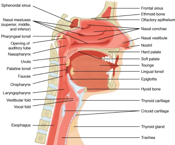
The oral cavity, larynx, pharynx and œsophagus are the structures used in swallow-ing. They are described by Bassols and Puech (2011), which will serve as the main reference for anatomy. Figure2.1shows the anatomy of the upper human digestive system.
2.1.1 Anatomy
The oral cavity is composed of several structures which are involved in swallowing: The lips close the oral cavity and maintain the food between the cheeks and teeth during chewing. The tongue with a mobile front is connected to the pharynx and the edge of the epiglottis by the glossoepiglottic fold on each side of which are located the valleys.The soft palate forms with its front and rear pillars and the palatal tonsils, the isthmus of the throat, passage between the oral cavity and the oropharynx. The mandible is mobilised by muscular activity in all three planes to crush the food with
14 Chapter 2. Anatomy and Physiology
Figure 2.1: Anatomy of human superior digestive system BCCam-pus (
https://opentextbc.ca/anatomyandphysiology/2303-anatomy-of-nose-pharynx-mouth-larynx/)
the teeth before swallowing: the pieces decrease in size and are unsalivated. The mouth floor is made up of three of the suprahyoid muscles that connect the mandible with hyoid bone: digastric, mylo-hyoidal and geniohyoidal.
The larynx is located at the front of the neck, it is moored at the top to the base of the tongue and bone hyoid, below it extends into the trachea. This musculocar-tilaginous duct communicates with the pharynx backwards. The larynx performs the functions of breathing, swallowing and phonation and can be considered as a valve that opens and closes the glottis. The larynx is divided into three levels. The supraglottic stage, or vestibule, is located above the vocal cords and includes the ventricular bands. The glottic layer corresponds to the plane of the vocal cords. The subglottic layer, located under the vocal cords, extends from the lower part of the vocal folds to the lower edge of the cricoid cartilage. The laryngeal skeleton consists of cartilage, among which two important joints can be mentioned. The crico-thyroid joint enables the thyroid cartilage to shift from the thyroid cartilage to the cricoid
cartilage, and thus to put the vocal cords under tension. The crico-arytenoid joint enables the arytenoid cartilage to shift in or out the cricoid cartilage to bring the strings closer or further apart, as well as a forward or backward shift to change their length. The larynx consists of two categories of muscles. The intrinsic muscles en-able adduction or abduction movements, as well as than tension or relaxation of the vocal cords. The constrictor and tightening muscles bring the vocal folds closer to-gether in order to close the glottis when swallowing, and the dilators open the glottis for breathing. The extrinsic muscles ensure the suspension and mobility of the lar-ynx in the neck. Some are subhyoidal and lower the larlar-ynx and hyoid bone, others are suprahyoids and elevators of the larynx and hyoid bone.
The pharynx is located behind the oral, nasal and laryngeal cavities, this con-duit musculomembranous consists of constrictor muscles, responsible for pharyn-geal peristalsis, and elevating muscles. It is divided into three floors. The rhinophar-ynx is located behind the nasal passages; its role is respiratory and it is closed by the velopharyngeal sphincter during the oral time of swallowing. The oropharynx, be-hind the oral cavity, is the crossroads of the aerodigestive pathways: the swallowed food bowl and the breathed air circulate through it. It contains the pillars anterior and posterior of the soft palate, the tongue base, the valleys and the face lingual epiglottis. The hypopharynx circles the larynx backwards and sideways and ends at the bottom with the œsophagus. It contains the piriform sinuses which are a depres-sion of the mucous membrane between the larynx and pharynx forming two cavities through which the food bolus passes during swallowing.
The œsophagus is a muscular tube connecting the pharynx with the stomach. The œsophagus is lined by moist pink tissue called mucosa. The upper œsophageal sphincter (UES) is a bundle of muscles at the top of the œsophagus. The muscles of the UES are under conscious control, used when breathing and eating and keep food and secretions from going down the trachea. The lower œsophageal sphincter (LES) is a bundle of muscles at the low end of the œsophagus, where it meets the stomach. The LES muscles are not under voluntary control.
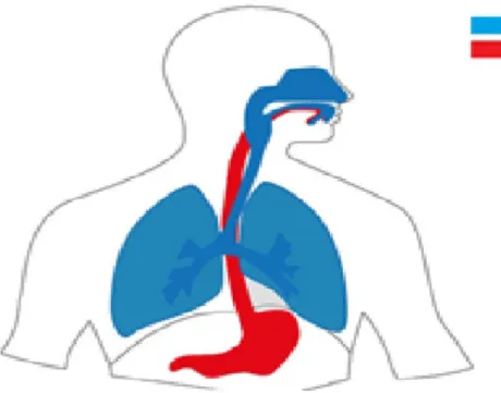
As acknowledged, swallowing and breathing are a paradoxical couple because of the common structure involved in both functions. The breathing air passes through the mouth or nose, pharynx, larynx and trachea. The air of phonation uses the lar-ynx, pharlar-ynx, mouth and nose. The swallowed food passes through the mouth, pharynx and œsophagus. Breathing, phonation and swallowing therefore share a
16 Chapter 2. Anatomy and Physiology
common crossroads: the mouth and pharynx. Figure2.2shows simultaneously the respiratory and digestive tracts.
Figure 2.2:Respiratory and digestive tracts in Human
2.1.2 Physiology
Swallowing is a process resulting from muscle contraction under nervous control. Swallowing is initiated by the sensory impulses transmitted as a result of the stim-ulation of receptors on the tongue, soft palate and posterior pharyngeal wall. Then, sensory impulses reach the brainstem first through the VII, IX and Xth cranial nerves, while the efferent function is mediated through the IX, X and XIIth cranial nerves. Then, the fact that the bolus reaches the posterior pharyngeal wall triggers the relax-ation and the opening of the cricopharyngeal sphincter which is reflexive.
The Aero-digestive junction is an area with complex but vital anatomy and func-tions. The aero-digestive junction is the crossroads of the airways that enables the passage of air between the outside of our body and the lungs and digestive tract that enables us to feed. Swallowing refers to the placement of food in the mouth, its mas-tication if necessary, prior to initiate swallow when the bolus is propelled backward by the tongue to the pharynx and to the œsophagus through the upper œsophagus sphincter.
Classically, swallowing is described in three phases: oral, pharyngeal and œsophageal. Swallowing is initiated by the introduction of the food into the oral cavity and its preparation by the process of mastication and insalivation. Once the bolus is formed and is ready to be expelled to the pharynx, the tip of the tongue is raised up and ap-plied against the alveolar ridge of the upper incisors and the tongue takes the form of a spoon where the bolus slips and forms a single mass. The latter moves backwards by the moving tongue which gradually applies to the palate from front to back. At this moment, the soft palate realises the closure of the oropharynx and prevents the penetration of the bolus into the pharynx, however, the larynx is still open. By the time the bolus penetrates the throat isthmus, the oral time (voluntary time) is over. The back of the tongue moves forward and forms an inclination allowing the bolus to move towards the oropharyngeal cavity. Thus, the pharyngeal phase is triggered by the contact of the bolus with the sensory receptors of the throat isthmus and of the oropharynx.
The pharyngeal process is a continuous phenomenon in time, considered as a reflex accompanied simultaneously by the velopharyngeal closure by the velum, by the laryngeal occlusion assured by the elevation of the larynx, and by the retreat of the tongue’s base, the movement at the bottom of the epiglottis, the pharyngeal peristalsis and finally the opening of the upper sphincter of the œsophagus allows the passage of the food bolus into the œsophagus. This phase lasts less than one second. The opening of the upper sphincter of the œsophagus is initiated by the arrival of the pharyngeal peristalsis and passage through the esophagus is ensured by the continuity between the pharyngeal and the œsophageal peristalsis.
The pharyngeal stage triggered by the end of the oral stage becomes involun-tary and is called the swallowing reflex. Velum is raised to close the nasal cavities and avoids any passage of food into the nose and facilitates the passage of the bolus downwards towards the œsophagus through the pharynx. Precisely at this moment, the passage of food into the trachea is avoided. The larynx opens during chewing, to allow breathing, and is closed as soon as the bolus arrives on the base of the tongue. At the same time, the vocal cords are embracing to ensure airway closure, the mov-ing cartilages of the larynx (arytenoids) swmov-ing forward in the laryngeal vestibule, covered by the rocking of the epiglottis. The larynx is pulled up and down by the hyoid muscles, which place it under the protection of the base of the tongue.
18 Chapter 2. Anatomy and Physiology
As a result, breathing is interrupted and at the same time the last stage of swal-lowing begins with the introduction of the bolus into the œsophagus and its progres-sion through the œsophageal peristaltic waves (muscle contractions) to the stomach. The œsophagus is indeed a muscular tube, the two extremities of which are sphinc-ter zones, the upper œsophageal sphincsphinc-ter and the lower œsophageal sphincsphinc-ter act as a valve preventing reflux of food or acidity from the stomach.
The pharyngeal and œsophageal phases constitute the reflex of swallowing, with-out voluntary control Bassols and Puech (2011), Guatterie and Lozano (2005), and Jones (2012). The swallowing reflex is the result of the laryngopharyngeal activity, which is triggered by intrapharyngeal and sometimes laryngeal stimuli and is in-troduced in humans during the fourth month of intrauterine life and observed by ultrasound from the 6th monthGuatterie and Lozano (2005) in order to protect air-ways from any passage of food.
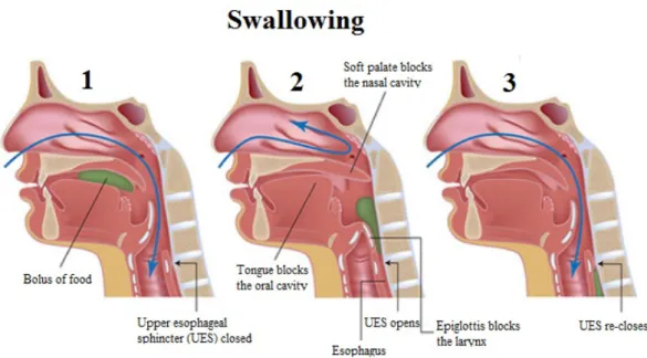
The functions associated (breathing and phonation) with swallowing are impor-tant in an evaluation of the swallowing since they involve the same neuro-anatomical structures. During the preparation of the food bolus, breathing continues through the nasal passages. Once the food bolus is prepared, it is time to propel it to the pharynx by back. First, the mandible closes and the food bowl is gathered on the back, from the tongue to the palatal vault. The apex is supported by the alveo-lar ridges, and the floor of the mouth contracts. The tongue exerts pressure by a movement anteroposterior and from bottom to top against the palate. The velopha-ryngeal sphincter closes (the palate veil rises and hardens until it comes into contact with the oropharyngeal wall) to avoid leakage of the food bolus to the nasal cavities, and therefore to the pharynx while the airways are still open. Finally, the tongue base contracts, and the food bolus slides to the pillars of the veil. The patient must strongly block their breathing before swallowing, which produces an early adduc-tion of the vocal cords before swallowing.
An unchanging coordination is well defined in order to avoid choking of the element being swallowed the wrong way(Figure2.3everydayhealth (2017)).
2.2
Dysphagia
Dysphagia or swallowing disorder is a defect of protection of the airway during the passage of the alimentary bolus towards the œsophagus. It results in a swallowing
Figure 2.3:Lateral view of bolus propulsion during swallowing
choking incident or inhalation of food in the airway, and a disorder of the transit of the alimentary bolus towards the œsophagus, leaving residues in the pharynx, which can leads to a secondary swallow choking incident. Dysphagia can be also manifested by difficulties of transport of the bolus into the œsophagus. The etiol-ogy of swallowing disorders is very varied insofar as any damage to the anatomical structures of the aerodigestive junction or structures allowing the neurological con-trol of swallowing can be involved. The etiology of dysphagia is dominated by neu-rological and tumoral pathologies of aerodigestive tract or brain tumors affecting swallowing functionality.
Two types of dysphagia are distinguished; oropharyngeal and œsophageal dys-phagia. Oropharyngeal dysphagia is defined as the difficulty of transferring the bolus from the mouth to the pharynx and the œsophagus. As for œsophageal dys-phagia, this occures during the third phase of the swallowing process whereby there is difficulty in passageof the bolus along the œsophagus to the stomach. Dysphagia can manifest in the form of a blockage to the passage of foods (stasis), a slowing down in the progression of the food bolus or in a lack of coordination of breathing and swallowing. In all these cases, aspiration can occur.
There are four different types of aspiration:
1. the aspiration before swallowing when the bolus passes before the swallow-ing reflex is triggered and this results in the absence of the swallowswallow-ing reflex
20 Chapter 2. Anatomy and Physiology
(when the bolus slides on the base of the tongue, but the absence of swallow-ing reflex does not trigger the closure of the larynx, which remains open, and therefore the respiratory function is active, and the aspiration occurs after ac-cumulations of food in the pharynx). This absence of swallowing reflex results in neuromotive disorganisation of this reflex. The aspiration can also occur due to a delayed swallowing reflex. In such cases, the swallowing reflex is slow to trigger, the bolus has time to flow on the base of the tongue and fill the bottom of the pharynx before swallowing is triggered.
2. Aspiration during swallowing when laryngeal protective structures are defi-cient.
3. Aspiration after swallowing (secondary or indirect aspiration) that occurs upon resumption of breathing, leading to food stasis in the trachea or nasal fossa.
4. Eventually, the most dangerous of aspiration is the silent one, which can occur before, during or after swallowing and does not cause reflex coughing. 71% of silent aspiration were found in elderly patients hospitalized in a long stay with acquired community pneumonia, compared to 10% for control popula-tion without pneumonia in a control study Marik and Kaplan (2003).
Among the clinical signs of the aspiration is the reflex cough that may be ab-sent (as in the case of silent aspiration), only coincidental and not very effective in clearing the channel if blocked. The alteration of the voice too can be a sign of aspiration. In addition, pain during food intake, vomiting, weight loss and recur-rent pneumopathies. Short-term complications can result in aspiration, respiratory complications, suffocation and severe lung infection. In the long term, the person expresses his disinterest and anxiety about the meal and its duration, which may be longer than normal DePippo, Holas, and Reding (1994) and Deborah J. C. Ramsey and Kalra (2003), which causes chronic malnutrition and inflammation of the lungs. Swallowing disorders in a chronic context may be related to chronic diseases such as stroke, degenerative diseases such as Alzheimer’s and Parkinson’s, Oto-Rhino-Laryngology surgery, drugs and aging structures and functions (presbyphagia).
Occasional difficulty in swallowing, which may occur when a person eats too fast or does not chew the food well is not a cause for concern. However, when it is persis-tent, it may indicates a serious medical condition requiring treatment and functional
rehabilitation. Persistent swallowing disorders cause many inconveniences, ranging from the deterioration of the quality of life of the patient to the risk of death Schmidt et al. (1994). The majority of dysphagic cases often remain undiagnosed and are therefore not treated. An undetected, untreated or inadequate swallowing disor-der definitely leads to malnutrition and/or dehydration, the consequences of which pose a risk of serious harm to the person. In fact, when swallowing is no longer be safe, the person experiences the pain of physical and social disability response to the loss of the pleasure of eating and drinking, and anorexia. The person may also have anxiousness associated with meals for themselves and for their relatives because of, for example, the drooling during food intake which consequently leads to social isolation.
2.2.1 Etiology of Dysphagia
The etiologies of swallowing disorders are numerous and its treatment depends on the cause. Dysphagia can result from a wide variety of diseases and disorders listed below.
Oral pathologies
Pathology affecting teeth, such as infection or caries, can also affect mastication of food. A congenital anomaly can also be one cause of dysphagia such as the malfor-mation of the œsophagus, Leflot et al. (2005), cleft lip and palate Tanaka et al. (2012) causing food and fluids drooling and reflux into nasal fossae when swallowing and the formation of bolus is also impaired. Upper œsophageal sphincter (UES) dysfunc-tion is the leading cause of pharyngeal dysphagia such as failure of UES relaxadysfunc-tion which is a motor disorder Cook (2006).
Xerostomia is also known as a cause of abnormally low volume of saliva which causes dysphagia. Furthermore, it causes the loss of the antibacterial protection that saliva affords. A person with xerostomia is more at risk of aspiration pneumonia if aspiration occurs due to a higher oral bacterial load Rofes et al. (2011).
Obstructions and diverticula
Obstructions, which can be caused by different conditions such as tumours of the head and/or neck, can cause swallowing difficulties since tumours can affect the
22 Chapter 2. Anatomy and Physiology
motility of structures involved in swallowing Logemann et al. (2006). Directly, if there exists a tumour in the neck in the form of obstructions blocking the tract of the bolus through the oral cavity or/and the pharynx. Indirectly, if there exists a tumour in the head causing damage to the nerves of oral cavity or pharynx Pauloski et al. (2000). Diverticula of the pharyngeal and œsophagus mucosa, pouches that protrude outward in a weak portion of the pharynx or œsophageal lining, are also a cause of dysphagia, namely Zenker’s diverticulum Sasegbon and Hamdy (2017), which is located just above the cricopharyngeal muscle causing complications that include aspiration and pneumonia Bergeron, Long, and Chhetri (2012) and Jonathan M. Bock and Blumin (2012).
Medications
Medications can be the origin of dysphagia. A recent study showed that up to 64% of patients with xerostomia take medications which cause this xerostomia Guggen-heimer and MOORE (2003). Chemical radiotherapy of head and neck cancers often results in delayed swallowing, decreased pharyngeal transport, and inefficient la-ryngeal protection Eisbruch et al. (2002) and Grobbelaar et al. (2004). In 2007, Elting et al. (2007) presented their study on a group of 204 patients receiving radiother-apy (RT) to head and neck primary cancers and showed that 91% of these patients developed oral mucositis, which is manifested by a painful swelling of the mucous membranes lining the digestive tract, which can lead to a different form of swallow-ing difficulties durswallow-ing mastication or the different stages of swallowswallow-ing. Also, 50% of the general elderly population use at least one anticholinergic medication Mul-sant et al. (2003) which is well recognised as a common cause of xerostomia Bostock and McDonald (2016). Another study showed that dysphagia in Parkinson’s pa-tients is induced by some medications Leopold (1996) causing abnormalities during all swallowing stages.
Neurological diseases
Neurological conditions that cause damage to the brain and nervous system can cause dysphagia, such as Parkinson’s disease and dementia Easterling and Robbins (2008) and Coates and Bakheit (1997), Huntington disease, Alzheimer’s dementia Secil et al. (2016), cerebral tumours Frank et al. (1989), stroke Paciaroni et al. (2004), cranial pairs damages and some sequelae of neurological interventions. Kalf and al.
Kalf et al. (2011) showed that the presence of oropharyngeal disease after stroke can be as high as 82%. Stroke is also known as the most common cause of oropharyngeal disease of acute onset Daniels (2006).
The most common cause of dysphagia is stroke. In France, one stroke occurs ev-ery 4 minutes, which implies 13 000 hospitalisations per year. Stroke causes severe sequelae. It represents the first cause of acquired handicap of the adult Santé (2012). Previous studies have suggested that complications in hospitalised stroke patients are frequent ranging from 40% to 96% of patients Langhorne et al. (2000). Dysphagia are frequent and are estimated at 42 to 76% following an acute stroke Group (1999) and Zhou (2009). Many studies have attempted to establish the incidence of dys-phagia after stroke with values ranging from 23% to 50% Singh and Hamdy (2006). Available statistics differ from one study to another, but their frequency seems high. In terms of economics, the hospitalisation of stroke patients is very expensive for health insurance-social security because the stroke is 100% covered by social secu-rity from the moment it is considered debilitating.
Swallowing is a complex process which requires the intervention of several mus-cles and cranial nerves with a very precise temporal coordination.
2.2.2 Semiology of dysphagia
Semiology is the study of the signs of diseases and so helps guide the diagnosis. For dysphagia, there is a specific semiology determining the physio-pathological mech-anisms of the swallowing disorder. Swallowing disorder or dysphagia is defined as difficulty of synchronisation between the progression of the alimentary bolus to the œsophagus and the protection of the airways, or / and mastication disorder.
Dysphagia is common in human pathology and can be caused by a variety of diseases affecting neural, motor and/or sensory systems that contribute to the swal-lowing function. It is due in a large number of cases to neurological damage Mann, Hankey, and Cameron (2000) and Counter and Ong (2018). Swallowing disorders can be described chronologically by classifying the various mechanisms according to airways protection defects and bolus transportation defects. Below are described the pathophysiological mechanisms present chronologically Bassols and Puech (2011):
• During the preparatory phase
24 Chapter 2. Anatomy and Physiology
∗ Forward: default of lip closure,
∗ Backward: default of oropharyngeal closure – Insalivation disorders
– Chewing disorders
• During the oral transport phase
– Disorders of initiation of oral time – Failure to close the oral cavity:
∗ Anterior ∗ Posterior
– Bolus control defects in the oral cavity – Bolus propulsion default
– Default of initiation of pharyngeal time – Failure to trigger pharyngeal time • During the pharyngeal phase
– Failure to protect the airways
∗ Upper = Default of velopharyngeal closure ∗ Lower =
· Default of laryngeal closure · Default of expulsion mechanisms – Pharyngeal transport defects:
∗ Failure of pharyngeal peristalsis ∗ Default of basi-lingual decline
– Dysfunction of the sphincter of the œsophagus • During œsophageal time
– Default of expulsion mechanisms
Swallowing disorders include several symptoms such as coughing, voice mod-ification and prolonged mealtimes. Dysphagia may not be accompanied by cough in many cases but is silent. Signs suggestive of swallowing disorder may be listed using a swallowing disorders scorecard (Annex 1) Desport et al. (2011).