HAL Id: hal-02279983
https://hal.archives-ouvertes.fr/hal-02279983
Submitted on 5 Jan 2021HAL is a multi-disciplinary open access
archive for the deposit and dissemination of sci-entific research documents, whether they are pub-lished or not. The documents may come from teaching and research institutions in France or abroad, or from public or private research centers.
L’archive ouverte pluridisciplinaire HAL, est destinée au dépôt et à la diffusion de documents scientifiques de niveau recherche, publiés ou non, émanant des établissements d’enseignement et de recherche français ou étrangers, des laboratoires publics ou privés.
Loss of endothelial sulfatase-1 after experimental sepsis
attenuates subsequent pulmonary inflammatory
responses
Kaori Oshima, Xiaorui Han, Yilan Ouyang, Rana El Masri, Yimu Yang, Sarah
Haeger, Sarah Mcmurtry, Trevor Lane, Pavel Davizon-Castillo, Fuming
Zhang, et al.
To cite this version:
Kaori Oshima, Xiaorui Han, Yilan Ouyang, Rana El Masri, Yimu Yang, et al.. Loss of endothe-lial sulfatase-1 after experimental sepsis attenuates subsequent pulmonary inflammatory responses. American Journal of Physiology - Lung Cellular and Molecular Physiology, American Physiological Society, 2019, �10.1152/ajplung.00175.2019�. �hal-02279983�
Title: Loss of endothelial sulfatase-1 after experimental sepsis attenuates subsequent
1
pulmonary inflammatory responses 2
Abbreviated title: Endothelial loss of Sulf-1 and post-septic CARS
3
Authors: Kaori Oshima1, Xiaorui Han2, Yilan Ouyang2, Rana El Masri3, Yimu Yang1, 4
Sarah M. Haeger1, Sarah A. McMurtry1, Trevor C. Lane1, Pavel Davizon-Castillo4,5, 5
Fuming Zhang2, Xinping Yue6, Romain R. Vivès3, Robert J. Linhardt2, Eric P. Schmidt1,7
6 7
Author affiliations:
8
1Department of Medicine, University of Colorado Denver, Aurora, Colorado, USA
9
2Departments of Chemistry and Chemical Biology, Chemical and Biological Engineering,
10
and Biomedical Engineering, Rensselaer Polytechnic Institute, Troy, New York, USA 11
3University of Grenoble Alpes, CNRS, CEA, IBS, Grenoble, France
12
4Department of Pediatrics, University of Colorado Denver, Aurora, Colorado, USA
13
5Hemophilia and Thrombosis Center, School of Medicine, University of Colorado,
14
Aurora, CO, USA 15
6Department of Physiology School of Medicine, Louisiana State University Health
16
Sciences Center, New Orleans, Louisiana, USA 17
7Department of Medicine, Denver Health Medical Center, Denver, Colorado, USA
18 19 Corresponding Author: 20 Eric P. Schmidt, MD 21
12700 E. 19th Avenue, Research Complex 2, Mail Stop C272 22 Aurora CO, 80045. 23 Phone: (303) 724-6106 24 E-mail: eric.schmidt@ucdenver.edu 25 26
27
Author Contributions:
28
K.O. and E.P.S. conceived and designed the study; K.O., Y.Y., S.M.H., S.A.M., T.L., 29
P.D-C, R.E.M. performed experiments; K.O., S.A.M, E.P.S. analyzed data; K.O., R.E.M., 30
R.R.V., and E.P.S. prepared figures; K.O. and E.P.S. drafted manuscript; K.O., Y.Y., 31
S.M.H., S.A.M., T.L., R.E.M., F.Z., X.Y., R.R.V., R.J.L., E.P.S. edited and revised 32
manuscript; K.O., Y.Y., S.M.H., S.A.M., T.L., R.E.M., F.Z., X.Y., R.R.V., R.J.L., E.P.S. 33
approved the final version of the manuscript. 34
35
Keywords:
36
Heparan sulfate, Sulfatase-1, Compensatory anti-inflammatory response syndrome 37
Abstract
38
Sepsis patients are at increased risk for hospital-acquired pulmonary infections, 39
potentially due to post-septic immunosuppression known as the compensatory anti-40
inflammatory response syndrome (CARS). CARS has been attributed to leukocyte 41
dysfunction, with an unclear role for endothelial cells. The pulmonary circulation is lined 42
by an endothelial glycocalyx, a heparan sulfate-rich layer essential to pulmonary 43
homeostasis. Heparan sulfate degradation occurs early in sepsis, leading to lung injury. 44
Endothelial synthesis of new heparan sulfates subsequently allows for glycocalyx 45
reconstitution and endothelial recovery. We hypothesized that remodeling of the 46
reconstituted endothelial glycocalyx, mediated by alterations in the endothelial 47
machinery responsible for heparan sulfate synthesis, contributes to CARS. 72 hours 48
after experimental sepsis, coincident with glycocalyx reconstitution, mice demonstrated 49
impaired neutrophil and protein influx in response to intratracheal lipopolysaccharide 50
(LPS). The post-septic reconstituted glycocalyx was structurally remodeled, with 51
enrichment of heparan sulfate disaccharides sulfated at the 6-O position of glucosamine. 52
Increased 6-O-sulfation coincided with loss of endothelial sulfatase-1 (Sulf-1), an 53
enzyme that specifically removes 6-O-sulfates from heparan sulfate. Intravenous 54
administration of Sulf-1 to post-septic mice restored the pulmonary response to LPS, 55
suggesting that loss of Sulf-1 was necessary for post-septic suppression of pulmonary 56
inflammation. Endothelial-specific knockout mice demonstrated that loss of Sulf-1 was 57
not sufficient to induce immunosuppression in non-septic mice. Knockdown of Sulf-1 in 58
human pulmonary microvascular endothelial cells resulted in downregulation of the 59
adhesion molecule ICAM-1. Taken together, our study indicates that loss of endothelial 60
Sulf-1 is necessary for post-septic suppression of pulmonary inflammation, representing 61
a novel endothelial contributor to CARS. 62
Introduction
63
Sepsis, defined as life-threatening organ dysfunction caused by a dysregulated host 64
immune response (34), is a leading cause of in-hospital mortality worldwide (12). 65
Classically, septic organ injury has been attributed to systemic, overwhelming hyper-66
inflammation. However, the failures of numerous clinical trials targeting inflammatory 67
signaling (18) led to the proposed concept that sepsis is not simply hyper-inflammation, 68
but also consists of a delayed period of immunosuppression, known as the 69
compensatory anti-inflammatory response syndrome (CARS) (1). This period of 70
suppressed inflammation is paradoxically harmful in sepsis, imparting an increased risk 71
for secondary infections (9, 15, 26, 35) such as hospital-acquired pneumonia (15, 39). 72
Evidence supporting the pathologic significance of post-septic CARS includes known 73
associations between mortality and increased plasma IL-10 and decreased HLA-DR 74
expression on leukocytes (10, 16, 21), decreased production of cytokines, such as 75
TNF, IFN-, IL-6, IL-10 in septic patients, and depletion of CD4+, CD8+, HLA-DR+ 76
cells in the spleen (2). CARS has therefore been largely attributed to dysfunctional 77
monocytes, including impaired cytokine production, decreased phagocytosis and 78
migration in response to inflammatory stimuli (23, 44). Despite the potential importance 79
of CARS, clinical trials targeting immunosuppression have been disappointing (18, 19, 80
25), perhaps reflecting an incomplete understanding of the mechanisms responsible 81
post-septic impairment in lung inflammation. 82
The endothelial glycocalyx is carbohydrate-rich endovascular layer that serves multiple 83
homeostatic functions at the endothelial surface. The major glycosaminoglycan 84
constituent of the glycocalyx is heparan sulfate (HS), a linear polysaccharide composed 85
of repeating glucosamine and hexuronic (glucuronic and iduronic) acid disaccharide 86
units. This disaccharide unit may be sulfated at the amino (N) and/or 6-O positions of 87
glucosamine and/or the 2-O position of hexuronic acid. The resultant pattern of 88
negative charge enables HS to bind to various cationic ligands and their cognate 89
receptors (11, 28), influencing multiple signaling processes responsible for organ injury 90
and repair. 91
We have previously reported that sepsis-induced degradation of HS from the pulmonary 92
endothelial glycocalyx mediates alveolar neutrophil adhesion and inflammatory lung 93
injury (31). During sepsis recovery (72 hours after cecal ligation and puncture (CLP) in 94
mice), endothelial synthesis of new HS allows for glycocalyx reconstitution, mediating 95
endothelial recovery (45). We postulated that post-septic changes in the endothelial 96
machinery responsible for endothelial HS synthesis lead to remodeling of the 97
endothelial glycocalyx, impairing pulmonary responses to subsequent inflammatory 98
stimuli. 99
In this report, we observed that mice demonstrated suppressed pulmonary inflammation 100
in response to intratracheal lipopolysaccharides (LPS) after experimental sepsis, 101
coincident with glycocalyx enrichment in 6-O-sulfated HS, a sulfation pattern implicated 102
in endothelial inflammation (27, 41). This remodeling was associated with 103
downregulation of pulmonary endothelial Sulf-1, an enzyme responsible for the 104
constitutive cleavage of extracellular 6-O-sulfo groups. We observed that loss of Sulf-1 105
in septic mice was necessary for the impaired pulmonary response to LPS characteristic 106
of CARS, but it was not sufficient to cause impaired inflammation in non-septic animals. 107
Knock down of Sulf-1 using siRNA in pulmonary microvascular endothelial cells resulted 108
in downregulation of ICAM-1 transcription. Our study therefore identifies post-septic 109
remodeling of the pulmonary endothelial glycocalyx as a novel contributor to CARS. 110
Materials and Method
112 113
Materials
114
We purchased LPS (from Escherichia coli O55:B5), heparinase I and III (from 115
Flavobacterium heparinum) from Sigma-Aldrich (St. Louis, MO) and reconstituted in 116
phosphate buffered saline (PBS). As controls, we heat-inactivated heparinase I and III 117
at 100°C for 20 min. We purchased protein assay dye reagent from Bio-Rad (Hercules, 118
CA). We purchased tamoxifen from Sigma-Aldrich and dissolved in corn oil with 5% 119
ethanol. For tissue digestion and fluorescence activated cell sorting, we purchased 120
collagenase type 2 from Worthington Biochemical Corporation (Lakewood, NJ), dispase 121
I from Sigma-Aldrich, and ACK lysing buffer from Gibco (Dublin, Ireland). We 122
purchased anti-mouse CD31-APC antibody (17-0311-82, Clone:390) from eBioscience 123
(San Diego, CA), and anti-mouse CD144-PE antibody (562243, Clone: 11D4.1) from 124
BD Biosciences (San Jose, CA) and DAPI from Invitrogen (Carlsbad, CA). For 125
quantitative Reverse Transcription-Polymerase Chain Reaction (qRT-PCR), we 126
purchased reagents for RNA extraction and DNase I from Qiagen (Hilden, Germany). 127
We purchased primary human pulmonary microvascular endothelial cells from 128
PromoCell (Heidelberg, Germany). We purchased iScript cDNA synthesis kit from Bio-129
Rad. We purchased siRNA targeted for sulfatase-1 (Sulf-1) from Dharmacon (Lafayette, 130
CO), and purchased transfection reagent, Lipofectamine RNAiMAX, from Thermo 131
Fisher Scientific (Waltham, MA). For production of recombinant HSulf-1, we purchased 132
all tissue culture reagents and media, from Thermo Fisher Scientific, and 133
chromatography reagents from GE Healthcare (Chicago, IL). 134
Animals
136
All experimental protocols were approved by the University of Colorado Institutional 137
Animal Care Use Committee, and all experiments were performed in accordance of 138
National Institutes of Health guideline. Wild type, male C57BL/6J mice (8-10 weeks) 139
were purchased from Jackson Laboratory. Sulf-1 and 2 double floxed mice (both males 140
and females, 8-12 weeks old) were used as previously described (37). VE-cadherin 141
CRE-ERT2 mice were provided by Dr. Ralf Adams at Max Planck Institute, Germany. 142
Sulf1/2 double homozygous mice with or without VE-cadherin-CRE-ERT2 (Sulf-1f/f Sulf-143
2f/f VEcadCreERT2- or+) were used for all knock out animal experiments. CRE-ERT2 144
translocation to nucleus was induced with intraperitoneal injections of tamoxifen 145
(dissolved in corn oil and 5% ethanol, sterile filtered), 1 mg/day, for 5 consecutive days. 146
Induction of sepsis in mice
147
We induced sepsis in 8-14 week-old mice with CLP as previously described (31). 148
Briefly, we anesthetized animals with 5% isoflurane inhalation. An incision of 149
approximately 1 cm was made in abdomen and cecum was exposed. We ligated the 150
cecum at 50 % of its length and punctured it through and through with a 22 G needle. 151
The cecum was then returned to the abdominal cavity, and the incision was closed with 152
suture. Buprenorphine (1.6 μg/ g body weight, i.p.) was given to each animal for pain 153
management, and 1 mL of sterile saline was given subcutaneously as fluid resuscitation. 154
Sham surgery was performed similarly, albeit without ligation and puncture of the cecum. 155
Intratracheal instillation
We anesthetized animals with 5 % isoflurane inhalation, and we instilled LPS at 3 g/g 157
body weight to trachea visualized with laryngoscope. Animals spontaneously breathed 158
throughout the instillation. 159
Blood analysis
160
We collected blood from the retro-orbital sinus using heparin-coated capillary tubes, and 161
determined complete blood counts (CBCs) using a veterinary hematology analyzer 162
(Heska, Loveland, CO). We collected blood anticoagulated with 3.8% citrate by cardiac 163
puncture and assessed global hemostasis using kaolin as activator by 164
thromboelastography (Haemonetics, BrainTree, MA). 165
Tissue and sample collection
166
We anesthetized mice with a lethal dose of ketamine/xylazine, and collected blood in 167
EDTA tubes by inferior vena cava cannulation. We collected plasma by centrifuging 168
whole blood at 1000 x g for 10 min. and stored it at -80 C until analysis. We 169
cannulated the trachea and performed bronchoalveolar lavage (BAL) 3 times with 1 mL 170
of PBS each. We flushed blood out of the lung by pulmonary artery perfusion, and the 171
right lung was snap frozen with liquid N2 andstored at -80 ºC until analysis. We inflated
172
the left lung with 1% low-melting agarose in PBS, fixed with 10% formalin overnight, and 173
processed the tissue for histological analysis. We determined total cells per mL in BAL 174
fluid using a hemocytometer. We centrifuged BAL fluid at 1200 rpm for 5 min. and 175
stored the supernatant at -80 ºC until analysis. We determined total protein 176
concentration in BAL fluid with a Coomassie Brilliant Blue-based colorimetric assay. 177
Cells in BAL fluid were mounted on a slide with cytospin at 600 rpm for 2 min., and 178
stained with Wright-Giemsa method for differential cell counting. 179
Isolated perfused mouse lung
180
We performed the isolated, perfused mouse lung as previously described (29). Briefly, 181
we deeply anesthetized mice with ketamine and xylazine. After confirming the loss of 182
toe-pinch reflex, we cannulated the trachea and ventilated mice with 21% O2 and 5%
183
CO2 at 125 breaths/min at tidal volume of 250 μL. We rapidly removed the sternum and
184
anterior chest wall, and then cannulated the pulmonary artery through an incision made 185
in the free wall of the right ventricle. We cannulated the left atrium through an incision 186
made at the left ventricular apex. We secured the cannulas in position with suture. We 187
then perfused the pulmonary circulation with endothelial cell growth media 188
supplemented with 4% (g/mL) bovine serum albumin at 1 mL/min flow. Perfusate was 189
kept at 37ºC with a water bath. We then added 0.5 unit/mL of heparinase I and III mix 190
to the perfusate and continued isogravimetric perfusion for 30 min. At the end of the 191
experiment, we collected perfusate and stored it at -80ºC until mass spectrometric 192
analysis. 193
Measurement of glycocalyx thickness with intravital microscopy
194
We performed intravital microscopy as previously described (31). Briefly, we 195
anesthetized animals with ketamine and xylazine, and placed a glass window 196
(coverslip) into the right anterior thoracic wall. We infused 150 kDa FITC-dextran into 197
the jugular vein to serve as vascular tracer that does not penetrate the glycocalyx. We 198
injected either heparinase I or III and captured pulmonary microvasculature for 1.5 h. 199
We used a custom-designed intravital microscope (31) to simultaneously measure total 200
vessel width (endothelial cell border to the opposite endothelial cell border, as defined 201
by differential interference contrast microscopy) as well as FITC-dextran width (which 202
does not include the glycocalyx). We determined glycocalyx thickness by subtracting 203
the FITC-dextran width from the total vascular width, then dividing by two. At least three 204
microvessels (< 20 µm width) were measured per each high-powered field. 205
Fluorescent Activated Cell (FAC) sorting of pulmonary endothelial cells
206
We excised the whole lung from each animal, and finely minced lung tissue with a 207
scalpel. We then digested the tissue with collagenase type 2 (1000 units/mL) and 208
dispase I (0.125 units/mL) as well as DNase I (0.01 Kunitz units/mL) in HBSS, with 209
agitation for 60 min. We filtered digested tissue through 70 μm filter to remove 210
undigested tissues, and lysed erythrocytes with ACK lysing buffer for 3 min. at 37 ºC. 211
We washed cells with PBS supplemented with 4% fetal bovine serum, then stained with 212
antibodies against CD31 and CD144 at 1:100 for 40 min. at 4ºC in dark. Live 213
(determined by negative staining for DAPI), double positive population, i.e. 214
CD31+/CD144+ endothelial cells, were then sorted and lysed immediately after the 215
sorting in lysis buffer supplied in RNeasy Mini Kit. Total RNA was extracted 216
immediately using RNeasy Mini Kit according to manufacturer’s protocol. RNA integrity 217
was tested with automated electrophoresis before further analysis. 218
RNA microarray
219
Total RNA from pulmonary endothelial cells (CD31+/CD144+) were used to synthesize
220
cDNA, and cDNA was hybridized to a microarray (Mouse Clariom D, Affymetrix) and 221
scanned with Affymetrix Genechip Scanner 3000. Samples with RNA integrity number 222
greater than 8.7 were used for analysis. 223
Quantitative Reverse Transcription-Polymerase Chain Reaction (qRT-PCR)
224
We synthetized cDNA using the iScript cDNA synthesis kit from total RNA isolated from 225
FAC sorted endothelial cells according to manufacturer’s protocol. Using Taqman 226
probes, we performed qRT-PCR with the Applied Biosystems 7300 Real-Time PCR 227
System. Cyclophilin (whose expression was unchanged between sham and CLP 228
groups, data not shown) was used as a housekeeping gene, and we analyzed data with 229
the 2^(-ΔΔCt) method (17). 230
Production of recombinant HSulf-1 protein
231
Recombinant HSulf-1 was expressed as previously described (7). Briefly, HSulf-1 232
coding sequence was inserted in a pcDNA3.1/Myc-His(-) vector, between SNAP and 233
6His tags at the N- and C-terminus, respectively. This vector was used to stably 234
transfect FreeStyle HEK 293-F cells. After harvesting the culture medium, we purified 235
HSulf-1 using two steps of cation-exchange (SP-Sepharose) and size-exclusion 236
chromatography (Superdex-200), as previously described for HSulf-2 (32). After 237
purification, the protein was concentrated over a 30 kDa centrifugal unit, supplemented 238
with 20% glycerol, aliquoted and stored at -20°C. 239
Enzyme activity was assessed as previously described (33), by analyzing the 240
disaccharide composition of untreated and Sulf-treated heparin in trisulfated [UA(2S)-241
GlcNS(6S)] disaccharide (substrate) and [UA-GlcNS(6S)] disulfated disaccharide 242
(product), using RPIP-HPLC coupled to 2-cyanoacetamide post-column fluorescent 243 derivatization (8). 244 In vitro assays 245
We cultured primary human pulmonary microvascular endothelial cells using 246
microvascular endothelial growth media. We knocked down Sulf-1 using siRNA 247
targeted to Sulf-1, delivered with Lipofectamine RNAiMax. siRNA was transfected when 248
cells were >80% confluent for 24 h. We harvested cells extracted total RNA using 249
RNeasy Mini kit according to manufacturer’s protocol. Cells that were passage 3-6 250
were used for the study. 251
Statistical analysis
252
Statistical analysis was performed with GraphPad Prism version 7.0. Single-comparison 253
analyses were performed by t-test; multiple comparisons were performed by one-way 254
ANOVA with Tukey's post-hoc multiple comparisons test. P-values less than 0.05 were 255
considered significant. The values are expressed as mean ± SD. 256
Results
257
Post-septic animals demonstrate impaired inflammatory response to intratracheal
258
LPS
259
To establish a model of post-septic impairment in pulmonary inflammatory responses, 260
we performed cecal ligation and puncture (CLP) or sham surgery. Three days later, we 261
administered 3 g/g intratracheal (IT) LPS to induce lung inflammation (Figure 1A). 262
Compared to sham-operated animals, post-CLP animals showed decreased alveolar 263
leukocyte infiltration (Figure 1B) and protein concentration (Figure 1C) 2 days after 264
intratracheal LPS. Lung histology from sham-operated animals demonstrated robust 265
intratracheal LPS-induced lung inflammation, including cellular infiltration and alveolar 266
flooding (Figure 1D), while such pathology was absent in the lungs of post-CLP animals 267
(Figure 1E). Taken together, these findings are consistent with a murine model of 268
CARS. 269
The post-septic, reconstituted pulmonary endothelial glycocalyx is remodeled
270
We have previously demonstrated that the pulmonary endothelial glycocalyx, degraded 271
during early sepsis, is reconstituted by 72 h after CLP (45), a time point coincident with 272
our observation of impaired pulmonary inflammatory responses (Figure 1). As a simple 273
screen for the presence of HS remodeling, we tested the sensitivity of the post-septic 274
glycocalyx to heparinases with different preferences for sulfated domains of HS. 275
Heparinase-I (which preferentially targets sulfated domains of HS (3)) readily degraded 276
the post-septic and post-sham glycocalyx (Figure 2A). In contrast, heparinase-III, 277
which targets undersulfated regions at the periphery of sulfated domains (4), was 278
unable to degrade the post-septic glycocalyx (Figure 2B). These findings suggested 279
structural remodeling of the post-septic endothelial glycocalyx. 280
We then directly examined the disaccharide composition of HS comprising the 281
reconstituted pulmonary endothelial glycocalyx (Figure 2C). We perfused lungs 282
isolated from mice 72 h after CLP (or sham) with both heparinase I and III for 30 min. 283
We collected the perfusate and measured disaccharide sulfation patterns with HPLC-284
mass spectrometry multiple reaction monitoring, a high-sensitivity approach capable of 285
detecting ng/ml concentrations of HS (30). HS extracted from the post-septic 286
endothelial glycocalyx demonstrated increased 6-O-sulfation compared to HS isolated 287
from the glycocalyx of sham mice (Figure 2D). This post-septic increase in 6-O-288
sulfation was similarly observed to occur in HS fragments circulating in the blood of 289
mice after CLP (Figure 2E). Taken together, these findings suggested that sepsis 290
alters the endothelial machinery responsible for HS disaccharide sulfation, favoring the 291
presence of 6-O-sulfation within the pulmonary vasculature. 292
Pulmonary endothelial cells in post-septic animals have decreased expression of
293
Sulf-1
294
To determine the presence of transcriptional changes in HS-modifying genes 295
(potentially responsible for the observed increase in 6-O-sulfation detailed in Figure 2), 296
we performed FAC sorting to isolate pulmonary endothelial cells (CD31+/CD144+
297
population) from lungs harvested 48 h after CLP (i.e. immediately prior to remodeling) or 298
sham (Figure 3A). Whole RNA transcriptome microarray analyses identified that post-299
septic pulmonary endothelial cells downregulated expression of sulfatase-1 (Sulf-1) 300
(Figure 3B), an enzyme that constitutively removes 6-O-sulfo groups from extracellular 301
HS-chains. Strikingly, there was no difference in other genes that modify 6-O-sulfation 302
of HS, namely sulfatase-2 or HS 6-O-sulfotransferase (microarray data are publically 303
available with GEO accession number: GSE129775). We confirmed these findings in a 304
separate cohort of mice by repeating FAC sorting of pulmonary endothelial cells, then 305
performing qRT-PCR of Sulf-1 (Figure 3C). 306
To determine if downregulation of endothelial Sulf-1 similarly occurs after non-septic 307
causes of glycocalyx degradation, we enzymatically degraded endothelial HS from 308
naïve mice by intravenous heparinase III injection (Figure 3D). This model leads to 309
rapid endothelial glycocalyx reconstitution within 24 h (45). Twelve hours after 310
heparinase-III (a time point prior to completion of reconstitution), qRT-PCR showed no 311
difference in endothelial Sulf-1 mRNA expression level between heat-inactivated 312
heparinase-III control and heparinase-III treatment groups (Figure 3E). These findings 313
suggest that downregulation of endothelial Sulf-1 is a sepsis-specific phenomenon. 314
Loss of Sulf-1 is necessary for post-septic suppression of pulmonary
315
inflammatory responses to intratracheal LPS
316
Given that loss of endothelial Sulf-1 immediately preceded immunosuppression in post-317
septic animals, we determined whether loss of Sulf-1 was necessary for CARS. We 318
produced recombinant, enzymatically active Sulf-1 as previously described (7). 319
Resultant Sulf-1 was enzymatically active as evidenced by the 6-O-desulfation of 320
trisulfated [UA(2S)-GlcNS(6S)] heparin disaccharides (NS2S6S) into [UA(2S)-GlcNS] 321
disulfated disaccharides (NS2S), monitored by RPIP-HPLC (Figure 4A). We induced 322
sepsis in mice by CLP and supplemented surviving animals with exogenous Sulf-1 323
intravenously (3 µg bolus), 2 h prior to intratracheal (IT) LPS challenge (Figure 4B). 324
Compared to diluent control group, Sulf-1 treated animals had increased alveolar 325
leukocyte (Figure 4C) and protein (Figure 4D) concentrations, suggesting reversal of 326
post-septic immunosuppression. Lung histology showed a modest, scattered increase 327
in leukocyte inflammation after IT LPS in post-CLP mice treated with intravenous Sulf-1 328
(Figure 4E, 4F). Of note, lungs from Sulf-1 treated animals showed marked 329
perivascular cuffing (Figure 4F, black arrows), suggesting restoration of an 330
inflammatory edematous response to LPS. These results suggest that post-septic loss 331
of Sulf-1 is necessary to suppress inflammatory responses to subsequent IT LPS. 332
Loss of Sulf-1 is not sufficient to induce immunosuppression in non-septic
333
animals
334
We created inducible, endothelial-specific, Sulf-1 and Sulf-2 double knockout mice by 335
breeding Sulf-1f/f Sulf-2f/f floxed mice with tamoxifen-inducible, endothelial specific 336
VEcadCreERT2 mice to determine whether loss of endothelial Sulf-1 is sufficient to 337
cause impaired pulmonary inflammation. Of note, as Sulf-2 is minimally expressed in 338
the pulmonary endothelium ((24), confirmed by our whole transcriptome experiments, 339
data not shown), endothelial-specific deletion of Sulf-2 is unlikely to impart significant 340
pulmonary impact. We induced recombination with tamoxifen injections for 5 341
consecutive days (1 mg/day, i.p.). After 2 weeks, we first confirmed inducible 342
endothelial-specific recombination with FAC sorting followed by DNA electrophoresis 343
(Figure 5A). The cell-specific recombination was confirmed with DNA extracted from 344
FAC sorted endothelial cells (CD31+/CD 144+) and non-endothelial cells (CD31-/CD 345
144-) from Sulf-1f/f
Sulf-2f/f VEcadCreERT2+ or Sulf-1f/f Sulf-2f/f VEcadCreERT2- mice, 346
both treated with tamoxifen (Figure 5B). These knockout animals had normal blood 347
leukocyte differential and normal coagulation, as measured by CBCs and 348
thromboelastography clot onset time (R-time) respectively (data not shown). There was 349
no evidence of increased pulmonary apoptosis in these mice after tamoxifen treatment 350
(TUNEL staining), and lung histology was unchanged (data not shown). We performed 351
intravital microscopy to determine if the sensitivity of the pulmonary endothelial 352
glycocalyx of tamoxifen-treated, Sulf-1f/f
Sulf-2f/f VEcadCreERT2+ mice to enzymatic 353
degradation was similar to that observed in post-septic wild-type mice (Figure 2B). The 354
pulmonary endothelial glycocalyx of these Sulf-1 and -2 double knockout mice 355
demonstrated resistance to heparinase-III (Figure 5C), but not against heparinase I 356
(data not shown), confirming similar glycocalyx remodeling as observed in post-septic 357
mice (Figure 2A, 2B). 358
We challenged tamoxifen-treated Sulf-1f/f
Sulf-2f/f VEcadCreERT2+ mice with IT LPS to 359
determine whether endothelial-specific Sulf-1/Sulf-2 knockout is sufficient to impair 360
pulmonary inflammatory responses in non-septic mice, (Figure 5A). There was no 361
difference between control group animals and double knockout animals in leukocytes 362
numbers (Figure 5D) and protein concentration (Figure 5E) in BAL fluid. Similarly, 363
there was no obvious difference observed in lung histology between the control group 364
(Figures 5F) and Sulf-1/2 knockout group (Figure 5G). Taken together, the data 365
indicated that loss of Sulf-1 is not sufficient to cause impaired pulmonary inflammation 366
in non-septic animals. 367
Sulf-1 silencing in human primary pulmonary microvascular endothelial cells
368
results in decreased mRNA expression of ICAM-1
6-O-sulfation may impact numerous biological pathways, spanning growth factor, 370
chemokine, and damage-associated molecular pattern signaling (5). We used siRNA 371
approaches to knock down Sulf-1 in primary human microvascular endothelial cells to 372
determine if the consequences of Sulf-1 loss on these signaling pathways converge to 373
impart a general anti-inflammatory phenotype to endothelial cells. We cultured primary 374
human lung microvascular endothelial cells in a 6-well plate, and transfected siRNA 375
using lipofectamine. We performed three biological replicates, each of which 376
represented cells collected from a separate human donor. Our transfection resulted in 377
greater than 83% knock down efficiency (Figure 6A). We examined whether a major 378
adhesion molecule for neutrophil adhesion, ICAM-1, was affected by this knockdown. 379
Loss of Sulf-1 resulted in downregulation of ICAM-1 expression in endothelial cells 380
(Figure 6B). These findings suggest that loss of Sulf-1 may drive pathways that 381
suppress endothelial activation. As these unstimulated endothelial cells expressed little 382
ICAM-1 protein at baseline (data not shown), we are unable to demonstrate a direct 383
effect of loss of Sulf-1 on endothelial-leukocyte adhesion. 384
385
Discussion
386
In this study, we demonstrated that post-CLP mice have impaired alveolar 387
inflammation in response to intratracheal LPS. This post-septic “immunoparalysis” 388
coincides with reconstitution of a remodeled pulmonary endothelial glycocalyx, as 389
demonstrated by differential sensitivity to enzymatic degradation during intravital 390
microscopy and enrichment in 6-O-sulfated HS monitored by mass spectrometry. 391
These changes coincide with selective pulmonary endothelial downregulation of Sulf-1, 392
an enzyme dedicated to the constitutive removal of 6-O-sulfo groups. We observed that 393
loss of Sulf-1 is necessary for impaired inflammation in post-septic animals, but it alone 394
was not sufficient to cause impaired inflammation in non-septic animals. Our data 395
therefore collectively indicate an endothelial role in CARS, a syndrome to date largely 396
relegated to post-septic leukocyte dysfunction. 397
Our findings indicate that sepsis suppresses endothelial Sulf-1, impairing 398
subsequent inflammatory responses after the resolution of septic endothelial injury. 399
Teleologically, this response may be designed to limit the degree of lung injury, shifting 400
endothelial signaling towards tissue repair. For example, an increase in 6-O-sulfation 401
promotes cell response to HS-binding growth factors, FGF2 and VEGF, which may 402
promote endothelial repair processes (6). However, this response may backfire if the 403
host is exposed to a secondary infection, by attenuating host-protective antimicrobial 404
responses. Post-septic patients are susceptible to secondary infection, with hospital-405
acquired pneumonia the most common complication (15, 39). Septic patients who later 406
acquired secondary infections experience more severe illness, longer length-of-stay, 407
and higher 1-year mortality (39, 40). Therefore, in the presence of hospital-acquired 408
infection, loss of endothelial Sulf-1 may be detrimental. Additionally, we found that post-409
septic animals had increased absolute neutrophil count in blood at the time of 410
intratracheal LPS (data not shown), indicating that decreased neutrophil infiltration 411
found in BAL fluid is not due to post-septic depletion of circulating neutrophils. 412
Interestingly, when the whole transcriptome profiles from circulating leukocytes were 413
compared between patients who acquired secondary infection and those who did not, 414
there was no difference in pro- or anti-inflammatory genes (39). However, there was 415
significant increase in plasma proteins, including circulating cytokines, such as IL-8 and 416
IL-10, and markers of vascular dysfunction and activation, such as E-selectin, 417
angiopoietins, ICAM-1, and proteins that promote coagulation in patients who 418
developed secondary infections, compared to the patients who did not (40). Taken 419
together, these findings suggest that CARS is most likely more complex than leukocyte 420
dysfunction, and warrant further investigations to determine contributions by other cells 421
and organs such as vascular endothelial cells. 422
Although we have shown one potential effect of loss of Sulf-1, i.e. endothelial 423
ICAM-1 downregulation, the mechanisms by which loss of Sulf-1 results in impaired 424
inflammation remain uncertain. Sulf-1 and Sulf-2 are unique in that they are the only 425
extracellular sulfatases modifying HS at the post-synthetic level, suggesting a critical 426
importance of 6-O-sulfation for the function of glycocalyx HS. Indeed, 6-O-sulfation may 427
influence numerous biological processes of consequence, including growth factor, 428
chemokine, and damage-associated molecular pattern signaling (5). One possible 429
mechanism is that a shift in 6-O-sulfation affects the affinity of endothelial HS to 430
cytokines that modulate neutrophil infiltration. Sulf-2 is shown to selectively mobilize not 431
only specific growth factors but also cytokines (such as SDF-1 and SLC) that impact 432
neutrophil migration (36, 38). Changes in endothelial HS 6-O-sulfation shape the 433
microenvironment and inflammatory response in multiple ways, given their multiple 434
biological roles. Changes in HS sulfation may result in altered systemic immunity, and 435
enhanced bacterial adhesion to endothelial cells (43). Although the roles of endothelial 436
selectins to neutrophil extravasation in pulmonary circulation are complex (13, 20), it 437
has been reported that changes in sulfation in endothelial HS, particularly 6-O-sulfation, 438
weakens neutrophil binding to L-selectin and P-selectin (41, 42). Accordingly, the 439
immunosuppressive effects of loss of Sulf-1 may arise from numerous potential 440
processes, potentially converging upon pathways such as adhesion molecule 441
expression (Figure 6). Future studies will screen for 6-O-sulfated HS binding proteins, 442
providing greater insight into the downstream mechanisms responsible for our observed 443
effects of Sulf-1 on post-septic pulmonary inflammation. 444
An additional consideration is that changes in endothelial Sulf-1/2 expression 445
may impact the dynamics of glycocalyx degradation. Indeed, our endothelial microarray 446
experiments (GEO accession number: GSE129775) demonstrated that post-septic (48 447
h) loss of Sulf-1 coincided with increased expression of matrix metalloproteinases 8, 9, 448
25, and disintegrin and metalloproteinase domain-containing proteins 8, 15, 23. No 449
changes were seen in endothelial expression of heparanase or matrix metalloproteinase 450
15 in these post-septic mice. Future studies will be required to investigate the presence 451
of sulfatase-sheddase cross-talk and its relevance to lung injury and repair. 452
Sulf-1 and Sulf-2 preferentially targets trisulfated, NS2S6S disaccharides of HS, 453
and to a lesser extent, NS6S (22). Loss of Sulf-1, therefore, would be expected to 454
result in an increase of trisulfated, NS2S6S disaccharides (substrate), with a decrease 455
in NS2S (product). However, we rather observed an increase of HS overall 6-O-456
sulfation (Figure 2D). This discrepancy may reflect the additional ability of Sulf-1 and 457
Sulf-2 to directly regulate HS biosynthesis enzymes (14). Therefore, it is possible that 458
our biological observations following Sulf-1 downregulation may be caused directly by 459
the loss of Sulf-1 activity, and/or indirectly, through a regulation of HS biosynthesis. Of 460
note, our recombinant Sulf-1 is of human origin. Based on the strong sequence 461
homology of HSulf-1 and MSulf-1, we do not expect any significant differences in 462
enzymatic activity (22), but we cannot exclude the possibility that enzymatic activity is 463
slightly different in vivo. 464
In conclusion, our study showed that post-septic pulmonary endothelial 465
glycocalyx HS undergoes structural remodeling, coincident with loss of endothelial Sulf-466
1. Although loss of Sulf-1 in endothelial cells is not sufficient to cause impaired 467
inflammation in non-septic mice, it contributes to post-septic CARS, offering a potential 468
new target for treating post-septic patients at risk for nosocomial pneumonia. 469
Acknowledgement
470
We would like to thank technical help from the Genomics and Microarray Core and from 471
the CU Cancer Center Flow Cytometry Shared Resource at University of Colorado 472
Denver. We would also like to thank Dr. Ralf Adams at Max Planck Institute, Germany, 473
for generously providing VEcadhein-CRE-ERT2 mice. 474
475
Funding
476
This work was supported by Department of Defense (CDMRP) grant PR150655 (to Dr. 477
Schmidt), NIH grant R01 HL125371 (to Drs. Schmidt and Linhardt). In addition, work 478
performed by Dr. Vivès was supported by the CNRS and the GDR GAG (GDR 3739), 479
the “Investissements d’avenir” program Glyco@Alps (ANR-15-IDEX-02), and by grants 480
from the Agence Nationale de la Recherche (ANR-17-CE11-0040) and Université 481
Grenoble-Alpes (UGA AGIR program). Hematologic analyses were supported by 482
Maternal Child Health Bureau Grant H30MC24049 (to Dr. Davizon-Castillo). 483 484 Disclosure 485 None. 486
Figure Legends
487
Figure 1: Post-CLP mice demonstrate suppressed inflammatory response to
488
intratracheal lipopolysaccharide. (A) We performed cecal ligation and puncture
489
(CLP) or sham surgery on day 0. Three days after surgery, we challenged animals with 490
intratracheal (IT) lipopolysaccharides (LPS) (3 μg/g body weight). Animals were 491
harvested 2 days after IT LPS. (B) Post-CLP animals had decreased number of 492
leukocytes per mL in bronchoalveolar lavage (BAL) fluid 2 days after intratracheal LPS. 493
(C) Post-CLP animals demonstrated less alveolar injury 2 days after intratracheal LPS, 494
based on protein concentrations in BAL fluid. (D) Lung histological section from a sham 495
animal (stained with H&E) showed evidence of lung inflammation 2 days after 496
intratracheal LPS. (E) Lung histological section from a CLP animal (stained with H&E) 2 497
days after intratracheal LPS showed minimal evidence of lung inflammation. Scale bars 498
on lower magnification images = 500 m and those on higher magnification images 499
=100 m. n= 5 each group, Student’s t-test, *P < 0.05. 500
Figure 2: The post-septic, reconstituted endothelial glycocalyx is remodeled. (A)
501
At the 72 h time point characterized by pulmonary endothelial HS reconstitution, we 502
injected 1 unit of heparinase I (Hep I) into the jugular vein, which degraded both sham 503
and post-CLP pulmonary endothelial glycocalyx heparan sulfate. (B) In contrast, 1 unit 504
of heparinase III (Hep III) into the jugular vein did not degrade post-septic pulmonary 505
endothelial heparan sulfate. (C) Major sulfation positions on heparan sulfate constituent 506
disaccharides. (D) Post-septic, reconstituted pulmonary endothelial heparan sulfate 507
had a significantly increase (P < 0.05) in disaccharides with 6-O-sulfation (percentage of 508
total sulfated disaccharides) as compared to heparan sulfate from sham animals. (E) 509
The percentage of 6-O-sulfated disaccharides increased over time in the plasma of 510
post-CLP mice. ESL: endothelial surface layer. n= 3-5 per group, Student’s t-test, *P < 511
0.05. 512
Figure 3: Post-septic pulmonary endothelial cells downregulate Sulf-1 mRNA. (A)
513
We collected pulmonary endothelial cells and extracted total RNA at day 2, a time point 514
immediately prior to glycocalyx reconstitution at day 3. (B) Pulmonary endothelial cells 515
in mice 48 h after CLP had decreased Sulf-1 expression detected with RNA microarray. 516
(C) We validated the downregulation of Sulf-1 in pulmonary endothelial cells by qRT-517
PCR using separate biological replicates. (D) We collected pulmonary endothelial cells 518
and extracted total RNA at 12 h after heparinase III injection, a time point immediately 519
prior to glycocalyx reconstitution after enzymatic, non-septic degradation. (E) mRNA 520
expression level of Sulf-1 in pulmonary endothelial cells were similar 12 h after 521
enzymatic degradation by heparinase III (Hep III) or heat-inactivated (HI) Hep III. n= 3-4 522
each group, Student’s t-test, *P < 0.05. 523
Figure 4: Loss of Sulf-1 contributes to post-septic CARS. (A) Recombinant Sulf-1
524
was enzymatically active and efficiently removed 6-O sulfates from heparin, as shown 525
by the decrease in trisulfated [UA(2S)-GlcNS(6S)] disaccharide content (NS2S6S) and 526
concomitant increase of [UA-GlcNS(6S)] disulfated disaccharide (NS2S) in Sulf-1 527
treated heparin (white bars) compared to untreated control (black bars). (B) We 528
performed CLP on mice on day 0. On day 3, surviving post-septic mice were treated 529
with intravenous (IV) Sulf-1 (3 μg bolus) or diluent, then challenged with intratracheal 530
(IT) LPS (3 μg/g body weight). Animals were harvested 2 days after IT LPS. (C) Sulf-1 531
treated animals had increased leukocytes in BAL fluid 2 days after IT LPS, as compared 532
to diluent treated animals 2 days after IT LPS. (D) Sulf-1 treated animals also showed 533
increased lung injury based on protein concentration in BAL fluid. (E) Lung histological 534
section from a post-septic, diluent treated animal (stained with H&E) showed very little 535
evidence of lung inflammation. (F) Lung histological section from a post-septic Sulf-1 536
treated animal (stained with H&E) showed increased tissue consolidation 2 days after IT 537
LPS. In addition, Sulf-1 treated animals had marked perivascular cuffs (black arrows). 538
Scale bars on lower magnification images = 500 m and those on higher magnification 539
images =100 m. n= 4-5 each group, Student’s t-test, *P < 0.05. 540
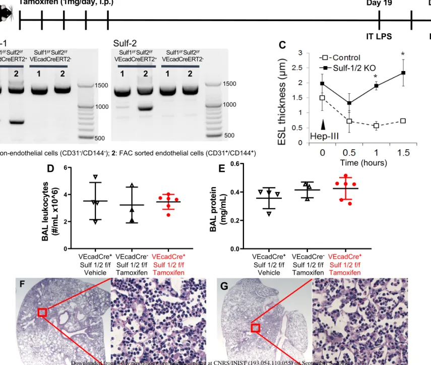
Figure 5: Loss of Sulf-1 is not sufficient to cause impaired inflammation in
non-541
septic animals. (A) Sulf-1f/f Sulf-2f/f VEcadCreERT2+ or – animals received tamoxifen or
542
vehicle control injections intraperitoneally for 5 consecutive days (1 mg/day). 543
Recombination of genes and pulmonary endothelial glycocalyx characteristics were 544
evaluated 2 weeks after the last injection of tamoxifen (or vehicle). Knockout or control 545
mice were alternatively challenged with IT LPS (3 μg/ g body weight) at the same time 546
point. (B) We confirmed cell-specific, inducible recombination of Sulf-1 and Sulf-2 with 547
DNA gels. Lane 1: DNA from pulmonary non-endothelial cells (CD31-/CD144-), Lane 2: 548
DNA from pulmonary endothelial cells (CD31+/CD144+). (C) Pulmonary endothelial 549
glycocalyx of Sulf-1/2 knockout animals was resistant to heparinase III (Hep III) 550
degradation, similar to the post-septic endothelial glycocalyx resistance to heparinase III 551
observed in wild-type mice (Figure 2B). Control animals used were Sulf-1f/f Sulf-2f/f
552
VEcadCreERT2–(floxed gene alone without Cre recombinase),treated with tamoxifen. 553
(D) Number of leukocytes in BAL fluid in Sulf-1/2 knockout animals did not differ from 554
control groups 2 days after IT LPS. (E) Protein concentration of BAL fluid was similarly 555
not different among the experimental groups. (F) Lung histological section from a 556
control animal (Sulf-1f/f Sulf-2f/f VEcadCreERT2–, treated with tamoxifen) had clear 557
consolidation. (G) Lung histological section from a Sulf-1/2 knockout animal similarly 558
had evidence of lung inflammation. Scale bars on lower magnification images = 500 m 559
and those on higher magnification images =100 m. n = 3-6 each group, One-way 560
ANOVA with post hoc Turkey test, *P < 0.05. 561
Figure 6: Knocking down Sulf-1 in pulmonary microvascular endothelial cells
562
decreases ICAM-1 expression. (A) We transfected cultured endothelial cells with
563
siRNA targeted to Sulf-1 using Lipofectamine RNAiMAX for 24 hours, which resulted in 564
greater than 83% knockdown of Sulf-1. (B) Knock down of Sulf-1 resulted in 565
downregulation of ICAM-1 expression in endothelial cells. Control cells were treated 566
with lipofectamine without siRNA. n = 3, each data point represents average of 2-3 567
wells per treatment. Student’s t-test, *P < 0.05. 568
References:
569
1. Bone RC, Grodzin CJ, and Balk RA. Sepsis: a new hypothesis for
570
pathogenesis of the disease process. Chest 112: 235-243, 1997. 571
2. Boomer JS, To K, Chang KC, Takasu O, Osborne DF, Walton AH, Bricker TL,
572
Jarman SD, 2nd, Kreisel D, Krupnick AS, Srivastava A, Swanson PE, Green JM,
573
and Hotchkiss RS. Immunosuppression in patients who die of sepsis and multiple
574
organ failure. JAMA 306: 2594-2605, 2011. 575
3. Desai UR, Wang HM, and Linhardt RJ. Specificity studies on the heparin lyases
576
from Flavobacterium heparinum. Biochemistry 32: 8140-8145, 1993. 577
4. Desai UR, Wang HM, and Linhardt RJ. Substrate specificity of the heparin
578
lyases from Flavobacterium heparinum. Arch Biochem Biophys 306: 461-468, 1993. 579
5. El Masri R, Seffouh A, Lortat-Jacob H, and Vives RR. The "in and out" of
580
glucosamine 6-O-sulfation: the 6th sense of heparan sulfate. Glycoconj J 34: 285-298, 581
2017. 582
6. Ferreras C, Rushton G, Cole CL, Babur M, Telfer BA, van Kuppevelt TH,
583
Gardiner JM, Williams KJ, Jayson GC, and Avizienyte E. Endothelial heparan sulfate
584
6-O-sulfation levels regulate angiogenic responses of endothelial cells to fibroblast 585
growth factor 2 and vascular endothelial growth factor. J Biol Chem 287: 36132-36146, 586
2012. 587
7. Heidari-Hamedani G, Vives RR, Seffouh A, Afratis NA, Oosterhof A, van
588
Kuppevelt TH, Karamanos NK, Metintas M, Hjerpe A, Dobra K, and Szatmari T.
589
Syndecan-1 alters heparan sulfate composition and signaling pathways in malignant 590
mesothelioma. Cell Signal 27: 2054-2067, 2015. 591
8. Henriet E, Jager S, Tran C, Bastien P, Michelet JF, Minondo AM, Formanek
592
F, Dalko-Csiba M, Lortat-Jacob H, Breton L, and Vives RR. A jasmonic acid
593
derivative improves skin healing and induces changes in proteoglycan expression and 594
glycosaminoglycan structure. Biochim Biophys Acta Gen Subj 1861: 2250-2260, 2017. 595
9. Hotchkiss RS, and Opal S. Immunotherapy for sepsis--a new approach against
596
an ancient foe. The New England journal of medicine 363: 87-89, 2010. 597
10. Hynninen M, Pettila V, Takkunen O, Orko R, Jansson SE, Kuusela P,
598
Renkonen R, and Valtonen M. Predictive value of monocyte histocompatibility
599
leukocyte antigen-DR expression and plasma interleukin-4 and -10 levels in critically ill 600
patients with sepsis. Shock 20: 1-4, 2003. 601
11. Jastrebova N, Vanwildemeersch M, Lindahl U, and Spillmann D. Heparan
602
sulfate domain organization and sulfation modulate FGF-induced cell signaling. J Biol 603
Chem 285: 26842-26851, 2010. 604
12. Kempker JA, and Martin GS. The Changing Epidemiology and Definitions of
605
Sepsis. Clin Chest Med 37: 165-179, 2016. 606
13. Kolaczkowska E, and Kubes P. Neutrophil recruitment and function in health
607
and inflammation. Nat Rev Immunol 13: 159-175, 2013. 608
14. Lamanna WC, Frese MA, Balleininger M, and Dierks T. Sulf loss influences N-, 609
2-O-, and 6-O-sulfation of multiple heparan sulfate proteoglycans and modulates 610
fibroblast growth factor signaling. J Biol Chem 283: 27724-27735, 2008. 611
15. Landelle C, Lepape A, Voirin N, Tognet E, Venet F, Bohe J, Vanhems P, and
612
Monneret G. Low monocyte human leukocyte antigen-DR is independently associated
613
with nosocomial infections after septic shock. Intensive Care Med 36: 1859-1866, 2010. 614
16. Lekkou A, Karakantza M, Mouzaki A, Kalfarentzos F, and Gogos CA.
615
Cytokine production and monocyte HLA-DR expression as predictors of outcome for 616
patients with community-acquired severe infections. Clin Diagn Lab Immunol 11: 161-617
167, 2004. 618
17. Livak KJ, and Schmittgen TD. Analysis of relative gene expression data using
619
real-time quantitative PCR and the 2(-Delta Delta C(T)) Method. Methods 25: 402-408, 620
2001. 621
18. Marshall JC. Why have clinical trials in sepsis failed? Trends Mol Med 20:
195-622
203, 2014. 623
19. Meisel C, Schefold JC, Pschowski R, Baumann T, Hetzger K, Gregor J,
624
Weber-Carstens S, Hasper D, Keh D, Zuckermann H, Reinke P, and Volk HD.
625
Granulocyte-macrophage colony-stimulating factor to reverse sepsis-associated 626
immunosuppression: a double-blind, randomized, placebo-controlled multicenter trial. 627
Am J Respir Crit Care Med 180: 640-648, 2009. 628
20. Mizgerd JP, Meek BB, Kutkoski GJ, Bullard DC, Beaudet AL, and
629
Doerschuk CM. Selectins and neutrophil traffic: margination and Streptococcus
630
pneumoniae-induced emigration in murine lungs. J Exp Med 184: 639-645, 1996. 631
21. Monneret G, Finck ME, Venet F, Debard AL, Bohe J, Bienvenu J, and
632
Lepape A. The anti-inflammatory response dominates after septic shock: association of
633
low monocyte HLA-DR expression and high interleukin-10 concentration. Immunol Lett 634
95: 193-198, 2004. 635
22. Morimoto-Tomita M, Uchimura K, Werb Z, Hemmerich S, and Rosen SD.
636
Cloning and characterization of two extracellular heparin-degrading endosulfatases in 637
mice and humans. J Biol Chem 277: 49175-49185, 2002. 638
23. Munoz C, Carlet J, Fitting C, Misset B, Bleriot JP, and Cavaillon JM.
639
Dysregulation of in vitro cytokine production by monocytes during sepsis. J Clin Invest 640
88: 1747-1754, 1991. 641
24. Nagamine S, Tamba M, Ishimine H, Araki K, Shiomi K, Okada T, Ohto T,
642
Kunita S, Takahashi S, Wismans RG, van Kuppevelt TH, Masu M, and Keino-Masu
643
K. Organ-specific sulfation patterns of heparan sulfate generated by extracellular
644
sulfatases Sulf1 and Sulf2 in mice. J Biol Chem 287: 9579-9590, 2012. 645
25. Nakos G, Malamou-Mitsi VD, Lachana A, Karassavoglou A, Kitsiouli E,
646
Agnandi N, and Lekka ME. Immunoparalysis in patients with severe trauma and the
647
effect of inhaled interferon-gamma. Crit Care Med 30: 1488-1494, 2002. 648
26. Otto GP, Sossdorf M, Claus RA, Rodel J, Menge K, Reinhart K, Bauer M,
649
and Riedemann NC. The late phase of sepsis is characterized by an increased
650
microbiological burden and death rate. Crit Care 15: R183, 2011. 651
27. Reine TM, Kusche-Gullberg M, Feta A, Jenssen T, and Kolset SO. Heparan
652
sulfate expression is affected by inflammatory stimuli in primary human endothelial cells. 653
Glycoconj J 29: 67-76, 2012. 654
28. Sarrazin S, Lamanna WC, and Esko JD. Heparan sulfate proteoglycans. Cold
655
Spring Harb Perspect Biol 3: 2011. 656
29. Schmidt EP, Damarla M, Rentsendorj O, Servinsky LE, Zhu B, Moldobaeva
657
A, Gonzalez A, Hassoun PM, and Pearse DB. Soluble guanylyl cyclase contributes to
658
ventilator-induced lung injury in mice. Am J Physiol Lung Cell Mol Physiol 295: L1056-659
1065, 2008. 660
30. Schmidt EP, Overdier KH, Sun X, Lin L, Liu X, Yang Y, Ammons LA, Hiller
661
TD, Suflita MA, Yu Y, Chen Y, Zhang F, Cothren Burlew C, Edelstein CL, Douglas
662
IS, and Linhardt RJ. Urinary Glycosaminoglycans Predict Outcomes in Septic Shock
663
and Acute Respiratory Distress Syndrome. Am J Respir Crit Care Med 194: 439-449, 664
2016. 665
31. Schmidt EP, Yang Y, Janssen WJ, Gandjeva A, Perez MJ, Barthel L,
666
Zemans RL, Bowman JC, Koyanagi DE, Yunt ZX, Smith LP, Cheng SS, Overdier
667
KH, Thompson KR, Geraci MW, Douglas IS, Pearse DB, and Tuder RM. The
668
pulmonary endothelial glycocalyx regulates neutrophil adhesion and lung injury during 669
experimental sepsis. Nat Med 18: 1217-1223, 2012. 670
32. Seffouh A, El Masri R, Makshakova O, Gout E, Hassoun ZEO, Andrieu JP,
671
Lortat-Jacob H, and Vives RR. Expression and purification of recombinant
672
extracellular sulfatase HSulf-2 allows deciphering of enzyme sub-domain coordinated 673
role for the binding and 6-O-desulfation of heparan sulfate. Cell Mol Life Sci 2019. 674
33. Seffouh I, Przybylski C, Seffouh A, El Masri R, Vives RR, Gonnet F, and
675
Daniel R. Mass spectrometry analysis of the human endosulfatase Hsulf-2. Biochem
676
Biophys Rep 18: 100617, 2019. 677
34. Singer M, Deutschman CS, Seymour CW, Shankar-Hari M, Annane D, Bauer
678
M, Bellomo R, Bernard GR, Chiche JD, Coopersmith CM, Hotchkiss RS, Levy MM,
679
Marshall JC, Martin GS, Opal SM, Rubenfeld GD, van der Poll T, Vincent JL, and
680
Angus DC. The Third International Consensus Definitions for Sepsis and Septic Shock
681
(Sepsis-3). JAMA 315: 801-810, 2016. 682
35. Stephan F, Yang K, Tankovic J, Soussy CJ, Dhonneur G, Duvaldestin P,
683
Brochard L, Brun-Buisson C, Harf A, and Delclaux C. Impairment of
684
polymorphonuclear neutrophil functions precedes nosocomial infections in critically ill 685
patients. Crit Care Med 30: 315-322, 2002. 686
36. Suratt BT, Petty JM, Young SK, Malcolm KC, Lieber JG, Nick JA, Gonzalo
687
JA, Henson PM, and Worthen GS. Role of the CXCR4/SDF-1 chemokine axis in
688
circulating neutrophil homeostasis. Blood 104: 565-571, 2004. 689
37. Tran TH, Shi X, Zaia J, and Ai X. Heparan sulfate 6-O-endosulfatases (Sulfs)
690
coordinate the Wnt signaling pathways to regulate myoblast fusion during skeletal 691
muscle regeneration. J Biol Chem 287: 32651-32664, 2012. 692
38. Uchimura K, Morimoto-Tomita M, Bistrup A, Li J, Lyon M, Gallagher J, Werb
693
Z, and Rosen SD. HSulf-2, an extracellular endoglucosamine-6-sulfatase, selectively
694
mobilizes heparin-bound growth factors and chemokines: effects on VEGF, FGF-1, and 695
SDF-1. BMC Biochem 7: 2, 2006. 696
39. van Vught LA, Klein Klouwenberg PM, Spitoni C, Scicluna BP, Wiewel MA,
697
Horn J, Schultz MJ, Nurnberg P, Bonten MJ, Cremer OL, van der Poll T, and
698
Consortium M. Incidence, Risk Factors, and Attributable Mortality of Secondary
699
Infections in the Intensive Care Unit After Admission for Sepsis. JAMA 315: 1469-1479, 700
2016. 701
40. van Vught LA, Wiewel MA, Hoogendijk AJ, Frencken JF, Scicluna BP, Klein
702
Klouwenberg PMC, Zwinderman AH, Lutter R, Horn J, Schultz MJ, Bonten MMJ,
703
Cremer OL, and van der Poll T. The Host Response in Patients with Sepsis
704
Developing Intensive Care Unit-acquired Secondary Infections. Am J Respir Crit Care 705
Med 196: 458-470, 2017. 706
41. Wang L, Brown JR, Varki A, and Esko JD. Heparin's anti-inflammatory effects
707
require glucosamine 6-O-sulfation and are mediated by blockade of L- and P-selectins. 708
J Clin Invest 110: 127-136, 2002. 709
42. Wang L, Fuster M, Sriramarao P, and Esko JD. Endothelial heparan sulfate
710
deficiency impairs L-selectin- and chemokine-mediated neutrophil trafficking during 711
inflammatory responses. Nat Immunol 6: 902-910, 2005. 712
43. Xu D, Olson J, Cole JN, van Wijk XM, Brinkmann V, Zychlinsky A, Nizet V,
713
Esko JD, and Chang YC. Heparan Sulfate Modulates Neutrophil and Endothelial
714
Function in Antibacterial Innate Immunity. Infect Immun 83: 3648-3656, 2015. 715
44. Xu PB, Lou JS, Ren Y, Miao CH, and Deng XM. Gene expression profiling
716
reveals the defining features of monocytes from septic patients with compensatory anti-717
inflammatory response syndrome. J Infect 65: 380-391, 2012. 718
45. Yang Y, Haeger SM, Suflita MA, Zhang F, Dailey KL, Colbert JF, Ford JA,
719
Picon MA, Stearman RS, Lin L, Liu X, Han X, Linhardt RJ, and Schmidt EP.
720
Fibroblast Growth Factor Signaling Mediates Pulmonary Endothelial Glycocalyx 721
Reconstitution. Am J Respir Cell Mol Biol 56: 727-737, 2017. 722
Figure 1 CARS model
Day 0 Sham/CLP Harvest Day 3 IT LPS Day 5A
B
C
Sham + IT LPS CLP + IT LPS Sham + IT LPS CLP + IT LPS*
Sham + IT LPS CLP+ IT L PS 0.0 0.5 1.0 1.5 2.0Figure 1 CARS BAL cell count (total leukocyte) 112018
# L e u k o c yt e/ m L ( x 10 ^ 6)
*
Sham + IT LPS CLP+ IT L PS 0.0 0.1 0.2 0.3 0.4 0.5Figure 1 CARS BAL Protein
m g /m L
D
E
BAL le u ko cy te s (# /m L x 10 ^ 6) BAL p ro te in (m g /m L )Figure 2 Post-septic, reconstituted glycocalyx acquires increased 6-O sulfation and newly found resistance against
heparinase III digestion, but not against heparinase I
0
0.5
1
1.5
2
2.5
3
0
0.5
1
1.5
E
S
L
th
ickn
ess
(µ
m
)
Time (hours)
72 h after sham
72 h after CLP
Hep-III*
*
*
0
0.5
1
1.5
2
2.5
3
0
0.5
1
1.5
E
S
L
th
ickn
ess
(µ
m
)
Time (hours)
72 h after sham
72 h after CLP
Hep-I*
A
B
D
72h after sham
72h after CLP
C
0 2 4 C ir c u la t in g H S a ft e r C L P % 6 -O s u lf a ti o nE
% 6 -O -su lf at io nFigure 3 Post-septic pulmonary endothelial cells downregulate Sulf-1
Sham CLP Re la ti ve e xp re ss io n o f Su lf -1 (2^ -ΔΔ Ct ) Sham CLP*
B
C
Sh a m EC 48 hr CL P E C 4 8 hr 0 .0 0 .5 1 .0 1 .5 e x c lu d e b a s t a r d _ d d C t _ s h a m /C L P E C s a t 4 8 h r G r o u p R e la ti v e e x p re s s io n*
E
Sham CL P 0.0 0.5 1.0 1.5 sulf 1 0.0 0.5 1.0 1.5qPCR FAC sorted cells 0
IV Hep III (or HI-Hep III)
Harvest 24 hr
D
Day 0 Sham/CLP Day 3 HarvestA
Day 1 Day 2 Glycocalyx Reconstitution 12 hr Glycocalyx Reconstitution Re la ti ve e xp re ss io n o f Su lf -1 (2^ -ΔΔ Ct ) Re la ti ve e xp re ss io n o f Su lf -1 (2^ -ΔΔ Ct )Figure 4 Sulf-1 is necessary for CARS
*
*
A
E
C
D
B
Day 0 CLP Harvest Day 3 IT LPS Day 5 IV Sulf-1 or diluent Diluent Sulf1 0.0 0.5 1.0 1.5 2.0 2.5BAL leukocyte Count
# L e u k o c yt e /m L ( x 10 ^ 6) Diluent Sulf1 0.0 0.1 0.2 0.3 0.4 BAL protein m g /m L
F
Heparin Heparin+ HSulf-1 BAL p ro te in (m g /m L ) BAL le u ko cy te s (# /m L x 10 ^ 6) Sulf-1 Diluent Sulf-1 DiluentFigure 5 Loss of Sulf-1 is not sufficient to cause CARS in non-septic mice
Day 0-5 Harvest Day 19 IT LPS Day 21 Sulf-1 Sulf1f/fSulf2f/f VEcadCreERT2+A
B
VEcadCre -Sulf 1/2 f/f Tamoxifen VEcadCre+ Sulf 1/2 f/f Vehicle VEcadCre+ Sulf 1/2 f/f TamoxifenC
D
FTamoxifen (1mg/day, i.p.)
Sulf1f/fSulf2f/f VEcadCreERT2 -1 1000 500 1500 1000 500 1500 2 1 2
1: FAC sorted non-endothelial cells (CD31-/CD144-); 2: FAC sorted endothelial cells (CD31+/CD144+)
Sulf-2 Sulf1f/fSulf2f/f VEcadCreERT2+ Sulf1f/fSulf2f/f VEcadCreERT2 -1 2 1 2 Vehi cle CR E+ IT LPS Tam x Cr e- I T LP S Taxm CRE + IT LPS 0 2 4 6 MS BAL leukocyte #/mL x10^6 Group # /m L ( x 1 0^ 6) Vehi cle CRE+ IT L PS Tam x Cr e- I T LP S Taxm CRE + IT LPS 0.0 0.2 0.4 0.6 MS BAL Protein Group m g /m L