HAL Id: tel-01751560
https://hal.univ-lorraine.fr/tel-01751560
Submitted on 29 Mar 2018
HAL is a multi-disciplinary open access
archive for the deposit and dissemination of
sci-entific research documents, whether they are
pub-lished or not. The documents may come from
teaching and research institutions in France or
abroad, or from public or private research centers.
L’archive ouverte pluridisciplinaire HAL, est
destinée au dépôt et à la diffusion de documents
scientifiques de niveau recherche, publiés ou non,
émanant des établissements d’enseignement et de
recherche français ou étrangers, des laboratoires
publics ou privés.
ZSF1 rat: a model of chronic cardiac and renal diseases
in the context of metabolic syndrome. Characterization
with anti-oxidant, mineralocorticoid receptor antagonist
and aldosterone synthase inhibitor
William Riboulet
To cite this version:
William Riboulet. ZSF1 rat: a model of chronic cardiac and renal diseases in the context of metabolic
syndrome. Characterization with anti-oxidant, mineralocorticoid receptor antagonist and aldosterone
synthase inhibitor. Human health and pathology. Université de Lorraine, 2015. English. �NNT :
2015LORR0041�. �tel-01751560�
AVERTISSEMENT
Ce document est le fruit d'un long travail approuvé par le jury de
soutenance et mis à disposition de l'ensemble de la
communauté universitaire élargie.
Il est soumis à la propriété intellectuelle de l'auteur. Ceci
implique une obligation de citation et de référencement lors de
l’utilisation de ce document.
D'autre part, toute contrefaçon, plagiat, reproduction illicite
encourt une poursuite pénale.
Contact : ddoc-theses-contact@univ-lorraine.fr
LIENS
Code de la Propriété Intellectuelle. articles L 122. 4
Code de la Propriété Intellectuelle. articles L 335.2- L 335.10
http://www.cfcopies.com/V2/leg/leg_droi.php
ECOLE DOCTORALE BioSE (Biologie-Santé-Environnement)
Thèse
Présentée et soutenue publiquement pour l’obtention du titre de
DOCTEUR DE L’UNIVERSITE DE LORRAINE
Mention « Sciences de la Vie et de la Santé »
par William RIBOULET
L
E RAT
ZSF1:
UN MODELE DE MALADIE CARDIO
-
RENALE ASSOCIEE AU
SYNDROME METABOLIQUE
.
C
ARACTERISATION PAR L
’
UTILISATION D
’
UN
ANTIOXYDANT
,
D
’
UN ANTAGONISTE DES RECEPTEURS DES
MINERALOCORTICOÏDES ET D
’
UN INHIBITEUR DE L
’
ALDOSTERONE SYNTHASE
.
Le 18 Mai 2015
Membres du jury :
Rapporteurs :
M Paulus MULDER
PU, INSERM U1096, Rouen
M Bernard JOVER DR, INSERM EA7288, Montpellier
Examinateurs :
Mme Catherine VERGELY-VANDRIESSE PU, INSERM UMR866, Dijon
M Pierre-Yves MARIE PU-PH, INSERM-U961, Vandœuvre-lès-Nancy
Mme Agnès BENARDEAU DR, Bayer AG, Wuppertal, co-directeur de thèse
Mme Isabelle LARTAUD PU, EA 3452, Nancy, directeur de thèse
EA 3452 CITHEFOR
ii
Laboratoire d’accueil de la thèse :
Cardiovascular and Metabolism Department
F.Hoffmann La-Roche AG,
Grenzacherstrasse 124,
4070 Basel, Switzerland
iii
REMERCIEMENTS
Je tiens à remercier tout particulièrement Mme Isabelle Lartaud d’avoir accepté d’être ma directrice de thèse sachant que les expériences seraient menées « à distance », ainsi que pour son écoute, ses précieux conseils, sa sympathie et l’efficacité de nos interactions.
Je souhaite également remercier Mr Jacques Mizrahi ainsi que Mme Karin Conde-Knape de m’avoir permis d’effectuer ce travail au sein du laboratoire in vivo Pharmacology du feu département Cardiovascular and Metabolism dont ils étaient responsables, tout en restant employé à part entière au sein de leur équipe.
Je remercie chaleureusement Mme Agnès Bénardeau qui a accepté d’être ma co-directrice de thèse au sein de l’entreprise Roche, avec qui travailler fut un réel plaisir, déjà avant la thèse, sein de son équipe, ainsi que pour sa bonne humeur, et son amitié. Je te souhaite, chère Agnès, une excellente continuation dans ta nouvelle vie allemande, en espérant qu’on se fera encore des bads de folie lors de tes visites en Alsace ;-).
Merci à Mr Philippe Verry alias « Pôpy » pour son soutien permanent dans les moments difficiles, pour son amitié, la manière dont il m’a aidé à évoluer, et sa sempiternelle bonne humeur :-). Je te souhaite également une bonne seconde vie dans laquelle je suis sûr tu es déjà bien occupé.
Je souhaite exprimer ma profonde gratitude à Mr Bernard Jover, et Mr Paulus Mulder pour avoir accepté d’être rapporteurs de ce manuscrit, ainsi qu’à Mme Catherine Vergely-Vandriesse et Mr Pierre-Yves Marie pour avoir accepté de faire partie du jury de thèse, malgré leur emploi du temps chargés.
iv
Ces travaux n’auraient pas pu voir le jour sans mes collègues et amis Mme Anne-Emmanuelle Salman (ou Choupette), Mme Emmanuelle Hainaut (ou Mômie), Mr Anthony Vandjour (je vais y aller mollo sur les surnoms sur ce coup là^^), ainsi que Mr Anthony Retournard (alias Kiki), un grand merci à vous pour les fous-rire, mais aussi les manips’, et en particulier pour les 60 cages métaboliques dont on ne se lasse pas ! Merci à mes autres collègues ainsi qu’à toutes les personnes ayant participé de près ou de loin à la réalisation de ces travaux.
Je remercie ma famille et mes amis pour leur aide et soutien en toutes circonstances, en particulier Yanick Vinot, mon ami de toujours, sans oublier PH, Joe et Sébouille pour … c’est vrai ça, pourquoi ?
Merci à Laetitia Pouzol d’avoir été un soutien important durant cette période chargée (bon ok plus sur le début que sur la fin ;-)), ainsi que m’avoir donné un fils magnifique, Evan.
v
SUMMARY
“ZSF1 rat: a model of chronic cardiac and renal diseases in the context of metabolic syndrome. Characterization with anti-oxidant, mineralocorticoid receptor antagonist and aldosterone synthase
inhibitor.”
In the context of metabolic syndrome, development of Type 2 Diabetes is associated with (and influenced by) cardiac and renal comorbidities linked to micro- and macro-vasculature alterations. To assess the efficacy of new compounds on targeted organs in the context of metabolic syndrome, the Zucker fatty/Spontaneously hypertensive heart failure F1 hybrid (ZSF1) rat model could be suitable assuming cardiorenal alterations would develop in a short timeframe. Actually, the ZSF1 rat model recapitulates features of human metabolic syndrome, but develops relatively late (1year-time) and moderate cardiac and renal dysfunctions. The aim of our work was to exacerbate cardiorenal impairments in terms of onset and extent. Two options were explored. On one hand, unilateral nephrectomy was performed in ZSF1 rats, and cardiac and renal functions were longitudinally assessed. This surgical insult only significantly deteriorated renal function, which was prevented by standard of care, lisinopril and new renal protective agent, bardoxolone. On the other hand, subcutaneous infusion of angiotensin II (AngII) was used in the aim to induce hemodynamic and hormonal stress and thus to enhance cardiorenal impairments. AngII-infused ZSF1 rats displayed significant hypertension and increased levels of circulating aldosterone. Moreover, renal and cardiac functions dropped, concomitantly. We showed in this model that an aldosterone synthase inhibitor induced overall better renal protection than the mineralocorticoid receptor antagonist eplerenone. Our data showed that ZSF1-AngII rat is a suitable model to evaluate the cardio and renoprotective effects of drugs in the context of metabolic syndrome.
Keywords: ZSF1, aldosterone synthase inhibitor, metabolic syndrome, unilateral nephrectomy, angiotensin II. RESUME
“Le rat ZSF1: un modèle de maladie cardio-rénale associée au syndrome métabolique. Caractérisation par l’utilisation d’un antioxydant, d’un antagoniste des récepteurs des
minéralocorticoïdes et d’un inhibiteur de l’aldostérone synthase.”
Chez les patients présentant un syndrome métabolique, le développement des comorbidités cardiaques et rénales associées au diabète de type 2 sont liées à des altérations au niveau de la micro- et macro-vasculature. Afin d’évaluer l’effet protecteur rénal et cardiaque de nouvelles molécules, le modèle de rat Zucker fatty/Spontaneously hypertensive heart failure F1 hybrid (ZSF1) semblait approprié. Cependant, son développement lent et les impacts rénaux et cardiaques modestes en limitaient son utilisation. Le but de nos travaux a consisté à exacerber et accélérer ces altérations par deux méthodes distinctes. Nous avons tout d’abord effectué une néphrectomie unilatérale chez le rat ZSF1 et évalué l’évolution des fonctions cardiaque et rénale en fonction de l’âge. Cette méthode n’a permis de mettre en évidence qu’une exacerbation de la dysfonction rénale, mais nous a néanmoins permis de démontrer l’effet protecteur rénal de l’inhibiteur de l’enzyme de conversion de l’angiotensine lisinopril ainsi que d’un composé antioxydant, le bardoxolone. Notre seconde stratégie a consisté à infuser de l’angiotensine II (AngII) durant 6 semaines, à des rats ZSF1. La conséquence de cette intervention a été un développement de l’hypertension déjà existante dans ce modèle, ainsi qu’une élévation significative du niveau d’aldostérone circulante. Dans ce contexte, les fonctions cardiaque et rénale ont été fortement dégradées. Enfin nous avons montré que dans ce modèle, un inhibiteur de l’aldostérone synthase induisait une meilleure protection rénale que l’antagoniste des récepteurs à l’aldostérone, éplérénone. Par l’ensemble des travaux effectués, nous avons mis en évidence que le rat ZSF1-AngII est un modèle de dysfonction cardio-rénale permettant d’évaluer l’effet protecteur de composés sur les fonctions rénale et cardiaque, dans un contexte de syndrome métabolique.
Mots-clés: ZSF1, inhibiteur de l’aldostérone synthase, syndrome métabolique, néphrectomie unilatérale,
1
TABLE OF CONTENTS
1. BIBLIOGRAPHY ... 12
1.1 Metabolic syndrome history and definition ... 15
1.2 Obesity, insulin resistance and type 2 diabetes ... 22
1.2.1 Obesity ... 22
1.2.2 Insulin resistance and Type 2 diabetes ... 23
1.2.3 Micro and macrovascular complications of hyperglycemia ... 30
1.3 Hypertension... 36
1.4 Impact of metabolic syndrome on kidney and heart ... 39
1.4.1 Diabetic nephropathy, chronic kidney disease, end stage renal failure and metabolic syndrome ... 39
1.4.2 Cardiovascular disease linked to chronic kidney disease and metabolic syndrome ... 43
1.5 Description and Involvement of renin angiotensin aldosterone system in metabolic syndrome ... 46
1.5.1 Systemic RAAS ... 46
1.5.2 Tissue RAAS and other sites of angiotensin synthesis ... 53
1.5.3 Link between RAAS, insulin resistance and metabolic syndrome ... 55
1.5.4 Targeting the RAAS for treatment of metabolic syndrome ... 59
1.6 Management of metabolic syndrome ... 64
1.7 New therapeutic approaches ... 71
2. EXPERIMENTAL WORK : EVALUATION OF THE CARDIAC AND RENAL FUNCTIONS OF THE MALE ZSF1 RAT ... 73
2.1 Animal models of metabolic syndrome ... 75
Editorial (Journal of Experimental and Applied Animal Science, 2013, 1(1):152-155) ... 76
2.2 Usefulness of the ZSF1 model and aim of the experimental work ... 83
2.3 ZSF1 rat background ... 85
2 Experimental study 1 (Journal of Experimental and Applied Animal Science, 2013,
1(1):125-139) ... 93
2.5 Alteration of renal function by unilateral nephrectomy in the ZSF1 rat model ... 123
2.5.1 Renal function of ZSF1 rat ... 123
2.5.2 Effect of unilateral nephrectomy on renal function of ZSF1 rats ... 124
Experimental study 2 (JASN, to be submitted in March) ... 129
2.5.3 Effect of the antioxidant bardoxolone on kidney function of unilateral nephrectomized ZSF1 rats ... 150
Experimental study 3 (Pflügers archiv, to be submitted in March) ... 153
2.6 Angiotensin II infusion in the ZSF1 rat model as a strategy to deteriorate cardiac and renal functions ... 202
Experimental study 4 (Kidney International, to be submitted in April) ... 205
3
LIST OF FIGURES
Figure 1 Schematic view of the Metabolic Syndrome. ... 19
Figure 2 Islet β cell failure and the natural history of type 2 diabetes. ... 26
Figure 3 Mechanisms leading to insulin resistance ... 28
Figure 4 Alteration of endothelial function in the context of type 2 diabetes ... 32
Figure 5 Mechanisms of hyperglycemia-induced vascular damage. ... 34
Figure 6 Proposed mechanisms responsible for hypertension onset in Metabolic Syndrome. ... 38
Figure 7 Current chronic kidney disease nomenclature used by kidney disease: improving global outcomes (KDIGO). ... 42
Figure 8 Current and potential targets for therapeutic interventions in the RAAS cascade ... 47
Figure 9 Schematic diagram showing the possible involvement of prorenin dependent system in the development of insulin resistance through angiotensin II-dependent and inII-dependent pathways. ... 49
Figure 10 Multiple roles of the RAAS in the pathogenesis of chronic kidney disease. ... 52
Figure 11 Effects of aldosterone on metabolic syndrome ... 56
Figure 12 Genomic and non-genomic actions of aldosterone and interaction with the mineralocorticoid receptor ... 58
Figure 13 Schematic of adrenal steroid biosynthesis. ... 64
Figure 14 Left ventricular M-mode spectrum of 32 weeks-old male ZSF1 rat obtained in a parasternal short axis view. ... 88
Figure 15 Left ventricular Doppler mode spectrum of 32 weeks-old male ZSF1 rat obtained in a four-chambers view. ... 89
4
LIST OF TABLES
Table 1 Evolution of the diagnostic criteria proposed for the clinical diagnosis of the
metabolic syndrome. ... 18
Table 2 Gender and age-specific waist circumference cut-offs. ... 20
Table 3 The natural history of cardiomyopathy... 46
5
LIST OF ABBREVIATIONS
11-DOC: 11-deoxycorticosterone
AACE: American Association of Clinical Endocrinologists ACE: Angiotensin Converting Enzyme
ACE2: Angiotensin Converting Enzyme 2 ADA: American Diabetes Association AGEs: Advanced Glycation End products AHA: American Heart Association Akt: Protein Kinase B
ALLHAT: Antihypertensive and Lipid-Lowering treatment to prevent Heart Attack Trial
ALPINE: Antihypertensive Treatment and Lipid Profile in the North of Sweden Efficacy Evaluation AngI: Angiotensin I
AngII: Angiotensin II AngIII: Angiotensin III AngIV: Angiotensin IV AP-1: Activator protein11 ApoB: Apolipoprotein B
ARB: Angiotensin Receptor Blocker ASi: Aldosterone Synthase Inhibitor AT1: Type 1 angiotensin II receptor AT2: Type 2 angiotensin II receptor AT3: Type 3 angiotensin II receptor AT4: Type 4 angiotensin II receptor
BEACON: Bardoxolone methyl EvAluation in patients with Chronic kidney disease and type 2 diabetes mellitus: the Occurrence of renal eveNts
BMI: Body Mass Index
CETP: Cholesterol Ester Transport Protein CKD: Chronic Kidney Disease
COX-2: Cyclooxygenase-2
CYP11B2: Aldosterone Synthase / Cytochrome P450, family 11, subfamily B, polypeptide 2 Cyst-C: Cystatin C
DASH: Dietary Approaches to Stop Hypertension
DEMAND: Developing Education on Microalbuminuria for Awareness of renal and cardiovascular risk in Diabetes
DRI: Direct Renin Inhibitor; MRA, mineralocorticoid receptor antagonist
ECDCDM: Expert Committee on the Diagnosis and Classification of Diabetes Mellitus ECM: Extracellular Matrix
EGIR: European Group for the Study of Insulin Resistance eNOS: Endothelial NO Synthase
ERK: Extracellular-signal Regulated Kinase ET-1: Endothelin-1
FFA: Free Fatty Acids
FoxO1: Forkhead box protein O1 GDF-15: Growth Differentiation Factor-15 GFR: Glomerular Filtration Rate GLUT-4: Glucose Transporter 4 HbA1C: Glycated hemoglobin HDL: High Density Lipoprotein
HDL-C: High Density Lipoprotein Cholesterol ICAM-1: Intracellular Cell Adhesion Molecule-1
6 IDF: International Diabetes Federation
IFG: Impaired Fasting Glucose IGT: Impaired Glucose Tolerance
IKK-β: Inhibitor of nuclear factor Kappa-B Kinase subunit Beta ILs: Interleukins
IRS-1: Insulin Receptor Substrate-1 JNC-VIII: Eighth Joint National Commission JNK: c-Jun amino-terminal Kinase
KDIGO: Kidney Disease: Improving Global Outcomes KDOQI: Kidney Disease Outcomes Quality Initiative KIM-1: Kidney injury molecule
LDL: Light Density Lipoprotein LV: Left Ventricle
Mas-R: Mas-receptor
MCP-1: Monocyte Chemo-attractant Protein-1 MR: Mineralocorticoïd Receptor
NADPH: Nicotinamide Adenine Dinucleotide Phosphate
NCEP ATP III: National Cholesterol Education Program – Third Adult Treatment Panel NCEP: National Cholesterol Education Program
NEFA: Non Esterified Fatty Acids NFkB: Nuclear Factor-kappa B NGT: Normal Glucose Tolerance
NHANES: National Health and Nutrition Examination Survey NIH: National Institute of Health
NO: Nitric Oxide O2-: Superoxide anion ONOO−: Peroxynitrite
OSA: Obstructive Sleep Apnea
PAI-1: Plasminogen Activator Inhibitor-1 PARP: Poly ADP Ribose Polymerase PGI2: Prostacyclin
PGIS: Prostacyclin Synthase PI3-kinase: Phosphoinositide 3 kinase PKC: Protein Kinase C
PPAR: Peroxisome Proliferator Activated Receptor (P)RR: Prorenin Receptor
RAAS: Renin Angiotensin Aldosterone System
RAGE: Receptors for Advanced Glycation End product RNA: Ribonucleic Acid
ROS: Reactive Oxygen Species
SGK1: Serum and Glucocorticoid-regulated Kinase 1 SHHF: Spontaneously Hypertensive Heart Failure SRAA: Système Rénine Angiotensine Aldostérone STOP-NIDDM: STOP-Noninsulin-Dependent Diabetes Mellitus SUN: Seguimiento University of Navarra
T2D: Type 2 Diabetes
T2DM: Type 2 Diabetes Mellitus TA2: Thromboxane A2 TF: Tissue Factor
TGF-β: Transforming Growth Factor-β TGs: Triglycerides
7 TNFα: Tumor Necrosis Factor α
TZD: Thiazolidinedione
UKPDS: UK Prospective Diabetes Study UNx: Unilateral Nephrectomy
USA: United States of America
V-ATPase: Vacuolar H+-Adenosin Tri-Phosphatase VCAM-1: Vascular Cell Adhesion Molecule-1 VEGF: Vascular Endothelial Growth Factor VLDL: Very Light Density Lipoprotein WC: Waist Circumference
WHO: World Health Organization ZDF: Zucker Diabetic Fatty
ZSF1: Zücker fatty/Spontaneously hypertensive heart failure F1 hybrid β2M: β2 Microglobulin
8
INTRODUCTION GENERALE
Le syndrome métabolique est caractérisé par une inflammation chronique accompagnée d’un risque accru de développer un diabète de type II et des maladies cardiovasculaires. Ce syndrome se définit par la combinaison de différents symptomes tels que la résistance à l’insuline, la dyslipidémie, une dysfonction endothéliale, l’hypertension ou encore le diabète de type 2 (Kaur 2014). Le diabète est une condition physiopathologique associée à d’importantes complications au niveau de la microvasculature et des gros vaisseaux. Les complications microvasculaires incluent le rétinopathie, la néphropathie, et la neuropathie; les complications au niveau des gros vaisseaux concernent les vaisseaux périphériques, et sont représentées par les maladies coronariennes ou encore les accidents vasculaires cérébraux, induisant des dommages tissulaires chez environ un tiers à la moitié des patients diabétiques (Cade 2008). Avec une prévalence du syndrome métabolique qui ne cesse d’augmenter, il est important et nécessaire d’établir de nouvelles approches thérapeutiques permettant aux patients concernés de voir leur statut métabolique amélioré. Etablir des modèles animaux (et notamment chez le rongeur) de syndrome métabolique n’est pas chose aisée compte-tenu de la complexité des maladies associées. Différents modèles ont été caractérisés, mais ne développent pas l’ensemble des symptomes définissant le syndrome métabolique, ce qui constitue un important fossé avec la réalité du syndrome retrouvé chez l’Homme. Au début des années 2000, le rat ZSF1 (Zücker fatty/Spontaneously hypertensive heart failure F1 hybrid) s’est montré prometteur dans ce domaine, compte-tenu du fait qu’il partage des caractéristiques communes avec le développement du syndrome humain. Ce modèle de rat obèse devient donc résistant à l’insuline, présente une hypertension modérée, et développe une néphropathie diabétique ainsi qu’une cardiomyopathie (Tofovic and Jackson 2003; Griffin et al. 2007; Joshi et al. 2009). Néanmoins, le degré de sévérité de ces maladies cardiaque et rénale associées est assez limité, et leur apparition ne survient que tardivement. Par consequent, ce modèle n’est pas forcément adapté à l’étude des effets cardio- et rénoprotecteur de nouveaux composés dans un contexte de syndrome métabolique en un temps optimal. Le but de ce travail a été d’améliorer les défauts majeurs de ce modèle de rat ZSF1 en augmentant le degré de sévérité de la maladie cardiorénale associée au syndrome métabolique, ainsi
9 que d’en réduire le temps de développement en vue de son utilisation pour la caractérisation des effets protecteurs de nouvelles molécules au niveau cardiaque et rénal.
En premier lieu, nous reviendrons sur les différentes composantes définissant le syndrome métabolique et leurs impacts notamment au niveau des fonctions rénale et cardiaque. Ensuite nous décrirons la manière dont nous avons évalué la fonction cardiaque du modèle ZSF1 au cours du temps, et évoquerons les connaissances actuelles sur l’évolution de leur fonction rénale. Dans une seconde partie nous développerons les deux stratégies utilisées pour exacerber les défaillances rénale et cardiaque chez ces animaux et ainsi améliorer le modèle: la première par néphrectomie unilatérale (approche chirurgicale), la seconde par l’infusion d’angiotensine II (approche hormonale). Enfin nous montrerons que ces approches ont permis d’améliorer le modèle ZSF1, et donc de tester la capacité à protéger la fonction rénale et/ou cardiaque, selon la méthode utilisée, de différents composés: le bardoxolone (antioxydant), l’eplerenone (antagoniste des récepteurs aux minéralocorticoïdes), et enfin un inhibiteur sélectif de l’aldostérone synthase.
10
GENERAL INTRODUCTION
Metabolic syndrome is a state of chronic low-grade inflammation that increases the risk of cardiovascular diseases and development of type 2 diabetes. This is recognized as a combination of pathophysiological disorders such as insulin resistance, dyslipidemia, endothelial dysfunction, genetic susceptibility, hypertension, hypercoagulable state, and chronic stress, and type 2 diabetes (Kaur 2014). Diabetes has been reported to be strongly associated with microvascular and macrovascular complications. Microvascular complications include retinopathy, nephropathy, and neuropathy, whereas macrovascular ones include ischemic heart disease, peripheral vascular disease, and cerebrovascular disease, resulting in organ and tissue damage in approximately one third to one half of people with diabetes (Cade 2008). With an increasing global prevalence of metabolic syndrome, it’s necessary to work on new therapeutics that could improve the metabolic status of patients. As a cluster of metabolic disturbances, animal models (especially rodents) that mimic the onset of metabolic syndrome do not exist per se and are not easy to set up; several models have been characterized, with only focus on one of the component of metabolic syndrome listed above. This constitutes a gap with the human situation. Quite recently, the ZSF1 (Zücker fatty/Spontaneously hypertensive heart failure F1 hybrid) rat has given some hope in the field of metabolic syndrome, as it resumes features and complications that are common with the human situation. It is characterized by insulin resistance, mild hypertension, obesity and develops diabetic nephropathy and cardiomyopathy (Tofovic and Jackson 2003; Griffin et al. 2007; Joshi et al. 2009). Nevertheless, the associated cardio-renal defects observed with metabolic syndrome are quite limited and develop on the long run only. In this condition it is not a suitable model to efficiently characterize new compounds, especially preventing cardiac and renal failure associated with metabolic syndrome in a reasonable time frame. The goal of the present work is to worsen and shorten the time frame of the cardio-renal defects in the ZSF1 model in order to facilitate new drugs’ expertise.
In the first chapter of this thesis work, we will describe the components of the metabolic syndrome and their consequences on health status, especially the way they impact cardiac and renal functions. Then, we
11 will describe how we longitudinally characterized cardiac function of the ZSF1 rat. In a second chapter we will develop how we improved (worsened) the ZSF1 rat model by two different approaches, i.e. surgical insult (by unilateral nephrectomy) and hormonal stress (by infusion of angiotensin II). These approaches worsened extent and onset of heart and kidney defects. Protective actions of compounds such as bardoxolone (anti-oxidant), eplerenone (mineralocorticoid receptor antagonist), and finally an in-house aldosterone synthase inhibitor, were explored. The treatments demonstrated efficacy in protecting the structure and function of kidney, and heart in a lesser extent.
12
1. BIBLIOGRAPHY
13 Ce premier chapitre bibliographique décrit en premier lieu la difficulté qu’a eue la communauté scientifique à trouver un consensus quant à la définition du syndrome métabolique, étant donné que ce n’est pas une maladie en soi, mais plutôt une combinaison de plusieurs altérations touchant différents organes. Un consensus ayant été adopté et proposé par l’International Diabetes Federation, nous décrirons les différentes composantes associées à ce syndrome, à savoir l’obésité, le diabète de type 2 et ses différentes étapes de développement, et comment elles mènent à la notion de résistance à l’insuline, centrale dans ce syndrome. L’hyperglycémie étant une conséquence du diabète de type 2 et de la résistance à l’insuline, nous décrirons dans quelle mesure elle est génératrice de complications au niveau des vaisseaux sanguins, et notamment au niveau endothélial par la production d’espèces réactives de l’oxygène. Nous poursuivrons par les conséquences pathologiques associés: une inflammation vasculaire, l’athérosclérose, et une vasoconstriction. L’hypertension induite est aussi une composante importante du syndrome métabolique, et est associée à l’obésité, au diabète, à la résistance à l’insuline, et à l’activation pathologique du système rénine angiotensine aldostérone (SRAA). Toutes ces altérations ont une incidence néfaste sur les fonctions rénale et cardiaque, et les patients présentant un syndrome métabolique peuvent être sujets au développement d’une néphropathie diabétique pouvant mener à une maladie rénale chronique voire à une insuffisance rénale, ainsi qu’à des risques cardiovasculaires accrus.
L’implication du SRAA est importante dans le syndrome métabolique, c’est pourquoi il est également décrit dans cette partie bibliographique, au niveau systémique, mais également tissulaire. Il est présenté dans le contexte du développement du syndrome, par exemple par son action sur la résistance à l’insuline, et sur l’hypertension. De plus, les effets de deux hormones clés du SRAA, l’angiotensine II (AngII) et l’aldostérone sont détaillés, ainsi que les stratégies pharmacologiques adoptées pour contrer les effets néfastes du SRAA sur le syndrome métabolique. De manière plus large enfin, nous développerons la manière dont ce syndrome est pris en charge chez les patients pour tenter de le réduire.
14 Pour faire le lien avec la recherche pharmaceutique, les différents modèles animaux disponibles pour étudier le syndrome métabolique ou tout du moins certaines composantes sont évoqués, ce qui a donné lieu à la publication d’un éditorial. Il en ressort que le modèle de rat ZSF1 nous a semblé le plus pertinent et a constitué l’objet de nos études, développées dans le second chapitre expérimental de ce manuscrit.
15
1.1
METABOLIC SYNDROME HISTORY AND DEFINITION
Metabolic syndrome is not a disease by itself but is defined by a cluster of metabolic disturbances which represent major risk factors for cardiovascular disease. These several abnormalities have already been observed and studied during the First World War, by two physicians, Karl Hitzenberger and Martin Richter-Quittner. They made clinical observations of metabolic abnormalities in patients and discussed the relationship between metabolism and vascular hypertension (Hitzenberger and Richter-Quittner 1921), as well as blood pressure and type 2 diabetes (Hitzenberger 1921). Then rapidly, two other physicians, Kylin and Marañon described in two different papers the coexistence of hypertension and type two diabetes in adults and defined mechanisms for the development of these disorders (Kylin 1921; Marañon 1922). In 1923, Kylin described for the first time the existence of a hypertension–hyperglycemia–hyperuricaemia syndrome (Kylin 1923). Then the notion of insulin resistance in patients with type 2 diabetes was pointed out by Himsworth in 1936 (Himsworth 1936). This notion will be later central in the understanding of the metabolic syndrome pathophysiology. More than 10 years later, the difference between android and gynaecoïd obesity was described and the link between the android form and diabetes, hypertension, gout and atherosclerosis were evoked (Vague 1947; Vague 1956). Then during the 1960s, two important points were described: the disturbances were associated with lifestyle and nutrition habits (Mehnert and Kuhlmann 1968), and the role of free fatty acids in the development of insulin resistance and type 2 diabetes were evoked (Randle et al. 1963). Despite the important research in this field, the name “metabolic syndrome” only appeared in 1977 with Haller (Haller 1977), and was used again in 1981 to talk about the association of type 2 diabetes, hyperinsulinemia, obesity, hypertension, hyperlipidemia, gout and thrombophilia (Hanefeld and Leonhardt 1981). In 1988, Reaven proposed the name “syndrome X” (Reaven 1988) to emphasize the fact that it was quite misunderstood, in which insulin resistance was central in the etiology. Importantly, one year later, Kaplan talked about the “deadly quartet” that included central obesity for the first time, on top of hypertriglyceridemia, hypertension, glucose intolerance (Kaplan 1989). Then many scientists contributed to several definitions of the metabolic syndrome, and some
16 talked about “insulin resistance syndrome” (DeFronzo and Ferrannini 1991; Haffner et al. 1992) with more or less the same criteria. This shows that at this time, still no international consensus had been found about what was included in the syndrome, despite it was already an important research topic in the 1990s (Sarafidis and Nilsson 2006). Nowadays, “metabolic syndrome” is the most common term used. Still, defining metabolic syndrome remains difficult, as several institutions have successively proposed definitions (Table 1). The World Health Organization (WHO) has suggested in 1999 a first working definition that was to be improved with time, and aimed to define the essential components of the syndrome and importantly the insulin resistance. Unfortunately this definition was very complex and inconvenient to work with. Less than one year later, the European Group for the Study of Insulin Resistance (EGIR) modified the WHO definition, insisted more on the abdominal obesity and skipped the type 2 diabetes component (Balkau and Charles 1999). Only two years later, the National Cholesterol Education Program – Third Adult Treatment Panel (NCEP ATP III) simplified the metabolic syndrome definition, with notably several differences with the WHO definition, such as insulin resistance which was not considered as mandatory anymore, or central obesity instead of abdominal obesity (NCEP, 2002). In a clinical point of view, this definition was much more practicable as the one from WHO: for example, albuminuria was no longer required to be measured. In 2003, the American Association of Clinical Endocrinologists (AACE) proposed to place impaired glucose tolerance, dyslipidemia, obesity and hypertension as the main components of metabolic syndrome (moreover they preferred the term “insulin resistance syndrome” as EGIR did), with clinical judgment as an important part. Most of worldwide researchers still preferred using the NCEP ATP III definition as it was the simplest and the more clinically relevant (Parikh and Mohan 2012). However, waist circumference cutoffs used in this definition were not adapted to many countries because of the morphology of the populations (Tan et al. 2004; Matsuzawa 2005; Zhou et al. 2005; Barbosa et al. 2006).
With all these controversies regarding diagnosis criteria and in the hope of finding a global definition, the International Diabetes Federation (IDF) proposed a worldwide consensus in 2005, as displayed in
17 Table 1 (IDF, 2005). It is stated in the definition that although the pathogenesis of each component is complex and not fully understood, central obesity and insulin resistance are key causative factors
of metabolic syndrome. Nevertheless, although insulin resistance is central in this syndrome is not an essential requirement as it is not easy to assess in a daily clinical practice.
18
Table 1 Evolution of the diagnostic criteria proposed for the clinical diagnosis of the metabolic syndrome.
From (Kaur 2014). Clinical
measures WHO (1998) EGIR (1999) ATPIII (2001) AACE (2003) IDF (2005)
Insulin resistance
IGT, IFG, T2DM, or lowered insulin
Sensitivitya
plus any 2 of the following Plasma insulin >75th percentile plus any 2 of the following None, but any 3 of the following 5 features IGT or IFG plus any of the following based on the clinical judgment None Body weight Men: waist-to-hip ratio >0.90; women: waist-to-hip ratio >0.85 and/or BMI > 30 kg/m2 WC ≥94 cm in men or ≥80 cm in women WC ≥102 cm in men or ≥88 cm in women BMI ≥ 25 kg/m2 Increased WC (population specific) plus any 2 of the following
Lipids TGs ≥150 mg/dL and/or HDL-C <35 mg/dL in men or <39 mg/dL in women TGs ≥150 mg/dL and/or HDL-C <39 mg/dL in men or women TGs ≥150 mg/dL HDL-C <40 mg/dL in men or <50 mg/dL in women TGs ≥150 mg/dL and HDL-C <40 mg/dL in men or <50 mg/dL in women TGs ≥150 mg/dL or on TGs Rx. HDL-C <40 mg/dL in men or <50 mg/dL in women or on HDL-C Rx Blood pressure ≥140/90 mm Hg ≥140/90 mm Hg or on hypertension Rx ≥130/85 mm Hg ≥130/85 mm Hg ≥130 mm Hg systolic or ≥85 mm Hg diastolic or on hypertension Rx
Glucose IGT, IFG, or T2DM IGT or IFG (but not diabetes)
>110 mg/dL (includes diabetes)
IGT or IFG (but not diabetes)
≥100 mg/dL (includes diabetes)b
Other
Microalbuminuria: Urinary excretion rate
of >20 mg/min or albumin: creatinine
ratio of >30 mg/g.
Other features of
19
aInsulin sensitivity measured under hyperinsulinemic euglycemic conditions, glucose uptake below
lowest quartile for background population under investigation.
bIn 2003, the American Diabetes Association (ADA) changed the criteria for IFG tolerance
from >110 mg/dl to >100 mg/dl (Shaw et al. 2006).
cIncludes family history of type 2 diabetes mellitus, polycystic ovary syndrome, sedentary lifestyle,
advancing age, and ethnic groups susceptible to type 2 diabetes mellitus.
BMI: body mass index; HDL-C: high density lipoprotein cholesterol; IFG: impaired fasting glucose; IGT: impaired glucose tolerance; Rx: receiving treatment; TGs: triglycerides; T2DM: type 2 diabetes mellitus; WC: waist circumference.
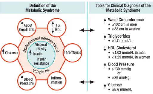
As a result and according to this definition, a patient will be diagnosed with metabolic syndrome if he displays central obesity plus two of the following components: raised triglycerides, reduced high density lipoproteins (HDL), raised fasting plasma glucose and hypertension (Figure 1).
Figure 1 Schematic view of the Metabolic Syndrome.
From (Barker 2012) – ApoB: apolipoprotein B; HDL: high density lipoprotein; LDL: low
20 Nevertheless, as the different ethnicities and populations have different norms for body weight, waist circumference, and also different risk to develop type 2 diabetes or cardiovascular disease, the IDF, has proposed a new set of criteria with ethnic/racial specific cut-offs in 2009 (Alberti et al. 2009) (Table 2).
Table 2 Gender and age-specific waist circumference cut-offs.
From (Alberti et al. 2009).
Country/ethnic group
Waist circumference cut-off
Male (cm) Female (cm)
Europids
In USA, the ATPIII values (102 cm males; 88 cm females) are likely to continue to be used for clinical purposes.
≥94 ≥80
South Asians
Based on a Chinese, Malay, and Asian-Indian population. ≥90 ≥80
Chinese ≥90 ≥80
Japanese ≥90 ≥80
Ethnic South and Central Americans Use South Asians recommendations until more specific data are available.
Sub-Saharan Africans Use European data until more specific data are available.
Eastern Mediterranean and Middle East (Arabs) population Use European data until more specific data are available.
21 According to the definition used, and ethnicity, age, sex or region, the prevalence of metabolic syndrome can be estimated from about 10% to more than 80% of the population (Desroches and Lamarche 2007; Kolovou et al. 2007). Worldwide, the IDF estimates that 25% of the adult population has the metabolic syndrome (IDF 2005). The prevalence has been shown to be influenced by several factors such as sedentary lifestyle, genetic background, physical activity, smoking, education, heredity of diabetes (Cameron et al. 2004) and ageing (Ford et al. 2002; Park et al. 2003). The National Health and Nutrition Examination Survey (NHANES) has shown that 5% of the subjects of normal weight, 22% among the overweight, and 60% among the obese had metabolic syndrome (Park et al. 2003) showing in the meantime that some non-obese people can have the syndrome. Palaniappan et al. have shown that each 11 cm increase in waist circumference was associated with a risk of developing metabolic syndrome within 5 years by 80% (Palaniappan et al. 2004). Moreover, the risk of developing metabolic syndrome increases with the number of metabolic disorders (Andreadis et al. 2007).
In 2009, it was reported that metabolic syndrome induces a 5-fold increase in the risk of developing type 2 diabetes and a 2-fold increase in the risk for cardiovascular disease over the next 5 to 10 years (Alberti et al. 2009). More precisely, stroke risks are increased by 2 to 4 fold, myocardial infarction by 3 to 4 fold, and death event by 2 fold compared to age-matched healthy population (Alberti et al. 2005).
As indicated before, metabolic syndrome is not a disease by itself but is rather defined by a cluster of disturbances that we will develop in the next section.
22
1.2
OBESITY, INSULIN RESISTANCE AND TYPE 2
DIABETES
As stated previously, the worldwide accepted consensus on the definition of metabolic syndrome proposed by the IDF stated that developing insulin resistance is not an essential requirement as it is not easy to assess in a daily clinical practice. Nevertheless, it is also stated that insulin resistance has been central in the development of this syndrome (IDF 2014), which was also recognized by
the former definitions we described in the previous chapter (Table 1).
In this section, we will define how insulin resistance develops in a context of obesity and dyslipidemia, both components of the metabolic syndrome, and the way it is linked to type 2 diabetes which leads to morbid consequences such as chronic kidney disease and cardiovascular disease.
1.2.1
Obesity
According to the World Health Organization, overweight and obesity are defined as abnormal or excessive fat accumulation that may impair health. The main cause of obesity and overweight is an energy imbalance between calories consumed and calories expended, essentially due to an increased consumption of high fat diet with an increase in physical inactivity and sedentary lifestyle. These two conditions are defined by the Body Mass Index (BMI), calculated as a person's weight in kilograms divided by the square of his height in meters. People are considered as overweight if their BMI is greater than or equal to 25 and obese if greater than or equal to 30. The main advantage of this index is that it is independent of the sex and age, but it may not correspond to the same degree of fatness in different individuals. Raised BMI is a major risk factor to develop type 2 diabetes, among other diseases and disorders (WHO 2014).
Worldwide, between 1980 and 2013, prevalence of combined overweight and obesity rose by 27.5% for adults and 47.1% for children. The number of overweight and obese individuals was estimated at
23 857 million in 1980, and to be 2.1 billion in 2013. Worldwide, the proportion of overweight people increased from 28.8% in 1980, to 36.9% in 2013 for men, and from 29.8% to 38.0% for women. In 2013, the prevalence of obesity was higher in women in developed and developing countries than in men (Ng et al. 2014).
The increase in the prevalence of type 2 diabetes is closely linked to central obesity: about 90% of type 2 diabetes is attributable to excess weight. Furthermore, it has been shown that approximately 197 million people worldwide had impaired glucose tolerance in 2007, most commonly because of obesity and the associated metabolic syndrome (Hossain et al. 2007).
1.2.2
Insulin resistance and Type 2 diabetes
According to the IDF, there are 387 million people with diabetes worldwide today with 179 million of undiagnosed people; by 2035 this number is expected to rise to more than 592 million people. In 2014 diabetes caused 4.9 million deaths, i.e. 1 every 7 seconds (IDF 2014). Diabetes Mellitus or Type 2 Diabetes represent 90% of Diabetes and is recognized as a worldwide epidemic whose progression unhalted. Prevalence of obesity, genetic susceptibility, urbanization and ageing contribute to important increase of the number of people suffering from diabetes mellitus. The proportion of obese and diabetic patients might slow down or even decrease in Europe and the United States while it should increase in Asia and Africa (ECDCDM Balarajan and Villamor 2009; Doak et al. 2012; Flegal et al. 2012; Tabaei et al. 2012). We could hypothesize that way of life is changing in all these regions, but not in the same way: in Asia and Africa, physical inactivity and rich diet consumption habits may increase with the economic development, whereas population in the United States starts to increase physical activity again.
People suffering from type 2 diabetes can be asymptomatic for many years. Early symptoms of type 2 diabetes may include infections that are more frequent and slower healing process, fatigue, hunger, increased thirst and urination, blurred vision or erectile dysfunction. However, type 2 diabetes
24 diagnosis is often made at an advanced stage through blood or urine bio-analysis, i.e. when micro-and macro-vascular defects already exist (ECDCDM report 2003; Wild et al. 2004; Danaei et al. 2011). Among the diabetic population two main co-morbidities have be identified: cardiovascular diseases (coronary heart disease, stroke, peripheral artery disease due to atherosclerosis) and kidney failure associated with elevated proteinuria (Janghorbani et al. 2007; Arboix et al. 2008; Preiss et al. 2011; Barkoudah et al. 2012).
Diagnosis is then confirmed by several measurements such as:
Fasting blood glucose level - diabetes is diagnosed if fasting blood glucose is higher than 126 mg/dL (7 mM) (when measured twice),
Glycated hemoglobin (HbA1C) of 6.5% or higher, this notion of glycation will be further developed in the next section,
During oral glucose tolerance test performed by drinking a special sugar drink, glycemia is higher than 200 mg/dL 2 hours after the glucose challenge (Pories et al. 2011) (American Diabetes Association 2013).
Several risk factors for development of type 2 diabetes exist, such as inactivity, central obesity, family history, age over 45 years, gestational diabetes, elevated glycemia, metabolic syndrome.
The onset of type 2 diabetes occurs mainly in people that are insulin resistant, have genetic predisposition, or in response to overweight generally as a consequence of sedentary lifestyle and over nutrition. Insulin resistance is defined as a pathophysiological condition in which a normal insulin concentration does not adequately produce a normal insulin response in the peripheral target tissues such as adipose tissue, muscle and liver, i.e. glucose uptake and storage is lowered in these organs, leading to high glycemia values.
25 With time, insulin resistance develops due to lipid accumulation (lipotoxicity) that can activate several pathways like unfolded protein response or immune pathways, which are linked to free fatty acids uptake, lipogenesis and energy expenditure. As a result, accumulation of lipid metabolites such as ceramides and diacyglycerols in skeletal muscles and liver impairs insulin signaling in the cells (Samuel and Shulman 2012).
Consequently, pancreatic β-cells in pancreatic Langerhans islets produce more insulin to compensate the impaired insulin signaling and to maintain euglycemia. This peculiar stage is called pre-diabetes, and is characterized by subjects who are still normally glucose tolerant. At this stage, exercise and appropriate diet can still reverse the process of β-cell signaling impairment. With time, if inactivity and overweight develops further, β-cells function tends to decline more; under these circumstances people evolve progressively towards impaired glucose tolerance, characterized by significant hyperglycemia, as insulin secretion signaling is impaired and targeted organs (mainly skeletal muscles) are desensitized (Kaur 2014). This vicious circle contributes progressively to β-cell failure. This is the step during which T2D is obviously diagnosed (Figure 2) (Kasuga 2006; Prentki and Nolan 2006).
In Langerhans islets, α-cells also have an important role, as they produce glucagon, a hormone that favors release of glucose in the blood. In type 2 diabetes, there’s a dysregulation of glucagon release, which is not suppressed after a meal. Whether this is a primary cause or a consequence of β-cell dysfunction is still not clear (Dunning and Gerich 2007).
26
Figure 2 Islet β-cell failure and the natural history of type 2 diabetes.
From (Prentki and Nolan 2006) – IGT: impaired glucose tolerance; NGT: normal glucose tolerance; T2D: type 2 diabetes.
27 As stated before, lipotoxicity plays an important role in the development of insulin resistance. In adipocytes, lipolysis is increased, which induces elevation of free fatty acids levels.
On one hand, the increased levels of free fatty acids also activate toll like receptor (TLR). These receptors have complex expression patterns in different cell types but their expression is particularly significant in different types of white blood cells like mast cells, macrophages, and dendritic cells (Christmas 2010). TLR are known to recognize pathogen-associated molecular patterns like the lipopolysaccharide of Gram-negative bacteria and activate the innate immune system and provoke inflammation. In particular, TLR4 signaling has been shown to be pivotal in the pathological lipid effects on cardiovascular and metabolic functions (Eguchi and Manabe 2014). As a result, TLR can be activated by free fatty acids, that:
induce, via nuclear translocation of nuclear factor-kappa B (NF-kB), up-regulation of the genes coding for interleukin 6 (IL-6) and tumor necrosis factor α (TNFα), and as a result induce a pro-inflammation status. TNFα has been reported to increase IL-18 that has a direct link with the development of insulin resistance (Krogh-Madsen et al. 2006).
induce phosphorylation of insulin receptor substrate-1 (IRS-1) via c-Jun amino-terminal kinase (JNK) and protein kinase C (PKC), blocking its action on phosphoinositide 3 kinase (PI3-kinase) and Akt that normally enhances the glucose transporter 4 (GLUT-4) synthesis necessary to internalize glucose into the cell (essentially expressed on skeletal muscle and adipose tissue). This results in insulin resistance (Figure 3) (Paneni et al. 2013).
28
Figure 3 Mechanisms leading to insulin resistance.
Adapted from (Paneni et al. 2013) – Akt: protein kinase B; FFA: free fatty acids; GLUT-4: glucose transporter 4; IKK-β: inhibitor of nuclear factor kappa-B kinase subunit beta; ILs: interleukins; IRS-1: insulin receptor substrate-1; JNK: c-Jun amino-terminal kinase; NFkB: nuclear factor-kappa B; PKC: protein kinase C; TLR: toll like receptor; TNFα: tumor necrosis factor α.
On a second hand, impaired insulin signaling induces dyslipidemia.
Free fatty acids are used in the liver to synthetize triglycerides. Elevation of free fatty acids promotes stabilization of apolipoprotein B (apoB) and as a consequence, promotes very light density lipoproteins (VLDL) production synthesis by the liver. In healthy conditions, apoB is degraded via insulin signaling, as well as VLDL degradation via lipoprotein lipase. VLDL is metabolized into remnant lipoproteins and light density lipoproteins LDL, both of which are atherogenic (Lewis and Steiner 1996). HDL normally possess anti-atherogenic properties, as it reverses the cholesterol transport from blood to the liver for excretion, on top of other protective activities such as antioxidative, anti-inflammatory, antithrombotic and vasodilatory actions (Assmann and Nofer 2003).
29 Under insulin resistance, hypertriglyceridemia and decreased activity of lipoprotein lipase induce a raise in HDL triglyceride content. In parallel to deterioration of LDL and VLDL metabolism, hypertriglyceridemia also induces an increase in HDL triglyceride content by an elevation of cholesterol ester transport protein (CETP) activity that exchanges the triglycerides from VLDL to HDL, and the cholesteryl esthers from HDL to VLDL (Le Goff et al. 2004). As a result, plasma HDL levels are decreased, as they are better degraded by hepatic lipase when associated to triglycerides. These mechanisms result in increased production of atherogenic particles, and decreased protective effects of HDL, which constitutes one of the feature of metabolic syndrome and type 2 diabetes (Lamarche et al. 1999). They are closely associated with oxidative stress and endothelial dysfunction, reinforcing the proinflammatory nature of macrovascular atherosclerotic disease (Kaur 2014).
The fact that abdominal obesity leads to increase in adipocytokines levels is a feature of metabolic syndrome. It results in adipose tissue inflammation that favors atherothrombosis and atherosclerosis, insulin resistance, liver steatosis and pancreatic β-cell function impairment (Kaur 2014).
On top of these mechanisms, adiponectin, recognized as a regulator of insulin sensitivity, food intake, and anti-inflammatory hormone (Liu and Liu 2009), is negatively associated to body weight and waist circumference (Xydakis et al. 2004) and low levels can be predictive for cardiovascular event (Pischon et al. 2004).
Another important contributing hormone is leptin, an adipokine responsible for satiety (Lau et al. 2005). Usually, its circulating levels increase in obese patients that progressively develop leptin resistance, as for insulin resistance. As a result, high plasma concentration cannot suppress the hunger anymore, via the leptin receptors located in the hypothalamus (Hutley and Prins 2005). Leptin has also been described as a hypertensive agent, through the sympathetic nervous system (Carlyle et al. 2002), and its action in increasing peripheral vascular resistance (Shirasaka et al. 2003).
30 It has also been reported that over synthesis of glucocorticoid (mainly cortisol) under chronic stress may lead to insulin hypersecretion, increase in body and visceral fat mass, and decrease in muscle mass, resulting in dyslipidemia, hypertension, and type 2 diabetes, in individuals with a genetic predisposition (Chrousos 2009).
All these mechanisms contribute to permanent inflammation that favors development and maintenance of insulin resistance in the body. Consequently, insulin resistance induce hyperglycemia responsible for micro and microvasculature defects that we will describe in the next section, leading to cardio-renal dysfunction, chronic diseases and finally can cause death (cardiovascular disease and chronic kidney disease).
1.2.3
Micro and macrovascular complications of
hyperglycemia
Among several consequences of hyperglycemia, a major impact effect of metabolic syndrome with type 2 diabetes is the damage of blood vessels that will be described in this section. Impact on blood vessels (structure and function), triggers several pathologies such as diabetic retinopathy, glaucoma, cataracts, diabetic neuropathy, cardiovascular diseases, sexual dysfunction and diabetic nephropathy (Cade 2008).
The endothelium is a cell layer that surrounds the vascular lumen. This monolayer is necessary to maintain vascular homeostasis acting as endocrine, paracrine and autocrine organ (Vallance 2001; Bonetti et al. 2003). Its functions include regulation of vessel integrity, vascular growth and remodeling, tissue growth and metabolism, immune responses, cell adhesion, angiogenesis, hemostasis and vascular permeability.
31 As a result, endothelium controls tissue perfusion and inflammatory responses and regulates blood viscosity (Félétou and Vanhoutte 2006; Moncada and Higgs 2006; Sena et al. 2013).
Vasodilation and vasoconstriction are accurately balanced by endothelium, as well as proliferation of smooth muscle cells, fibrinolysis, thrombogenesis and adhesion and aggregation of platelets. This balance is very fragile and any disturbance has a major impact on vascular function and structure that can cause dramatic consequences on the organs function, like heart and kidney (Sena et al. 2013).
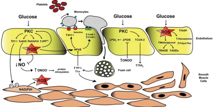
As displayed in Figure 4, the combination of hyperglycemia, high lipid levels and insulin resistance that characterizes metabolic syndrome, negatively impacts blood vessel structure and function through several mechanisms such as oxidative stress, activation of receptors for advanced glycation end product (RAGE) and PKC activation. The concept of glycemic continuum, i.e. the sequence of pre-diabetes, diabetes and cardiovascular risk shows that low levels of glycemia that are still under the threshold of overt diabetes already have negative effects on the vessels, and more particularly on the endothelium (Wei et al. 1998; Coutinho et al. 1999; Bartnik and Cosentino 2009; Rutter and Nesto 2011).
32
Figure 4 Alteration of endothelial function in the context of type 2 diabetes.
From (Creager et al. 2003). Schematic representation of cellular mechanisms triggered by hyperglycemia, elevated free fatty acids or insulin resistance, alone or in combination. These insults include increased oxidative stress, disturbances of intracellular signal transduction (such as activation of PKC) and activation of Receptor for Advanced Glycation Endproducts (RAGE). The consequences are decreased availability of nitric oxide (NO), increased production of endothelin (ET-1), activation of transcription factors such as NF-κB and activator protein-1 (AP-1), and increased production of pro-thrombotic factors such as tissue factor (TF) and plasminogen activator inhibitor-1 (PAI-1).
In the context of hyperglycemia and insulin resistance, endothelial dysfunction is then a consequence of enhanced overproduction and accumulation of reactive oxygen species (ROS) by mitochondria. This phenomenon is considered as the main link between hyperglycemia and diabetic vascular defects (Nishikawa et al. 2000; Creager et al. 2003).
33 High glycaemia activates PKC that induces alterations in cellular permeability, inflammation, angiogenesis, cell growth, extracellular matrix expansion, and apoptosis at the level of the endothelium (Geraldes and King 2010). Four main mechanisms of endothelial cellular damages have been described (Figure 5):
a- Superoxide anion (O2−) is produced via NADPH oxidase (Inoguchi et al. 2000) and, with nitric
oxide (NO), induces the formation of oxidative agent peroxynitrite (ONOO−) that leads to nitration (introduction of a nitro group into an aromatic chemical compound, generally in tyrosine) of endothelial NO synthase (eNOS) and antioxidant enzymes (Hink et al. 2001). Moreover, PKCβ2 has been recently shown to phosphorylate p66Shc, which causes an accumulation of ROS in the
mitochondria (Cosentino et al. 2008; Paneni et al. 2012) and which was reported to enhance the NADPH activity, overall leading to production of ROS (Tomilov et al. 2010; Shi et al. 2011)
b- PKC activation enhances eNOS activity but as NO is also used as a reactive to produce ONOO−, as described above (Cosentino et al. 1997; Du et al. 2001; Alp and Channon 2004; Geraldes and King 2010), this leads to decrease NO levels. The lack of NO coupled to activation of PKC causes synthesis of ET-1 and prostanoids like thromboxane A2 (TA2), which are vasoconstrictors and inducers of platelet aggregation (Hink et al. 2001; Geraldes and King 2010).
c- Vascular inflammation is also triggered by PKC-dependent ROS production via NF-kB pathway, that induces elevation of monocyte chemo-attractant protein-1 (MCP-1), and adhesion proteins like vascular cell adhesion molecule-1 (VCAM-1) and intracellular cell adhesion molecule-1 (ICAM-1) (Kouroedov et al. 2004; Giacco and Brownlee 2010). As a result, activated macrophages produce cytokines that maintain the inflammatory status, and participate to atherosclerotic process by the formation of foam cells.
34 d- Methylglyoxal and advanced glycation end products (AGEs) synthesis are also a result of ROS production and are known to be highly involved in the process of endothelial dysfunction by increasing the production of ROS (American Diabetes Association 2013) (Brouwers et al. 2010; Yan et al. 2010; Sena et al. 2012).
Figure 5 Mechanisms of hyperglycemia-induced vascular damage.
From (Paneni et al. 2013) – AGEs: advanced glycation end products; COX-2: cyclooxygenase-2; eNOS: endothelial nitric oxide synthase ; ET-1: endothelin-1; ICAM-1: intracellular cell adhesion molecule-1; ILs: interleukins; MCP-1: monocyte chemo-attractant protein-1; NF-ƙB: nuclear factor kappa binding; NO: nitrogen monoxide; ONOO-: peroxynitrite; PARP: poly ADP ribose polymerase; PGI
2: prostacyclin; PGIS:
prostacyclin synthase; PKC: protein kinase C; RAGE: receptor for advanced glycation endproducts; ROS: reactive oxygen species; TNFα: tumor necrosis factor α; TXA2: thromboxane A2; VCAM-1: vascular cell adhesion molecule-1.
Glycation was discovered by Maillard and consists in the chemical (non-enzymatic) reaction of reducing sugars with amino groups (Maillard 1912). Glycation generally impairs the function of biologic molecules, in contrast to enzymatic glycosylation of proteins, which is physiological. As an example, hemoglobin glycation leads to the formation of glycated hemoglobin (HbA1c). It results from
35 the formation of an adduct between glucose and the valine amino group of the β chain of hemoglobin. The determination of HbA1c in diabetes patients is a reliable indicator for monitoring glycemic control in diabetes patients (Wautier and Schmidt 2004). HbA1c is a marker used to follow the long term effects of a treatment on glycemia, as it integrates longer modifications than glycemia.
The initial glycation reaction is followed by a cascade of chemical reactions resulting in the formation of intermediate products (Schiff base, Amadori and Maillard products) and finally to a variety of derivatives named AGEs.
Moreover, AGEs have been detected in atherosclerotic plaques from diabetic patients showing that AGEs are closely linked to plaques formation (Niwa et al. 1997). Atherosclerosis consists in deposition of aggregates on arterial walls, and can be responsible for vessel occlusion and myocardial infarction (when plaques rupture happens). In atherogenesis, AGEs can bind arterial collagens that can trap plasma proteins or lipoproteins like LDL, to participate to atheroma formation in diabetes (Brownlee et al. 1985). Another important point for the atherosclerotic process is proliferation of smooth muscle cells. This cell proliferation is promoted by cytokines like insulin-like growth factor-I and platelet-derived growth factor whose secretion is promoted by AGEs as well (Ahmed 2005). Moreover, the RAGE can bind several ligands like AGEs, and is expressed on several cell types such as endothelial cells, monocytes and vascular smooth muscle cells (Kilhovd et al. 1999; Schmidt et al. 1999). The binding of ligands to RAGE have been reported to induce inflammatory responses in several chronic diseases including atherosclerosis (Naka et al. 2004). One other mechanism that is believed to induce damage to vessels is linked to Aldose reductase. Aldose reductase is the initial enzyme in the intracellular polyol pathway that converts glucose into sorbitol. Hyperglycemia increases the activity of the polyol pathway. As a result, sorbitol accumulates in cells and that could be responsible for the development of diabetic microvascular complications by creating an osmotic stress. It has been shown in preclinical studies that sorbitol accumulation has
36 been linked to micro-aneurysm formation, thickening of basement membranes, and loss of pericytes (Gabbay 1975; Fong et al. 2004).
On top of these mechanisms, platelet aggregation is a consequence of insulin resistance as well. Indeed, lack of insulin signaling in platelets also impairs the IRS1/PI3K pathway resulting in Ca2+ accumulation in the cytosol necessary for platelet aggregation (Paneni et al. 2013). This
phenomenon coupled with impaired fibrinolysis, mostly due to elevated levels of PAI-1, is a feature of type 2 diabetes (Collier et al. 1992; McGill et al. 1994). Moreover, elevated levels of coagulation activation markers, including thrombin-antithrombin complexes, have been found in patients with type 2 diabetes (Davì et al. 1992; Nagai 1994; López et al. 1999). Overall, these mechanisms favor hypercoagulable state and atherosclerosis.
To summarize, three main defects occur due to hyperglycemia, via ROS production: vasoconstriction, vascular inflammation and atherosclerosis. These three phenomena can contribute to development of hypertension, chronic inflammation and thrombosis.
1.3
HYPERTENSION
Hypertension is one of the main defects that characterize the metabolic syndrome, and it has been reported that metabolic syndrome is present in up to one third of the hypertensive patients (Cuspidi et al. 2004; Schillaci et al. 2004). The IDF defined blood pressure as a risk factor from 130 mm Hg of systolic blood pressure or diastolic blood pressure from 85 mm Hg
(IDF 2005) (Table 1). In the
metabolic syndrome, visceral obesity has been proposed to be the main trigger of hypertension. As stated previously, leptin, TNF-α, (IL-6), angiotensinogen, and free fatty acids are overproduced with fat accumulation. These entities also favor the development of hypertension, on top of the development of insulin resistance (Katagiri et al. 2007). Insulin has also been shown to induce renal37 sodium reabsorption, and this anti-natriuretic effect seems to be exacerbated in an insulin resistant state, which can explain the link between hyperinsulinemia and development of hypertension (Sechi 1999). Other studies have shown that insulin induces production of ET-1, which as stated before is a potent vasoconstrictor that also favors hypertension (Sarafidis and Bakris 2007). In a previous section we described the interdependence between insulin resistance and oxidative stress and decrease of NO, which respectively induce sodium retention and decrease the vasodilation capacity of the vessels. Together, these features favor the onset of hypertension.
Abdominal obesity has also been involved in the overactivity of 2 main systems that enhance the elevation of blood pressure: the renin angiotensin aldosterone system and the sympathetic nervous system.
On one hand, insulin resistance, in combination with hyperinsulinemia, hyperleptinemia and obesity have been shown to induce activation of sympathetic nervous system. That activation induces sodium reabsorption by the renal proximal tubules which induce increase in volemia, and increase cardiac output. Moreover, obese people have impaired renal-pressure natriuresis, explaining the fact that these people need an increased blood pressure to maintain the sodium homeostasis (Hall 1997). Leptin is also linked to sympathetic activity, as leptin has been shown to increase its activity through the ventromedial and dorsomedial hypothalamus, and consequently increase blood pressure (Carlyle et al. 2002; Marsh et al. 2003). Obstructive sleep apnea has been also related to an increased prevalence of metabolic syndrome, along with enhancement of sympathetic activity (Wolk et al. 2003; Coughlin et al. 2004), reinforcing its hypertensive action.
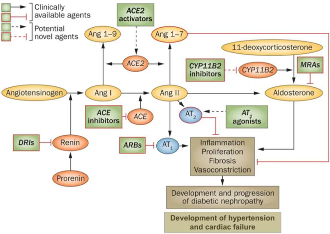
On the other hand, hyperinsulinemia coupled to hyperglycemia have been reported to activate key components of the Renin Angiotensin Aldosterone System (RAAS), such as angiotensinogen, angiotensin II (AngII), and its receptor isoform AT1, that are all involved in hypertensive process, via sodium retention and promotion of insulin resistance (Malhotra et al. 2001). RAAS will be further
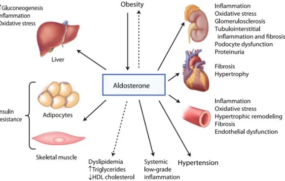
38 described in the next section. More recently, it has been reported that AngII can trigger aldosterone synthesis by adipocytes, meaning that obese patients are more responsive to AngII as compared to fit individuals (Briones et al. 2012) ( Figure 6).
Figure 6 Proposed mechanisms responsible for hypertension onset in Metabolic Syndrome.
From (Yanai et al. 2008) – IL6 : interleukin 6 ; NEFA: non esterified fatty acids (or free fatty acids); OSA: obstructive sleep apnea; TNF-α : tumor necrosis factor α.
Finally, adiposity is a trigger for inflammation, insulin resistance, enhancement of sympathetic system and RAAS. All these defects are closely linked and together favor the development of hypertension, via elevation of volemia, vasoconstriction and decrease in arterial vasodilation capabilities.