National Park Service
Paleontological Research
United States Department of the Interior•National Park Service•Geological Resource Division
Edited by Vincent L. Santucci and Lindsay McClellandTechnical Report NPS/NRGRD/GRDTR-99/03
NPS Fossil Resources
Copies of this report are available from the editors. Geological Resources Division
12795 West Alameda Parkway Academy Place, Room 480
Lakewood, CO 80227
Please refer to: National Park Service D-1341 (October 1999).
Cover Illustration
Life reconstruction of Metamynodon planifrons and an extinct crowned crane, Balearica sp. in a riparian habitat during the early Oligocene. Based on work conducted in Badlands National Park. The original painting was produced by Carl Buell.
NATIONAL PARK SERVICE
PALEONTOLOGICAL RESEARCH
VOLUME 4
EDITED BY
Vincent L. Santucci
National Park Service
PO Box 592
Kemmerer, WY 83101
AND
Lindsay McClelland
National Park Service
Room 3223 - Main Interior
1849 C Street, N.W.
Washington, DC 20240-0001
Geologic Resources Division Technical Report
NPS/NRGRD/GRDTR-99/03
To Dr. Michael Soukup,
National Park Service
Associate Director for
Natural Resource Stewardship & Science
CONTENTS
INTRODUCTION ... vii BADLANDS NATIONAL PARK
Vertebrate paleontology of the Pierre Shale and Fox Hills formations (Late Campanian - Late Maastrichtian) of Badlands National Park, South Dakota
David J. Cicimurri, Gordon L. Bell, Jr. and Philip W. Stoffer ... 1 Locomotor adaptations in Metamynodon planifrons compared to other amynodontids (Perissodactyla,
Rhinocerotoidea)
William P. Wall and Kristen L. Heinbaugh ... 8
BIGHORN CANYON NATIONAL RECREATION AREA
A preliminary assessment of paleontological resources at Bighorn Canyon National Recreation Area, Montana and Wyoming
Vincent L. Santucci, David Hays, James Staebler, and Michael Milstein ... 18
CANYONLANDS NATIONAL PARK
An aetosaur (Reptilia:Archosauria) from the Upper Triassic Chinle Group, Canyonlands National Park, Utah
Andrew B. Heckert, Spencer G. Lucas and Jerald D. Harris ... 23
CHANNEL ISLANDS NATIONAL PARK
Giant island/pygmy mammoths: The Late Pleistocene prehistory of Channel Islands National Park
Larry D. Agenbroad and Don P. Morris ... 27
CHESAPEAKE AND OHIO CANAL NATIONAL HISTORIC PARK
Stratigraphic and paleontologic record of the Sauk III regression in the central Appalachians
David K. Brezinski, John E. Repetski and John F. Taylor ... 32
CURECANTI NATIONAL RECREATION AREA
Non-marine trace fossils from the Morrison Formation (Jurassic) of Curecanti National Recreation Area, Colorado Anthony R. Fiorillo ... 42
DENALI NATIONAL PARK & PRESERVE
All is not quiet on the paleontological front in Denali National Park
R.B. Blodgett and Phil Brease ... 47
FLORISSANT FOSSIL BEDS NATIONAL MONUMENT
Fossil birds of Florissant, Colorado: With a description of a new genus and species of cuckoo
Robert M. Chandler ... 49
FOSSIL BUTTE NATIONAL MONUMENT
Paleoecology and paleoenvironments during the intial stages of Eocene Fossil Lake, SW Wyoming
Roberto E. Biaggi and H. Paul Buchheim ... 54
Vegetational history and climatic transition in an Eocene intermontane basin: Plant microfossil evidence from the Green River Formation, Fossil Basin, Wyoming
Robert A. Cushman ... 66 Caddisfly (Trichoptera) larval cases from Eocene Fossil Lake
Mark A. Loewen, V. Leroy Leggit and H. Paul Buchheim ... 72
PETRIFIED FOREST NATIONAL PARK
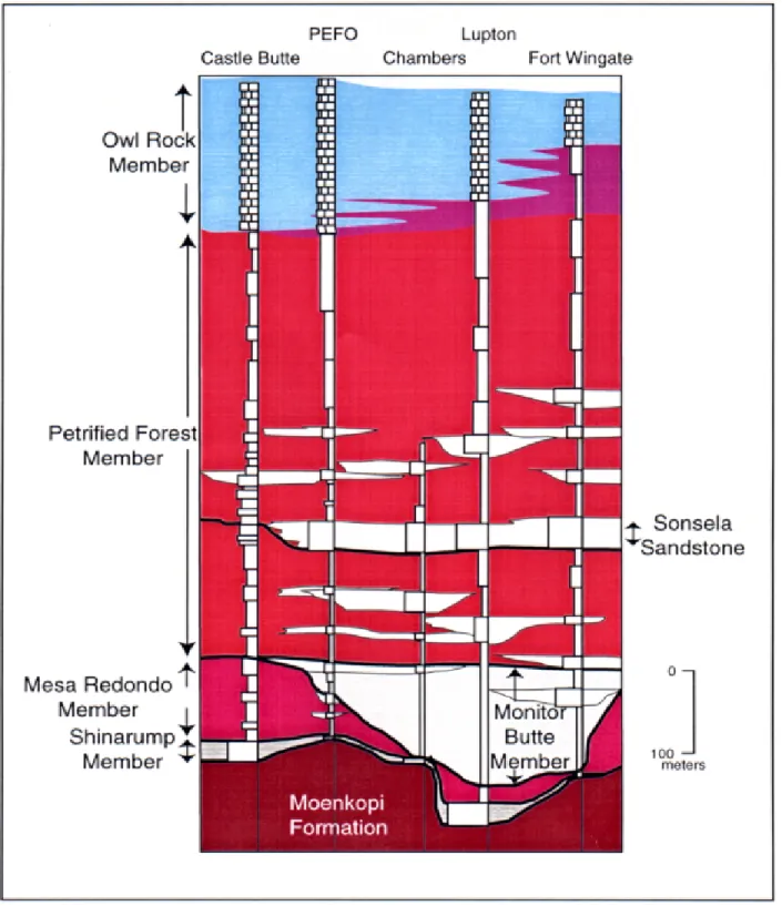
Incised valley fills in the lower part of the Chinle Formation, Petrified Forest National Park, Arizona: Complete measured sections and regional stratigraphic implications of Upper Triassic Rocks
Russel F. Dubiel, Stephen T. Hasiotis and Timothy M. Demko ... 78
Probable reptile nests from the Upper Triassic Chinle Formation, Petrified Forest National Park, Arizona
Stephen T. Hasiotis and Anthony J. Martin ... 85 Occurrences of Zamites powelli in oldest Norian strata in Petrified Forest National Park, Arizona
Alisa S. Herrick, David E. Fastovsky and Gregory D. Hoke ... 91
New discoveries of Late Triassic dinosaurs from Petrified Forest National Park, Arizona
Adrian P. Hunt and Jeremiah Wright ... 96
The oldest Triassic strata exposed in Petrified Forest National Park, Arizona revisited
Francois Therrien, Matthew M. Jones, David E. Fastovsky, Alisa S. Herrick, and Gregory D. Hoke ... 101
TIMPANOGOS CAVE NATIONAL MONUMENT
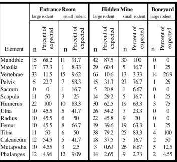
An investigation of the Late Pleistocene fauna of Timpanogos Cave National Monument, Utah
Christian O. George ... 109
WALNUT CANYON NATIONAL MONUMENT
An inventory of paleontological resources from Walnut Canyon National Monument, Arizona
Vincent L. Santucci and V. Luke Santucci, Jr. ... 118
MULTIPLE PARKS
Continental ichnofossils from the Upper Jurassic Morrison Formation, Western Interior, USA: What organism behavior tells us about Jurassic environments and climates
Stephen T. Hasiotis ... 121
APPENDIX ... 126
INTRODUCTION
During this last year of the century, the list of National Park Service areas identified with paleontological resources has grown to 134. Along with redwoods, grizzlies, geysers, and ancient ruins, the national parks preserve a remarkable record of life extending back over a billion years. The rich paleontological resources found in parks have attracted con-siderable research interest. Paleontological research from within national parks is reported regularly at scientific con-ferences and provides numerous graduate students with the-sis projects.
This fourth National Park Service Paleontological Re-search Volume compiles 20 papers representing paleonto-logical research in 12 different National Park Service areas. The individual reports reflect a cross-section of the types of paleontological research being conducted throughout the National Park System by academic scientists, their students, and U.S. Geological Survey staff. The contributions from each of the investigators and their research teams are recog-nized and acknowledged in this volume.
I am again proud to include reports documenting a wide diversity of paleontological research in the national parks. The volume continues to include a number of papers focusing on the biostratigraphy of Triassic sediments at Pet-rified Forest National Park. A student from Franklin and Marshall College has prepared a report on his research on the cave fauna uncovered at Timpanogos Cave National Monument. Other papers in this volume include work on the Pleistocene mammoths from Channel Islands National Park, marine reptiles from Badlands National Park, and a descrip-tion of a new bird from Florissant Fossil Beds Nadescrip-tional Monu-ment.
Thanks to Sid Ash, Ron Blakey, Ken Carpenter, Bill Cobban, Russell Dubiel, Dave Gillette, Steve Hasiotis, Adrian Hunt, Clay Kyte, Greg McDonald, Steve Mitchelson, Don Prothero, Tom Olson, Kris Thompson, William Wall, and Michael Whalen, for their willingness to review manuscripts. Additional thanks to Dave Shaver, Bob Higgins, Dave McGinnis, Arvid Aase, Kris Thompson, Graeme MacDonald, Erin Retelle, Marikka Hughes and Bianca Santucci for their suggestions and support relative to this research publica-tion. I am indebted to Lindsay McClelland, the co-editor of this volume, for many contributions that helped to promote the management, protection and research of paleontological resources in the national parks.
This volume is dedicated to Mike Soukup, Associate Director for Natural Resource Stewardship and Science in the National Park Service. His leadership in building support for science-based decisionmaking has strengthened the man-agement and protection of all park natural resources. Fossils have been key beneficiaries of these policies, as parks in-creasingly recognize the importance of paleontological re-search and the value of paleontological resources.
Finally, through the combined efforts of the women and men already mentioned, along with many others, the NPS Paleontological Resource Program continues to grow. Many research questions remain to be explored within the national parks and monuments. Likewise, the increasing num-bers of paleontological inventories being initiated in the parks continue to uncover new evidence about the biological past. A holistic approach to managing paleontological resources, which includes research, is becoming the standard practice in national parks.
Vincent L. Santucci National Park Service
VERTEBRATE PALEONTOLOGY OF THE PIERRE SHALE
AND FOX HILLS FORMATIONS
(LATE CAMPANIAN - LATE MAASTRICHTIAN) OF
BADLANDS NATIONAL PARK, SOUTH DAKOTA
DAVID J. CICIMURRI1, GORDEN L. BELL, JR.2, AND PHILIP W. STOFFER3 1Bob Campbell Geology Museum, Clemson, South Carolina 29634
2Museum of Geology, South Dakota School of Mines and Technology, Rapid City, South Dakota 57701 3U.S.G.S. Menlo Park, San Jose, California 95192
____________________
ABSTRACT—Recent field investigations were concentrated in the Pierre Shale and Fox Hills formations (Late Cretaceous)
exposed in Badlands National Park (BADL). Here we describe the occurrence of vertebrate fossils from the two lithostratigraphic units within BADL. Specimens include a tooth of the sand tiger shark, Odontaspis; a teleost tooth and scales; a partial left maxilla and associated dorsal vertebrae of a juvenile Mosasaurus conodon; and an isolated anterior caudal vertebra of a large unidentified mosasaur. A rich and varied invertebrate assemblage was also found that includes: ammonites, nautiloids, gastropods, pelecypods, scaphopods, decapods, inarticulate brachiopods, bryozoa, and scleractinian corals.
The juvenile specimen of Mosasaurus conodon and the teleost tooth were collected from the Baculites cuneatus biozone of the Verendrye Member, Pierre Shale. The teleost scales were associated with Baculites clinolobatus and Hoploscaphites
burkelundi, and were found in the Mobridge Member, Pierre Shale. The Odontaspis tooth was collected from the Elk Butte
Member, Pierre Shale, whereas the isolated mosasaur caudal vertebra was collected from the upper part of the Fox Hills Formation.
____________________
INTRODUCTION
D
uring much of the Late Cretaceous, a vast north-south trending epicontinental sea existed in the Western Interior of North America. The eastern margin of the seaway was formed by the low-lying stable Canadian Shield, while the entire western margin was flanked by an unstable cordilleran highland (MacDonald and Byers, 1988).Rapid sea level rise during the late Early Campanian re-sulted in a shift from chalk deposition of the Niobrara Sea-way to muds of the Pierre SeaSea-way (McGookey et al., 1972). During the existence of the Pierre Seaway, several minor trans-gressive/regressive events occurred that are recorded in the rocks of the Pierre Shale exposed in Badlands National Park. Retreat of the seaway began in early Maastrichtian time due to an increase in both tectonic activity and rate of coarse clastic deposition (McGookey et al., 1972). The Fox Hills Formation represents a nearshore transition between the ma-rine environments of the Pierre Shale and terrestrial environ-ments of the Hell Creek Formation.
The Pierre Shale and Fox Hills Formation are exposed today in the northern and southern parts of Badlands Na-tional Park of southwestern South Dakota (Figure 1). The park is well known for Eocene - Miocene mammalian assem-blages in the White River Group. However, little is known about the fossil occurrences, especially of vertebrates, within Cretaceous rocks exposed throughout the park.
FIGURE 1— A, Map of Badlands National Park. Symbols indicate
vertebrate fossil localities; B, Map of southwestern South Dakota showing the location of the park. B modified from U.S.G.S. 1:50,000-scale map of Badlands National Park.
Abbreviations - BADL, Badlands National Park, SDSM,
Museum of Geology, South Dakota School of Mines and Technology. All specimens are stored in the Museum of Geology. Precise stratigraphic and geographic information is on record at Badlands National Park.
HISTORY OF COLLECTING
Very little is known about the fossil resources of Creta-ceous rocks exposed in BADL. Meek and Hayden (1862) were the first to recognize the presence of the “Fort Pierre Formation” (Pierre Shale) in the Sage Creek area of the Park (North Unit). Since then, this formation has received little attention. However, recent field research has documented fossils within Cretaceous strata of BADL, including ammo-nites, pelecypods, gastropods, scaphopods, crustaceans, brachiopods, nautiloids, bryozoans, corals, scaphopods, and belemnites (Table 1). The majority of the fossils are from the Pierre Shale, but several ammonites and the belemnites, as well as a lobster tail, were recovered from the Fox Hills Forma-tion. Meager vertebrate remains, consisting of isolated te-leost scales and teeth, a single mosasaurine caudal vertebra, and vertebrae and jaw fragment of Mosasaurus conodon, were also discovered. This small assemblage probably re-flects a collecting bias (as exposure surface is limited), rather than actual low abundance.
Several important marine reptile discoveries have been made outside the park boundary. The type specimen of
Prognathodon overtoni (KU 950) was collected from “... near
the top of the Pierre deposits of the Cheyenne River of South Dakota” (Russell, 1967; Williston, 1897, p. 95), and an addi-tional specimen (SDSM 3393) was recovered from “... the Virgin Creek Member, Upper Pierre Shale Formation ... south-west of Cuny Table, Shannon County, South Dakota” (Russell, 1967). The preservation of SDSM 3393 indicates that the bones were collected from the Yellow Mounds Paleosol.
An additional mosasaur skeleton, probably Mosasaurus, was collected by SDSMT personnel over 30 years ago north of Scenic, South Dakota. The specimen consists of a nearly complete skeleton. Unfortunately, only the skull, limbs, and part of the tail were collected at the time of discovery. The bones are encased in hard, yellowish limestone derived from the Yellow Mounds Paleosol. This material has been pre-pared with acetic acid to dissolve the limestone. Several teeth of the dogfish, Squalus, were also discovered in the lime-stone. This association may indicate, as has been docu-mented from the Pierre Shale of the Missouri River area of South Dakota, that the mosasaur carcass was scavenged by a school of dogfish (Bell et al., in press).
In 1926, a stratodont osteichthyan, probably Stratodus (SDSM 2674 and 2675), was collected from Cuny Table, Sh-annon County, by the Museum of Geology. The remains consist of a complete dentary, premaxilla, edentulous jaw frag-ments with a double row of equally sized alveoli, isolated teeth, and scales. Some of the bones are encased in hard yellow and pink phosphatic nodules that are characteristic of the Yellow Mounds Paleosol.
CRETACEOUS STRATIGRAPHY OF BADLANDS NATIONAL PARK
Cretaceous rocks of BADL consist of the Pierre Shale, and bluish, fine-grained glauconitic sandstone of the Fox Hills Formation. Extensive outcrops of the Pierre Shale are found throughout the Sage Creek Wilderness area of the North Unit and within tributaries of the Cheyenne River in the South Unit. Overlying the Pierre Shale is a yellow weath-ered unit, variously referred to as the “Interior Zone”, “Inte-rior Formation”, Rusty Member” (Stoffer et al., 1998), and “Yellow Mounds Paleosol” (Pettijohn, 1965). Dunham (1961) recognized that pre-Eocene weathering was responsible for the bright yellow sediments of the “Interior Formation”. However, it was uncertain as to whether these sediments belonged to the Pierre Shale or Fox Hills Formation (Agnew and Tychsen, 1965). Recent work by Stoffer et al. (1998) has established that the “Yellow Mounds Paleosol” is the result of meteoric weathering of both the upper Pierre Shale and Fox Hills Formation. Exposures of the Fox Hills Formation are
FIGURE 2— Composite stratigraphic section of Cretaceous rocks of
Badlands National Park. Eo = Eocene. Modified from Stoffer et al. (1998).
found throughout the North Unit of BADL.
Collecting efforts have been concentrated in the Pierre Shale during the past several years, and several ammonite biozones have been recognized, including: Didymoceras
cheyennense, Baculites compressus, B. cuneatus, B. reesidei, B. jenseni and B. eliasi, B. baculus, B. grandis, B. clinolobatus, and Hoploscaphites burkelundi (oldest to
youngest). These biozones have been used to subdivide the Pierre Shale into several biostratigraphic units. In BADL, the lithologic composition correlates with the DeGrey, Verendrye (Crandall, 1958), Virgin Creek, Mobridge, and Elk Butte (Searight, 1937) members of the Pierre Shale of central South Dakota (Figure 2).
Fossils are scarce in the Fox hills Formation, but this may reflect a collecting bias because this unit has largely been neglected. Those fossils that do occur are generally in poor condition because they have been subjected to several episodes of subaerial exposure. The Fox Hills Formation of BADL has been divided into lower and upper units (Stoffer
et al., 1998), although correlation of the Fox Hills with the
type area in Central South Dakota (Waage, 1961) is hindered by the poor preservation of invertebrate fossils. The lower unit is no older than late early Maastrichtian, because this interval overlies the Baculites clinolobatus biozone of the Pierre Shale (Cobban et al., 1994). Sr87/SR86 values of
belem-nites found in the upper unit yielded an age of 67 mya (Stoffer
et al., 1998).
SYSTEMATIC PALEONTOLOGY
Class Chondrichthyes Huxley, 1880 Order Lamniformes Berg, 1958 Family Odontaspididae Muller and Henle, 1839
Genus Odontaspis Agassiz, 1838
Odontaspis sp.
Figure 3, A-C
Referred Specimens - BADL 9935, lower anterior tooth found
as float in the Elk Butte Member, Pierre Shale.
Description - The tooth possesses a tall, narrow cusp with a
pointed apex. The cusp is slightly sigmoid in lateral profile, and distally inclined. Its labial crown face is smooth and nearly flat, whereas the lingual face is smooth and greatly convex. The cutting edges are smooth and continuous. The enameloid of the labial face extends basally onto root lobes. The lingual dental band is wide and a prominent lingual boss bears a nutritive groove. The root is incomplete, but was bilobate with meso-distally thin lobes and a u-shaped interlobe area. Several foramina are located on the boss and within nutritive groove.
Discussion – Broken surfaces indicate that at least one pair
of lateral cusplets was originally present at the base of each side of the central cusp. This character distinguishes BADL 9935 from lower anterior teeth of the Mitsukurinidae (goblin sharks). Also, the smooth crown faces separate the tooth from both mitsukurinids and striated odontaspids
(“Synodontaspis”). The crown morphology is similar to
Odontaspis hardingi from the Upper Campanian of New
Jer-sey (Cappetta and Case, 1975), but one specimen is not ad-equate for precise taxonomic assignment.
Teeth of Odontaspis are well suited for a piscivorous diet. The lower teeth pierce and hold, while the upper teeth cut into the prey (Cappetta, 1987). Recent odontaspids are found in shallow bays and coastal waters, to a depth of 200 meters (Tricas et al., 1997).
FIGURE 3— Lower anterior tooth of Odontaspis sp. (A-C), BADL
9935. A, Labial view; B, Mesial view; C, Lingual view. Scale bar = 3 cm.
Class Osteichthyes Order indeterminate
Figure 4, A-C
Referred specimens - BADL 8189, single tooth found as
float in the Baculites cuneatus biozone, Verendrye Member, Pierre Shale; BADL 8172, BADL 8179, BADL 8181, BADL 8182, BADL 8183, BADL 8184, BADL 8185, BADL 8186, BADL 8187, BADL 8188, all isolated scales collected in situ from the Baculites clinolobatus biozone, Mobridge Mem-ber, Pierre Shale.
Description - The tooth is laterally compressed and bisected
into equal labial and lingual faces by unserrated anterior and posterior carinae. Fine striations are located on the lower half of the tooth. The posterior carina is vertical, whereas the anterior carina is strongly sloping. The apex is broken and there is a deep basal pulp cavity. Enamel covers only the upper two thirds of the crown.
Two types of cycloid scales have been collected. One is oval and is taller than long (Figure 4-B). The other type is subequal in length and height, and has three “denticles” on the posterior edge (Figure 4, C).
Discussion - An enamel-free basal section and deep pulp
cavity indicates the tooth belonged to a fish with a thec-odont dentition. The tooth came from a bioturbated lime-stone and was associated with broken Inoceramus and
Lucina. This specimen was collected near Blindman Table,
South Unit.The scales were collected from a limestone bed and are associated with inarticulate brachiopods (Lingula). BADL 8180 is similar to scales of the Ichthyodectidae de-scribed by Bardack (1965, p. 51). However, the lack of asso-ciated skeletal material makes taxonomic assignment diffi-cult. These scales were collected from just north of the Sage Creek Primitive Campground, North Unit.
based, taper distally to a smooth rounded end, and project slightly anteriorly. The prezygopophyses are narrow with a smooth, ovate, upward oriented articulation.
Discussion – Preservation of the vertebrae inhibits
descrip-tion of postzyopophyses, and the presence of zygosphenes and zygantra are unknown. However, the length of the synapophyses suggests that the vertebrae are medial or pos-terior dorsals. In addition, the length of the vertebrae and the size and fibrous texture of the maxilla indicates that the speci-men was a juvenile. Serrations and faceting are less devel-oped than in more derived mosasaurines such as Mosasaurus
dekay, M. maximus, and M. missouriensis (Goldfuss, 1845;
Russell, 1967). BADL 9831 is geologically younger than speci-mens of M. conodon collected by the Museum of Geology from the Pierre Shale of central South Dakota.
FIGURE 4— Osteichthyes tooth (A) and scales (B-C). A, Lateral
view of tooth, BADL 8189. Scale bar = 1 mm; B, Type 1 scale, BADL 8180. Scale bar = 1 cm; C, Type 2 scale, BADL 8186. Scale bar = 1 mm. Anterior is left for all specimens.
Class Reptilia Linnaeus, 1758 Order Squamata Oppel, 1811 Family Mosasauridae Gervais, 1853 Genus Mosasaurus Conybeare, 1822
Mosasaurus conodon (Cope, 1881)
Figure 5
Referred specimen - BADL 9831, partial left maxilla and seven
dorsal vertebrae collected as float from the Baculites cuneatus biozone, Verendrye Member, Pierre Shale. Collected approxi-mately one half mile south of the Sage Creek Primitive Camp-ground, North Unit.
Description - Only the middle portion of the left dentary,
including seven teeth, is preserved. Seven foramina are lo-cated near the ventral border of the maxilla. This edge of the jaw is wide to accomodate the teeth. The bone thins dorsally and curves strongly medially. A small portion of the external naris is preserved.
The teeth are bicarinate and divided into nearly equal labial and buccal parts. Irregular serrations are uniformly distributed along the length of the anterior and posterior carinae. Both the labial and buccal crown faces are convex and weakly faceted. The teeth are recurved and the apices are internally inclined, becoming more pronounced anteroposteriorly. Crown height decreases anteroposteriorly; labial and buccal convexity increases.
Each dorsal vertebra measures 4.6 cm in length. The cotyle is deeply concave and has a sharp perimeter. The condyle is convex with a circular ventral border, and has flat dorsal and dorsolateral sides. The neural spines are tall, broad, nearly flat walled, and posteriorly inclined. Synapophyses are dorsoventrally compressed and broad
FIGURE 5— Mosasaurus conodon, juvenile, BADL 9831. View of
concretion showing incomplete left maxilla (bottom) and five dorsal vertebrae. Neural spine of a sixth vertebra can be seen at left. Scale bar = 10 cm.
Subfamily Mosasaurinae (Gervais, 1853) Williston, 1897 Gen. et sp. indet.
Figure 6, A-C
Referred Specimen - BADL 9934, isolated anterior caudal
vertebra found as float from the Fox Hills Formation just south of Robert’s Prairie Dog Town, North Unit.
Description - Maximum length of the centrum is 4.1 cm,
whereas maximum width and height are equal at 3.2 cm. The cotyle and condyle have a sub-triangular shape. The neural
spine is tall (7.8 cm as preserved) and nearly vertical, with convex sides. Transverse processes are dorsoventrally com-pressed, anteroposteriorly narrow, slightly posteriorly in-clined, and ventrally oriented. The haemal arch is incom-plete, but the base was fused to the midlength of the centrum’s ventral surface.
Discussion - Anterior-most mosasaur caudal vertebrae have
long transverse processes but lack haemal arches. Size and length of the centrum and transverse prosesses diminishes posteriorly. The orientation and morphology of the trans-verse processes of BADL 9934 indicate that it is an anterior caudal vertebra with a haemal arch. The haemal arch was fused to the centrum, indicating a taxon within Mosasaurinae.
CONCLUSIONS
Cretaceous vertebrate fossils of Badlands National Park consist of an isolated mosasaur caudal vertebra, a partial maxilla and dorsal vertebrae of Mosasaurus conodon, an iso-lated tooth of the sand tiger shark, Odontaspis, and a tooth and scales of osteichthyes. The paucity of vertebrate mate-rial may reflect a collecting bias, as prospectable exposures of Cretaceous rocks generally occur as near-vertical sections.
Invertebrate fossils were abundant and diverse, espe-cially in the Verendrye Member of the Pierre Shale. This assemblage, as well as heavily bioturbated limestones and shales, indicate well oxygenated water and an abundant food supply. Gill and Cobban (1966) suggested that deposition of the Pierre Shale was relatively fast, preventing the dissolu-tion of mollusc shells, thus allowing their fossilisadissolu-tion. The shells of ammonites became hardgrounds for bryozoans and gastropods, and living chambers of Baculites were found to contain fecal pellets, indicating these were used as homes by some invertebrates. Disturbed bentonites and linearly ori-ented baculites in concretions provides evidence that bot-tom currents were active. The substrate was heavily bioturbated which, coupled with current action, led to the disarticulation and chaotic orientation of pelecypod remains. An abundance of invertebrates, including bryozoans, and juvenile mosasaur remains suggest relatively shallow water. Sohl (1966) reported that the presence of ostreid bivalves in some parts of the Pierre Shale indicated a shallow water envi-ronment.
The fish tooth and scales were collected from a lime-stone bed of the Mobridge Member, Pierre Shale. These
FIGURE 6— Mosasaurinae gen. indet. caudal vertebra (A-C), BADL 9934. A, Posterior view; B, Left lateral view; C, Ventral view. Anterior
is at left. Scale bars = 10 cm (in A-C).
remains were associated with abundant inarticulate brachio-pods (Lingula). Craig (1952) noted that extant lingulid bra-chiopods are shallow water forms that are most common in waters less than 18.7 m, and only rarely occur at depths up to 37.5 m. Abundant ammonites, gastropods, and pelecypods indicate normal marine conditions.
Yellow, thin-bedded sandstones above the Pierre Shale are lithologically equivalent to the Fox Hills Formation. How-ever, invertebrate fossils indicate that these strata are tempo-rally equivalent to the Elk Butte Member of north-central South Dakota. As evidenced by drag and tool marks, current action was strong. The presence of sand tiger shark remains suggests nearshore marine conditions (Tricas et al., 1997).
ACKNOWLEDGEMENTS
This work would not have been possible without the cooperation of Badlands National Park, especially Rachel Benton and Bruce Bessken. Thanks are also extended to Paula Messina, Niel Landman, John Chamberlain, Christian Maloney Cicimurri, and Patti Bell for their help in the field and with ammonite identification. Barb Rowe assisted with fig-ures 3, 5, and 6. Many thanks to the anonymous reviewers for their editorial suggestions.
REFERENCES
AGNEW, A.F. AND P.C. TYCHSEN, 1965. A Guide to the
Stratig-raphy of South Dakota. Bulletin, South Dakota Geologi-cal Survey 14:1-195.
BARDACK, D., 1965. Anatomy and Evolution of Chirocentrid
Fishes. Paleontological Contributions, Vertebrata, Uni-versity of Kansas 10:1-88.
BELL, G.L., JR., J.E. MARTIN, AND D.R. RUX, In Press. Squaline
Shark Scavenging of a Mosasaur and Other Large Late Cretaceous Vertebrates. Palaios.
CAPPETTA, H., 1987. Chondrichthyes II: Mesozoic and
Ceno-zoic Elasmobranchii. In H.P.Schultz and O. Khun (eds.), Handbook of Paleoichthyology, vol. 3B, Stuttgart, New York, 193 p.
______, AND CASE, G.R. 1975. Contribution a l’etude des
selaciens du groupe Monmouth (Campanien-Maestrichtien) du New Jersey. Palaeontographica, ser. A, 151(1-3): 1-46, 9 plates.
COBBAN, W.A., E.A. MEREWETHER, T.D. FOUCH, AND J.D.
OBRADOVICH, 1994. Some Cretaceous Shorelines in the
Western Interior of the United States. In M.V. Caputo, J.A. Peterson, and K.J. Franzyk (eds.), Mesozoic Sys-tems of the Rocky Mountain Region, U.S.A. Rocky Moun-tain Section, SEPM, p. 393-413.
CRAIG, G.Y., 1952. A Comparative Study of the Ecology and
Palaeoecology of Lingula. Edinburgh Geological Soci-ety Transactions 15:110-120, 4 plates.
CRANDALL, D.R., 1958. Geology of the Pierre Area, South
Dakota. U.S. Geological Survey, Professional Paper 307:1-79.
DUNHAM, R.J., 1961. Geology of the Uranium in Chadron
Area, Nebraska and South Dakota. U.S. Geological Sur-vey Open File Report, p. 55-98.
GILL, J.R. AND W.A. COBBAN, 1966. The Redbird Section of
the Upper Cretaceous Pierre Shale in Wyoming. U.S. Geological Survey, Professional Paper 393-A:1-73. GOLDFUSS, A., 1845. Der Shadelbau des Mosasaurus, durch
Beschreibung einer neuen Art dieser Gattung erlautert. Nova Acta Acad. Caes. Leopoldino-Carolinae Germanicae Nat. Curiosorum 21:173-200.
MCGOOKEY, D.P, J.D. HAUN, L.A. HALE, H.G. GOODELL, D.G.
MCCUBBIN, R.J. WEIMER, AND G.R. WULF, 1972.
Creta-ceous System. In Geologic Atlas of the Rocky Moun-tain Region, U.S.A.: Rocky MounMoun-tain Association of Geologists, Denver, Colorado, p. 190-228.
MACDONALD, H.R. AND C.W. BYERS, 1988. Depositional
His-tory of the Greenhorn Formation (Upper Cretaceous), Northwestern Black Hills. The Mountain Geologist 25:71-85.
MEEK, F.B. AND F.V. HAYDEN, 1862. Descriptions of new
Lower Silurian (Primordial), Jurassic, Cretaceous, and Ter-tiary fossils, collected in Nebraska...with some remarks on the rocks from which they were obtained. Proceed-ings Philadelphia Academy of Natural Sciences 13:415-447.
PETTIJOHN, W.A., 1965. Eocene soil profile in the Northern
Great Plains. Proceedings of the South Dakota Acad-emy of Science 44:80-87.
RUSSELL, D.A., 1967. Systematics and morphology of
Ameri-can mosasaurs. Bulletin Yale Peabody Museum, Yale University 23:1-240.
SEARIGHT, W.V., 1937. Lithologic stratigraphy of the Pierre
Formation of the Missouri Valley in South Dakota. South Dakota Geological Survey, Report of Investigation 27:1-63.
SOHL, N.F., 1966. Upper Cretaceous Gastropods from the
Pierre Shale at Red Bird, Wyoming. U.S. Geological Sur-vey, Professional Paper 393-B:1-46.
STOFFER, P.W., P. MESSINA, AND J.A. CHAMBERLAIN, JR., 1998.
Upper Cretaceous Stratigraphy of Badlands National Park, South Dakota: Influence of Tectonism and Sea Level Change on Sedimentation in the Western Interior Seaway. in Dakoterra, vol. 5, p. 55-62.
TRICAS, T.C., K. DEACON, P. LAST, J.E. MCCOSKER, T.I. WALKER, AND L. TAYLOr, 1997. The Nature Company Guide to
Sharks and Rays. Time Life Publishing, U.S.A., 288 p.
WAAGE, K.M., 1961. The Fox Hills Formation in its Type
Area, Central South Dakota. Wyoming Geological As-sociation Guidebook, p. 229-240.
WILLISTON, S.W., 1897. Brachysaurus, a new genus of
Invertebrata Coelenterata Scleractinia Micrabaciidae Micrabacia sp. Arthropoda Crustacea Decapoda Dakoticancridae Dakoticancer sp. Family indeterminate (shrimp abdomen) Mollusca Pelecypoda Grypaeidae Pycnodonte sp. Inoceramidae Inoceramus sp. Ostreidae Lopha sp. Ostrea sp. Lucinidae Lucina occidentalis Nuculanidae Nuculana sp. Nuculidae Nucula cancellata Scaphopoda Dentaliidae Dentalium sp. Gastropoda Naticidae Natica sp. Family indeterminate Turris contortus Vanikoridae Vanikoro ambiqua Vanikoropsis sp. Aphorridae Drepanochilus nebrascensis Siphonariidae
Anisomyon aff. borealis
Cephalopoda Nautilidae Eutrephoceras dekayi Belemnitellidae Belemnitella sp. Placenticeratidae Placenticeras meeki Nostoceratidae Didymoceras cheyennense Scaphitidae Hoploscaphites burkelundi Jeletzkytes nodosus Baculitidae Baculites compressus B. cuneatus B. reesidei B. jenseni B. eliasi B. grandis B. baculus B. clinolobatus Brachiopoda Inarticulata Lingulidae Lingula sp. Bryozoa Gymnolaemata Order indeterminate
Ichnites (trace fossils) Diplocraterion sp. Nerites sp. Vertebrata Elasmobranchii Odontaspididae Odontaspis sp. Osteichthyes
Order indeterminate (tooth and scales) Reptilia Squamata Mosasauridae Mosasaurus conodon Family indeterminate (caudal vertebra)
TABLE 1 — Pierre Shale and Fox Hills Formation invertebrate and vertebrate fossils collected in Badlands National Park, South Dakota.
LOCOMOTOR ADAPTATIONS IN METAMYNODON
PLANIFRONS COMPARED TO OTHER AMYNODONTIDS
(PERISSODACTYLA, RHINOCEROTOIDEA)
WILLIAM P. WALL AND KRISTEN L. HEINBAUGH
Dept. of Biology, Georgia College & State University, Milledgeville, GA 31061
____________________
ABSTRACT—The association of Metamynodon specimens with channel sandstones (particularly with the Orellan section ex-posed in the southern unit of Badlands National Park) has contributed heavily to the common perception that all amynodontid rhinoceroses were semi-aquatic. An analysis of anatomical traits in a variety of amynodontids was conducted to determine the most likely mode(s) of life for these extinct perissodactyls. The characters providing the most useful information on life habits in amynodontids are: orbital position on the skull (high or low); relative development of the nuchal ligament (as determined by thoracic spine size); the relative size of the olecranon process compared to the length of the radius; and reconstruction of hindlimb musculature with reference to locomotor adaptations. Based on these results primitive amynodontids were subcursorial terrestrial mammals similar to a variety of Eocene ungulates. Cadurcodontines were tapir-like terrestrial mammals. Only one group of amynodontids, the Metamynodontini, was adapted to a semi-aquatic mode of life. The genus Metamynodon possibly represents the extreme stage in amynodontid evolution toward this mode of life. Middle Eocene metamynodontines are found in both North America (Megalamynodon) and Asia (Paramynodon). Migration between these two areas may be a significant factor in tracing the lineage culminating in the hippo-like Metamynodon.
____________________
INTRODUCTION
A
mynodontids are commonly called swamp rhinocer-oses or aquatic rhinocerrhinocer-oses in reference to their presumed amphibious life style. Although aquatic habits for amynodontids are firmly ingrained in the paleonto-logical literature today, this has not always been the case. Marsh’s (1877) original description of a skull of Amynodonadvenus (Uintan, Eocene) made no mention of aquatic
hab-its. Scott and Osborn (1882) likewise did not discuss aquatic habits when they described a skull of Orthocynodon (=
Amynodon) and raised the amynodontids to a separate
fam-ily within the Rhinocerotoidea. Even when a skull and skel-eton of Metamynodon was described (Scott and Osborn, 1887, and Osborn and Wortman, 1894) no reference was made to aquatic habits in amynodontids. Osborn (1898), in his monograph on rhinoceroses, stated for the first time that amynodontids were aquatic. Osborn must have assumed that a semi-aquatic mode of life for amynodontids was com-mon knowledge since he did not justify his statement. Taphonomic evidence may have contributed to the interpre-tation of aquatic habits for amynodontids.
The vast majority of Metamynodon specimens are found in or near Orellan (early Oligocene) channel sandstones (see Retallack, 1983, and 1992). These channels are particularly well exposed in the Southern Unit of Badlands National Park (Prothero and Whittlesey, 1998). For twenty-one years prior to Osborn’s statement on aquatic habits, amynodontids had never been described in the literature as amphibious animals. Since Osborn’s paper, however, no one questioned the aquatic mode of life for all amynodontids until Wall (1980). The
pur-pose of this paper is to look in detail at various lines of ana-tomical evidence alluding to aquatic habits in amynodontids.
MATERIALS AND METHODS
Fossil specimens used in this study are housed in the American Museum of Natural History (AMNH); Georgia College & State University Vertebrate Paleontology Collec-tion (GCVP); the Museum of Comparative Zoology, Harvard (MCZ); the South Dakota School of Mines and Technology (SDSM); and the University of Florida (UF). Modern mam-mals from the American Museum of Natural History (AMNH); Georgia College & State University Mammalogy Collection (GCM); and the University of Massachusetts, Amherst (UMA) were used for comparative purposes. All measure-ments were taken with Helios dial calipers. General informa-tion on name, origin, inserinforma-tion, and funcinforma-tion of muscles comes from Sisson and Grossman (1953). Amynodontid taxonomy is based on Wall (1989).
ANATOMICAL EVIDENCE
Previous attempts (Troxell, 1921; Scott, 1941) at analyz-ing evidence for aquatic habits in amynodontids were based solely on the genus Metamynodon. In the discussion below we have analyzed the characters presented by Troxell and Scott (as well as others they did not mention) from a broader perspective, looking at the entire range of anatomical fea-tures present in amynodontids. Where appropriate we have included two well known sympatric North American Miocene rhinocerotids that are generally regarded as having distinctly different life habits, Aphelops, a terrestrial browser, and
Teleoceras, an amphibious grazer (see Prothero, 1998), to
test the general applicability of our biomechanical interpreta-tions.
Dentition
Scott (1941) stated that resemblance between the large canine tusks of Metamynodon, Hippopotamus, and
Astrapotherium was probably due to their similar aquatic life
style. Scott did not mention why large canines would be indicative of aquatic habits in mammals. Recent hippos use their canines as weapons and for intraspecific display (Her-ring, 1975), a function that is also true of many terrestrial mammals including pigs and peccaries (Herring, 1972). Al-though metamynodontines exhibit an extreme in canine size for the family, large canines are typical of amynodontids in general (including the tapir-like cadurcodontines, Wall, 1980; 1989). Canine size in amynodontids varies in a manner imply-ing sexual dimorphism. If that is the case, canine size prob-ably had a behavioral function independent of the animal’s other life habits. The tusk-like lower incisors of the presum-ably terrestrial Aphelops are relatively larger than those of the supposed semi-aquatic Teleoceras. Canine size does not appear to be of any value in deciding whether amynodontids were aquatic or terrestrial.
Cranial Characters
There are a series of skull characters that can be used to help determine whether amynodontids were aquatic. Most
of these characters have been used with variable success by other authors dealing with aquatic adaptations in fossil ver-tebrates.
POSITIONOFNARIALOPENINGS
Troxell (1921) believed the shortened preorbital region of the skull and large external nares indicated that
Metamynodon probably had a prehensile upper lip. Troxell
further stated that since Hippopotamus had a similar prehen-sile upper lip the presence of the same structure in
Metamynodon indicated that it was aquatic as well.
Analy-sis of snout structure in amynodontids (Wall, 1980) is in agree-ment with Troxell’s interpretation of a prehensile upper lip in
Metamynodon. We do not agree with Troxell, however, that
a prehensile upper lip implies aquatic habits. A variety of terrestrial mammals also have a prehensile upper lip, includ-ing the black rhinoceros (Diceros bicornis).
The position of the external nares on the skull could provide evidence for aquatic habits. Typically, aquatic mam-mals have external nares, which open high on the snout. Perhaps the best comparison for amynodontids is with
Hip-popotamus (Figure 1A). The nasal bones in the modern hippo
skull are retracted and do not overhang the external nares. In addition, the lateral borders of the external nares slope gradu-ally backward. As a result of these cranial modifications the nostrils of Hippopotamus open dorsally on the snout. A wide range of snout configurations can be recognized within the Amynodontidae (Wall, 1980). Skulls of Metamynodon,
FIGURE 1— Lateral views of skulls of A, Hippopotamus; B, Metamynodon; C, Cadurcodon; and D, Rostriamynodon.
Cadurcodon, and Rostriamynodon are also illustrated
(Fig-ures 1B, 1C, and 1D respectively). Rostriamynodon, a primi-tive amynodontid (Wall and Manning, 1986), has elongate nasal bones, which extend far over the external nares. It is unlikely therefore that the nostrils could have opened dor-sally on the skull. A certain amount of nasal retraction is apparent in both Cadurcodon and Metamynodon, but the overall construction of the snout region in the two amynodontids is different. Cadurcodon has a vertically heightened nasal opening that is partially roofed by thick-ened nasal bones. Numerous cranial features of Cadurcodon are convergent with tapir skulls (Wall, 1980) therefore it is likely that advanced cadurcodontines probably had a pro-boscis. Since the nostril openings are invariably at the distal end of a proboscis, this structure would rule out any possi-bility that cadurcodontines had a dorsally positioned nasal opening. Metamynodon, however, does show some similar-ity to the snout region of Hippopotamus. Figure 1 shows some nasal overhang above the external nares, but this is not always the case in Metamynodon. In some skulls the nasal bones do not overhang the external nares at all. The configu-ration of the snout region in Metamynodon does allow for the possibility of a dorsal opening for the nostrils.
Troxell (1921) believed that the far posterior placement of the internal nares in amynodontids was an adaptation to allow a continuous passage of air from nostrils to larynx when the mouth was under water. It is true that in all amynodontids the internal narial opening is far back on the palate (at the level of the M3 protoloph), but this does not in itself prove that the larynx had an unbroken soft tissue connection with the internal nares. A second problem with interpreting the posterior position of the internal nares as an aquatic charac-ter is that direct connection of the larynx to the incharac-ternal nares also may be advantageous in a terrestrial mammal. As Troxell pointed out, horses have direct connections between the larynx and external nares. Troxell believed this adaptation prevented dust from entering the lungs while the horse was eating. Since Troxell realized that the same respiratory ar-rangement could be found in terrestrial and aquatic mam-mals, his interpretation of amynodontid internal nares posi-tion as an aquatic character was based solely on his prior bias that amynodontids were aquatic.
The only reliable narial character for interpreting aquatic life habits appears to be the relative position of the nostrils. Using this character to interpret amynodontid life habits, three “groups” of amynodontids can be recognized. A primitive group, including Rostriamynodon, lacked any modifications beyond the primitive perissodactyl condition in nostril posi-tion. Cadurcodontines were derived but the nostrils prob-ably opened low on the face at the end of a short proboscis. Snout structure in metamynodontines does allow for dorsal opening of the nostrils; therefore this is the only amynodontid group showing modifications of the snout for aquatic life. REDUCEDOLFACTORYABILITYINAQUATICMAMMALS
Poor sense of smell has commonly been regarded as a by-product of adopting aquatic habits (see Howell, 1930;
and Mitchell and Tedford, 1973). Troxell (1921) believed that, because of lateral constriction by preorbital fossae and ven-tral constriction resulting from a highly concave secondary palate, the snout region of amynodontids could not have provided space for abundant nasal epithelium. Direct evi-dence on olfactory ability in amynodontids is limited. Amynodontid endocranial anatomy is poorly known; only a single brain cast has been made (that of Amynodon figured in Marsh, 1886). Olfactory bulbs in Amynodon show no signifi-cant reduction in size compared to an endocranial cast of
Hyrachyus (although the cerebral hemispheres in Amynodon
were relatively larger than in Hyrachyus).
Troxell’s indirect evidence regarding reduced olfactory ability in amynodontids is open to interpretation. It is true that laterally positioned preorbital fossae reduce the internal surface area of the snout, but we believe Troxell was mis-taken as to the function of the fossae (he believed they were for snout muscle attachment; see Wall, 1980 for snout muscle attachment sites). If amynodontid preorbital fossae housed enlarged nasal diverticula (as asserted by Gregory, 1920a), there still would be ample room for nasal epithelium. Thus, ascertaining the function of preorbital fossae in amynodontids is an integral part of determining whether these animals had reduced olfactory abilities. There are only two likely functions of preorbital fossae in amynodontids: the fossae provided space for either nasal diverticula or scent glands.
Gregory (1920a) argued that preorbital fossae in some extinct horses (such as Onohippidium) were developed to allow room for large, laterally directed nasal diverticula. As evidence for his theory, Gregory cited similar fossa develop-ment in modern tapirs that (as shown by dissected animals) clearly contain a nasal diverticulum. Gregory applied a nasal diverticula function to a host of fossil mammals exhibiting preorbital fossa. Although this may be true of some fossil mammals, evidence from amynodontids does not entirely support Gregory’s viewpoint. In tapirs, the preorbital fossa connects with the external nares via a distinct groove, which provides passage for the nasal diverticulum. No such con-nection exists in amynodontids; in fact, the large canine root produces a maxillary bulge, which might have formed a bar-rier to migration of nasal diverticula into the preorbital fossa. An alternative function for preorbital fossae in amynodontids is that they housed scent glands of some type. Gregory disregarded this idea because the shape of most preorbital fossae were not as circular or as distinctly rimmed as the depression housing the larmier gland in deer and ante-lopes. Clearly the preorbital fossa in amynodontids is not homologous to the larmier fossa in artiodactyls, however, that does not rule out similarity in function.
Both of the most probable functions for the amynodontid preorbital fossa are associated with a good sense of smell. If the fossa is well developed it can be assumed that olfactory ability was also acute. Figure 2 illustrates the relative devel-opment of preorbital fossae in the three tribes within the Amynodontinae. The primitive preorbital fossa condition is seen in Amynodon; in this animal the fossa is large but
be-cause of the length of the snout it does not extend medial to the orbit. The fossa in Cadurcodon remains large, but due to shortening of the snout region, the fossa extends far medial to the orbit. Metamynodon, however, has a relatively small preorbital fossa, and in spite of reduction in snout length and hypertrophy of the canines the preorbital fossa does not extend medial to the orbit. Assuming there is a correlation between olfactory ability and preorbital fossa size, metamynodontines had a poorer sense of smell than other amynodontids. The original statement of reduced olfactory ability implying aquatic habits would therefore only apply to the tribe Metamynodontini.
REDUCTIONINSIZEOFTHELACRIMALBONE
Many aquatic mammals have reduced or lost the lacri-mal bone and lacrilacri-mal foramen (for a review see Mitchell and
Tedford, 1973). Although this character is not universal among aquatic mammals (for example, the hippo,
Hippopota-mus amphibius, has a large lacrimal, see Gregory, 1920b) it
may be of some use in amynodontids. A broad contact be-tween the lacrimal and nasal is a primitive character for peris-sodactyls. A naso-lacrimal contact is retained in most amynodontids but in at least Zaisanamynodon and
Metamynodon (Figure 3) the lacrimal is reduced and its
con-tact with the nasal is broken by a backward extension of the maxilla, which contacts the frontal. If reduction in size of the lacrimal is indicative of aquatic habits this trait applies only to the Metamynodontini.
MUZZLEBREADTH
Howell (1930) stated that many aquatic mammals tend to have relatively broad muzzles. He believed that an increase in muzzle breadth was related to development of a nasal clo-sure mechanism, which “crowded” the narial opening by a large fibro-muscular pad (see for example phocids and ot-ters). Mitchell and Tedford (1973) also argued that a broad muzzle was an aquatic adaptation in Enaliarctos believing that it provided additional space for warming inspired air.
Metamynodontines have the largest muzzles in the fam-ily, but they are also relatively more brachycephalic than other amynodontids. Muzzle width is probably correlated with di-etary habits (see Mead and Wall, 1998, for a review of this character). We do not believe this character provides useful information on the question of aquatic versus terrestrial mode of life in amynodontids.
FIGURE 2— Preorbital fossa development in A, Cadurcodon; B,
Metamynodon; and C, Amynodon.
FIGURE 3— Lacrimal development in A, Amynodontopsis; and B,
Metamynodon. Abbreviations: F, frontal; L, lacrimal; MX, maxilla;
N, nasal; P, premaxilla.
ORBITALPOSITION
High placement of the orbit on the skull is a likely adap-tation to a semi-aquatic mode of life. Rostriamynodon (Fig-ure 1D) and Amynodon show no significant change in orbital position from other primitive perissodactyls (like Hyrachyus), and it is likely that both of these early amynodontids were terrestrial. Derived amynodontids exhibit two strikingly dif-ferent orbital patterns. Cadurcodon (Figure 1C) represents one extreme in which the orbit is located low on the skull. Expansion of the frontal sinuses in cadurcodontines has el-evated the nasals and skull roof far above their position in
Rostriamynodon. Such unusual skull proportions in
cadurcodontines can best be explained as proboscis modifi-cations in this group (Wall, 1980). Metamynodon (Figure 1B) typifies the opposite pattern. In this genus the orbit is lo-cated high on the skull, practically even with the anterior skull roof, a position consistent with an amphibious mode of life.
SUMMARYOFCRANIALCHARACTERS
There is no single skull pattern that can be defined as typically amynodontid. Since there are several different skull configurations it is likely that different amynodontids were adapted to different modes of life. Amynodontid cranial anatomy indicates a dichotomous evolutionary pattern stem-ming from a common ancestral skull form. This dichotomy is illustrated in Figure 4 using distorted coordinates to depict evolutionary change from the primitive amynodontid,
Rostriamynodon. Cadurcodontines remained terrestrial but
modified the skull for a proboscis. Only metamynodontines, shifted to an aquatic mode of life, and cranial anatomy in this group converged toward a Hippopotamus-like pattern.
POST-CRANIAL CHARACTERS
It is easy to differentiate a cursorial terrestrial mammal from a permanently aquatic one on the basis of skeletal anatomy. Most of the difference between these extremes can be attributed to two major factors. First, there are differences in mode of locomotion, appendicular in the terrestrial mam-mal and axial in the aquatic mammam-mal. Second, is the differing effect of gravity on the two body forms. All land mammals must constantly support their own body weight. A column of water, however, passively supports aquatic mammals. The majority of mammals fall somewhere between extremes of cursoriality and permanently aquatic. Less specialized ter-restrial and aquatic mammals are more difficult to differenti-ate. For example, can the life habits of Ceratotherium simum (white rhinoceros) and Hippopotamus amphibius be accu-rately determined solely from a study of postcranial anatomy? Both the rhino and the hippo move entirely by appendicular locomotion and, since the hippo feeds on land, each is sub-jected to gravitational force, but the two animals lead very different lives. Scott (1937) stated that “Short of the devel-opment of flippers, there seems to be no general character of skeleton which distinguishes aquatic from terrestrial mam-mals.” We disagree with Scott’s statement. Although skel-etal differences may be subtle, they must exist if terrestrial
and amphibious animals are optimally adapted to their differ-ent environmdiffer-ents.
STRENGTHOFTHORACICSPINES
Scott (1941) commented that the neural spines in
Metamynodon were “remarkably short and weak, another
indication of aquatic habits.” Scott (1937) also interpreted the unusually weak neural spines of Astrapotherium as an aquatic adaptation in this extinct South American ungulate. In neither paper did Scott explain why he thought weakness of neural spines was an aquatic adaptation. We assume, however, that Scott believed that head weight was partially supported by the surrounding water. The neural spines of large terrestrial ungulates are enlarged in the withers to serve as attachment sites for a powerful nuchal ligament support-ing the neck and head. Two factors influence the size of the nuchal ligament, neck length, and head weight. The strong nuchal ligament in Equus is primarily due to its elongate neck. The nuchal ligament is better developed in oxen (Sisson and Grossman, 1953) where enlargement is primarily due to the larger skull size and addition of horns.
Figure 5 illustrates the skeletons of several modern and fossil ungulates. The six animals pictured are arranged in decreasing relative size of thoracic neural spines from top to bottom and left to right. Brontops (an extinct titanothere) and Rhinoceros, the Indian rhinoceros, exhibit the greatest development of neural spines. Both of these animals had relatively large heads requiring a well-developed nuchal liga-ment for weight support. Amynodon and Hippopotamus have neural spines intermediate in size between Rhinoceros and the next size group below. Although the hippo skull is much larger than the skull of Amynodon, its neural spines are
Figure 4— Distortion grid showing cranial modifications in A,
Cadurcodon and B, Metamynodon based on the primitive
only slightly better developed than in this primitive amynodontid. Based solely on the large size of its skull, the hippo should have neural spines larger than the rhino and roughly equal to that of the titanothere. Since it does not, the hippo probably depends on periodic support from water to relieve stress on neck musculature and the nuchal ligament. The neural spines of Metamynodon are even more weakly developed than in Hippopotamus and show a clear size re-duction from the condition seen in Amynodon. As pointed out by Scott (1937), Astrapotherium shows an extreme re-duction in neural spine size. Part of this weakness could be due to the small size of the skull, but even the lightly built tapir has better neural spine development than
Astrapotherium.
There is a clear association between well-developed tho-racic neural spines and terrestrial habits in large ungulates. A reduction in neural spine size could be related to acquisition of amphibious habits. Based on this character, Amynodon and Sharamynodon (a basal cadurcodontine whose com-plete skeleton is illustrated in Osborn, 1936) fall into a me-dium-sized terrestrial ungulate range, whereas Metamynodon neural spine development indicates an aquatic mode of life for this taxon.
RIBCAGEDIAMETER
The broad, expansive rib cage of Metamynodon has been compared to that of Hippopotamus as additional evidence for aquatic habits in amynodontids (see for example, O’Harra, 1920; Troxell, 1921; and Scott, 1941). However, Howell (1930) did not believe there was any relationship between aquatic habits and development of a barrel-like chest cavity in
Hip-popotamus. Instead, Howell suggested that the food habits
of hippos required an enormous gut, which expanded the rib cage.
Although increased space for an enlarged digestive tract may be the proximal cause for ribcage expansion, the ultimate factor allowing such a modification to occur may still have been a shift to aquatic habits. Coombs (1975) presented a mechanical analysis of weight forces acting on a round-bod-ied tetrapod and a narrow-bodround-bod-ied sauropod. His analysis showed that weight is supported by serratus musculature originating along the ribcage and inserting on the scapula. Contraction of the serratus musculature creates a force pull-ing the rib cage outward. This force is resisted by ligaments spanning the articular surfaces between the ribs and verte-bral column and by ventral rib attachment to the sternum (Coombs, 1975). Rotational force or moment is the product of
FIGURE 5—Skeletons of various ungulates illustrated in order of decreasing size of thoracic neural spines (a good indicator of nuchal
ligament size) relative to skull size and neck length. A, Brontops (Scott, 1941); B, Rhinoceros (Young, 1962); C, Amynodon (Wall, 1998); D, Hippopotamus (Young, 1962); E, Metamynodon (Scott, 1941); and F, Astrapotherium (Scott, 1937). Not to scale.
a force times its lever arm. Rotational force for a given body weight will be greater in a round-bodied animal than in a narrow-bodied one. Therefore a round-bodied animal must either develop stronger resistance forces to compensate for its rib cage mechanical disadvantage or find some other method of reducing rotational force on the ribs (or both). Coombs pointed out that resistance force at the ribs can be increased by enlarging the lever arm of Rp (this is accom-plished by increasing the distance between rib tuberculum and capitulum). Since the transverse processes (capitulum attachment site) on thoracic vertebrae in Metamynodon are relatively large (Scott, 1941), this animal has shown some selection for increase in resistance force acting on the ribs. If
Metamynodon were aquatic however, additional relief from
rotational stress at the ribcage would result from water buoy-ancy. It is conceivable that the ability to at least temporarily relieve the ribcage from body load stress by entering water made body cavity expansion mechanically feasible in both
Metamynodon and Hippopotamus.
Ribcage evidence implies that Metamynodon could have been semi-aquatic. Amynodon and Sharamynodon have considerably narrower bodies than Metamynodon, and the ribs themselves were more like the characteristic t-shape of terrestrial mammals. It seems likely therefore that at least primitive amynodontids were terrestrial.
LIMBPROPORTIONS
The relative lengths of appendicular skeletal elements provide useful insights into the locomotor adaptations of mammals (see discussion in Wall and Hickerson, 1995). Lo-comotor differences between large terrestrial and aquatic ungulates should be discernable. The large size of both rhi-nos and hippos requires a significant locomotor out force to overcome inertia during changes in motion. There can be differences, however, in the amount of outward force that is actually used in propulsion and the amount that is “wasted.” The nature of the substrate the animal is traveling on is an important factor. A hard, packed substrate, as on dry land, provides firmer footing, and relatively little energy is lost in moving across it. A muddy river or marsh bottom, however, will give when the animal tries to push off, decreasing pro-pulsive force. In addition deep mud requires additional force to slog through it. Another factor influencing the amount of force required for locomotion is the medium through which the animal is moving. A terrestrial rhino meets little resis-tance from surrounding air compared to the water resisresis-tance faced by a submerged aquatic mammal.
Although the mode of locomotion is the same in the rhino and hippo the amount of force required to produce movement will be different, therefore modifications of the skeleton should be visible in the hippo to provide greater force. The magnitude of the propulsive force produced by the limbs pushing off the ground is related to the amount of input force and the lever arm lengths of these two forces. This relationship can be formulated as: Fo = FiIi/Io where Fo is force output, or as in this case propulsive force; Fi is force input (which for the front limb comes primarily from
contrac-tion of the triceps muscle); Ii is the input lever arm, or the perpendicular distance from the fulcrum (elbow joint) to the line of action of the muscle; and Io. is the output lever arm, or distance from the fulcrum to ground contact. The formula indicates that output or propulsive force can be increased either by increasing input force or input lever arm, decreas-ing the output lever arm (for example Teleoceras), or a combi-nation of these factors. To simplify analysis, manus length and triceps force have not been included in this study. Table 1 gives the length of the olecranon process (proportional to Ii) and total radius length (proportional to Io) for a series of ungulates. The index presented in Table 1 shows the relative size of the input lever arm compared to the majority of output lever arm. Two groups can be distinguished from the index values presented. Animals with a high index are
Metamynodon, Teleoceras, Choeropsis (pygmy
hippopota-mus), and Hippopotamus. All other mammals listed in Table 1 have small indices but some increase is visible based on overall body size and probably reduced cursorial habits. Thus
Rangifer (caribou) has the lowest index measured in this study
while the most ponderous animal measured, Brontops, has the highest index for a terrestrial mammal.
Based on evidence from limb proportions it appears that both Metamynodon and Teleoceras were aquatic and that
Aphelops was terrestrial. The relatively low index of Amynodon places it not only with terrestrial mammals but
also suggests that it was relatively cursorial. Paramynodon is interesting in that although it falls within the terrestrial group it is intermediate in proportions between Amynodon and Metamynodon (an idea first recognized by Colbert, 1938). Since Paramynodon is a primitive metamynodontine its limbs may have been only marginally adapted for aquatic life. Con-tinued selection for aquatic adaptations therefore probably resulted in the condition seen in Metamynodon.
MUSCLERECONSTRUCTION
A thorough reconstruction of body musculature in amynodontids is beyond the scope of this paper, but relative development of certain muscles may be useful in differentiat-ing between terrestrial and aquatic life habits. Of particular
TAXON OLECRANON RADIUS INDEX
(mm) (mm) (O/Rx100) Rangifer (AMNH 24206) 6 2 2 8 8 21.53 Tapirus (UMA 24) 6 0 1 9 4 30.9 Ceratotherium (GCM 575) 1 2 2 3 6 7 33.3 Aphelops (UF 26043) 1 1 2 3 4 6 32.37 Brontops (SDSM 523) 1 9 0 4 7 6 39.9 Amynodon (AMNH 1961) 7 3 28 6 25.52 Paramynodon (AMNH 20013) 9 5 2 9 8 31.9 Metamynodon (MCZ 11968) 1 3 8 2 8 1 49.11 Teleoceras (UF 26038) 1 1 0 2 3 0 47.8 Choeropsis (AMNH 148452) 8 7 1 6 3 53.3 Hippopotamus (AMNH 15898) 1 3 7 2 8 2 48.58
TABLE 1 — Comparative forelimb proportions in some aquatic and terrestrial ungulates.
interest are several muscles in the hind limb: Mm. popliteus, gastrocnemius, soleus, extensor digitalis longus, and pero-neus tertius.
The same selection factors bringing about proportional changes in limb elements of aquatic and terrestrial mammals will also have an affect on the musculature operating the limbs. Relative muscle development can be determined by examination of the muscle’s site of origin and insertion.
The popliteus originates in a pit on the lateral epicondyle of the femur. In Hippopotamus, Teleoceras, and
Metamynodon, this pit is large and distinct. In Ceratotherium
and Aphelops the popliteus pit is shallower and not as dis-tinct. Difference in pit size between these two groups implies that Mm. popliteus is performing differently in these two groups of animals. The popliteus inserts high on the tibia, functionally it can act as a synergist, aiding the Mm. gastroc-nemius/soleus complex (which inserts on the calcaneum) in plantar flexion of the foot. Enlargement of the gastrocne-mius/soleus musculature in Hippopotamus, Teleoceras, and
Metamynodon is also indicated by increased roughness of
the femoral supracondyloid crests and head of the calca-neum (the calcaneal tuber in Metamynodon is also relatively longer than in terrestrial rhinos). The reason for the differ-ences cited above becomes apparent from a study of hind limb mechanics. The ankle joint is functionally analogous to the elbow joint (they can both act as Class I levers) and the same relationship between in forces and out forces described above holds true for the ankle as well. The amount of propul-sive force applied to the ground (Fo) is proportional to the input force and lever arm. For plantar flexion at the ankle joint the input force is provided by Mm. popliteus, gastrocne-mius, and soleus. The input lever arm is the length of the calcaneum tuber. Since both of these components are en-larged in Hippopotamus, Teleoceras, and Metamynodon, these animals could produce greater propulsive force than is possible in the relatively equal-sized terrestrial rhinos. As mentioned above, an aquatic animal meets more resistance while walking than a terrestrial mammal. This evidence sup-ports an amphibious mode of life for Metamynodon and
Teleoceras.
Mm. extensor digitalis longus and peroneus tertius are important in maintaining the stifle joint which locks the hind limb in place while the animal is standing (as in horses). Both of these muscles originate from the extensor fossa on the distal end of the femur just posterior to the lateral ridge of the trochlea. Ceratotherium and Aphelops have an expanded, distinct extensor fossa. Hippopotamus, Metamynodon and
Teleoceras, however, have a reduced extensor fossa. As
mentioned above, Mm. extensor digitalis longus and pero-neus tertius help maintain the stifle-joint, an important weight bearing adaptation in terrestrial ungulates. The relatively poor development of this mechanism in Hippopotamus,
Metamynodon and Teleoceras could be due to acquisition of
aquatic habits, which provided weight support from surround-ing water.
The only amynodontid available for comparison with
Metamynodon is the primitive genus, Amynodon. In
Amynodon the popliteus pit is distinct, but the crests along
the supracondyloid fossa are not enlarged, the fossa itself is shallow and the calcaneum tuber is relatively smaller than in
Metamynodon. These skeletal characters suggest that Amynodon had a large popliteus but that its gastrocnemius/
soleus complex was not enlarged. Most cursorial ungulates, including Equus, have a large popliteus. Terrestrial mediportal mammals generally show a reduction in size of M. popliteus, whereas large amphibious ungulates increase the size of the popliteus. Apparently the popliteus is serving a different purpose in all three groups (cursorial, mediportal, and semi-aquatic). In cursorial mammals M. popliteus increases spring in the leg, particularly in saltators like Gazella. Heavy terres-trial mammals do not rely on speed to as great an extent and therefore M. popliteus is reduced. In semi-aquatic mammals M. popliteus adds to plantar flexion force (acting with the gastrocnemius and soleus), and therefore would be large in these mammals. Amynodon also has a well-developed exten-sor fossa indicating this animal probably had an efficient stifle joint.
Summarizing characters of hind limb musculature,
Metamynodon and Teleoceras show aquatic adaptations
simi-lar to those of Hippopotamus. Amynodon, however, does not; this animal shows characters more typical of a cursorial or subcursorial mammal. It seems evident therefore that primi-tively amynodontids were subcursorial, terrestrial mammals and that metamynodontines shifted to a semi-aquatic mode of life.
ADAPTIVE RADIATION OF METAMYNODONTINES
Intermediate evolutionary stages between Amynodon and Metamynodon are seen in two Asiatic amynodontids,
Paramynodon and Zaisanamynodon and one North
Ameri-can genus, Megalamynodon. These genera show a clear trend toward increasing adaptation for an amphibious mode of life. Zaisanamynodon in particular comes close to (but does not equal) Metamynodon in a number of these charac-ters. The initial radiation of metamynodontines occurred during the middle Eocene. Megalamynodon and
Paramynodon exhibit roughly equivalent adaptive stages in
North America and Asia. Unfortunately, the relatively poor fossil record for both of these primitive metamynodontines does not allow for a definitive systematic review of the rela-tionship between these two taxa.
Historically, Megalamynodon is viewed as the ancestor of Metamynodon (Scott, 1945), however, the Asiatic
Zaisanamynodon shares more derived characters with Metamynodon (Wall, 1989). Migration between Asia and
North America was a significant factor in amynodontid evo-lution from the middle Eocene to the middle Oligocene (Wall, 1998). Hanson (1996) has assigned the amynodontid speci-mens from Hancock Quarry (upper Clarno Formation, Duchesnean) to the Asiatic taxon Procadurcodon. Hanson suggested that Procadurcodon could be a sister taxon to
Zaisanamynodon. This scenario would open up the
possi-bility that Metamynodon is derived from an Asiatic source rather than descending from Megalamynodon.