HAL Id: hal-02988704
https://hal.archives-ouvertes.fr/hal-02988704
Submitted on 16 Dec 2020
HAL is a multi-disciplinary open access
archive for the deposit and dissemination of
sci-entific research documents, whether they are
pub-lished or not. The documents may come from
teaching and research institutions in France or
abroad, or from public or private research centers.
L’archive ouverte pluridisciplinaire HAL, est
destinée au dépôt et à la diffusion de documents
scientifiques de niveau recherche, publiés ou non,
émanant des établissements d’enseignement et de
recherche français ou étrangers, des laboratoires
publics ou privés.
control nutrient transporter endocytosis in response to
amino acids
Vasyl Ivashov, Johannes Zimmer, Sinead Schwabl, Jennifer Kahlhofer, Sabine
Weys, Ronald Gstir, Thomas Jakschitz, Leopold Kremser, Günther Bonn,
Herbert Lindner, et al.
To cite this version:
Vasyl Ivashov, Johannes Zimmer, Sinead Schwabl, Jennifer Kahlhofer, Sabine Weys, et al..
Comple-mentary a-arrestin-ubiquitin ligase complexes control nutrient transporter endocytosis in response to
amino acids. eLife, eLife Sciences Publication, 2020, 9, �10.7554/elife.58246�. �hal-02988704�
*For correspondence: [email protected] †These authors contributed equally to this work Competing interests: The authors declare that no competing interests exist. Funding:See page 33 Received: 24 April 2020 Accepted: 01 August 2020 Published: 03 August 2020 Reviewing editor: Maya Schuldiner, Weizmann Institute, Israel
Copyright Ivashov et al. This article is distributed under the terms of theCreative Commons Attribution License,which permits unrestricted use and redistribution provided that the original author and source are credited.
Complementary a-arrestin-ubiquitin ligase
complexes control nutrient transporter
endocytosis in response to amino acids
Vasyl Ivashov
1†, Johannes Zimmer
1†, Sinead Schwabl
1, Jennifer Kahlhofer
1,
Sabine Weys
1, Ronald Gstir
2, Thomas Jakschitz
2, Leopold Kremser
3,
Gu¨nther K Bonn
2, Herbert Lindner
3, Lukas A Huber
1,2, Sebastien Leon
4,
Oliver Schmidt
1*, David Teis
11
Institute for Cell Biology, Medical University of Innsbruck, Innsbruck, Austria;
2ADSI
– Austrian Drug Screening Institute GmbH, Innsbruck, Austria;
3Division of Clinical
Biochemistry, ProteinMicroAnalysis Facility, Medical University of Innsbruck,
Innsbruck, Austria;
4Universite´ de Paris, CNRS, Institut Jacques Monod, Paris,
France
Abstract
How cells adjust nutrient transport across their membranes is incompletely understood. Previously, we have shown that S. cerevisiae broadly re-configures the nutrient transporters at the plasma membrane in response to amino acid availability, through endocytosis of sugar- and amino acid transporters (AATs) (Mu¨ller et al., 2015). A genome-wide screen nowrevealed that the selective endocytosis of four AATs during starvation required the a-arrestin family protein Art2/Ecm21, an adaptor for the ubiquitin ligase Rsp5, and its induction through the general amino acid control pathway. Art2 uses a basic patch to recognize C-terminal acidic sorting motifs in AATs and thereby instructs Rsp5 to ubiquitinate proximal lysine residues. When amino acids are in excess, Rsp5 instead uses TORC1-activated Art1 to detect N-terminal acidic sorting motifs within the same AATs, which initiates exclusive substrate-induced endocytosis. Thus, amino acid excess or starvation activate complementary a-arrestin-Rsp5-complexes to control selective endocytosis and adapt nutrient acquisition.
Introduction
Cells regulate the import of amino acids, glucose, and other nutrients to fuel metabolism, sustain growth or maintain homeostasis. For the selective transport of amino acids across the plasma mem-brane (PM) and other cellular memmem-branes, the human genome encodes more than 60 known amino acid transporters (AATs). Mutations in AATs cause severe defects of amino acid metabolism, and the deregulation of AATs is linked to a range of human pathologies, including neurodegenerative dis-eases, diabetes and cancer (Kandasamy et al., 2018;Smith, 1990;McCracken and Edinger, 2013; Zhang et al., 2017).
AATs belong to the family of solute carriers (SLCs) and form selective pores that change from an outward- to an inward-facing conformation and thereby transport their amino acid substrates across membranes. Hence, the addition of AATs to the PM or their selective removal by endocytosis deter-mines quantity and quality of amino acid transport. In human cells, the molecular mechanisms lead-ing to the selective endocytosis of AATs are largely unclear.
The mechanisms for the endocytic down-regulation of AATs are beginning to emerge in the bud-ding yeast, S. cerevisiae. Yeast cells frequently experience acute fluctuations in amino acid availabil-ity (Broach, 2012) and can rapidly remodel their 21 PM-localized AATs to optimize the import of amino acids with regard to the quantity and quality of the nitrogen source available (Grenson et al.,
1966;Grenson, 1966;Andre´, 2018;Van Belle and Andre´, 2001;Bianchi et al., 2019). The selec-tive, ubiquitin-dependent endocytosis of AATs and other integral PM proteins frequently requires the HECT-type ubiquitin ligase Rsp5, the orthologue of human Nedd4 (Hein et al., 1995; Dupre´ et al., 2004; Belgareh-Touze´ et al., 2008). To specifically recognize and ubiquitinate PM proteins, Rsp5 interacts with the a-arrestin proteins, a family of ubiquitin ligase adaptors (Nikko et al., 2008;Lin et al., 2008). The budding yeast genome encodes 14 a-arrestin proteins (arrestin-related trafficking adaptors, ARTs; Art1-10, Bul1-3 and Spo23), which function in a partially redundant manner (Becuwe et al., 2012a; MacGurn et al., 2012; Babst, 2020; O’Donnell and Schmidt, 2019; Nikko and Pelham, 2009). Six a-arrestins have been identified in humans (Alvarez, 2008). a-Arrestin proteins use PPxY (PY) motifs to interact directly with the three WW domains of Rsp5/Nedd4 (Becuwe et al., 2012a;Rauch and Martin-Serrano, 2011). How a-arrestins recognize their cargoes in general, and more specifically AATs, is only partially understood, but appears to involve their arrestin domains. These domains have large, disordered loop- and tail-like insertions that contribute to the function of ARTs (Baile et al., 2019).
The activity of a arrestins is controlled by complex post-translational modifications (PTMs) involv-ing (de-)ubiquitination and (de-)phosphorylation. In several cases, the bindinvolv-ing of a arrestins to Rsp5 results in their ubiquitination, which is required for function (Lin et al., 2008;Becuwe et al., 2012b; Hovsepian et al., 2017). In case of Art1, ubiquitination determines its localization (Lin et al., 2008). Additional ubiquitination of ARTs can target them for proteasomal degradation, which is counter-acted by deubiquitinating enzymes (Ho et al., 2017). Several a arrestins are activated by dephos-phorylation events in response to nutrient availability or other cellular stresses (Becuwe et al., 2012a; O’Donnell et al., 2013; Alvaro et al., 2014; Becuwe et al., 2012b;Becuwe and Le´on, 2014;Merhi and Andre´, 2012). One example are high levels of amino acids, which activate the tar-get of rapamycin complex 1 (TORC1). This leads to inhibition of the kinase Npr1 and thereby pro-motes the activity of Art1 (MacGurn et al., 2011). At the PM, Ppz phosphatases activate Art1 by dephosphorylation of its Npr1-dependent phosphorylation sites (Lee et al., 2019) and thereby stim-ulate ubiquitination and endocytosis of the Art1 cargoes Mup1 (methionine transporter), Can1 (argi-nine transporter), Tat2 (tyrosine and tryptophan transporter) and Lyp1 (lysine transporter) in response to substrate excess. Similarly, TORC1 signaling also promotes endocytosis of the general amino acid permease (Gap1) via the ARTs Bul1/2 (Merhi and Andre´, 2012). It seems that nutrient-dependent TORC1 activation controls a arrestin-mediated ubiquitin-nutrient-dependent AAT endocytosis to adjust amino acid influx to metabolic needs.
Activated Art1 recognizes specific sorting signals in AATs. The flux of methionine through Mup1 or arginine through Can1 requires conformational changes into the inward-facing conformation, which probably dislodge a C-terminal plug that otherwise ‘seals’ the pore. At the same time, it exposes an N-terminal acidic patch that is recognized by activated Art1. Art1 interacts with this acidic patch and then orients Rsp5 to ubiquitinate nearby lysine residues (Guiney et al., 2016; Gournas et al., 2018;Busto et al., 2018;Ghaddar et al., 2014). Similar results have been obtained for the uracil transporter Fur4 (Keener and Babst, 2013). A further layer of complexity may be added by stimulus-induced phosphorylation of nutrient transporters (Marchal et al., 1998; Paiva et al., 2009), akin to the recognition of G-protein-coupled receptors (GPCRs) by b–arrestins in mammalian cells (Nikko et al., 2008;Nobles et al., 2011). Hence, a molecular basis of a arrestin-dependent nutrient transporter endocytosis in response to excess nutrients is emerging. We refer to this process as substrate-induced endocytosis.
Yet, not only substrate transport can induce nutrient transporter endocytosis, but also nutrient limitation, acute starvation or rapamycin treatment (Crapeau et al., 2014;Hovsepian et al., 2017; Jones et al., 2012;Laidlaw et al., 2020; Lang et al., 2014;Mu¨ller et al., 2015; Schmidt et al., 1998;Yang et al., 2020). Starvation-induced endocytosis is part of a coordinated catabolic cascade. Together with proteasomal degradation and with macro- and micro-autophagy, it maintains amino acid homeostasis to allow entry into quiescence for cell survival during nutrient limitation. This cata-bolic cascade at the onset of starvation appears to be conserved in metazoans (Mejlvang et al., 2018;Edinger and Thompson, 2002a;Edinger and Thompson, 2002b;Vabulas and Hartl, 2005; Suraweera et al., 2012).
The molecular mechanisms of starvation-induced endocytosis are not clear. Hence, there is a sig-nificant knowledge gap in understanding how cells control nutrient transporter abundance upon amino acid and nitrogen scarcity. Here, we have characterized how amino acid abundance alters
several nutrient transporters at the PM of the budding yeast S. cerevisiae. We find that TORC1 sig-naling and the general amino acid control (GAAC) pathway toggle Art1- or Art2-Rsp5 complex activ-ities to induce endocytosis of the same set of four different AATs depending on amino acid availability. In these AATs, activated Art1- or Art2-Rsp5 complexes recognize distinct acidic sorting signals in the N-terminal (Art1 sorting signal) or C-terminal (Art2 sorting signal) regions to initiate Rsp5-dependent ubiquitination and subsequent endocytic down-regulation. Using such complemen-tary a arrestin switches in combination with distinct acidic sorting motifs in a single nutrient trans-porter could represent a more general mechanism to adjust transport across cellular membranes and thereby to meet metabolic demands.
Results
Amino acid availability induces selective endocytosis of nutrient
transporters
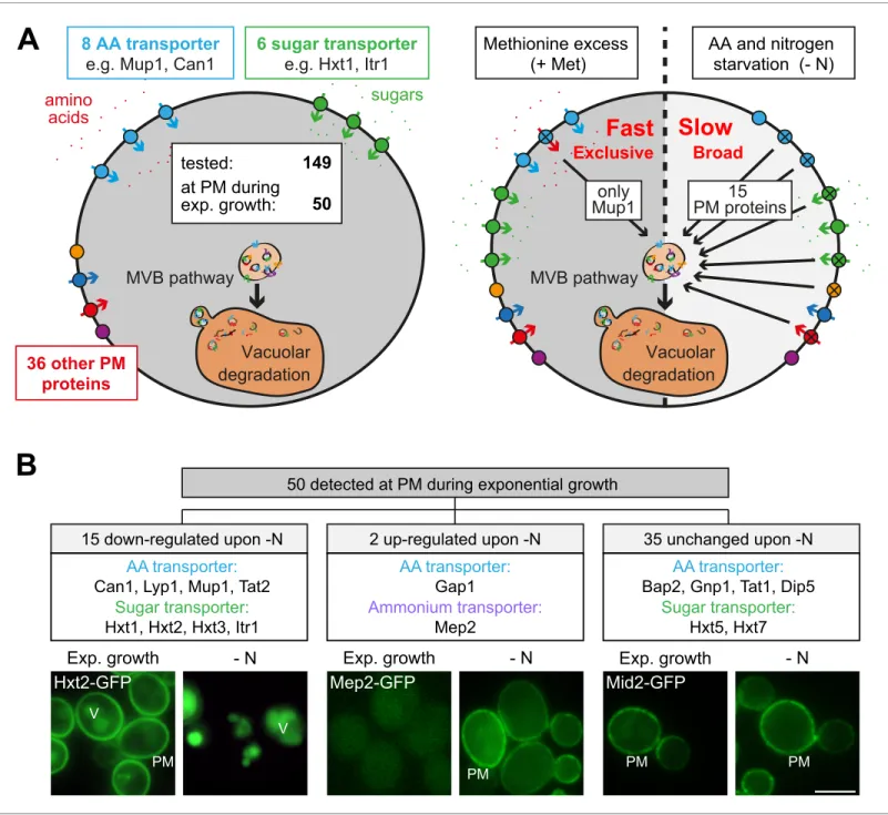
We employed S. cerevisiae as a model system to address how eukaryotic cells adjust their nutrient transporters at the plasma membrane (PM) to nutrient availability. First, we used live cell fluores-cence microscopy to analyze in yeast cells the localization of 149 putative PM proteins that were C-terminally GFP-tagged at their native chromosomal locus (Saier et al., 2016;Babu et al., 2012; Breker et al., 2014;Huh et al., 2003). This collection included 16 different amino acid transporters (AATs) out of the 21 AATs that localize to the PM (Bianchi et al., 2019). In cells growing exponen-tially under defined (rich) conditions, we detected 50 GFP-tagged proteins at the PM, including eight different AATs and six different carbohydrate transporters (Figure 1A,Figure 1—figure sup-plement 1A,Supplementary file 1). A fraction of these proteins was additionally detected inside the vacuole (Figure 1B,Figure 1—figure supplement 1A), suggesting continuous turnover.
Others and we had shown earlier that amino acid and nitrogen starvation (hereafter starvation) induced the degradation of membrane proteins via the MVB pathway (Mu¨ller et al., 2015; Jones et al., 2012). Consistently, in response to starvation 15 (out of 50) different PM proteins were selectively removed and transported into the vacuole, including four AATs (Mup1, Can1, Lyp1, Tat2) and four carbohydrate transporters (Hxt1, Hxt2, Hxt3, Itr1) (Figure 1B,Supplementary file 1). The general amino acid permease Gap1 and the ammonium permease Mep2 were up-regulated and now localized to the PM. The localization of 35 GFP-tagged proteins to the PM remained largely unchanged, although in some instances the vacuolar GFP fluorescence was increased (Figure 1A,B, Figure 1—figure supplement 1A,Supplementary file 1). For five AATs, we did not detect signals under rich or starvation conditions (Supplementary file 1).
Next, we examined how amino acid availability could regulate AAT endocytosis. Therefore, we used the high-affinity methionine transporter Mup1 as a model cargo because its regulation in response to nutrient excess is well characterized (Busto et al., 2018; Lee et al., 2019; Gournas et al., 2018;Guiney et al., 2016;Baile et al., 2019). Moreover, Mup1 is one of the most abundant PM proteins and it is easy to follow its endocytosis and subsequent transport into the vac-uole (Busto et al., 2018). In absence of methionine Mup1-GFP localized to the PM (Figure 1—figure supplement 2A). Low levels of methionine in the growth medium did not efficiently trigger its endo-cytosis (Figure 1—figure supplement 2A,B). Yet, above a critical methionine concentration the vast majority of Mup1-GFP was removed from the PM by endocytosis and was subsequently transported to the vacuole via the multivesicular body (MVB) pathway (Figure 1—figure supplement 2A,B). This was confirmed by western blot (WB) analysis from total cell lysates. Without methionine or with low levels of methionine in the growth medium only full-length Mup1-GFP was detected (Figure 1—fig-ure supplement 2B, lanes 1+2). Once the critical methionine concentration was surpassed, Mup1-GFP was delivered into the vacuole. The proteolytic degradation of Mup1-Mup1-GFP inside the vacuole then released free GFP, which remained stable and could be monitored by western blotting ( Fig-ure 1—figFig-ure supplement 2B, lane 4). Substrate-induced endocytosis was rapid and efficient: within 60–90 min after methionine addition Mup1 was almost quantitatively delivered to the vacuole. Excess methionine appeared to exclusively induce endocytosis of Mup1 since all other tested PM proteins remained at the cell surface (Figure 1A, Supplementary file 1). This exclusive substrate-induced endocytosis of Mup1 was also dependent on its methionine transport activity as shown by using the Mup1-G78N mutant, which cannot transition to the open-inward conformation
B
A
only
Mup1
PM proteins
15
AA and nitrogen
starvation (- N)
Methionine excess
(+ Met)
MVB pathway
Vacuolar
degradation
amino
acids
sugars
MVB pathway
Vacuolar
degradation
tested:
36 other PM
proteins
6 sugar transporter
e.g. Hxt1, Itr1
8 AA transporter
e.g. Mup1, Can1
50
at PM during
exp. growth:
149
Fast
Slow
2 up-regulated upon -N
35 unchanged upon -N
15 down-regulated upon -N
50 detected at PM during exponential growth
AA transporter:
Can1, Lyp1, Mup1, Tat2
Sugar transporter:
Hxt1, Hxt2, Hxt3, Itr1
AA transporter:
Gap1
Ammonium transporter:
Mep2
AA transporter:
Bap2, Gnp1, Tat1, Dip5
Sugar transporter:
Hxt5, Hxt7
Exp. growth
- N
Exp. growth
- N
Exp. growth
- N
Hxt2-GFP
Mep2-GFP
Mid2-GFP
V PM PM PM V PMBroad
Exclusive
Figure 1. Amino acid and nitrogen starvation triggers broad but specific endocytosis and lysosomal degradation of plasma membrane proteins. (A) Left: a library of 149 yeast strains expressing chromosomally GFP-tagged membrane proteins was tested for plasma membrane (PM) localization during nutrient replete exponential growth. Right: verified PM proteins were starved 6–8 hr for amino acids and nitrogen (- N) or treated with 20 mg/ml L-methionine (+Met) after 24 hr of exponential growth. The localization of GFP was assayed by fluorescence microscopy. (B) Summary of the
phenotypes of GFP-tagged PM proteins during starvation. Indicated are numbers of PM proteins that are down-regulated, up-regulated or unchanged compared to the exponential growth phase, each exemplified by one representative strain. PM: plasma membrane; V: vacuole. Scale bars = 5 mm. See alsoFigure 1—figure supplements 1and2andSupplementary file 1.
The online version of this article includes the following figure supplement(s) for figure 1:
Figure supplement 1. Localization of PM proteins during exponential, rich growth and starvation. Figure supplement 2. Characterization of starvation- and substrate-induced endocytosis of Mup1.
(Busto et al., 2018; Figure 1—figure supplement 2C). These results are consistent with earlier reports showing that the high-affinity AATs Mup1, Can1, Tat2 and Lyp1 undergo exclusive endocytic down-regulation in response to excess of their respective substrates but remained stable at the PM when the substrate of another permease was present in excess (Gournas et al., 2017;Busto et al., 2018;Nikko and Pelham, 2009).
In contrast, the starvation-induced endocytosis of Mup1 was independent of its capability to transport its substrate (Figure 1—figure supplement 2C) and was also observed upon starvation for individual essential amino acids (Figure 1—figure supplement 2D). We re-evaluated the starvation-induced endocytosis of Mup1, Can1, Lyp1, Tat2, Ina1, Fur4, Hxt1, Hxt2 and Hxt3 in a different genetic background (SEY6210;Figure 1—figure supplement 2A, Figure 3C,Figure 3—figure sup-plement 1C,D,Figure 6—figure supplement 2A). The overall response was similar.
Hence, in response to starvation, a broad range of nutrient transporters, including four AATs (Mup1, Can1, Lyp1, Tat2) and four carbohydrate transporters (Hxt1, Hxt2, Hxt3, Itr1) as well as other membrane proteins, were selectively removed from the PM by endocytosis and transported into the vacuole within 3–6 hr. In contrast, substrate-induced endocytosis of AATs was quick and exclusive (Figure 1A). Earlier work showed that substrate-induced endocytosis of the AATs Mup1, Can1 and Lyp1 required TORC1 signaling to allow ubiquitination of the transporters by the HECT-type ubiqui-tin ligase Rsp5 in complex with the a-arresubiqui-tin Art1 (Lin et al., 2008; Guiney et al., 2016; Busto et al., 2018;Gournas et al., 2018;MacGurn et al., 2011). Remarkably, the same set of AATs underwent simultaneous starvation-induced endocytosis. The molecular mechanism of their starva-tion-induced endocytosis was not clear.
A genome-wide screen identifies regulators of starvation-induced
endocytosis of AATs
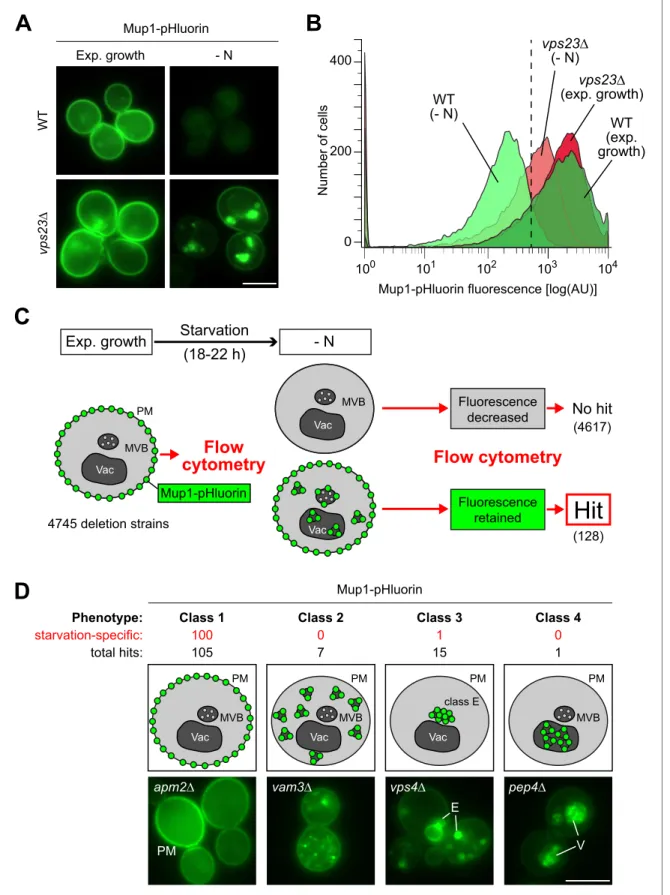
To identify the genes that are required for the starvation-induced endocytosis of Mup1, we con-ducted a fluorescence-based genome-wide screen. We measured Mup1 sorting into the lumen of vacuoles via the MVB pathway by fusing the pH-sensitive GFP variant pHluorin to the C-terminus of Mup1 (Mup1-pHluorin) (Henne et al., 2015; Prosser et al., 2010). Fluorescence microscopy and flow cytometry (Figure 2A,B) showed a strong signal of Mup1-pHluorin at the PM of cells growing under defined rich conditions. In response to starvation, the fluorescence of Mup1-pHluorin was effi-ciently quenched in the acidic vacuoles of wild type (WT) cells, but not when the MVB pathway was blocked (e.g. ESCRT-I mutant vps23D) (Figure 2A,B). Hence Mup1-pHluorin is a suitable reporter to identify mutants that block trafficking of Mup1 from the PM into the vacuole.
Next, Mup1-pHluorin was introduced into the yeast non-essential knock-out collection. 4745 mutants expressing Mup1-pHluorin were grown in selection medium to exponential phase before they were subjected to starvation for 18–22 hr (Figure 2C). Automated flow cytometry was used to measure Mup1-pHluorin fluorescence intensity of at least 15,000 cells before and after starvation. The vast majority of mutants efficiently quenched the fluorescence of Mup1-pHluorin after starva-tion, but 128 mutants still exhibited Mup1-pHluorin fluorescence after starvation (Figure 2C, Supplementary file 2). To determine at which step Mup1-pHluorin transport into vacuoles was blocked in these mutants, we used live cell fluorescence microscopy. This allowed us to classify four phenotypes (Figure 2D,Supplementary file 2). Class one mutants retained Mup1-pHluorin at the PM after starvation (105 mutants, e.g. apm2D mutants). In class two mutants, Mup1-pHluorin was detected on small intracellular objects (seven mutants, e.g. vam3D mutants). Class three mutants accumulated Mup1-pHluorin in larger class E compartment-like objects (15 mutants, e.g. vps4D mutants). One Class four mutant (pep4D) did not efficiently quench Mup1-pHluorin fluorescence inside the vacuole, as it is deficient in the main lysosomal protease. Gene ontology (GO) analysis confirmed that our screen identified major general regulators of endosomal transport (e.g. the ESCRT complexes) and was enriched for proteins that are annotated as components of the PM, endosomes and vacuoles (Figure 2—figure supplement 1,Supplementary file 3) suggesting that it was successful. In addition, we identified many genes and components of protein complexes that would not be predicted to be involved in the regulation of endocytosis (Figure 2—figure supple-ment 1,Supplementary file 2,3).
Next, we aimed to narrow down genes that function specifically during starvation-induced endo-cytosis and to distinguish them from general endocytic regulators (i.e. core components of endocytic trafficking machineries). Therefore, most mutants (124) were exposed to methionine excess for 90
C
A
D
B
WT (exp. growth) WT (- N) vps23∆ (- N) vps23∆ (exp. growth)Mup1-pHluorin fluorescence [log(AU)]
100 101 102 103 104 N u mb e r o f ce lls 0 400 200 Exp. growth W T vp s2 3 ∆ - N Mup1-pHluorin Mup1-pHluorin Class 1 100 105 apm2∆ PM Class 2 0 7 vam3∆ Class 4 0 1 pep4∆ V Class 3 1 15 vps4∆ MVB Vac PM MVB Vac PM class E Vac PM MVB PM E Phenotype: starvation-specific: total hits:
Exp. growth
Starvation
(18-22 h)
Flow
cytometry
- N
MVB Vac MVB Vac PM 4745 deletion strainsFlow cytometry
Fluorescence decreased Fluorescence retained Mup1-pHluorinHit
No hit
Vac (4617) (128)Figure 2. A genome wide screen revealed genes affecting Mup1-pHluorin endocytosis during starvation. (A) Live-cell fluorescence microscopy analysis of WT (BY4742) and vps23D cells expressing MUP1-pHluorin from plasmid and starved (- N) for 18–22 hr. The images exemplify quenched pHluorin fluorescence in vacuoles of wild type (WT)-like cells and retained fluorescence in mutants with defects in the starvation-induced endocytosis of Mup1-pHluorin. (B) The strains from (A) were exponentially grown in 96-well plates for 5 hr and starved (- N) for 18–22 hr. At least 15,000 cells from each strain Figure 2 continued on next page
min and then subjected to live cell fluorescence microscopy. Of the mutants falling into classes 2–4, the majority was required for Mup1 sorting into the vacuole in response to both starvation and methionine excess. Among them were many mutants in subunits of well-characterized protein com-plexes that constituted the core machinery of endo-lysosomal trafficking (Figure 2—figure supple-ment 1, Supplementary file 2, 3). However, the majority (100) of the class one mutants (Mup1-pHluorin retained at the PM) was specifically required for starvation-induced endocytosis but not for methionine-induced endocytosis (Figure 2D,Supplementary file 2).
The a arrestin Art2 is specifically required for amino acid transporter
endocytosis in response to starvation
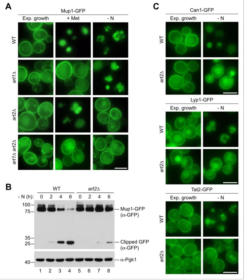
A key step for the endocytic down-regulation of PM proteins is their selective ubiquitination. This selectivity is mediated by a arrestin molecules that direct the HECT-type ubiquitin ligase Rsp5 to ubiquitinate specific PM proteins (Babst, 2020). Among the 14 a arrestins encoded by the yeast genome, our screen identified only Ecm21/Art2 to be specifically required for starvation-induced endocytosis of Mup1.
We compared Art2-dependent starvation-induced endocytosis to Art1-dependent substrate-induced endocytosis of Mup1-GFP. Efficient methionine-substrate-induced endocytosis of Mup1 required Art1 but not Art2 (Figure 3A), consistent with earlier observations (Guiney et al., 2016;MacGurn et al., 2011; Busto et al., 2018). Starvation-induced endocytosis of Mup1 was slower than methionine-induced endocytosis. In WT cells, 240–360 min after onset of starvation the majority of Mup1-GFP was removed from the PM and sorted into vacuoles for degradation (Figure 3A,B,Figure 3—figure supplement 1A). In art2D mutants, but not in art1D mutants, the vast majority of Mup1-GFP remained at the PM and was no longer delivered to vacuoles (Figure 3A). This was confirmed by WB analysis. In WT cells, the levels of full length Mup1-GFP decreased, while free GFP accumulated over time (Figure 3B, lane 2–4; Figure 3—figure supplement 1A), whereas in art2D mutants the majority of Mup1-GFP remained stable and only little free GFP could be detected (Figure 3Blane 7–8;Figure 3—figure supplement 1A). Re-expression of Art1 or Art2 in the corresponding mutants restored methionine- or starvation-induced Mup1 endocytosis, respectively (Figure 3—figure sup-plement 1B). Importantly, Art2 was required for the efficient starvation-induced endocytosis of all four AATs, Mup1, Can1, Lyp1, and Tat2 (Figure 3A,C). Interestingly, the same four AATs (Mup1, Can1, Lyp1 and Tat2) also depend on Art1 for efficient substrate-induced endocytosis (Gournas et al., 2017;MacGurn et al., 2011;Lin et al., 2008;Nikko and Pelham, 2009). Starva-tion-induced endocytosis of Ina1 was also dependent on Art2 (Figure 3—figure supplement 1C), while other PM proteins (e.g. the uracil transporter Fur4 or the hexose transporters Hxt1 and Hxt2) were largely independent of Art2 (Figure 3—figure supplement 1D).
These results suggested a swap between the a arrestins Art1 and Art2 by amino acid availability as a rule for the regulation of four AATs. The starvation-induced endocytosis of Mup1, Can1, Lyp1 and Tat2 required Art2. Substrate-induced endocytosis of Mup1, Can1 and Lyp1 required Art1, whereas in case of Tat2 substrate-induced endocytosis was less stringent and could be mediated either by Art1 or Art2 (Nikko and Pelham, 2009).
Figure 2 continued
and condition were analyzed by flow cytometry. The exemplified histograms display decrease of fluorescence in wild type (WT)-like strains and fluorescence retention in mutants with defects in the starvation-induced endocytosis of Mup1-pHluorin (e.g. vps23D). (C) Workflow of the flow-cytometry-based genome-wide screen for mutants defective in starvation-induced endocytosis of Mup1-pHluorin. (D) Summary of phenotypes of all mutants scored in the starvation-induced endocytosis screen (C) as determined by fluorescence microscopy. Class one mutants retain Mup1-pHluorin fluorescence at the plasma membrane (PM); class two mutants in small cytosolic objects; class three mutants in class E-like objects (E); class four mutants within vacuoles (V). Each phenotype is exemplified by one representative deletion mutant. Indicated are the numbers of strains that share a similar phenotype and the number of hits specific for starvation-induced endocytosis of Mup1-pHluorin (red). Scale bars = 5 mm. See alsoFigure 2— figure supplement 1andSupplementary file 2.
The online version of this article includes the following figure supplement(s) for figure 2:
A
Mup1-GFP
+ Met
Exp. growth
W
T
- N
a
rt
1
∆
a
rt
2
∆
a
rt
1
∆
a
rt
2
∆
B
C
Tat2-GFP
Exp. growth
- N
C
C
C
C
C
C
C
C
C
C
W
T
a
rt
2
∆
C
Can1-GFP
Exp. growth
W
T
a
rt
2
∆
- N
Lyp1-GFP
Exp. growth
- N
W
T
a
rt
2
∆
100
75
35
25
Mup1-GFP
(α-GFP)
0
2
4
6
0
2
4
6
Clipped GFP
(α-GFP)
art2∆
WT
- N (h):
40
α
-Pgk1
1
2
3
4
5
6
7
8
Figure 3. Art1 and Art2 are non-redundant in promoting substrate- and starvation-induced endocytosis of amino acid transporters. (A) Live-cell fluorescence microscopy analysis of Mup1-GFP endocytosis in wild type (WT), art1D, art2D and art1D art2D cells expressing MUP1-GFP from plasmid. Cells were treated with 20 mg/ml L-methionine (+ Met) for 1.5 hr or starved (- N) for 6 hr after 24 hr exponential growth. (B) SDS PAGE and western blot analysis with the indicated antibodies of whole cell protein extracts from wild type (WT) and art2D cells expressing MUP1-GFP that were starved (- N) for Figure 3 continued on next page
Up-regulation of Art2 by the GAAC pathway drives starvation-induced
endocytosis of Mup1
Our experiments so far demonstrated that Art1 and Art2 mediate the endocytic down-regulation of Mup1, Can1 and Lyp1 in response to changes in amino acid availability in a mutually exclusive man-ner. Hence, their activity must be strictly regulated. a arrestin proteins are subject to complex multi-level regulation and their activation can require transcriptional regulation, (de-)phosphorylation and ubiquitination (Hovsepian et al., 2017;Lin et al., 2008;Becuwe et al., 2012b). WB analysis of total cell lysates indicated that 6xHis-TEV-3xFlag-tagged Art1 protein levels were unchanged in response to starvation (Figure 4A, lanes 2,3), whereas the protein levels of the functional 170kD pro-tein Art2-HTF (Figure 4—figure supplement 1A) were up-regulated in response to starvation (Figure 4A, lanes 5,6;Figure 4—figure supplement 1B). Quantitative reverse transcription (RT) PCR analysis indicated that also the ART2 mRNA levels increased during starvation (Figure 4B).
In search of the pathway responsible for induction of Art2, we queried the results of our screen and identified the eIF2 kinase Gcn2 and several more GCN genes encoding key components of the general amino acid control (GAAC) pathway (Supplementary file 2). Gcn2 is activated during amino acid starvation by unloaded tRNAs and subsequently phosphorylates eIF2a to reduce protein syn-thesis in general, but thereby stimulates specifically the translation of the transcription factor Gcn4. Gcn4 then activates the transcription of genes, many of which are involved in amino acid metabolism (Hinnebusch, 2005). gcn4D was not scored in our genome-wide screen because it failed the quality control due to its slow growth phenotype. Interestingly, the promoter of ART2 contained predicted Gcn4-binding sites (Venters et al., 2011;Schuldiner et al., 1998). Disrupting the GAAC pathway (gcn4D) eliminated the induction of Art2 in response to starvation at mRNA and protein levels (Figure 4B,C,Figure 4—figure supplement 1C). Consistently, the starvation-induced endocytosis of Mup1-GFP was hampered in gcn4D cells and several other gcn mutants (Figure 4D,Figure 4—fig-ure supplement 1D). Also, starvation-induced endocytosis of Can1 was dependent on the GAAC pathway (Figure 4—figure supplement 1E). When we introduced mutations in the predicted Gcn4-binding sites in the ART2 promoter, Art2 protein levels no longer increased in response to starva-tion, and starvation-induced endocytosis of Mup1-GFP was impaired (Figure 4E,Figure 4—figure supplement 1F). When the ART2 promoter region was used to replace the ART1 promoter, it also induced the expression of Art1 during starvation (Figure 4—figure supplement 2A). Yet, the upre-gulation of Art1 protein levels driven by the ART2 promoter failed to restore starvation-induced endocytosis of Mup1-GFP in art2D cells (Figure 4—figure supplement 2B). This construct was func-tional for substrate-induced endocytosis of Mup1-GFP in art1D cells (Figure 4—figure supplement 2B).
The expression of a constitutively translated Gcn4C construct (Mueller and Hinnebusch, 1986) increased Art2 protein levels already under rich conditions, as revealed by WB analysis (Figure 4F, compare Art2 protein levels in lanes 1 and 3,Figure 4—figure supplement 1G), and drove unsched-uled Mup1-GFP endocytosis (Figure 4F, lower panel). Consistently, over-expression of Art2 in WT cells or in gcn4D mutants using the strong and constitutively active TDH3 promoter (Figure 4—fig-ure supplement 2C) initiated Mup1-GFP endocytosis already under nutrient replete conditions (Figure 4G) and bypassed the requirement of Gcn4 during starvation.
These results indicated that during amino acid starvation, Art2 transcription was induced via the GAAC pathway, leading to the up-regulation of Art2 protein levels. Moreover, it seemed that the up-regulation of Art2 protein levels was sufficient to drive Mup1 endocytosis. Hence, under amino acid replete conditions Art2 must be tightly repressed to prevent AAT endocytosis.
Figure 3 continued
the indicated times after 24 hr exponential growth. Quantification inFigure 3—figure supplement 1A. (C) Live-cell fluorescence microscopy analysis of wild type (WT) and art2D cells expressing CAN1-GFP, LYP1-GFP or TAT2-GFP. Cells were starved (- N) for 6 hr after 24 hr exponential growth. Scale bars = 5 mm. See alsoFigure 3—figure supplement 1.
The online version of this article includes the following figure supplement(s) for figure 3:
A
C
B
D
F
0 3 WT - N (h): 170 0 3 gcn4∆ 40 Art2-HTF (α-Flag) α-Pgk1 1 2 3 4 0 3 +pART2-ART2 - N (h): 170 0 3 art2∆ 40 Art2-HTF α-Pgk1 +pART2*-ART2 1 2 3 4 *(α-Flag) Mup1-GFP - N Exp. growth W T g cn 4 ∆ Mup1-GFP - N Exp. growth p AR T 2*
-AR T 2 Mup1-GFP - N Exp. growth G C N 4 CE
Mup1-GFP - N Exp. growth g cn 4 ∆ p T D H 3 -AR T 2 p T D H 3 -AR T 2G
1.0 0 3 F o ld ch a n g e AR T 2 mR N A re la ti ve t o PG K1 3.0 2.0 2.5 1.5 0.5 0.0 WT gcn4∆ 0 3 3.5 p = 0.03 p = 0.43 -N (h): (n=11) (n=9) (n=8) (n=7) - N (h): Art2-HTF α-Pgk1 40 100 130 170 0 3 6 0 3 6 Art1-HTF * ART1-HTF ART2-HTF (α-Flag) 1 2 3 4 5 6 0 3 + GCN4 - N (h): 170 0 3 gcn4∆ 40 Art2-HTF (α-Flag) α-Pgk1 + GCN4C 1 2 3 4Figure 4. The general amino acid control pathway promotes starvation-induced endocytosis Mup1 by up-regulating Art2. (A) SDS PAGE and western blot analysis with the indicated antibodies of whole cell protein extracts from WT cells expressing ART1-HTF or ART2-HTF. Cells were starved (- N) for the indicated times after 24 hr exponential growth. The asterisk indicates a non-specific background band of the FLAG antibody. Quantification in
Figure 4—figure supplement 1B. (B) RT-qPCR analysis of ART2 transcript levels (normalized to the stable PGK1 transcript) in wild type (WT) and gcn4D Figure 4 continued on next page
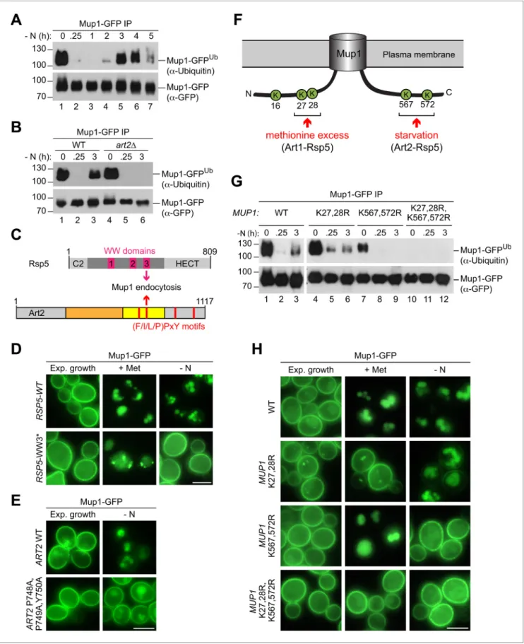
Art2 directs Rsp5-dependent ubiquitination of C-terminal lysine
residues in Mup1
To determine how Art2 contributed to Mup1 endocytosis, we examined its role in Mup1 ubiquitina-tion. WT cells were harvested and Mup1-GFP was immunoprecipitated in denaturing conditions before and at different time points after starvation. Equal amounts of immunoprecipitated full-length Mup1-GFP were subjected to SDS-PAGE and WB analysis to compare the extent of its ubiquitination at different time points (Figure 5A). This analysis indicated that a pool of Mup1 was ubiquitinated prior to the onset of starvation (Figure 5A, lane 1). At the onset of starvation, ubiquitination of Mup1-GFP appeared to decrease for some time (Figure 5Alanes 2–4), until ubiquitination of Mup1-GFP began to increase again after 2–3 hr during starvation (Figure 5A lanes 4–6), temporally coin-ciding with Art2 induction and starvation-induced endocytosis. Mup1-GFP was still ubiquitinated in
art2D cells growing under rich conditions (Figure 5Blane 4) and seemingly de-ubiquitinated at the onset of starvation, but the increase of ubiquitination during starvation was no longer observed (Figure 5B, lane 6). Hence, Art2 was essential for the starvation-induced ubiquitination of Mup1.
a Arrestins use PY motifs to bind to at least one of the three WW domains of Rsp5 (Lin et al., 2008). Starvation-induced endocytosis of Mup1 (but not methionine-induced endocytosis) was par-ticularly dependent on the WW3 domain of Rsp5 (Figure 5C,D,Figure 5—figure supplement 1A). Art2 has four putative PY motifs (Figure 5C). Mutations in the PY motif that resides within the pre-dicted arrestin fold of Art2 (P748,P749,Y750) reduced starvation-induced endocytosis of Mup1-GFP (Figure 5E). The Art2P748A,P749A,Y750A mutant was expressed at similar levels than the WT protein (Figure 5—figure supplement 1B). We suggest that interaction between WW3 in Rsp5 and the PY motif (748-750) of Art2 was required for the efficient starvation-induced endocytosis of Mup1.
To identify lysine residues in Mup1 that were ubiquitinated in response to starvation, Mup1-GFP was immunoprecipitated before and 3 hr after starvation, digested and subjected to liquid chroma-tography-mass-spectrometry (LC-MS) (Figure 5—figure supplement 1C). Two N-terminal lysine resi-dues (K16 and K27) and two C-terminal lysine resiresi-dues (K567 and K572) in Mup1 were found to be ubiquitinated (Figure 5F.Figure 5—figure supplement 1C), consistent with available high-through-put proteomic datasets (Swaney et al., 2013;Iesmantavicius et al., 2014). Earlier reports showed that two N-terminal lysine residues (K27, K28) in Mup1 were ubiquitinated by Art1-Rsp5 and required for methionine-induced endocytosis (Guiney et al., 2016; Busto et al., 2018; Ghaddar et al., 2014;Figure 5F). The role of ubiquitination at C-terminal lysine residues was not clear.
To determine how these N- and C-terminal lysine residues contributed to Mup1 ubiquitination under different growth conditions, we mutated the N-terminal (K27, K28) or C-terminal (K567, K572) lysine residues to arginine and additionally generated a quadruple mutant (K27,28,567,572R). Equal amounts of immunoprecipitated Mup1-GFP, Mup1K27,28R-GFP, Mup1K567,572R-GFP and Mup1K27,28,567,572R-GFP from exponentially growing cells or after starvation or methionine treatment were subjected to SDS-PAGE and WB analysis to compare their ubiquitination (Figure 5G, Fig-ure 5—figFig-ure supplement 1D). Preventing the ubiquitination of the C-terminal lysine residues (K567,572R) abrogated starvation-induced ubiquitination of Mup1 (Figure 5G, lanes 7–9), whereas Mup1K27,28R-GFP was still ubiquitinated after starvation (Figure 5G, lanes 4–6). Upon methionine
Figure 4 continued
cells. Cells were starved (- N) for 3 hr after 24 hr exponential growth. Values are presented as fold-change of the starting values (t = 0). Error bars represent the standard deviation. Statistical significance was assessed by Student’s t-test. (C) SDS PAGE and western blot analysis with the indicated antibodies of whole cell protein extracts from the indicated strains expressing ART2-HTF. Cells were starved (- N) for 3 hr after 24 hr exponential growth. Quantification inFigure 4—figure supplement 1C. (D) Live-cell fluorescence microscopy analysis of the indicated strains expressing MUP1-GFP from plasmid. Cells were starved (- N) for 6 hr after 24 hr exponential growth. (E), (F) The indicated strains were analyzed as in C) (upper panels) and D) (lower panels). Quantification of western blots inFigure 4—figure supplement 1F,G. (G) Live-cell fluorescence microscopy analysis of art2D or gcn4D cells expressing pRS415-MUP1-GFP and pRS416-pTDH3-ART2 starved (- N) for 6 hr after 24 hr exponential growth. Scale bars = 5 mm. See also
Figure 4—figure supplements 1and2.
The online version of this article includes the following figure supplement(s) for figure 4:
Figure supplement 1. The general amino acid control pathway promotes starvation-induced endocytosis Mup1 by up-regulating Art2 – supporting experiments and quantifications.
Figure 5. Art2-Rsp5 mediates the starvation-induced ubiquitination of Mup1-GFP at two specific C-terminal lysine residues. (A), (B) SDS PAGE and western blot analysis with the indicated antibodies of immunoprecipitated Mup1-GFP from WT cells or art2D cells starved for the indicated times after 24 hr of exponential growth. Equal amounts of immunoprecipitated Mup1-GFP were loaded to compare the extent of ubiquitination. (C) Scheme depicting the domain arrangement of Rsp5 and Art2, indicating the localization of the WW domains and PY motifs required for starvation-induced Figure 5 continued on next page
treatment, Mup1K567,572R-GFP ubiquitination was comparable to WT Mup1 (Figure 5—figure sup-plement 1D, lanes 1–4), while the ubiquitination of Mup1K27,28Rwas impaired as reported previously (Figure 5—figure supplement 1D, lanes 5,6) (Guiney et al., 2016; Busto et al., 2018; Ghaddar et al., 2014). The quadruple mutant Mup1K27,28,567,572R-GFP always appeared to be devoid of ubiquitination (Figure 5G, lanes 11,12;Figure 5—figure supplement 1D, lanes 7,8), sug-gesting that the two N-terminal and the two C-terminal lysine residues are the major sites for ubiqui-tination. Consistently, the quadruple lysine mutation rendered Mup1K27,28,567,572R-GFP refractory to methionine- and starvation-induced endocytosis (Figure 5H). Mutations in the N-terminal lysine resi-dues (K27,28R) blocked methionine-induced endocytosis, but not starvation-induced endocytosis. In contrast, starvation-induced endocytosis of Mup1K567,572R-GFP was specifically blocked, but not its methionine-induced endocytosis (Figure 5H).
Collectively, these results suggested that Art2 directs Rsp5 to the ubiquitination of the C-terminal lysine residues K567 and K572 during starvation, whereas Art1-Rsp5 complexes ubiquitinated Mup1 on the two N-terminal lysine residues K27 and K28 in response to methionine excess (Figure 5F).
The C-terminus of Mup1 contains an acidic sorting signal for Art2
In addition to ubiquitination, our mass-spectrometry analysis of immunoprecipitated Mup1-GFP from starved cells identified potential phosphorylation sites at the N-terminus (S9,23,31,42 and T34) and at the C-terminus of Mup1 (T552 and S568,573) (Figure 6A,Figure 5—figure supplement 1C). Additional phosphorylation on T560 was reported in other studies (Swaney et al., 2013; Iesmantavicius et al., 2014). Of note, the C-terminal phosphorylation sites were proximal (T552, T560) or directly adjacent (S568, S573) to lysine residues K567 and K572 that were ubiquitinated in an Art2-dependent manner in response to starvation (Figure 6A,Figure 5—figure supplement 1C). Mutation of the C-terminal threonine residues to alanine (T552,560A) to prevent phosphorylation specifically blocked starvation-induced endocytosis of Mup1T552,560A-GFP, but not methionine-induced endocytosis (Figure 5—figure supplement 1E). Mutation of these phosphorylation sites also prevented ubiquitination of Mup1 during starvation (Figure 5—figure supplement 1F). Block-ing the phosphorylation of S568 and S573 also impaired starvation-induced endocytosis of Mup1S568,573A-GFP (Figure 5—figure supplement 1E), yet starvation-induced ubiquitination could still be detected (Figure 5—figure supplement 1F). Mutation of most N-terminal serine and threo-nine residues, including all experimentally determined phosphorylation sites, to alathreo-nine (T6,25,26,34A,S9,22,23,24,31,33,42A) did not affect starvation-induced endocytosis of Mup1 ( Fig-ure 5—figFig-ure supplement 1G). It seemed that the phosphorylation of C-terminal threonine residues (T552, 560) contributed to the starvation-induced Art2-Rsp5 ubiquitination of Mup1 and thus might add an additional layer of regulation, similar to the recognition of GPCRs by b–arrestins in mamma-lian cells (Shukla et al., 2014;Nikko et al., 2008;Marchal et al., 2000;Hicke et al., 1998).
An extended acidic Art1 sorting motif at the N-terminus of Mup1 is required for ubiquitination of K27,28 and subsequent Mup1 endocytosis in response to methionine excess (Guiney et al., 2016; Busto et al., 2018). Consistent with our model involving two distinct mechanisms for Mup1 endocy-tosis, mutations in the acidic N-terminal Art1 sorting motif of Mup1 (D43A,G47A,Q49A,T52A,L54A) blocked Art1-Rsp5-dependent methionine-induced endocytosis, but not Art2-Rsp5-dependent star-vation-induced endocytosis (Figure 6—figure supplement 1A).
Figure 5 continued
endocytosis of Mup1. (D) Live-cell fluorescence microscopy analysis of rsp5D cells expressing pRS416-MUP1-GFP and pRS415-HTF-RSP5-WT (wild type) or pRS415-HTF-RSP5-WW3*. Cells were treated with 20 mg/ml L-methionine (+ Met) for 1.5 hr or starved (- N) for 6 hr after 24 hr exponential growth. (E) Live-cell fluorescence microscopy analysis of art2D cells expressing MUP1-GFP and pRS416-ART2 (WT) or pRS416-ART2 P748A,P749A,Y750A. Cells were starved (- N) for 6 hr after 24 hr exponential growth. (F) Scheme of Mup1 topology with the N- and C-terminal ubiquitination sites targeted during substrate excess by Art1-Rsp5 and during starvation by Art2-Rsp5. Ubiquitinated lysines (K) shown in green with numbers corresponding to amino acid positions in the Mup1 sequence. (G) WT cells expressing MUP1-GFP WT or the indicated MUP1-GFP mutants starved for the indicated times after 24 hr of exponential growth analyzed as in B). (H) Live-cell fluorescence microscopy analysis of cells expressing MUP1-GFP (wild type (WT)), MUP1 K27,28R-GFP, MUP1 K567,572R-GFP or MUP1 K27,28,567,572R-GFP as in D). Scale bars = 5 mm. See alsoFigure 5—figure supplement 1.
The online version of this article includes the following figure supplement(s) for figure 5:
The C-terminus of Mup1 also contains an acidic patch (D549-D555), close to the ubiquitination sites K567 and K572 and the C-terminal threonine phosphorylation sites (T552, 560) involved in star-vation-induced endocytosis (Figure 6A,B). Mutation of the acidic residues in this region to basic amino acids (R) demonstrated that this C-terminal acidic region was specifically required for starva-tion-induced endocytosis. Live cell fluorescence microscopy revealed that Mup1D549R,D551R,E554R, D555R
-GFP remained at the PM in response to starvation, whereas methionine-induced endocytosis was not impaired (Figure 6B). Even Art2 overexpression failed to induce endocytosis during expo-nential growth or starvation when the C-terminal acidic patch in Mup1 was mutated (Figure 6—fig-ure supplement 1B). Moreover, immunoprecipitation of Mup1D549R,D551R,E554R,D555R-GFP and subsequent SDS-PAGE and WB analysis revealed that it was no longer efficiently ubiquitinated (Figure 6C, lanes 4–6), suggesting that the C-terminal acidic patch was essential for the Art2-Rsp5-dependent ubiquitination during starvation.
Comparing the amino acid sequences of the C-terminal tails of the four Art2-dependent AATs (Mup1, Can1, Tat2 and Lyp1) and of Ina1 indicated similar acidic patches (Figure 6D). To analyze if the acidic patch in Can1 also contributed to starvation-induced endocytosis, we mutated D567, E569, E574 and E575 to arginine. Mutant Can1D567R,E569R,E574R,D575R-GFP mostly localized to the PM under growing conditions. Importantly, the Art2-dependent starvation-induced down-regulation of Can1D567R,E569R,E574R,D575R-GFP was impaired (Figure 6—figure supplement 1C). These results imply that Mup1, Can1 and potentially also Lyp1, Tat2 and Ina1 have acidic amino acid sequences at their C-termini that could serve as sorting signal for Art2-mediated starvation-induced endocytosis.
The C-terminal acidic sorting signal of Mup1 is sufficient for
Art2-dependent starvation-induced endocytosis
It seemed that the last 26 amino acid residues (aa 549–574) of Mup1 harbor three features that are collectively required specifically for starvation-induced endocytosis: putative phosphorylation sites, the acidic patch and the ubiquitination sites. Hence, we tested if the C-terminal region of Mup1 was sufficient to convert an Art2-independent cargo into an Art2 cargo. We selected the low-affinity glu-cose transporter Hxt3, which was efficiently removed from the PM in response to starvation ( Fig-ure 6—figFig-ure supplement 2A,Figure 1B,Supplementary file 1). Live cell fluorescence microscopy and WB analysis showed that starvation-induced endocytosis of Hxt3-GFP was independent of Art2, but instead required Art4 (Figure 6E,Figure 6—figure supplement 2A). In art4D mutants, but not in art2D mutants, Hxt3-GFP remained mostly at the PM (Figure 6—figure supplement 2A) in response to starvation, and its vacuolar degradation was impaired (Figure 6—figure supplement 2B, lane 4, Figure 6—figure supplement 2C). However, when the C-terminal 30 amino acids of Mup1 (aa 545–574) were fused onto the C-terminus of Hxt3 (Figure 6E), they restored starvation-induced endocytosis in art4D mutants. This Hxt3-Mup1-C-GFP chimera was now efficiently removed from the PM and transported to the vacuole in the vast majority of art4D cells (86%, n = 76 cells) (Figure 6E), most likely because it became an Art2 substrate. Indeed, the additional deletion of
Figure 6. The C-terminus of Mup1 harbors a transplantable, starvation-responsive acidic degron. (A) Left: scheme of Mup1 topology with N- and C-terminal ubiquitination sites and acidic patches targeted by Art1-Rsp5 and Art2-Rsp5, respectively, and the C-terminal phosphorylation sites of Mup1 that promote its starvation-induced endocytosis. Ubiquitinated lysines (K) shown in green and phosphorylated serines (S) and threonines (T) in red with numbers corresponding to amino acid positions in the Mup1 sequence. Right: Hxt3 as an Art4-Rsp5 dependent cargo during nitrogen starvation. (B) Live-cell fluorescence microscopy analysis of Mup1-GFP endocytosis in cells expressing MUP1-GFP (wild type (WT)) or MUP1 D549R,D551R,E554R, D555R-GFP. Cells were treated with 20 mg/ml L-methionine (+ Met) for 1.5 hr or starved (- N) for 6 hr after 24 hr exponential growth. (C) SDS PAGE and western blot analysis with the indicated antibodies of immunoprecipitated Mup1-GFP from cells expressing MUP1-GFP (WT) or MUP1 D549R,D551R, E554R,D555R-GFP starved for the indicated times after 24 hr of exponential growth. Equal amounts of immunoprecipitated Mup1-GFP were loaded to compare the extent of ubiquitination. (D) Amino acid sequence alignment of the C-terminal acidic patches of Mup1, Can1, Lyp1, Tat2 and Ina1. The boxes indicate acidic residues (red) and phosphorylation sites (blue), which are required for Art2-dependent starvation-induced endocytosis. Red letters illustrate further acidic residues. (E) Live-cell fluorescence microscopy analysis of wild type (WT), art4D and art2D art4D cells expressing HXT3-GFP (top), HXT3-MUP1-C-GFP (second row), HXT3-MUP1-C K567,572R-GFP (third row) or HXT3-MUP1-C D549R,D551R,E554R,D555R-GFP (bottom). Cells were starved (- N) for 6 hr after 24 hr exponential growth. Scale bars = 5 mm. See alsoFigure 6—figure supplements 1and2.
The online version of this article includes the following figure supplement(s) for figure 6: Figure supplement 1. The role of acidic patches in AATs for starvation-induced endocytosis. Figure supplement 2. Art4-dependent starvation-induced endocytosis of Hxt3.
ART2 (art2D art4D) blocked starvation-induced endocytosis and demonstrated that Art2 now
medi-ated the degradation of Hxt3-Mup1-C-GFP in art4D mutants. The degradation of Hxt3-Mup1-C-GFP and the concomitant accumulation of free GFP were further examined by SDS-PAGE and WB analy-sis from total cell extracts (Figure 6—figure supplement 2D, E). This analysis showed that the majority of the full-length Hxt3-Mup1-C-GFP chimera was degraded in WT cells, art2D and art4D mutants during starvation (Figure 6—figure supplement 2D, lanes 2, 4, 8), but only poorly in art2D
art4D double mutants (Figure 6—figure supplement 2D, lane 6,Figure 6—figure supplement 2E). The Art2-dependent endocytosis of the Hxt3-Mup1-C-GFP chimera required two key features provided by the C-terminus of Mup1 (the acidic patch and the two C-terminal lysine residues), since in art4D cells starvation-induced endocytosis of Hxt3-Mup1-CK567,572R-GFP and Hxt3-Mup1-CD549R, D551R,E554R,D555R
-GFP was blocked (Figure 6E).
Taken together, these results demonstrate that the C-terminus of Mup1 (aa 545–574) encodes a portable acidic sorting signal that can be recognized by Art2 and directs Rsp5 to ubiquitinate specif-ically two proximal lysine residues to promote starvation-induced endocytosis.
A basic patch of Art2 is required for starvation-induced degradation of
Mup1
After having defined that the C-terminus of Mup1 (and possibly also the C-terminus of further Art2-targets) provides a degron sequence for Art2-Rsp5 complexes, we addressed how it could be recog-nized. Upon inspection of the predicted arrestin domain in Art2, we noted a stretch of positively charged residues within the arrestin-C domain (Figure 7A). Converting these basic residues into an acidic patch (Art2K664D,R665D,R666D,K667D) abolished starvation-induced endocytosis of Mup1 (Figure 7B). Western blot analysis of total cell lysates showed that the Art2 basic patch mutant pro-tein was expressed at similar levels as WT Art2 and was also upregulated after 3 hr of starvation ( Fig-ure 7—figFig-ure supplement 1A, lane 6). In addition, the Art2 basic patch mutant also impaired, at least partially, starvation-induced endocytosis of Can1, Lyp1 and Ina1, while the endocytosis of Tat2 was independent of the basic patch (Figure 7B,Figure 7—figure supplement 1B,C).
Overall, it seemed that Art2 employed a positively charged region in its arrestin fold to recognize C-terminal acidic patches in at least three different AATs (Mup1, Can1, Lyp1) and thus mediate their endocytosis in starvation conditions.
Discussion
We have made progress toward understanding how yeast cells selectively re-configure their reper-toire of transporters at the PM in response to their nutritional status. The model inFigure 7C pro-vides the conceptual framework for the mutually exclusive but complementary action of Art1-Rsp5 and Art2-Rsp5 ubiquitin ligase complexes for mediating the selective endocytosis of the methionine transporter Mup1 in response to changes in amino acid availability. Similar concepts may apply for the arginine transporter Can1, the lysine transporter Lyp1 and the tryptophan and tyrosine trans-porter Tat2. Based on our results and work from others, the endocytic down-regulation of these AATs can be described by the following set of rules: (1) Reciprocal (de-)activation of the a arrestins Art1 and Art2. (2) Art1 or Art2 each recognize different acidic sorting signals on their client AATs that may require additional phosphorylation. (3) To do so, they employ basic patches in their extended arrestin C-domains and defined PY motifs to orient Rsp5 with high specificity toward prox-imal lysine residues. These rules satisfy the plasticity required for different a arrestin and AAT inter-actions that drive exclusive or relatively broad substrate specificity depending on the metabolic context.
While both Art1 and Art2 lead to the degradation of AATs, they answer to distinct metabolic cues and are thus wired into distinct signaling pathways. Activation of Art1 by amino acid influx requires the coordinated interplay of TORC1 signaling to inactivate Npr1 (a kinase that negatively regulates Art1) and the action of phosphatases (Gournas et al., 2017; Lee et al., 2019; MacGurn et al., 2011;Tumolo et al., 2020). In response to amino acid limitation, TORC1 is no lon-ger active. This will activate Npr1 to phosphorylate Art1, thereby inactivating it. At the same time, the lack of amino acids will activate the eIF2a kinase Gcn2. Gcn2 will phosphorylate eIF2a, which leads to the global down-regulation of translation, but enables specific translation of the transcrip-tion factor Gcn4 (Hinnebusch, 2005). Gcn4 then induces transcription of genes required for amino
A
B
18 6 23 8 31 9 38 8 44 2 47 8 55 4 70 0 82 3 87 7 N C PY 659-LDFASKRRKERSVS-672 ~~~~~rr~keR~l~ Art2 sequence: HH-Pred consensus: basic patch Art2/Ecm21 (1117 aa) Arrestin-N Arrestin-CC
AR T 2 a rt 2 ∆ AR T 2 K6 6 4 D ,R 6 6 5 D , R 6 6 6 D ,K6 6 7 D Mup1-GFP Exp. growth - N Lyp1-GFP Exp. growth - N Can1-GFP Exp. growth - N K K 567 572 C Plasma membrane Mup1S S 568 573 T 552 acidic patch 2 T 560 K K 27 28 acidic patch 1 Cytoplasm Extracellular space N
Art
1
Rsp
5
Art
2
Rsp
5
Endocytosis
Lysosomal degradation
(Can1, Lyp1)TORC1
Npr1
ART2 TGACTCGcn4
Starvation
Nutrient
excess
GAAC
Nutrient sufficiencyPpz
Figure 7. A basic patch of Art2 promotes the starvation-induced endocytosis of Mup1, Can1 and Lyp1. (A) Scheme of Art2 topology with arrestin-N domain in orange, arrestin-C domain in yellow and tails and interspersed extended loops (Baile et al., 2019) in light grey. The basic amino acid residues shown in blue mediate the starvation-induced endocytosis of Mup1, Can1 and Lyp1 (numbers correspond to amino acid positions in the Art2 sequence). Below is aligned the HHpred consensus sequence for Art2/Ecm21 derived from three HHblits iterations (Zimmermann et al., 2018), Figure 7 continued on next page
acid biosynthesis and of ART2, which causes an increase in Art2 protein levels and thus formation of Art2-Rsp5 complexes. This appears as primary means to activate Art2, since unscheduled increase in Art2 protein levels was sufficient to drive Art2-dependent nutrient transporter endocytosis already in cells growing under rich conditions. When amino acids become available again, the system can effi-ciently reset. TORC1 is reactivated resulting in Art1 reactivation. Conversely, Gcn4 will become insta-ble and rapidly degraded by the ubiquitin proteasome system (Kornitzer et al., 1994; Meimoun et al., 2000; Irniger and Braus, 2003), and thus, the transcription of ART2 will cease. Interestingly, two de-ubiquitinating enzymes (Ubp2, Ubp15) de-ubiquitinate Art2 to influence its pro-tein stability (Ho et al., 2017;Kee et al., 2006). Inhibiting their activity could provide additional con-trol to repress Art2-dependent endocytosis in cells growing under rich conditions. Our screen identified also two de-ubiquinating enzymes, Doa4 and Ubp6 to be specifically required for starva-tion-induced endocytosis of Mup1. They could act directly on Art2 or Mup1 or help to maintain homeostasis of the ubiquitin pool during starvation.
Art2 is subject to extensive post-translational modification, including ubiquitination and phos-phorylation. Database searches and our own proteomic experiments identified 68 phosphorylation sites and 20 ubiquitination sites in Art2 (data not shown) (Swaney et al., 2013;Albuquerque et al., 2008;Holt et al., 2009). How these modifications help to control the activity of Art2 remains a com-plex and open questions. Several arrestins were found to be phospho-inhibited in specific conditions (MacGurn et al., 2011; Becuwe et al., 2012b; O’Donnell et al., 2013;Hovsepian et al., 2017; Merhi and Andre´, 2012;Llopis-Torregrosa et al., 2016), the common molecular basis of which is unknown. An exciting hypothesis would be that a-arrestin hyper-phosphorylation would add nega-tive charges, and thereby prevent the recognition of acidic patches on transporters through electro-static repulsion. Interestingly, our screen identified the pleitropic type 2A-related serine-threonine phosphatase Sit4 as a class one hit. Hence, Sit4 may be linked directly or indirectly to de-phosphory-lation of Art2 and controlling its activity as reported recently for the Art2-dependent regude-phosphory-lation of vitamin B1 transporters (Savocco et al., 2019).
Through the complementary activation of Art1 and Art2, cells can coordinate amino acid uptake through at least four high-affinity amino acid transporters with amino acid availability. The regulation of hexose transporters by glucose availability appears to be conceptually related, with distinct a arrestin-Rsp5 complexes in charge of down-regulating the same transporters at various glucose concentrations with distinct mechanisms and kinetics (Hovsepian et al., 2017;Nikko and Pelham, 2009). In particular, the endocytosis of high-affinity hexose transporters during glucose starvation involves Art8, the closest paralogue of Art2, whose expression is also controlled by nutrient-regu-lated transcription (Hovsepian et al., 2017). Altogether, a picture emerges in which the transcrip-tional control of a arrestin expression by nutrient-signaling pathways is critical to cope with nutrient depletion. This becomes part of the complex regulation of the ART-Rsp5 ubiquitin ligase network.
Our work also extends on previous findings regarding the determinants of a arrestin/transporter interaction, indicating communalities between starvation- and substrate-induced endocytosis. Art1-Rsp5 and Art2-Art1-Rsp5 complexes both recognize specific acidic sequences on Mup1 (Figure 7C). Acti-vated Art1-Rsp5 complexes recognize an acidic stretch in the N-terminus of Mup1 and Can1, close to the first transmembrane domain. The exposure of these N-terminal acidic patches is linked to
Figure 7 continued
suggesting conservation of the basic patch in the Art2 protein family. (B) Live-cell fluorescence microscopy analysis of art2D cells expressing MUP1-GFP, CAN1-GFP or LYP1-GFP and pRS416-ART2-WT, empty vector or pRS416-ART2 K664D,R665D,R666D,K667D. Cells were starved (- N) for 6 hr after 24 hr exponential growth. (C) Scheme for the regulation of the substrate- and starvation-induced endocytosis of Mup1. During substrate excess TORC1 inhibits the Npr1 kinase which otherwise would phosphorylate and inhibit Art1. Art1-Rsp5 becomes dephosphorylated and subsequently binds the acidic patch 1 at the N-terminal tail of Mup1, leading to the ubiquitination of K27 and K28 and degradation of Mup1 (left). During amino acid and nitrogen starvation, the general amino acid control (GAAC) pathway upregulates the ubiquitin ligase adaptor Art2 via the transcriptional regulator Gcn4. The ensuing Art2-Rsp5 complex binds with its basic patch to the acidic patch 2 of Mup1 leading to the ubiquitination of K567 and K572 and degradation of Mup1 (right). At the same time, starvation causes TORC1 inhibition and activation of Npr1 and inhibits Art1-dependent ubiquitination. Ubiquitinated lysines (K) shown in green and phosphorylated serines (S) and threonines (T) in red with numbers corresponding to amino acid positions in the Mup1 sequence. Scale bars = 5 mm. See alsoFigure 7—figure supplement 1.
The online version of this article includes the following figure supplement(s) for figure 7: Figure supplement 1. Additional characterization of the basic patch mutant of Art2.
substrate transport and conformational transitions in the transporters from the outward-open to the inward-open conformation, which in Mup1 also includes the so-called ‘C-plug’ (aa 520–543) (Busto et al., 2018;Guiney et al., 2016;Gournas et al., 2017). This conformational switch drives lateral re-localization of Mup1 and Can1 into a disperse PM compartment, where they are ubiquiti-nated by Art1-Rsp5 (Gournas et al., 2018;Busto et al., 2018). Art2 recognizes specifically an acidic patch in the C-terminal tail of Mup1, and thereby directs Rsp5 to ubiquitinate two juxtaposed C-ter-minal lysine residues. The C-plug is very close to the C-terC-ter-minal acidic patch but is not part of the C-terminal Mup1 degron. We speculate that in the absence of nutrients AATs will spend more time in the outward open state with the C-Plug in place. In this state, activated Art2-Rsp5 complexes can still engage the C-terminal acidic patches. Hence, toggling Art1/Art2 activation in combination with accessibility of N- or C-terminal acidic sorting signals in AATs, in part regulated by their conforma-tional state, must fall together to allow selective endocytosis.
An additional layer of regulation for endocytosis is provided by phosphorylation of AATs close to the acidic sorting signal. At the moment, we can only speculate about the kinase responsible for the phosphorylation of the C-terminal serine or threonine sites of Mup1. Perhaps, constitutive PM-asso-ciated kinases such as the yeast casein kinase one pair (Yck1/2) are involved, which are known to rec-ognize rather acidic target sequences and to regulate endocytosis (Hicke et al., 1998;Paiva et al., 2009;Nikko et al., 2008;Marchal et al., 2002).
a-Arrestins lack the polar core in the arrestin domain that is used for cargo interactions in b-arrestins (Aubry et al., 2009;Polekhina et al., 2013). Instead Art1-Rsp5 and Art2-Rsp5 complexes each use a basic region in their arrestin C-domain to detect the acidic sorting signal in their client AATs. Studies on the interaction between GPCRs and b-arrestins revealed a multimodal network of flexible interactions: The N-domain of b-arrestin interacts with phosphorylated regions of the GPCR, their finger loop inserts into the transmembrane domain bundle of the GPCR and loops at the C-ter-minal edge of b-arrestin engage the membrane (Staus et al., 2020;Huang et al., 2020). Perhaps, a similar concept also holds true for a-arrestins. This is not unlikely given that their arrestin fold appears to be interspersed with disordered loops and very long, probably unstructured N- and/or C-terminal tails, some of which participate in cargo recognition or membrane interactions (Baile et al., 2019).
Despite the possible plasticity in substrate interactions, the selectivity of Art1-Rsp5 and Art2-Rsp5 complexes in ubiquitinating lysine residues proximal to the acidic patches of Mup1 is remarkable. Mup1 has 19 lysine residues at the cytoplasmic side: four at the N-terminal tail, six in the C-terminal tail and nine in the intracellular loops of the pore domain. Yet, Art1-Rsp5 complexes only ubiquiti-nate K27 and K28, whereas Art2-Rsp5 complexes only ubiquitiubiquiti-nate K567 and K572. Even in the Hxt3-Mup1-C chimeric protein, Art2-Rsp5 complexes ubiquitinated only the lysine residues close to the acidic patch, despite six further lysine residues in the directly adjacent C-terminal tail of Hxt3. How is this possible? We speculate that these two a-arrestin-Rsp5 complexes orient the HECT domain of Rsp5 with high precision toward the lysine residues that are spatially close to the acidic patches. Once ubiquitinated, the AAT can engage the endocytic machinery to be removed from the PM.
In conclusion, Art1-Rsp5 complexes act rapidly to prevent the accumulation of excess amino acids, whereas the Art2-Rsp5 complexes help to degrade idle high-affinity amino acid transporters over longer periods of starvation to recycle their amino acid content. Starvation-induced endocytosis and the subsequent degradation of membrane proteins is required to maintain intracellular amino acid homeostasis (Mu¨ller et al., 2015;Jones et al., 2012). As such, it is well suited that Art2 activity and thus starvation-induced endocytosis of AATs is co-regulated and coordinated with de novo amino acid biosynthesis via the GAAC pathway. The down-regulation of AATs together with glucose transporters and further PM proteins could also free up domains at the PM that are populated by selective nutrient transporters (Spira et al., 2012; Grossmann et al., 2008) for transporters with broader substrate specificity such as the general amino acid permease Gap1 and the ammonium transporter Mep2, which are strongly up-regulated during starvation. Hence starvation-induced endocytosis could prepare cells – anticipatory – for non-selective nutrient acquisition, as soon as nutrients become available again. Consistent with this notion and a role of Art2 during starvation, yeast cells with mutations in ART2 show an altered adaptation to repetitive cycles of starvation (Wloch-Salamon et al., 2017). However, at the moment, it is not clear how these physiological defects are linked to the role of Art2 in starvation-induced endocytosis. Hence, despite the
emergence of regulatory mechanisms, we are still far from understanding how each individual ART-Rsp5 complex controls nutrient acquisition and how this process translates into fine-tuning of metab-olism, cell growth and survival. Even more unclear is how the activities of the entire ART-Rsp5 net-work are harmonized to achieve a coordinated cellular response to nutrient availability. Since a more global disruption of the ART-Rsp5 network causes proteotoxic stress with strong growth defects (Nikko and Pelham, 2009;Zhao et al., 2013;Hein et al., 1995), addressing these important ques-tions will require new approaches.
Altogether, our results provide a better understanding of the molecular mechanisms that couple metabolic signaling and nutrient availability to nutrient transporter endocytosis in budding yeast. This might provide basic insights and principles for the vastly more complex regulation of the nutri-ent transporter repertoire in human cells under normal as well as pathophysiological conditions.
Materials and methods
Key resources table Reagent type (species) or resource Designation Source or reference Identifiers Additional information Strain, strainbackground Saccharomyces cerevisiae SEY6210 Robinson et al., 1988
SEY6210 Parental yeast strain (genotype: MATa leu2-3,112 ura3-52 his3-D200 trp1-D901 suc2-D9 lys2-801 GAL), obtained from the Emr lab Strain, strain
background Saccharomyces cerevisiae
SEY6210.1 Robinson et al., 1988
SEY6210.1 Parental yeast strain (genotype: MATa leu2-3, 112 ura3-52 his3-D200 trp1-D901
suc2-D9 lys2-801 GAL), obtained from the Emr lab Strain, strain background Saccharomyces cerevisiae SEY6210 rsp5D::HIS3 pRS415-6xHIS-TEV-3xFLAG-RSP5
This paper DTY557 RSP5 wild
type yeast strain, obtained from
the Emr lab (previously unpublished) Strain, strain background Saccharomyces cerevisiae SEY6210 rsp5D::HIS3 pRS415- 6xHIS-TEV- 3xFLAG-RSP5-WW1*
This paper DTY558 RSP5 mutant yeast strain,
obtained from the Emr lab (previously unpublished) Strain, strain background Saccharomyces cerevisiae SEY6210 rsp5D::HIS3 pRS415-6xHIS- TEV-3xFLAG-RSP5-WW2*
This paper DTY559 RSP5 mutant yeast strain, obtained from
the Emr lab (previously unpublished) Strain, strain background Saccharomyces cerevisiae SEY6210 rsp5D::HIS3 pRS415-6xHIS- TEV-3xFLAG-RSP5-WW3*
This paper DTY560 RSP5 mutant yeast
strain, obtained from
the Emr lab (previously unpublished)