Fatty Acid Phytyl Ester Synthesis in Chloroplasts
of Arabidopsis
WFelix Lippold,
a,1,2Katharina vom Dorp,
a,1Marion Abraham,
b,3Georg Hölzl,
aVera Wewer,
aJenny Lindberg Yilmaz,
cIda Lager,
dCyrille Montandon,
eCéline Besagni,
eFelix Kessler,
eSten Stymne,
dand Peter Dörmann
a,4aInstitute of Molecular Physiology and Biotechnology of Plants, University of Bonn, 53115 Bonn, Germany bMax Planck Institute of Molecular Plant Physiology, 14476 Potsdam, Germany
cScandinavian Biotechnology Research, S-23053 Alnarp, Sweden
dSwedish University of Agricultural Sciences, Department of Plant Breeding and Biotechnology, S-23053 Alnarp, Sweden eLaboratory of Plant Physiology, Institute of Biology, University of Neuchâtel, 2000 Neuchatel, Switzerland
During stress or senescence, thylakoid membranes in chloroplasts are disintegrated, and chlorophyll and galactolipid are broken down, resulting in the accumulation of toxic intermediates, i.e., tetrapyrroles, free phytol, and free fatty acids. Chlorophyll degradation has been studied in detail, but the catabolic pathways for phytol and fatty acids remain unclear. A large proportion of phytol and fatty acids is converted into fatty acid phytyl esters and triacylglycerol during stress or senescence in chloroplasts. We isolated two genes (PHYTYL ESTER SYNTHASE1 [PES1] and PES2) of the esterase/lipase/ thioesterase family of acyltransferases from Arabidopsis thaliana that are involved in fatty acid phytyl ester synthesis in chloroplasts. The two proteins are highly expressed during senescence and nitrogen deprivation. Heterologous expression in yeast revealed that PES1 and PES2 have phytyl ester synthesis and diacylglycerol acyltransferase activities. The enzymes show broad substrate specificities and can employ acyl-CoAs, acyl carrier proteins, and galactolipids as acyl donors. Double mutant plants (pes1 pes2) grow normally but show reduced phytyl ester and triacylglycerol accumulation. These results demonstrate that PES1 and PES2 are involved in the deposition of free phytol and free fatty acids in the form of phytyl esters in chloroplasts, a process involved in maintaining the integrity of the photosynthetic membrane during abiotic stress and senescence.
INTRODUCTION
Chloroplasts are characterized by the presence of an intricate membrane system, the thylakoids, which contain a unique set of lipids and harbor the photosynthetic pigment protein complexes (Joyard et al., 1998). As semiautonomous organelles, chlo-roplasts contain the enzymes of thefinal biosynthetic steps for chloroplast lipids and carry the entire set of enzymes required for de novo fatty acid synthesis (Douce, 1974; Ohlrogge et al., 1979; Soll et al., 1985; Beale, 1999). During leaf senescence, thylakoid membranes are disintegrated and the pigment-protein complexes of photosynthesis are disassembled. Chlorophyll and galactolipids are degraded (Harris and Arnott, 1973; Hörtensteiner, 2006), accompanied by the accumulation of to-copherol (vitamin E) and triacylglycerols (TAGs). During senes-cence, the number and sizes of plastoglobules, lipid protein particles localized to the stroma of chloroplasts, increase
(Tuquet and Newman, 1980; Zbierzak et al., 2010). Proteomic studies revealed the presence of a large number of structural proteins and biosynthetic enzymes in these lipid-protein par-ticles (Vidi et al., 2006; Ytterberg et al., 2006; Bréhélin et al., 2007). Plastoglobules contain different nonpolar lipids, including tocopherol and TAG (Tevini and Steinmüller, 1985; Vidi et al., 2006), and are believed to be surrounded by a galactolipid monolayer membrane (Austin et al., 2006). In this regard, plas-toglobules are structurally similar to oil bodies in the cytosol of the plant cell, as oil bodies provide storage capacity for non-polar lipids, in particular TAG. TAG has also been shown to rapidly increase after exposure of the leaves to ozone, freezing conditions, osmotic or salt stress, or drought (Sakaki et al., 1990; Kaup et al., 2002; Moellering et al., 2010).
The chlorophyll molecule consists of a tetrapyrrole ring with a central magnesium cation and an ester-linked, hydrophobic side chain, an isoprenoid alcohol that was designated phytol (Willstätter and Hocheder, 1907; Fleming, 1967). During chlo-rophyll degradation, the magnesium is first removed by Mg dechelatase, and the remaining pheophytine moiety is dephy-tylated by pheophytinase (Harris and Arnott, 1973; Schelbert et al., 2009). Chlorophyll is the most abundant photosynthetic pigment in cyanobacteria, green algae, and plants; synthesis and turnover of chlorophyll represent important physiological processes (Hendry et al., 1987). In recent years, much effort has been devoted to elucidating the degradation pathway for chlo-rophyll. The metabolic fate of the tetrapyrrole ring has been studied in detail (Hörtensteiner, 2006). Tetrapyrrole catabolites
1 These authors contributed equally to this work.
2 Current address: Aevotis GmbH, Hermannswerder 20a, 14473 Potsdam, Germany.
3 Current address: Dongseo University, Division of Health Science, Jurye-2 Dong, Sasang-gu, 617-764 Busan, South Korea.
4 Address correspondence to doermann@uni-bonn.de.
The author responsible for distribution of materials integral to the findings presented in this article in accordance with the policy described in the Instructions for Authors (www.plantcell.org) is: Peter Dörmann (doermann@ uni-bonn.de).
are highly detrimental to thylakoid proteins and lipids due to their phototoxic properties. However, the catabolic pathway for the phytyl group remained enigmatic. Free phytol, a primary C20 isoprenoid alcohol, is highly toxic to proteins and membranes due to its detergent-like characteristics. Therefore, it is likely that the metabolism of free phytol is tightly regulated.
In Arabidopsis thaliana, free phytol derived from chlorophyll degradation is channeled into tocopherol and fatty acid phytyl ester synthesis. These two phytol-containing lipids accumulate in thylakoids and plastoglobules during senescence or chlorotic stress (e.g., nitrogen deprivation) (Ischebeck et al., 2006; Vidi et al., 2006; Gaude et al., 2007). A certain proportion of phytol is phosphorylated to phytyl-phosphate and phytyl-diphosphate, a precursor for tocopherol synthesis (Ischebeck et al., 2006). Thus, tocopherol synthesis capacity is compromised in an Arabidopsis mutant affected in phytol kinase activity (vte5) (Valentin et al., 2006). Phytol is presumably degraded by a- and b-oxidation as described in animals, where phytol is oxidized in peroxisomes and mitochondria (Verhoeven et al., 1998). In Arabidopsis, elevated amounts of phytanoyl-CoA, a phytol oxi-dation product, were found in etfqo mutants, indicating the presence of similar oxidation pathways in plants (Ishizaki et al., 2005).
Fatty acid phytyl esters werefirst detected in yellowed leaves of Acer platanoides (Grob and Csupor, 1967; Csupor, 1971). Phytyl esters are of low abundance in green leaves, but accu-mulate to high amounts during senescence. Csupor (1971) al-ready established the link between the release of phytol from chlorophyll during senescence and its incorporation into fatty acid phytyl esters, but the mechanism involved in this process remained enigmatic. Fatty acid phytyl esters were also detected in marine bacteria, in particular after growth on phytol-containing medium (Rontani et al., 1999; Holtzapple and Schmidt-Dannert, 2007). It is believed that bacteria accumulate fatty acid phytyl esters for carbon and energy storage. Phytyl esters were found in green algae and hornworts (Buchanan et al., 1996; Rager and Metzger, 2000), in some Amazonian plant species (Pereira et al., 2002), and in the seed oils of sunflower and olive oil (Reiter and Lorbeer, 2001). Etiolated barley (Hordeum vulgare) seedlings ac-cumulate phytyl esters after exposure to light, suggesting that this lipid class takes up excess phytol produced during deetiolation (Liljenberg, 1977). Patterson et al. (1993) demonstrated that fatty acid phytyl esters accumulate in an Arabidopsis mutant (chilling sensitive1), but not in the wild type, after exposure to low tem-perature. The fact that phytyl ester accumulation during stress or senescence represents a general phenomenon in plants was described later (Ischebeck et al., 2006; Gaude et al., 2007). The function and biosynthetic pathway for this unusual lipid class remain unclear. The majority of acyl groups in phytyl esters of Arabidopsis are hexadecatrienoic acid (16:3) and medium-chain fatty acids (10:0, 12:0, and 14:0) (Patterson et al., 1993; Ischebeck et al., 2006; Gaude et al., 2007). Other plant species contain dif-ferent acyl groups in their phytyl esters, including a-linolenic acid (18:3; A. platanoides, potato [Solanum tuberosum], and rice [Oryza sativa]) (Csupor, 1971; Gaude et al., 2007) or palmitic acid (16:0; barley) (Liljenberg, 1977).
The genes encoding enzymes of chlorophyll headgroup deg-radation have been isolated in recent years. Here, we present the
isolation of two Arabidopsis genes that catalyze the synthesis of phytyl esters in chloroplasts of Arabidopsis and thus regulate the content of free phytol and free fatty acids during stress.
RESULTS
Acyltransferase Candidate Genes for Chloroplast Phytyl Ester Synthesis
Extraplastidic wax esters and TAG are synthesized by acyl-transferases from acyl-CoA and long-chain alcohols or di-acylglycerol (DAG), respectively. Therefore, the candidate genes for the synthesis of chloroplastidic phytyl esters and TAG most likely represent genes with sequence similarity to acyltransferases. Arabidopsis contains more than 40 acyl-transferase-related sequences (Beisson et al., 2003). The families of Acinetobacter-type/bifunctional acyltransferases (11 members in Arabidopsis) and of the jojoba-type acyl-transferases (12 members) were included in further analysis because the respective enzymes from Acinetobacter and jojoba harbor wax ester synthesis activity (Lardizabal et al., 2000; Kalscheuer and Steinbüchel, 2003). Furthermore, a family of six putative acyltransferases with sequence similarity to esterases/ lipases/thioesterases (ELT) was included in the analysis (see Supplemental Figure 1 online). Because phytyl esters accumu-late during leaf senescence or nitrogen deprivation, the ex-pression pattern of the 29 acyltransferases during senescence was analyzed in expression databases (Genevestigator; www. genevestigator.com) (see Supplemental Figure 1 online). The expression of three genes, one Acinetobacter-type/bifunctional gene (At3g49120) and two ELT genes (At1g54570 and At3g26840), was strongly (fivefold to 16-fold) upregulated in senescent versus green leaves.
Next, the localization of the candidate proteins was analyzed using a prediction program for subcellular localization (TargetP; www.cbs.dtu.dk/services/). The sequence of the Acinetobacter-type/bifunctional protein At3g49120 is devoid of an apparent N-terminal transit peptide. By contrast, the two proteins of the ELT family harbor an N-terminal extension predicted to contain targeting information for the chloroplast. Further evidence for the chloroplast localization of these two ELT enzymes came from plastoglobule proteomics studies because the two proteins At1g54570 and At3g26840 were identified in the proteome of plastoglobules from Arabidopsis (Vidi et al., 2006; Ytterberg et al., 2006). Therefore, the two proteins At1g54570 and At3g26840 were selected as candidates for chloroplastic acyltransferases involved in phytyl ester synthesis and tentatively designated PHYTYL ESTER SYNTHASE1 (PES1) and PES2.
To confirm the subcellular localization experimentally, im-port experiments were performed with isolated pea (Pisum sativum) chloroplasts. After in vitro synthesis in the presence of [35S]Met and analysis by SDS-PAGE and phosphor
imag-ing, a band of;80 kD was observed in each of the translation products in agreement with the calculated masses (78.2 and 78.6 kD for PES1 and PES2, respectively). PES1 and PES2 were taken up by chloroplasts, accompanied by the appear-ance of additional bands at;70 kD, indicating the removal of
the chloroplast transit sequence (Figure 1). Resistance to thermolysin degradation after chloroplast uptake confirmed that the two proteins were imported into the interior of chlo-roplasts. Taken together, data from chloroplast import ex-periments and the earlier proteomics studies indicate that PES1 and PES2 localize to chloroplasts and likely to plasto-globules (Vidi et al., 2006; Ytterberg et al., 2006). The similarity of the two PES proteins was high throughout the entire se-quence with the exception of the first ;100 amino acids. Because the signal sequences of related proteins show a low degree of sequence similarity, the start of the two mature proteins was presumed to be at positions Lys-100 and Arg-95 for PES1 and PES2, respectively (Figure 2A; see Supplemental Figure 2 online).
The Family of ELT Proteins in Plants
Arabidopsis contains six ELT sequences, two pairs of highly re-lated genes localized in tandem on chromosomes 3 (At3g26820 and At3g26840/PES2) and 5 (At5g41120 and At5g41130), and two additional sequences, At3g02030 and At1g54570/PES1, on chromosomes 3 and 1. The latter two sequences show the lowest similarity to the other four sequences (Figure 2B; see Supplemental Figure 2 online). The six proteins are 550 to 700 amino acids long. Orthologous sequences for PES1 and PES2 are found in all plants, including monocotyledons (rice) and dicotyledons, in mosses (Physcomitrella patens), in lyco-phytes (Selaginella), in green algae (Chlamydomonas rein-hardtii), and in brown algae (Ectocarpus) (Figure 2B ; see Supplemental Data Set 1 online). However, ELT genes are absent from nonphotosynthetic organisms, such as animals (rat, mouse, Drosophila, and Caenorhabdites), yeast (Saccha-romyces), and bacteria (Escherichia coli and Staphylococcus aureus). Therefore, the presence of phytyl ester synthases/ELT genes is restricted to photosynthetic, chlorophyll-containing organisms.
Substrate Specificities of PES1 and PES2
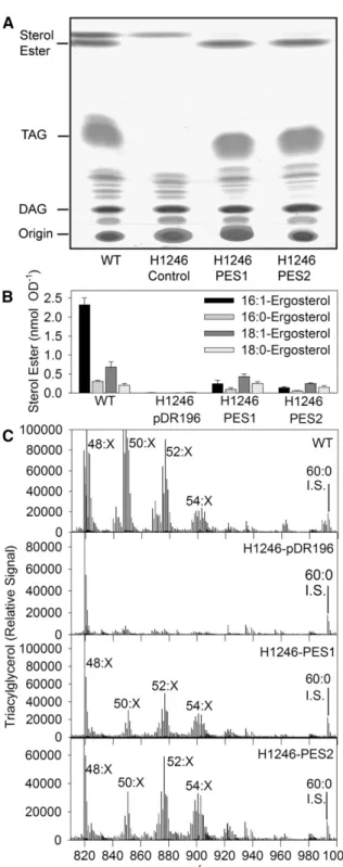
To study the substrate specificity of the PES1 and PES2 enzymes, the two sequences were transferred into yeast for heterologous expression. The yeast quadruple mutant H1246 (Sandager et al., 2002) deficient in four acyltransferase genes and therefore lacking all TAG and sterol ester synthesis activity was used as a host for expression. The transformed yeast cells were harvested and lipids extracted and separated by thin layer chromatography (TLC). Introduction of PES1 or PES2 into the H1246 mutant led to the accumulation of lipids comigrating with TAG and sterol esters (Figure 3A). To corroborate the identity of the two bands, the lipids were analyzed by quadrupole time-of-flight mass spectrometry (Q-TOF MS). The band designated as sterol esters contained ergosterol esters of 16:1, 16:0, 18:1, and 18:0 in the PES1- and PES2-expressing lines (Figure 3B). The Figure 1. Import of Radiolabeled PES1 and PES2 Preprotein into Pea
Chloroplasts.
The ribulose-1,5-bisphosphate carboxylase/oxygenase small subunit (SSU) was used as control. After uptake, chloroplasts were repurified and imported proteins analyzed by SDS-PAGE followed by phosphor imag-ing. 1, Translation products; 2, import after 0 min; 3, import after 15 min; 4, chloroplasts treated with thermolysin after import for 15 min. The precursor (pre) and mature (mat) forms of the proteins are separated.
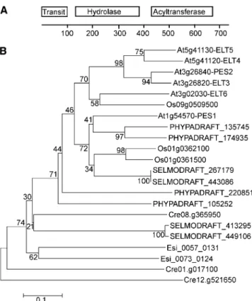
Figure 2. Protein Domains and Phylogram of ELT Sequences. (A) Domain structure of ELT sequences. BLAST searches with the full-length protein sequence of PES1 against the National Center for Biotechnology Information GenBank protein database revealed the presence of a hydrolase-like sequence (amino acids 120 to 380) and of an acyltransferase-like sequence (amino acids 420 to 670). The pre-dicted transit peptide (amino acids 1 to 100) is also shown.
(B) Phylogram of ELT protein sequences from different organisms. An unrooted phylogenetic tree was constructed using the neighbor-joining method from ELT protein sequences from Arabidopsis (At), rice (Os), Selaginella moellendorffii (SELMODRAFT), P. patens (PHYPADRAFT), C. rheinhardtii (Cre), and Ectocarpus siliculosus (Esi) (MEGA 5.0). The bootstrap values next to the branches were calculated from 1000 repli-cates. In the x-dimension, branch length represents evolutionary dis-tance based on the number of amino acid differences per site.
TAG band derived from the PES1- or PES2-expressing strains was rich in molecular species with total acyl chain lengths of 48: X, 50:X, 52:X, and 54:X (Figure 3C). Therefore, the introduction of PES1 or PES2 into the yeast H1246 mutant resulted in the complementation of TAG and sterol ester synthesis capacity, demonstrating that the two genes encode functionally active acyltransferases with broad substrate specificities.
The accumulation of TAG in yeast indicated that DAG can serve as a substrate for PES1 and PES2. Microsomes of PES1-or PES2-expressing H1246 cells were incubated with radioactive DAG (dihexanoylglycerol) and 12:0-CoA and lipids extracted, separated by TLC, and visualized by autoradiography. Figure 4A shows that PES1 and PES2 were capable of producing TAG and therefore harbor DAG acyltransferase (DGAT) activity. The re-action products of enzyme assays were quantified by electronic autoradiography after separation by TLC. Acyltransferase as-says with dihexanoylglycerol and radioactive 14:0-CoA re-vealed that PES1 and PES2 activity was highest with 1,2-DAG, but it was lower with 1,3-DAG and undetectable for mono-acylglycerol (Figure 4A). No DGAT activity was detected in H1246 cells carrying the empty vector (Figure 4A). PES2 activity was measured with different acyl donors, such as 14:0-CoA and 14:0-acyl carrier protein (ACP), each at three different concen-trations. The activity with 14:0-CoA was much higher than with 14:0-ACP at all concentrations (Figure 4B). Furthermore, the activity with 14:0-CoA was compared with that of the 14:0 free fatty acid. No acyltransferase activity was detected in assays with free fatty acids (Figure 4B). Assays with acyl-CoA esters of different chain lengths and with different degrees of unsatura-tion revealed that PES1 and PES2 show broad specificities for medium- to lo2ng-chain, saturated, and unsaturated acyl-CoAs (Figure 4C).
To test whether complex lipids can be employed as substrates by the PES enzymes, radioactive [14C]MGDG (for
monogalacto-syldiacylglycerol) was used as acyl donor in DGAT assays with yeast microsomes. Figure 4D shows that recombinant PES1 and PES2 are capable of transferring acyl groups from [14C]MGDG
onto DAG, while no acyltransferase activity was observed in the control. In addition, free fatty acids were released from [14C]MGDG
by PES1 or PES2, while only low amounts of free fatty acids were found in the control. The [14C]MGDG-dependent acyltransferase
and lipase activities of PES1 and PES2 were in the range of 0.01 to 0.02 nmol mg21protein min21and, therefore, much lower than the acyl-CoA–dependent activities (Figures 4B and 4D).
To address the question of whether PES1 and PES2 accept phytol as a substrate, acyltransferase assays were done with radioactive 14:0-CoA in the presence of free phytol. Figure 4E
Figure 3. Heterologous Expression of Arabidopsis PES1 or PES2 Re-sults in the Accumulation of TAG and Sterol Esters in Yeast.
Expression constructs for PES1 and PES2 and an empty vector control were introduced into the yeast quadruple mutant H1246.
(A) Lipids were extracted, separated by TLC, and stained with iodine vapor. Lipids were identified by cochromatography with standards and by Q-TOF MS. WT, the wild type.
(B) Identification of sterol esters in PES1 and PES2 expressing yeast cells. Sterol esters were quantified by Q-TOF MS using internal standards. The
bars show the amounts of ergosterol esters (mean andSDoffive mea-surements).
(C) Identification of TAG after heterologous expression of PES1 and PES2 in yeast. The four panels show the Q-TOF MS spectra without fragmentation in the mass-to-charge ratio (m/z) range of 810 to 1000. The identity of the TAG molecular species (48:X, 50:X, 52:X, and 54:X) was confirmed by MS/MS experiments of the individual peaks. Total amount of TAG was normalized to the internal standard triarachidin (60:0).
shows that recombinant PES1 and PES2 are capable of fatty acid phytyl ester synthesis. Addition of phytol resulted in phytyl ester production, while at the same time the synthesis of TAG based on the endogenous DAG from yeast was suppressed. The enzyme activity with phytol was in the range of 0.003 nmol mg21protein min21, which is much lower than the DGAT activity (Figures 4B and 4E).
Isolation of pes1 and pes2 Mutants of Arabidopsis
Arabidopsis mutants carrying T-DNA insertions in the two genes At1g54570 (PES1) and At3g26840 (PES2) were obtained from stock centers and homozygous lines identified by PCR (see Supplemental Figure 3A online). Two mutant lines were selected, SALK_034549 and SALK_071769, which contain T-DNAs inserted in exon 7 and exon 1 of the PES1 and PES2 genes, respectively. The two lines were crossed and double homozygous pes1 pes2 lines selected. Analysis by RT-PCR revealed that the expression of PES1 and PES2 was undetect-able in the pes1 pes2 double mutant (see Supplemental Figure 3B online). Therefore, the two mutants pes1 and pes2 most likely represent null alleles. Growth and overall morphology of the single mutants pes1 and pes2 and of the double mutant pes1 pes2 were not affected when the plants were grown on soil or in tissue culture. Expression of PES1 and PES2 was upregulated during nitrogen deprivation in the wild type (see Supplemental Figure 3B online). This result was in accordance with data in the gene expression databases that show that the two genes are induced during senescence and nitrogen deprivation (see Supplemental Figure 1 online).
To unravel whether chlorophyll degradation was affected in the pes1 pes2 mutant, the progression of senescence was studied by exposing detached leaves to darkness. After 5 d, chlorophyll in detached wild-type leaves was almost completely degraded (see Supplemental Figure 4A online). Chlorophyll degradation was slightly delayed in the pes1 pes2 double mu-tant, which still showed green leaves after 5 d of dark exposure
Figure 4. Acyltransferase Specificity of PES1 and PES2.
DGAT activity was measured with microsomes from H1246 yeast cells harboring an empty vector (control [C]) or expression constructs for
PES1 or PES2. Lipids were extracted, separated by TLC, and quantified by electronic autoradiography. Each bar represents the mean andSDof three tofive replicas.
(A) Specificity of PES1 and PES2 for different DAG or monoacylglycerol acceptors: [14C]1,2-dihexanoylglycerol (1,2-di6:0-DAG), [14 C]1,3-dihex-anoylglycerol (1,3-di6:0-DAG) or [14C]monohexanoylglycerol (6:0-MAG), each in combination with 14:0-CoA.
(B) Substrate specificity for acyl-CoA, acyl-ACP, or free fatty acids. DGAT assays of PES2 were done with three different amounts of acyl substrates (1,2-dihexanoylglycerol [1,2-di6:0-DAG] and [14C]14:0-CoA or [14C]14:0-ACP; left panel). The right panel shows DGAT assays of PES2 with [14C]1,2-dihexanoylglycerol (1,2-di6:0-DAG) and 5 nmol of 14:0-CoA or 14:0 free fatty acid (FFA).
(C) Specificity of DGAT activity for different acyl-CoAs. DGAT activity for PES1 (black bars) and PES2 (gray bars) was measured with [14C] 1,2-dihexanoylglycerol (1,2-di6:0-DAG) and different acyl-CoAs. (D) Acyltransferase activity of PES1 and PES2 with MGDG as acyl donor. Acyltransferase activity was measured with [14C]MGDG and endogenous DAG from the yeast microsomes.
(E) Phytyl ester synthesis activity was measured with free phytol and [14C]16:0-CoA.
with a reduction of the chlorophyll content to ;50% (see Supplemental Figure 4B online). Supplemental Figure 5 online shows electron micrographs of chloroplasts from leaves at days 0 and 7 after dark exposure. Wild-type and pes1 chloroplasts contain starch granules and plastoglobules after 7 d of dark exposure. Less starch and plastoglobules accumulate in the pes2 single mutant, and chloroplasts of the pes1 pes2 double mutant are similar at days 0 and 7 of dark exposure. Therefore, the senescence-induced disassembly of the photosynthetic membranes is slightly delayed in the pes1 pes2 double mutant, and the contribution of pes2 to the retardation of senescence is larger than that of pes1. However, after long-term dark-induced senescence or after growth under nitrogen deprivation for 10 or more days, chlorophyll degradation in pes1 pes2 progresses to an extent similar to the wild type.
Fatty Acid Phytyl Esters Are Decreased in the pes1 pes2 Double Mutant
The amount of fatty acid phytyl esters in leaves of the Arabi-dopsis wild type is usually very low, but it increases during ni-trogen deprivation (Csupor, 1971; Gaude et al., 2007). Therefore, Arabidopsis wild-type, pes1, pes2, and pes1 pes2 plants were grown on synthetic medium with nitrogen and then transferred to nitrogen-free medium. After 2 weeks, phytyl ester content and composition were measured by gas chromatography–mass spectrometry (GC-MS). The phytyl ester content in leaves of plants grown in the presence of nitrogen was similar in the wild type and pes1 but was strongly reduced in pes2 and in pes1 pes2 (Figure 5A). Growth in the absence of nitrogen resulted in a strong increase in phytyl ester content in the wild type. Under these conditions, the phytyl ester content in pes1 and pes2 single mutants was reduced by;25 and 75%, respectively, and it was reduced by 85% in pes1 pes2 compared with the wild type. These results demonstrate that the two proteins PES1 and PES2 rep-resent the predominant activities for phytyl ester synthesis and that PES2 shows a relatively higher contribution than PES1.
Fatty acid phytyl ester composition was determined for leaves of plants grown in the absence of nitrogen (Figure 5B). In the wild type, more than 50% of phytyl esters contained hexadecatrienoic acid (16:3); the remaining acyl groups were of medium-chain lengths (10:0, 12:0, and 14:0) and palmitic acid (16:0). This acyl composition was very similar in the pes1 mutant. The pes2 mutant also contained 16:3-phytol as predominant phytyl ester and minor amounts of 14:0-phytol and 16:0-phytol but was devoid of 10:0-phytol and 12:0-10:0-phytol. The pes1 pes2 double mutant was totally devoid of 16:3-phytol and medium-chain fatty acid phytyl esters. Instead, this plant contained low amounts of 16:0-phytol, 18:1-phytol, and 18:3-18:1-phytol, which are barely detectable in the wild type. The fact that the pes1pes2 mutant, in contrast with the wild type and the pes1 and pes2 single mutants, does not contain medium-chain and 16:3-phytyl esters, demonstrates that PES1 and PES2 share overlapping specificities and that PES1 and PES2 represent the only enzymes involved in the synthesis of these specific phytyl ester forms.
An alternative approach to study phytyl ester synthesis is the feeding of free phytol to seedlings of Arabidopsis in liquid me-dium (Ischebeck et al., 2006). Free phytol is readily taken up and
incorporated into phytyl esters. In addition to medium-chain and 16:3-phytyl esters accumulating during senescence or nitrogen deprivation, 16:0-phytol, 18:1-phytol, and 18:3-phytol accumu-late in wild-type seedlings incubated in the presence of phytol (Ischebeck et al., 2006). During phytol feeding, the pes1 pes2 mutant seedlings accumulated only;40% of wild-type levels of total phytyl esters. The phytyl ester composition was dominated by the acyl groups 16:0, 18:1, and 18:3, while medium-chain and 16:3-phytol were missing (see Supplemental Figure 6 online). Taken together, these data demonstrate that PES1 and PES2 produce the predominant proportion of phytyl esters (i.e., medium-chain and 16:3-phytyl esters).
Fatty Acid Phytyl Esters Are Involved in Adaptation to Transient Stress Conditions
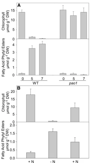
During senescence, large amounts of chlorophyll are broken down, and at the same time, fatty acid phytyl esters accumulate in the chloroplast (Csupor, 1971; Ischebeck et al., 2006). To address the question of whether the phytyl moiety incorporated into phytyl esters is derived from chlorophyll, fatty acid phytyl esters were measured during senescence in the pheophytin a oxidase1 (pao1) mutant, which is deficient in a key enzyme of chlorophyll degradation and therefore shows a stay green phenotype (Pruzinská et al., 2003). As shown in Figure 6A, chlorophyll was rapidly degraded in wild-type leaves during dark-induced senescence, while the pao1 mutant showed no chlorophyll degradation. Phytyl esters accumulated to large amounts in the wild type during senescence, but not in pao1. This experiment provides genetic evidence for the conclusion that the phytyl moiety accumulating in fatty acid phytyl esters is exclusively derived from chlorophyll degradation.
To address the question of whether chlorophyll degradation and phytyl ester accumulation in leaves are reversible, Arabi-dopsis wild-type plants were grown on complete medium for 14 d before transfer to nitrogen deficiency medium for 10 d. Sub-sequently, the plants were returned to full nitrogen medium for another 5 d. The amount of chlorophyll strongly decreased during nitrogen deprivation and recovered after transfer to full nutrition. However, the amount of phytyl esters strongly accu-mulated during 2N conditions and decreased after the plants were returned to full nutrition (Figure 6B). These changes in phytyl esters were even more severe for 16:3-phytol, the major molecular species of phytyl esters in leaves of Arabidopsis. The amount of 16:3-phytol increased from 0.0856 0.035 µmol g21 dry weight (DW) under +N conditions to 0.6916 0.122 µmol g21 DW at2N, and it decreased to 0.301 6 0.082 µmol g21 DW when plants were returned to full nutrition. These results indicate that fatty acid phytyl esters represent a transient sink for phytyl groups and acyl moieties accumulating during stress and that they can be hydrolyzed, releasing phytol and fatty acids. These metabolites might be employed for the synthesis of chlorophyll and membrane lipids, when growth conditions have improved.
Free Phytol Accumulates in the pes1 pes2 Double Mutant During senescence, large amounts of chlorophyll are broken down, and the phytol released is to a large extent incorporated
into tocopherol and fatty acid phytyl ester synthesis (Ischebeck et al., 2006). Therefore, the deficiency in phytyl ester synthesis in the pes1 pes2 mutant was expected to result in an accumulation of phytol. As a result, the phytol pool not used for phytyl ester synthesis in pes1 pes2 might be employed for tocopherol syn-thesis. Tocopherols and plastochromanol-8 were measured by fluorescence HPLC in wild-type and pes1 pes2 leaves after ni-trogen deprivation (see Supplemental Figure 7 online) and in the seeds. The amount of tocopherol increased in wild-type and pes1
pes2 leaves to comparable extents after nitrogen deprivation, with a-tocopherol showing the strongest increase. The amount and the composition of tocopherols in the seeds of the wild type and pes1 pes2 were very similar, with g-tocopherol representing the most abundant form. These results show that the pool of phytol not used for fatty acid phytyl ester synthesis in pes1 pes2 was not employed for tocopherol synthesis.
Therefore, the amount of free phytol was measured in leaves by GC-MS. The free phytol content was very low in leaves of wild-type plants grown under control conditions (Figure 7). After nitrogen deprivation, free phytol accumulated in the wild type. In
Figure 5. Fatty Acid Phytyl Ester Content of Arabidopsis pes1 and pes2 Mutants during Nitrogen Deprivation.
(A) Fatty acid phytyl ester content in leaves of plants grown in the presence (+N, black bars) or absence (2N, gray bars) of nitrogen. WT, the wild type.
(B) Acyl composition of fatty acid phytyl esters from leaves of plants grown in the absence of nitrogen (2N). Phytyl esters were measured by GC-MS. The values represent mean andSDof at least four measurements.
Figure 6. Changes in Chlorophyll and Fatty Acid Phytyl Ester Content during Senescence and Stress.
(A) Detached leaves of the wild type (WT) and the pao1 mutant of Arab-idopsis were incubated on wetfilter papers in the darkness for a different number of days as indicated.
(B) Arabidopsis plants were grown on synthetic full-nutrient medium (+N) before transfer to nitrogen-free medium (2N). After 10 d, plants were returned to nitrogen containing medium (+N). The top and bottom panels show the contents of chlorophyll and fatty acid phytyl esters during senescence, respectively. Chlorophyll was determined photometrically, and fatty acid phytyl esters were measured by GC-MS. Data represent mean andSDoffive measurements each.
the pes1 pes2 mutant, the amount of free phytol was higher than in the wild type during growth under control conditions. After nitrogen deprivation, the free phytol content increased to very high amounts in pes1 pes2 (approximately fourfold) compared with the wild type.
Taken together, these results show that free phytol released from chlorophyll degradation during senescence increases only to low amounts in the wild type. In the pes1 pes2 double mutant, free phytol increases strongly during senescence, demonstrat-ing that the capacity for phytol degradation or incorporation into tocopherol is limited.
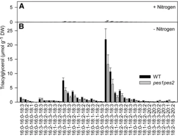
TAG Accumulation in the pes1 pes2 Double Mutant TAG is known to accumulate in the leaves after exposure to different stress conditions (Sakaki et al., 1990; Kaup et al., 2002). Furthermore, TAG in leaf mesophyll cells can be localized to different subcellular compartments, the cytosol and the chlo-roplasts. Therefore, TAG extracted from whole leaves is pre-sumably in part derived from the cytosol and chloroplasts. TAG molecular species were quantified by Q-TOF MS in whole leaves of the wild type and the pes1 pes2 mutant after nitrogen dep-rivation (Figure 8). Nitrogen deficiency led to a strong accumu-lation of different TAG molecular species in the wild type. The total TAG content in pes1 pes2 leaves during nitrogen de-ficiency was also increased, but it was lower by ;30% com-pared with the wild type. This difference was observed to roughly equal extents for the different molecular species and was obtained in three independent biological experiments. These results demonstrate that a deficiency in the chloroplast acyltransferases PES1 and PES2 in the pes1 pes2 double mutant affects in vivo TAG accumulation in leaves after ex-posure to nitrogen deficiency.
DISCUSSION
Plant cells harbor a number of biosynthetic pathways that are organized in parallel and are localized to different compart-ments. For example, membrane lipid assembly is localized to the chloroplast and to the endoplasmic reticulum (ER) (Browse et al., 1986b). The pathways for wax ester and TAG synthesis in the plant cell are associated with the ER. Here, we show that chloroplasts contain two acyltransferases, PES1 and PES2, in-volved in phytyl ester and TAG synthesis. Therefore, in addition to the ER, chloroplasts also harbor the biosynthetic capacity to produce long-chain acyl esters and TAG.
The ELT Acyltransferase Family
Arabidopsis contains two families of acyltransferases involved in wax ester synthesis. The bifunctional enzymes were identified based on their sequence similarity to the Acinetobacter acyl-transferase, which is specific for DAG and long-chain alcohols (Kalscheuer and Steinbüchel, 2003). The bifunctional acyl-transferase family contains 11 members in Arabidopsis. One protein, Wax Ester Synthase/Acyl-Coenzyme A:Diacylglycerol Acyltransferase1, was recently shown to be involved in wax ester synthesis in stems (Li et al., 2008). Furthermore, wax ester synthases with sequence similarity to the Acinetobacter acyltransferases were isolated from Marinobacter hydro-carbonoclasticus (Holtzapple and Schmidt-Dannert, 2007). The Marinobacter gene is involved in phytyl ester synthesis for car-bon storage when the cells are grown on phytol-containing medium under nitrogen or phosphate limitation. A second Figure 7. Phytol Content in Leaves of the pes1 pes2 Double Mutant.
Free phytol in leaves of wild-type (WT) and pes1 pes2 plants grown under normal (+N, black bars) and nitrogen deficient conditions (2N, gray bars) was measured by GC-MS of trimethylsilyl derivatives. Data represent the mean andSDof four measurements.
Figure 8. TAG Accumulation in Leaves of the Wild Type and pes1 pes2. Lipids were extracted from whole leaves of the wild type (WT) and pes1 pes2 and used for the quantification of TAG molecular species by Q-TOF MS. Plants were grown on Murashige and Skoog medium for 3 weeks and then transferred to synthetic medium containing nitrogen ([A]; +Ni-trogen) or to nitrogen deficient medium ([B]; 2Nitrogen) for 2 weeks. Data represent mean andSDof six measurements. The experiment was repeated two times with an additional set of six measurements each with the same result.
independent family of acyltransferases with sequence similarity to the jojoba seed wax ester synthase contains 12 genes in Arabidopsis (Lardizabal et al., 2000). However, analysis of gene expression data and of the phytyl ester content of Arabidopsis mutants revealed that plant phytyl ester synthases are not re-lated to Acinetobacter or jojoba-type acyltransferases but be-long to a different family of ELT-like proteins (Figure 2; see Supplemental Figure 2 online). Therefore, the Arabidopsis phytyl ester synthases are phylogenetically distinct from the paralo-gous genes from Marinobacter.
The family of ELT genes harbors six members in Arabidopsis. The ELT proteins contain two domains, one hydrolase/ELT-like sequence, and a second, DGAT/acyltransferase domain. All six ELT proteins harbor an N-terminal extension. For some of the proteins, including PES1 and PES2, this extension is predicted to provide chloroplast targeting. ELT sequences carrying two domains can be retrieved from all plants, including dicot-yledonous (Arabidopsis) and monocotdicot-yledonous species (rice), mosses (P. patens), and lycophytes (Selaginella). Furthermore, ELT sequences are present in green algae (C. reinhardtii) and brown algae (Ectocarpus). However, ELT genes carrying the two domains are absent from nonphotosynthetic organisms, such as animals (rat, mouse, Drosophila, and Caenorhabdites), yeast (Saccharomyces), and bacteria (E. coli and S. aureus) (data not shown). Therefore, the presence of phytyl ester synthases/ELT genes is restricted to photosynthetic, chlorophyll-containing organisms where phytol has to be metabolized during chloro-phyll degradation.
Phytyl Ester Synthesis in Arabidopsis
Phytyl esters were identified in all plants analyzed to date (Gaude et al., 2007). Phytyl ester content in leaves is usually very low but strongly increases during chlorophyll degradation, such as in senescence or abiotic or biotic stress (Csupor, 1971; Patterson et al., 1993; Pereira et al., 2002; Ischebeck et al., 2006). Fatty acid phytyl esters are intracellular wax esters that accumulate in the plastoglobules of chloroplasts. Considerable amounts of medium-chain fatty acids (10:0, 12:0, and 14:0) accumulate in the phytyl ester fraction. These acyl groups are presumably derived from 10:0-ACP, 12:0-ACP, and 14:0-ACP, intermediates of fatty acid de novo synthesis (Figure 9). It is possible that the acyl groups are hydrolyzed from ACP during senescence or stress to terminate fatty acid synthesis under unfavorable conditions. Acyl-ACPs can also directly be em-ployed as substrates by PES1 and PES2 (Figure 4B).
Hexadecatrienoic acid (16:3) in phytyl esters is presumably derived from the chloroplast galactolipid MGDG, which is known to contain 16:3 at the sn-2 position (Browse et al., 1986b). MGDG can directly serve as acyl donor for the acyltransferase reaction by PES1 and PES2 (Figure 4D). Thus, PES1/PES2 might be specific for the sn-2 position of MGDG or for the acyl group 16:3. The radioactive substrate employed for acyl-transferase assays, [14C]MGDG, was produced in spinach
(Spinacia oleracea) leaves after labeling with [14C]acetic acid.
This way both acyl groups in MGDG are labeled, and it is not possible to draw a conclusion on positional (sn-2) or acyl (16:3) specificity of PES1/PES2. In Arabidopsis, 16:3 amounts to
approximately one-third of the acyl groups at the sn-2 position of MGDG, the remainder being mostly 18:3 (Browse et al., 1986b). Furthermore, the Arabidopsis act1 mutant, which is devoid of 16:3 and contains mostly 18:3 at the sn-2 position of MGDG, accumulates phytyl esters that are free of 16:3 but do not contain 18:3 (Gaude et al., 2007). These results indicate that the accumulation of 16:3 in phytyl esters cannot be explained by a specificity of PES1/PES2 for the sn-2 position of MGDG. Acyltransferase assays with different acyl-CoAs revealed that PES1/PES2 show a broad specificity for different acyl chains, including 16:3. Although PES1/PES2 do not show a strong preference for 16:3-CoA, it is still possible that the enzymes are specific for other containing lipid substrates (e.g., 16:3-MGDG or 16:3-ACP). Furthermore, it is possible that 16:3 is released from MGDG by a specific galactolipase and in-corporated into the CoA or ACP pool, before being used for phytyl ester synthesis. In this scenario, the high proportion of 16:3 in phytyl esters would not be caused by the substrate specificity of PES1/PES2 per se but rather by the available pool size of 16:3-containing CoA or ACP substrates.
PES1 and PES2 harbor two distinct domains: a hydrolase or ELT domain (related to the a/b-hydrolase superfamily) at amino acids 120 to 380 (of the PES1 sequence) and an acyltransferase (related to the DGAT family) sequence between amino acids 420 and 670 (Figure 2A). Therefore, the ELT proteins possibly harbor two enzymatic functions, for the cleavage of an ester linkage and for the transfer of an acyl group onto an acceptor hydroxy group. This two-domain structure is in agreement with the finding that the enzymes can catalyze the transfer of acyl groups from a complex lipid (MGDG) onto an acceptor molecule (e.g., Figure 9. Synthesis of TAG and Fatty Acid Phytyl Esters by PES1 and PES2.
Free phytol is produced during chlorophyll degradation. Galactolipid degradation results in the release of DAG and free fatty acids (FFA). Free phytol and DAG can be employed for fatty acid phytyl ester and TAG synthesis, respectively, by PES1 and PES2. Acyl-CoAs, acyl-ACPs, and MGDG can serve as acyl donors for the PES1 and PES2 reactions.
DAG). It is also possible that PES1/PES2 contain a tightly bound cofactor, such as CoA, that is transiently acylated before the acyl group is transferred onto phytol.
Acyl-CoAs rather than MGDG or acyl-ACPs were the pre-ferred substrates for recombinant PES1 and PES2 in in vitro assays. Free fatty acids were not employed for phytyl ester synthesis by PES1/PES2. However, it is possible that free fatty acids are activated as CoA or ACP esters in an ATP-dependent manner by a chloroplastic acyl-CoA or acyl-ACP synthetase or possibly by PES1/PES2, prior to phytyl ester synthesis.
Acyl-CoA esters are important substrates for acyltransferases at the ER. Consequently, the acyl-CoA pool of the plant cell is dominated by long-chain acyl groups presumably localized to the cytosol (Larson and Graham, 2001). Short-chain acyl-CoAs, in particular acetyl-CoA and malonyl-CoA, are abundant in chloroplasts of spinach and pea (Post-Beittenmiller et al., 1992). At present, it remains unclear whether chloroplasts contain long-chain acyl-CoAs. Thus, the exact nature of the in vivo acyl-donor for the phytyl ester synthesis by PES1/PES2 remains unclear (Figure 9).
The pes1 pes2 double mutant still contains residual amounts of phytyl esters during nitrogen deprivation. This PES1- and PES2-independent phytyl ester synthesis might originate from other ELT enzymes, some of which might also be chloro-plast localized. However, expression of the other ELT genes is not induced during senescence or nitrogen deprivation (see Supplemental Figure 1 online), suggesting that their contribution to phytyl ester synthesis during nitrogen deprivation might be minor. It is also possible that ER-localized acyltransferases from the Acinetobacter or jojoba families are involved in the synthesis of phytyl esters outside of the chloroplast. This scenario is in agreement with thefinding that the phytyl ester composition of wild-type seedlings after feeding with free phytol differs from the pattern observed after nitrogen starvation (Figure 5; see Supplemental Figure 6 online). The phytyl ester 16:3-phytol is by far the most abundant after nitrogen deprivation, while other phytyl esters containing 16:0, 18:1, and 18:3 are more abundant after phytol feeding.
Regulation of Phytol Metabolism in Arabidopsis
Phytol represents a C20 isoprenoid alcohol derived from ger-anylgeraniol by reduction of three double bonds. Therefore, phytol originates from precursors of the plastidial methylery-thritol phosphate pathway of isoprenoid synthesis. During chlorophyll degradation, a large amount of phytol is released, but its further catabolic pathway is still not understood. Free phytol can be converted into phytol-diphosphate by action of two kinases (Ischebeck et al., 2006; Valentin et al., 2006). Phytol-diphosphate can then be used for the synthesis of tocopherol, phylloquinone, and chlorophyll (Ischebeck et al., 2006). Phytyl ester synthesis represents an alternative route for phytol catabo-lism during chlorotic growth conditions. As phytyl ester synthesis depends on the transfer of an activated acyl group onto free phytol, no phosphorylation of phytol is required.
Final degradation of phytol is believed to be mediated by a- and b-oxidation in the peroxisomes and mitochondria, similar to phytol catabolism in animals (Mukherji et al., 2003). The
accumulation of phytol and fatty acids in the form of phytyl esters might be explained by a limitation of phytol degradation capacity. In line with this scenario, the mutations that block the capacity of phytyl ester synthesis in pes1 pes2 result in an in-crease in the free phytol content indicating that free phytol cannot immediately be metabolized by oxidation in the perox-isomes or mitochondria. Furthermore, the fact that the amount of tocopherol in the leaves of the pes1 pes2 mutant was not increased is in agreement with thefinding that free phytol cannot be employed for tocopherol synthesis. It is possible that under chlorotic conditions in leaves, the two kinase reactions are limiting for the conversion of phytol into phytyl-diphosphate and thus restrict the amount of phytol that can be channeled into tocopherol synthesis.
The phytyl ester content decreases and the amount of chlo-rophyll increases when plants are returned from nitrogen dep-rivation to full nutrition medium (Figure 6). Therefore, it is likely that fatty acid phytyl esters represent a transient sink for free phytol and free fatty acids that are released from chlorophyll and galactolipids during chlorotic stress and are reutilized for the synthesis of chlorophyll and galactolipids when the stress has disappeared. At present, the mechanisms for phytyl ester break-down during the recovery of plants after stress and the presumed incorporation of phytol into chlorophyll, and of acyl groups into galactolipids, remain unclear.
TAG Synthesis in pes1 pes2
TAG analysis of the wild type and pes1pes2 was done with a lipid fraction isolated from whole leaves, thus presumably representing a mixture of cytosolic and chloroplastic molecular species. Molecular species analysis revealed that the TAG ac-cumulating in wild type during nitrogen deficiency lacks 16:3. This is in contrast with TAG molecules accumulating in the leaves after ozone, drought, or freezing stress (Sakaki et al., 1990; Moellering et al., 2010). It is believed that chloroplastic TAG is enriched with 16:3, but it is possible that different chlo-roplastic TAG pools exist with different molecular species composition. Although the TAG accumulation in leaves of pes1 pes2 is decreased by;30%, there is still a substantial amount of TAG produced during nitrogen deficiency. It is possible that the deficiency in the PES1/PES2 pathway causes a general perturbation of chloroplast lipid metabolism, which in turn af-fects TAG accumulation during stress. The increase in TAG synthesis during stress might originate from other ELT enzymes in the chloroplast. A certain proportion of stress-dependent TAG accumulation might also be extraplastidic. Therefore, ER-localized acyltransferases, including DGATs (DGAT1 and DGAT2) or phospholipid:diacylglycerol acyltransferase are presumably in-volved in stress-dependent TAG production in leaves (Zou et al., 1999; Dahlqvist et al., 2000; Lardizabal et al., 2001).
METHODS
Plant Material and Growth Conditions
Plants were grown on synthetic medium containing 0.8% agarose, 1% Suc, and Murashige and Skoog salts at 120µmol m22s21light (16 h light/
day) (Murashige and Skoog, 1962). After 2 weeks, the plants were transferred to Petri dishes with fresh synthetic medium (+N) or medium lacking nitrogen (2N) (Estelle and Somerville, 1987; Gaude et al., 2007). The two T-DNA insertion lines SALK_034549 (At1g54570, pes1) and SALK_071769 (At3g26840, pes2) were obtained from the Nottingham Arabidopsis Stock Center. Homozygous mutant lines were identified by PCR using oligonucleotide primers designed to the genomic sequence (PES1: PD706, PD707; PES2: PD708, PD709) and to the left T-DNA boarder (PD857; see Supplemental Table 1 online). The two homozygous mutant lines were crossed and double homozygous plants selected in the F2 generation by PCR. The pao1 mutant (At3g44880) was isolated pre-viously (Pruzinská et al., 2003).
Expression of PES1 and PES2 in Yeast
ESTs for At1g54570/PES1 and At3g26840/PES2 (clones U60915 and U09399, respectively) were obtained from the ABRC (Columbus, Ohio). The coding regions without apparent transit peptides were amplified by PCR using the following primers: Bn187 (PES1 forward), Bn188, (PES1 reverse), Bn189 (PES2 forward), and Bn190 (PES2 reverse) (see Supplemental Table 1 online). The PCR products were cloned into pGEM-T-Easy (Promega). The fragments were released with EcoRI-PstI and li-gated into pDR196 allowing expression in yeast under the control of the constitutive PMA1 promoter (Rentsch et al., 1995). The cDNAs for PES1 and PES2 were introduced into the yeast strain H1246 deficient in TAG and sterol ester synthesis for heterologous expression (Sandager et al., 2002).
Acyltransferase Assays
Recombinant yeast cells were grown in synthetic uracil drop-out medium at 30°C. After 24 h, yeast cells were harvested, washed with water, and disrupted by homogenization with glass beads using a Mini Beadbeater-8 (Biospec Products). A microsomal yeast fraction was prepared and protein concentrations determined using the BCA Protein Assay (Dahlqvist et al., 2000) (Thermo Scientific).
DGAT activity was measured in a total volume of 100mL with 50 µg of microsomal protein in assay buffer (50 mM HEPES-KOH, pH 7.2, and 5 mM MgCl2) at 30°C under shaking (650 rpm). The following substrates were added to the reaction: 5 nmol (or different amounts as indicated) [14C]acyl-CoA or [14C]acyl-ACP, 5 nmol dihexanoylglycerol (di6:0-DAG), or 5 nmol unlabeled acyl-CoA and 5 nmol [14 C]dihex-anoylglycerol (di6:0-DAG). The assays were terminated after 3 min if not stated otherwise.
For phytyl ester synthesis assays, phytol was dissolved in benzene and 5 nmol was added to 50µg of lyophilized microsomal protein. Sub-sequently, the benzene was evaporated under a stream of N2gas, leaving the phytol in direct contact with the yeast membranes. [14C]acyl-CoA (30 nmol) was then added in assay buffer to afinal volume of 100 µL. The reaction was incubated at 30°C for 60 min.
For MGDG-dependent assays, [14C]MGDG (100 nmol) was added to 1 mg of lyophilized microsomal enzyme in the same way, followed by addition of 100mL of assay buffer and incubation at 30°C for 60 min. DGAT activity in MGDG-dependent assays was based on endogenous DAG derived from the yeast microsomes, as no extra DAG was added to the reaction.
Reactions were terminated and lipids were extracted by addition of 100 mL of 0.15 M acetic acid and 500 mL of chloroform/methanol (1:1). Lipids in the chloroform phase were harvested and separated on TLC plates using hexane/diethylether/acetic acid (70:30:1) for assays with MGDG or hexane/diethylether/acetic acid (85:15:1) for DGAT and phytyl ester synthesis assays. Radioactivity was monitored and quantified using electronic autoradiography (Instant Imager; Packard Instruments).
Lipid Substrates
Acyl-CoAs, DAG, and acyl-ACPs with different chain lengths were chemically or enzymatically synthesized as previously described (Sánchez et al., 1973; Ohlrogge et al., 1978; Kanda and Wells, 1981). The DAG substrates [14C]1,2-dihexanoyldiacylglycerol (1,2-di6:0-DAG) and [14 C]1,3-dihexanoyldiacylglycerol (1,3-di6:0-DAG) or [14C]monohexanoylglycerol (6:0-MAG) were at a specific activity of 3000 dpm/nmol. The specific ac-tivities for the other substrates were [14C]14:0-CoA, 13,000 dpm/nmol; and [14C]14:0-ACP, 8000 dpm/nmol. Radioactive [14C]MGDG was synthesized by applying [14C]acetic acid (Perkin-Elmer) to young leaves of spinach (Spinacia oleracea) daily for a period of 5 d. Lipids were then extracted (Bligh and Dyer, 1959) and separated by TLC in chloroform/methanol/acetic acid/ water (90:15:10:3). [14C]MGDG was eluted from the silica gel with methanol: chloroform (2:1), extracted into chloroform, and quantified by GC and liquid scintillation. The specific activity of [14C]MGDG was 500 dpm/nmol. Measurements of TAG
Lipids were isolated from yeast or from frozen leaves with 2 volumes of methanol/chloroform/formic acid (1:1:0.1) and 1 volume of 1 M KCl, 0.2 M H3PO4. The solvent was evaporated and the lipids dissolved in 100% chloroform. Nonpolar lipids, including sterol esters and TAG, were iso-lated from the crude lipid extracts by solid phase extraction using Strata silica columns (Phenomenex) by elution with chloroform. Sterol esters were measured by direct infusion nanospray Q-TOF MS (Wewer et al., 2011).
The TAGs in the nonpolar lipid fraction were dissolved in methanol/ chloroform/aqueous 300 mM ammonium acetate (665:300:35) (Welti et al., 2002) and measured by Q-TOF MS. Molecular species composition of the TAGs (ammonium adducts) was determined after collision-induced dissociation with nitrogen gas at a collision energy of 20 V. The tandem mass spectrometry (MS/MS) signals were normalized to respective standards (saturated or unsaturated; see below) and to the DW of the leaves. Four internal standards were added: tridecanoin (tri-10:0), triar-achidin (tri-20:0), trierucin (tri-22:1D13cis) (Larodan), and triundecenoin (tri-11:1D10cis). The standard triundecenoin (tri-11:1D10cis) was synthesized from the acid chloride of undecenoic acid (11:1D10cis; Larodan) (obtained from the free acid by oxalyl chloride treatment) and glycerol and purified by TLC. Lipid standards were quantified by GC of fatty acid methyl esters (Browse et al., 1986a). The two saturated TAG standards were used to calculate the relative amounts of saturated TAGs in the sample. A linear regression line between the two standard signals was calculated to ac-count for the differences in ionization, which depends on the masses of TAG molecular species (i.e., acyl chain lengths) (Han and Gross, 2001). Unsaturated TAG molecular species show different MS and MS/MS re-sponses compared with saturated TAGs due to differences in ionization. Therefore, the two unsaturated TAG standards were employed for the quantification of all unsaturated TAG molecular species. As for the sat-urated standards, a linear regression line between the two unsatsat-urated standard MS signals was calculated.
RT-PCR
RNA was isolated from leaves of wild-type and pes1 pes2 mutant seedlings grown in the presence or absence of nitrogen using the RNA extraction kit (Roboklon) according to the instructions. An additional DNase (Fermentas) digest was performed. RNA (250 ng) was used for cDNA synthesis with the First-Strand cDNA Synthesis kit (Fermentas). RT-PCR was done for PES1 (primers Bn1256 and Bn1257), PES2 (Bn1258 and Bn1259), and actin (ACT2, At3g18780) (Bn358 and Bn359) (see Supplemental Table 1 online). The annealing temperature was optimized using a gradient PCR and was set to 59°C. Twenty-eight cycles of PCR were performed with 62 ng cDNA (PES1 and PES2) and 16 ng cDNA (ACT2).
Quantification of Chlorophyll, Tocopherol, Fatty Acid Phytyl Esters, and Free Phytol
Chlorophyll concentration was determined photometrically (Porra et al., 1989). Tocochromanols were determined as described (Zbierzak et al., 2010). Briefly, tocochromanols were extracted with 200 mL diethylether and 100mL 1 M KCl and 0.2 M H3PO4. Tocol (0.5µg) was added as internal standard. Tocochromanols were dissolved in hexane, sepa-rated by HPLC (Lichrospher 100 diol, 5µm, 3 mm 3 25 cm; Knauer) with hexane/tertiary butylmethyl ether (96:4) at aflow rate of 0.75 mL min21 and quantified by fluorescence detection (excitation, 290 nm; emission, 330 nm). Fatty acid phytyl esters were extracted from leaves in the presence of an internal standard (15:0-phytol) and quantified by GC-MS (Ischebeck et al., 2006; Gaude et al., 2007). For phytol measurements, lipids were extracted from frozen leaves with chloroform/methanol/ formic acid (1:1:0.1) and 1 M KCl and 0.2 M H3PO4as described above. The chloroform was evaporated with N2gas, and lipids were silylated with N-methyl-N-(trimethylsilyl)trifluoroacetamide. Trimethylsilylated phytol was quantified using 5 µg oleyl alcohol (Sigma-Aldrich) as internal standard by GC-MS (Agilent HP6890 Series GC with 5973 inert mass selective detector) using the same column and conditions as for fatty acid phytyl ester analysis (Ischebeck et al., 2006). The results were converted to a DW basis employing a fresh weight-to-DW ratio of 14.36 2.3 for plants grown in Petri dishes and a fresh weight-to-DW ratio of 11.86 1.3 for detached leaves.
Chloroplast Import Experiments and Electron Microscopy
Chloroplasts were isolated from pea (Pisum sativum) and employed for import experiments with [35S]Met-radiolabeled PES1 or PES2 preprotein (Smith et al., 2002). After uptake, chloroplasts were washed. Proteins were analyzed by SDS-PAGE followed by Coomassie Brilliant Blue staining and phosphor imaging (Agne et al., 2009). Leaves werefixed with glutaraldehyde and osmium tetroxide, and ultrathin sections were ana-lyzed by electron microscopy (Vidi et al., 2006).
Phylogenetic Analysis
Protein sequences from Arabidopsis thaliana (At), rice (Oryza sativa; Os), Selaginella moellendorffii, Physcomitrella patens, Chlamydomonas rheinhardtii and Ectocarpus siliculosus were obtained from GenBank (National Institutes of Health). Phylogenetic analyses were performed with MEGA 5.0 (Tamura et al., 2011). The sequences were aligned using the Clustal algorithm. An unrooted phylogenetic tree was constructed using the neighbor-joining method (p-distance method), and the bootstrap values were derived from 1000 replicates.
Accession Numbers
The EST sequence data for PES1 and PES2 can be found in the GenBank/ EMBL databases under the accession numbers U60915 (At1g54570/ PES1) and U09399 (At3g26840/PES2). The predicted amino acid sequences for At1g54570/PES1, At3g26840/PES2, At3g26820/ELT3, At5g41120/ELT4, At5g41130/ELT5, and At3g02030/ELT6 can be re-trieved at the Munich Information Center for Protein Sequences database.
Supplemental Data
The following materials are available in the online version of this article. Supplemental Figure 1. Acyltransferase Candidate Genes for Fatty Acid Phytyl Ester Synthesis in Chloroplasts of Arabidopsis.
Supplemental Figure 2. The Family of Esterase/Lipase/Thioesterase Proteins of Arabidopsis.
Supplemental Figure 3. Genomic Structure of the Arabidopsis pes1 and pes2 Mutants and Expression of the PES1 and PES2 Genes under Nitrogen Deprivation.
Supplemental Figure 4. Chlorophyll Degradation during Senescence in the pes1 pes2 Mutant.
Supplemental Figure 5. Chloroplast Ultrastructure in Leaves of the Wild Type, pes1, pes2, and pes1 pes2.
Supplemental Figure 6. Fatty Acid Phytyl Ester Content in pes1 pes2 Plants Supplemented with Free Phytol.
Supplemental Figure 7. Tocopherol Content and Composition in the Arabidopsis pes1 pes2 Mutant.
Supplemental Table 1. Oligonucleotide Primers Used in This Study Supplemental Data Set 1. Text File of Alignment Corresponding to Phylogenetic Analysis in Figure 2.
ACKNOWLEDGMENTS
Funding was in part provided by the European Union Industrial Crops producing added value Oils for Novel chemicals Project (FP7 project reference 211400). We thank Marcel Kristen and Simon Stilling (Univer-sity of Bonn) for help with plant growth and lipid analyses.
AUTHOR CONTRIBUTIONS
F.K., S.S., and P.D. designed the research. F.L., K.v.D., M.A., G.H., V.W., J.L.Y., I.L., C.M., and C.B. performed research. All authors analyzed data, and P.D. wrote the article.
REFERENCES
Agne, B., Infanger, S., Wang, F., Hofstetter, V., Rahim, G., Martin, M., Lee, D.W., Hwang, I., Schnell, D., and Kessler, F. (2009). A toc159 import receptor mutant, defective in hydrolysis of GTP, supports preprotein import into chloroplasts. J. Biol. Chem. 284: 8670–8679.
Austin, J.R., II, Frost, E., Vidi, P.A., Kessler, F., and Staehelin, L.A. (2006). Plastoglobules are lipoprotein subcompartments of the chloroplast that are permanently coupled to thylakoid membranes and contain biosynthetic enzymes. Plant Cell 18: 1693–1703. Beale, S.I. (1999). Enzymes of chlorophyll biosynthesis. Photosynth.
Res. 60: 43–73.
Beisson, F. et al. (2003). Arabidopsis genes involved in acyl lipid metabolism. A 2003 census of the candidates, a study of the dis-tribution of expressed sequence tags in organs, and a web-based database. Plant Physiol. 132: 681–697.
Bligh, E.G., and Dyer, W.J. (1959). A rapid method of total lipid ex-traction and purification. Can. J. Biochem. Physiol. 37: 911–917. Bréhélin, C., Kessler, F., and van Wijk, K.J. (2007). Plastoglobules:
Versatile lipoprotein particles in plastids. Trends Plant Sci. 12: 260–266.
Browse, J., McCourt, P.J., and Somerville, C.R. (1986a). Fatty acid composition of leaf lipids determined after combined digestion and fatty acid methyl ester formation from fresh tissue. Anal. Biochem. 152: 141–145.
Browse, J., Warwick, N., Somerville, C.R., and Slack, C.R. (1986b). Fluxes through the prokaryotic and eukaryotic pathways of lipid syn-thesis in the‘16:3’ plant Arabidopsis thaliana. Biochem. J. 235: 25–31. Buchanan, M.S., Hashimoto, T., and Asakawa, Y. (1996). Phytyl estsers and phaeophytins from the hornwort Megacerosflagellaris. Phytochemistry 41: 1373–1376.
Csupor, L. (1971). Das Phytol in vergilbten Blättern. Planta Med. 19: 37–41.
Dahlqvist, A., Ståhl, U., Lenman, M., Banas, A., Lee, M., Sandager, L., Ronne, H., and Stymne, S. (2000). Phospholipid:diacylglycerol acyltransferase: an enzyme that catalyzes the acyl-CoA-independent formation of triacylglycerol in yeast and plants. Proc. Natl. Acad. Sci. USA 97: 6487–6492.
Douce, R. (1974). Site of biosynthesis of galactolipids in spinach chloroplasts. Science 183: 852–853.
Estelle, M.A., and Somerville, C. (1987). Auxin-resistant mutants of Arabidopsis thaliana with an altered morphology. Mol. Gen. Genet. 206: 200–206.
Fleming, I. (1967). Absolute configuration and the structure of chlo-rophyll. Nature 216: 151–152.
Gaude, N., Bréhélin, C., Tischendorf, G., Kessler, F., and Dörmann, P. (2007). Nitrogen deficiency in Arabidopsis affects galactolipid composition and gene expression and results in accumulation of fatty acid phytyl esters. Plant J. 49: 729–739.
Grob, E.C., and Csupor, L. (1967). Zur Kenntnis der Blattlipide von Acer platanoides L. während der herbstlichen Vergilbung. Cell. Mol. Life Sci. 23: 1004–1005.
Han, X., and Gross, R.W. (2001). Quantitative analysis and molecular speciesfingerprinting of triacylglyceride molecular species directly from lipid extracts of biological samples by electrospray ionization tandem mass spectrometry. Anal. Biochem. 295: 88–100. Harris, J.B., and Arnott, H.J. (1973). Effects of senescence on
chloroplasts of the tobacco leaf. Tissue Cell 5: 527–544.
Hendry, G.A.F., Houghton, J.D., and Brown, S.B. (1987). Tansley Review No. 11. The degradation of chlorophyll—A biological enigma. New Phytol. 107: 255–302.
Holtzapple, E., and Schmidt-Dannert, C. (2007). Biosynthesis of isoprenoid wax ester in Marinobacter hydrocarbonoclasticus DSM 8798: Identification and characterization of isoprenoid coenzyme A synthetase and wax ester synthases. J. Bacteriol. 189: 3804–3812. Hörtensteiner, S. (2006). Chlorophyll degradation during senescence.
Annu. Rev. Plant Biol. 57: 55–77.
Ischebeck, T., Zbierzak, A.M., Kanwischer, M., and Dörmann, P. (2006). A salvage pathway for phytol metabolism in Arabidopsis. J. Biol. Chem. 281: 2470–2477.
Ishizaki, K., Larson, T.R., Schauer, N., Fernie, A.R., Graham, I.A., and Leaver, C.J. (2005). The critical role of Arabidopsis electron-transfer flavoprotein:ubiquinone oxidoreductase during dark-induced starvation. Plant Cell 17: 2587–2600.
Joyard, J., Teyssier, E., Miège, C., Berny-Seigneurin, D., Maréchal, E., Block, M.A., Dorne, A.-J., Rolland, N., Ajlani, G., and Douce, R. (1998). The biochemical machinery of plastid envelope mem-branes. Plant Physiol. 118: 715–723.
Kalscheuer, R., and Steinbüchel, A. (2003). A novel bifunctional wax ester synthase/acyl-CoA:diacylglycerol acyltransferase mediates wax ester and triacylglycerol biosynthesis in Acinetobacter cal-coaceticus ADP1. J. Biol. Chem. 278: 8075–8082.
Kanda, P., and Wells, M.A. (1981). Facile acylation of glycer-ophosphocholine catalyzed by trifluoroacetic anhydride. J. Lipid Res. 22: 877–879.
Kaup, M.T., Froese, C.D., and Thompson, J.E. (2002). A role for diacylglycerol acyltransferase during leaf senescence. Plant Phys-iol. 129: 1616–1626.
Lardizabal, K.D., Mai, J.T., Wagner, N.W., Wyrick, A., Voelker, T., and Hawkins, D.J. (2001). DGAT2 is a new diacylglycerol acyl-transferase gene family: Purification, cloning, and expression in insect cells of two polypeptides from Mortierella ramanniana with diacylglycerol acyltransferase activity. J. Biol. Chem. 276: 38862– 38869.
Lardizabal, K.D., Metz, J.G., Sakamoto, T., Hutton, W.C., Pollard, M.R., and Lassner, M.W. (2000). Purification of a jojoba embryo wax synthase, cloning of its cDNA, and production of high levels of wax in seeds of transgenic Arabidopsis. Plant Physiol. 122: 645–655.
Larson, T.R., and Graham, I.A. (2001). Technical Advance: A novel technique for the sensitive quantifcation of acyl CoA esters from plant tissues. Plant J. 25: 115–125.
Li, F., Wu, X., Lam, P., Bird, D., Zheng, H., Samuels, L., Jetter, R., and Kunst, L. (2008). Identification of the wax ester synthase/acyl-coenzyme A:diacylglycerol acyltransferase WSD1 required for stem wax ester biosynthesis in Arabidopsis. Plant Physiol. 148: 97–107.
Liljenberg, C. (1977). The occurrence of phytylpyrophosphate and acyl esters of phytol in irradiated dark-grown barley seedlings and their possible role in biosynthesis of chlorophyll. Physiol. Plant. 39: 101–105.
Moellering, E.R., Muthan, B., and Benning, C. (2010). Freezing tol-erance in plants requires lipid remodeling at the outer chloroplast membrane. Science 330: 226–228.
Mukherji, M., Schofield, C.J., Wierzbicki, A.S., Jansen, G.A., Wanders, R.J., and Lloyd, M.D. (2003). The chemical biology of branched-chain lipid metabolism. Prog. Lipid Res. 42: 359–376. Murashige, T., and Skoog, F. (1962). A revised medium for rapid
growth and bioassays with tobacco tissue culture. Physiol. Plant 15: 473–497.
Ohlrogge, J.B., Kuhn, D.N., and Stumpf, P.K. (1979). Subcellular localization of acyl carrier protein in leaf protoplasts of Spinacia oleracea. Proc. Natl. Acad. Sci. USA 76: 1194–1198.
Ohlrogge, J.B., Shine, W.E., and Stumpf, P.K. (1978). Fat metabo-lism in higher plants. Characterization of plant ACP and acyl-CoA hydrolases. Arch. Biochem. Biophys. 189: 382–391.
Patterson, G., Hugly, S., and Harrison, D. (1993). Sterols and phytyl esters of Arabidopsis thaliana under normal and chilling temper-atures. Phytochemistry 33: 1381–1383.
Pereira, A.S., Siqueira, D.S., Elias, V.O., Simoneit, B.R., Cabral, J.A., and Aquino Neto, F.R. (2002). Three series of high molecular weight alkanoates found in Amazonian plants. Phytochemistry 61: 711–719.
Porra, R.J., Thompson, W.A., and Kriedemann, P.E. (1989). De-termination of accurate extinction coefficients and simultaneous equations for assaying chlorophylls a and b extracted with four different solvents: Verification of the concentration of chlorophyll standards by atomic absorption spectroscopy. Biochim. Biophys. Acta 975: 384–394.
Post-Beittenmiller, D., Roughan, G., and Ohlrogge, J.B. (1992). Regulation of plant fatty acid biosynthesis: Analysis of acyl-coenzyme a and acyl-acyl carrier protein substrate pools in spinach and pea chloroplasts. Plant Physiol. 100: 923–930.
Pruzinská, A., Tanner, G., Anders, I., Roca, M., and Hörtensteiner, S. (2003). Chlorophyll breakdown: Pheophorbide a oxygenase is a Rieske-type iron-sulfur protein, encoded by the accelerated cell death 1 gene. Proc. Natl. Acad. Sci. USA 100: 15259–15264. Rager, M.-N., and Metzger, P. (2000). Six novel tetraterpenoid
ethers, lycopanerols B-G, and some other constituents from the green microalga Botryococcus braunii. Phytochemistry 54: 427–437.
Reiter, B., and Lorbeer, E. (2001). Analysis of the wax ester fraction of olive oil and sunflower oil by gas chromatography and gas chroma-tography-mass spectrometry. J. Am. Oil Chem. Soc. 78: 881–888. Rentsch, D., Laloi, M., Rouhara, I., Schmelzer, E., Delrot, S., and
Frommer, W.B. (1995). NTR1 encodes a high affinity oligopeptide transporter in Arabidopsis. FEBS Lett. 370: 264–268.
Rontani, J.-F., Bonin, P.C., and Volkman, J.K. (1999). Production of wax esters during aerobic growth of marine bacteria on isoprenoid compounds. Appl. Environ. Microbiol. 65: 221–230.
Sakaki, T., Kondo, N., and Yamada, M. (1990). Pathway for the synthesis of triacylglycerols from monogalactosyldiacylglycerols in ozone-fumigated spinach leaves. Plant Physiol. 94: 773–780. Sánchez, M., Nicholls, D.G., and Brindley, D.N. (1973). The
re-lationship between palmitoyl-coenzyme A synthetase activity and esterification of sn-glycerol 3-phosphate in rat liver mitochondria. Biochem. J. 132: 697–706.
Sandager, L., Gustavsson, M.H., Ståhl, U., Dahlqvist, A., Wiberg, E., Banas, A., Lenman, M., Ronne, H., and Stymne, S. (2002). Storage lipid synthesis is non-essential in yeast. J. Biol. Chem. 277: 6478–6482.
Schelbert, S., Aubry, S., Burla, B., Agne, B., Kessler, F., Krupinska, K., and Hörtensteiner, S. (2009). Pheophytin pheophorbide hy-drolase (pheophytinase) is involved in chlorophyll breakdown during leaf senescence in Arabidopsis. Plant Cell 21: 767–785.
Smith, M.D., Fitzpatrick, L.M., Keegstra, K., and Schnell, D.J. (2002). In vitro analysis of chloroplast protein import. In Current Protocols in Cell Biology, J.S. Bonifacino, M. Dasso, J. Lippincott-Schwartz, J.B. Harford, and K.M. Yamada, eds (New York: John Wiley & Sons), pp. 11.16.11–11.16.21.
Soll, J., Schultz, G., Joyard, J., Douce, R., and Block, M.A. (1985). Localization and synthesis of prenylquinones in isolated outer and inner envelope membranes from spinach chloroplasts. Arch. Bio-chem. Biophys. 238: 290–299.
Tamura, K., Peterson, D., Peterson, N., Stecher, G., Nei, M., and Kumar, S. (2011). MEGA5: Molecular evolutionary genetics analysis using maximum likelihood, evolutionary distance, and maximum parsimony methods. Mol. Biol. Evol. 28: 2731–2739.
Tevini, M., and Steinmüller, D. (1985). Composition and function of plastoglobuli. II. Lipid composition of leaves and plastoglobuli during beech leaf senescence. Planta 163: 91–96.
Tuquet, C., and Newman, D.W. (1980). Aging and regreening in soybean cotyledons. 1 Ultrastructural changes in plastids and plastoglobuli. Cytobios 29: 43–59.
Valentin, H.E., Lincoln, K., Moshiri, F., Jensen, P.K., Qi, Q., Venkatesh, T.V., Karunanandaa, B., Baszis, S.R., Norris, S.R., Savidge, B., Gruys, K.J., and Last, R.L. (2006). The Arabidopsis vitamin E pathway gene5-1 mutant reveals a critical role for phytol kinase in seed tocopherol biosynthesis. Plant Cell 18: 212–224. Verhoeven, N.M., Roe, D.S., Kok, R.M., Wanders, R.J., Jakobs, C.,
and Roe, C.R. (1998). Phytanic acid and pristanic acid are oxidized by sequential peroxisomal and mitochondrial reactions in cultured fibroblasts. J. Lipid Res. 39: 66–74.
Vidi, P.A., Kanwischer, M., Baginsky, S., Austin, J.R., Csucs, G., Dörmann, P., Kessler, F., and Bréhélin, C. (2006). Tocopherol cyclase (VTE1) localization and vitamin E accumulation in chloro-plast chloro-plastoglobule lipoprotein particles. J. Biol. Chem. 281: 11225– 11234.
Welti, R., Li, W., Li, M., Sang, Y., Biesiada, H., Zhou, H.-E., Rajashekar, C.B., Williams, T.D., and Wang, X. (2002). Profiling membrane lipids in plant stress responses. Role of phospholipase D a in freezing-induced lipid changes in Arabidopsis. J. Biol. Chem. 277: 31994–32002.
Wewer, V., Dombrink, I., vom Dorp, K., and Dörmann, P. (2011). Quantification of sterol lipids in plants by quadrupole time-of-flight mass spectrometry. J. Lipid Res. 52: 1039–1054.
Willstätter, R., and Hocheder, F. (1907). Untersuchungen über Chlorophyll; III. Über die Einwirkung von Säuren und Alkalien auf Chlorophyll. Liebigs Ann. Chem. 354: 205–258.
Ytterberg, A.J., Peltier, J.B., and van Wijk, K.J. (2006). Protein profiling of plastoglobules in chloroplasts and chromoplasts. A surprising site for differential accumulation of metabolic enzymes. Plant Physiol. 140: 984–997.
Zbierzak, A.M., Kanwischer, M., Wille, C., Vidi, P.A., Giavalisco, P., Lohmann, A., Briesen, I., Porfirova, S., Bréhélin, C., Kessler, F., and Dörmann, P. (2010). Intersection of the tocopherol and plas-toquinol metabolic pathways at the plastoglobule. Biochem. J. 425: 389–399.
Zou, J., Wei, Y., Jako, C., Kumar, A., Selvaraj, G., and Taylor, D.C. (1999). The Arabidopsis thaliana TAG1 mutant has a mutation in a diacylglycerol acyltransferase gene. Plant J. 19: 645–653.