ORIGINAL ARTICLE
FDG uptake in vaginal tampons is caused by urinary
contamination and related to tampon position
Irene A. Burger&David A. Scheiner&David W. Crook&
Valerie Treyer&Thomas F. Hany&
Gustav K. von Schulthess
Received: 19 July 2010 / Accepted: 2 September 2010 / Published online: 21 September 2010 # Springer-Verlag 2010
Abstract
Purpose The aim of the study was to determine the aetiology of FDG uptake in vaginal tampons (VT), a known artefact in premenopausal women evaluated by PET/CT.
Methods This Institutional Review Board approved study consisted of retrospective and prospective parts. The retrospective analysis included 685 women examined between January 2008 and December 2009 regarding VT presence. PET/CT images were analysed to determine the localization and the standardized uptake value (SUV) of VTs. We prospectively recruited 24 women (20–48 years old) referred for staging or follow-up in an oncology setting between February and April 2010, who were provided a commercial VT to be used during the entire examination after obtaining written informed consent. After image acquisition, VTs were individually analysed for creatinine concentration and blood traces. Statistical significance was tested with the Mann-Whitney U test.
Results In the retrospective part, 38 of 685 women were found to have a VT of which 17 (45%) were FDG positive. A statistically significant correlation was found between FDG activity and VT position below the pubococcygeal line (PCL) (13±11.2 mm). In the prospective study, 7 of 24
(29%) women had increased FDG activity in their VTs (SUV 18.8±11 g/ml) but were not menstruating. FDG-positive VTs were significantly lower in position (14.6± 11.4 mm,below the PCL) than FDG-negative VTs (p=0.039). The creatinine concentration was significantly increased in all seven positive VTs (931±615 μmol/l). Conclusion FDG uptake in VTs is caused by urine contamination, which is likely related to localization below the PCL resulting in contact with urine during voiding. Keywords FDG PET/CT . Vaginal tampon .
Misplacement . Urinary contamination . Spillover artefact . FDG contamination
Introduction
Increased18F-fluorodeoxyglucose (FDG) activity in vaginal tampons (VT) is a commonly encountered artefact in PET/ CT [1,2]. Moderately increased FDG activity is observed frequently in the uterine cavity during menstrual bleeding [3–5]; thus, FDG activity in VTs due to menstrual blood is plausible. However, in routine PET/CT, FDG activity in VTs is often in the range of brain or urine activity, rather than the blood pool, and localized mainly at the base of the VT. Although VTs are not expected to contain urine in cases of proper intravaginal placement, we hypothesized that increased FDG uptake in VTs is caused by urine. Urine may reach the VT by capillary effect via its cotton string or its proximity to the voiding pathway.
Although substantial FDG activity in a VT can be easily localized and identified in PET/CT examinations, high FDG uptake in VTs may obscure FDG activity of surrounding structures [6]. This has been described as a spillover artefact and occurs in the surroundings of high activity I. A. Burger (*)
:
D. W. Crook:
V. Treyer:
T. F. Hany:
G. K. von Schulthess
Division of Nuclear Medicine, Department of Medical Radiology, University Hospital of Zurich,
Ramistr. 100,
8091 Zurich, Switzerland e-mail: [email protected] D. A. Scheiner
Division of Obstetrics and Gynecology, University Hospital of Zurich, Zurich, Switzerland
structures in PET and SPECT [7,8]. Therefore, especially in women with cervicogenital carcinoma, this uptake should be prevented. To facilitate local staging of cervical cancer in MRI, several authors have proposed the use of VTs to distend the vaginal vault during imaging [9, 10]. However, due to increased resolution of MRI imaging, the use of VTs is no longer recommended [11]. The use of VTs for local staging and follow-up of cervicogenital malig-nancy has not yet been reported for PET/CT, but in order to analyse a potential benefit of VTs for PET/CT examinations of cervicogenital malignancies, high FDG activities in the VT must be prevented.
The aim of the present study was to determine whether FDG uptake in VTs is caused by menstrual blood, urine contamination or vaginal secretion and to find a way to prevent it.
Materials and methods Patients
This study was approved by the Institutional Review Board of the hospital and consisted of a retrospective and a prospective part. In the retrospective part, 685 PET/CT exams of women 20–48 years old scanned between 3 January 2008 and 30 December 2009 were reviewed for VT presence and position in relation to the pubococcygeal line (PCL) as well as VT FDG activity. Medical charts were reviewed with particular attention to obstetric history.
In the prospective part between February and April 2010, we recruited premenopausal women with known or suspected malignancies who regularly used VTs during menstruation and thus were familiar with their use. Exclusion criteria were cervical or genital carcinoma. To assess urine as a potential source of FDG activity in VTs, commercial VTs with normal cotton strings and modified VTs with absorbable strings were examined in non-menstruating women. After having obtained written informed consent, medical history was taken on the day of examination, including last menstruation date and parity. PET/CT acquisition and analysis
All women were examined using our standard protocol on a dedicated PET/CT scanner (GE Healthcare Discovery ST 16 or Discovery VCT 64, 7–8 frames, frame time 1.5 or 2 min) with optional administration of 1 l dilute oral contrast agent approximately 1 h before the scan, injection of 350–370 MBq FDG 40–45 min prior to the examination (approximate total dose equivalent for the entire PET/CT examination=10 mSv) and voiding just before the exami-nation to eliminate bladder activity [12].
In the retrospective analysis, PET/CT images were analysed for the maximum standardized uptake value (SUVmax) of VTs. The SUVmax (g/ml) was calculated by
dividing the maximum FDG uptake (kBq/ml) by the injected dose (MBq) and multiplication with the lean body weight (kg). Furthermore, the distance from the base of the VT to the PCL was measured, with negative values indicating a position above and positive values indicating a position below the PCL (Fig.1).
In the prospective part, women were provided with a commercial VT (Ob® Regular, dimensions 5×1.3×1.3 cm, Johnson & Johnson, New Brunswick, NJ, USA) and asked to insert it. Every second VT was modified by replacing the cotton string with a sterile, non-absorbent nylon monofila-ment (Dermalon 3-0, Tyco Healthcare, Mansfield, MA, USA). The investigators were blinded to string type. All patients were asked to insert the VTs before injection. After the FDG uptake phase and prior to imaging, patients were asked to void with the VT in situ. PET/CT images then were analysed for VT activity and position relative to the PCL.
Analysis of the VT
After completing imaging, all VTs were weighed and analysed for FDG activity by using an activity meter. The change in weight was calculated by subtracting 2.9 g (average weight of unused Ob® Regular VTs). VTs were cut into equal thirds (base, middle and top), which were
Fig. 1 Sagittal FDG PET/CT through the pelvis demonstrating the analysis of the VT position in a 37-year-old woman with breast cancer. The perpendicular distance (x) from the base of the VT to the PCL (bold white line) was determined, with negative values denoting VT position above and positive values denoting VT position below the PCL. U uterus, C cervix, B bladder, filled with urine with a high FDG activity
soaked separately for 3–4 h in 10 ml NaCl 9%. An aliquot of this sample was then evaluated for creatinine concen-tration using the kinetic Jaffe method [13].
Statistical analysis
An SUVmax greater than 3 g/ml was defined as positive
FDG uptake. An SUVmax greater than 2.5 g/ml in soft
tissue is considered to be abnormal [14]. However, due to the proximity to the highly active bladder, background uptake can be higher in the vaginal area. We therefore selected a cutoff of 3 g/ml to prevent false-positive VTs. Statistic evaluation was undertaken by means of the non-parametric Mann-Whitney U test for unpaired, non-normal distributed data and the Pearson chi-square test for binomial or categorical data (contingency tables), as appropriate. A p value less than 0.05 was considered statistically significant (two-sided).
Results
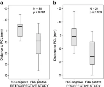
In the retrospective arm, VTs were found in 38 of 685 evaluated women, of which 17 (45%) demonstrated increased FDG uptake. Patient details are listed in Table1. Thirteen FDG-positive VTs were located below the PCL (13±11.2 mm), one 7 mm above,and three VTs were just at the level of the PCL. In contrast, 12 of the FDG-negative VTs were above and 9 slightly under the PCL (−1.4±6.9 mm) (Fig. 2a). A statisti-cally significant positive correlation was found between positive FDG uptake and position below the PCL (p=0.001).
Twenty-four premenopausal women were included in the prospective study of which four were in the menstrual phase (patient details listed in Table 1). In seven patients (29%), VTs were FDG positive (SUVmax18.8±11.9 g/ml),
none with blood traces evident at visual inspection. Five FDG-positive VTs were localized below (20±7.5 mm), one at the level of and one 3 mm above the PCL. In contrast, 9 of 17 inactive VTs were at the level of or above the PCL
Retrospective Prospective p
Number of patients 38 24
-Age (years±SD) 36±8.4 38±8.5 0.175
Menstrual bleeding n.a. 4 (16.6%)
-Body mass index (kg/m2± SD) 23.7±4.9 22.5±4.8 0.731
Parity 0.7±1 0.6±0.84 0.664 Primary tumour Breast cancer 13 7 GIST 1 1 Melanoma 10 6 Colon carcinoma 2 2 Lymphoma 6 4
Focal nodular hyperplasia 0 1
Bronchial carcinoma 0 2 Thyroid carcinoma 1 1 Osteosarcoma 1 0 Ewing’s sarcoma 1 0 Pharyngeal carcinoma 1 0 Infection 2 0
Table 1 Patient demographics
SD standard deviation, GIST gastrointestinal stromal tumour
Fig. 2 PET/CT data analysis. Correlation between VT position and FDG activity, illustrated with box plots for negative vs FDG-positive VTs. a In the retrospective study (n=38) for VT position above (negative) or below the PCL (mm) (p=0.001, Mann-Whitney U test) and b analogous in the prospective part (p=0.039, Mann-Whitney U test)
(−6.6±5.7 mm) and 8 inactive VTs were slightly below the PCL (6±4.7 mm) (Fig. 2b). A statistically significant positive correlation was found between VT FDG uptake and position below the PCL (p=0.039). Figure3illustrates
all seven FDG-active VTs of the prospective study and one FDG-negative VT (Fig. 3a) for direct comparison. FDG activity was found at the base of all seven positive VTs, extending into the middle portion in two cases (SUVmax
Fig. 3 Sagittal fused PET/CT images through the pelvis of eight non-menstruating women in the prospective study. a VT position relative to the PCL in a patient with an FDG-negative VT (*), with its base located at the level of the PCL (white line); creatinine concentration was not measurable. b–h All seven FDG-positive VTs of the
prospective study had a high creatinine concentration, mainly at the base (728±871μmol/l). Five VTs were located clearly below the PCL (b–f), one just at the level of the PCL (g) and one 3 mm above the PCL (h)
Fig. 4 Box plots for correlation of FDG activity in VTs with age, BMI or height. Statistical comparison with the Mann-Whitney U test for FDG-negative vs FDG-positive VTs in the pooled data (n=62): a for age (p=0.169), b BMI (p=0.903) and c height (p=0.453). No correlation was found
37.5 g/ml, 23.7 g/ml; Fig. 3b, c) and into the superior portion in just one patient (SUVmax 24 g/ml; Fig. 3d). Of
these seven positive VTs, six had the original manufac-turer’s cotton string in place (SUVmax 15.7±9.4 g/ml),
while one was modified (i.e. cotton string replaced by nylon monofilament), with uptake at its base and middle portion (SUVmax 37.5 g/ml; Fig.3c). Of 24 women in the
prospective study, 4 had blood traces on their VTs without increased FDG uptake.
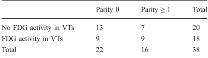
Pooling the retrospective and prospective data resulted in a total of 62 cases with FDG-active VTs. Thirty-four VTs (54.8%) were located below the PCL, of which 18 (53%) showed increased FDG uptake. On the other hand, 17 VTs were located above the PCL, of which only 2 (11%) were FDG positive. In the pooled data, no statistically significant correlation was found regarding VT FDG uptake and age (p=0.169), body mass index (BMI) (p=0.285) or height (p=0.453) (Fig. 4). The obstetric history of 38 (61%) patients was known. No statistically significant correlation was found for parity and VT FDG uptake (p=0.350,
Pearson chi-square). Table 2 shows the distribution of FDG-positive and FDG-negative VTs.
In the prospective arm, activity in the VTs after removal ranged from 202 to 919 kBq, with an average of 518± 308 kBq. Creatinine concentration was increased in all seven FDG-positive VTs, which was consistently found at the base, and to a lesser degree in the middle and superior portions [base 728±871 μmol/l, middle 469±595 μmol/l, top (positive only for five of seven VTs) 216±149μmol/l]. In contrast, in none of the three FDG-negative VTs, which served as negative controls, were creatinine concentrations within detectable limits (<18 μmol/l). A high correlation was noted between the SUVmax and VT creatinine
concen-tration (p<0.001, R2=0.772, y=0.016x+2.842) (Fig. 5a). Active VTs showed a significantly greater increase in weight after use (3.416±2.478 g, range 1.35–8.5 g) compared to FDG-negative VTs (0.501±0.311 g, range 0.026–1.032 g; p<0.001) (Fig. 5b). Calculating with a specific gravity of 1 g/ml for urine, and subtracting 0.5 g for vaginal secretion (i.e. average increase in weight of FDG-negative VTs), the average absorbed urinary volume is estimated at 2.8–2.9 ml, with a maximum of 7.97 ml.
Discussion
This study provides strong evidence that urinary contami-nation is the cause of FDG-positive VTs in PET/CT scanning. Particularly in non-menstruating women, creati-Table 2 Previous vaginal delivery versus FDG activity in VTs
Parity 0 Parity≥ 1 Total
No FDG activity in VTs 13 7 20
FDG activity in VTs 9 9 18
Total 22 16 38
p=0.350 (Pearson chi-square test)
Fig. 5 Results of the VT analysis regarding creatinine concentration and weight. a Correlation between FDG uptake and creatinine concentration in ten VTs. All seven VTs with increased FDG uptake (SUV >3 g/ml) had increased creatinine concentrations which correlated well with FDG uptake/SUVmax. Three VTs with an SUV
<3 g/ml served as negative controls and were not found to have a
creatinine concentration within detectable limits (<18μmol/l). b Box plot illustrating weight increases after use for all patients without menses, with no overlap between FDG-negative and FDG-positive VTs and a significant correlation between increased weight and activity (p<0.001)
nine concentration and FDG uptake in VTs correlated well with the increase in weight of up to 8.5 g, proving the presence of urine in the VT. In addition, the local distribution of creatinine mainly at the base with lower concentration in the middle or top of the VT correlated well with the imaging findings, which always showed FDG accumulation at the base. While urinary contamination of VTs is not a priori expected, the increased FDG uptake in VTs cannot be explained by menstrual blood, because of dominant radioactivity in the caudal portions of the VTs and because seven non-menstruating women also had FDG-positive VTs. If vaginal secretion of FDG or F-active metabolites were causative for VT activity, one would first expect an equal activity distribution over the whole surface of the VTs and therefore also in their top portions. Second, FDG activity would be expected to be increased in the vaginal lumen even in patients without VTs. Third, vaginal secretion has an average glucose concentration of approx-imately 5.0 mM, which is in the range of typical fasting blood sugar levels. Hence, VT FDG concentration in vaginal secretion should not be higher than blood pool values (SUV 1.5–2.5 g/ml) [15].
The prevalence of involuntary leakage of urine for premenopausal women aged 20–44 years is 9–23% and is mainly due to stress urinary incontinence [16]. Involuntary leakage in premenopausal women during the uptake phase in a resting supine position without physical activity is unlikely. Therefore, contamination of the VT is likely to occur during voiding, either by direct contamination due to VT positioning facilitating its contact with urine or by capillary action of the VT string positioned in the voiding pathway.
Direct contamination can only occur when at least a part of the VT is in the voiding pathway. A good correlation was seen between FDG uptake and position of the VT base at or below the PCL. Therefore, such a caudal position seems to be a risk factor for direct urinary contamination. Possible explanations for a low VT position could be poor place-ment of the VT or a weakness of the pelvic floor, and therefore related to age or parity. However, we could not show such correlations in our premenopausal collective.
A contributory factor of capillary action by the cotton string is questionable. The observations in the prospective group suggest that a modified string could reduce contam-ination since only one modified and six normal VTs showed increased activity. However, sheer capillary action of the cotton strings cannot explain quantities of VT urine content of up to 8 ml.
Very intense FDG activity can obscure adjacent structures due to the so-called blooming or spillover artefact [6–8]. Especially in women with cervicogenital malignancies such artefacts could impair diagnostic accuracy. In addition, also the incidental finding of
pathological activity in the cervical region could be obscured by a highly FDG-avid VT. Therefore, all possible measures should be taken to avoid FDG uptake in VTs. As a consequence of the evidence that urinary contamination during voiding is the reason for FDG accumulation in VTs, we suggest three options to prevent this uptake. Firstly, the VT has to be replaced immediately before PET/CT and after voiding. Secondly, the VT has to be removed prior to PET/CT, or thirdly VTs will have to be designed differently.
The fact that urine can be found in 39% of the VTs after voiding raises also concerns for the general female population using VTs. What impact this urine contamina-tion can have on comfort and hygiene, however, cannot be answered with the present study and needs further inves-tigations.
A limitation of this study is the incomplete obstetric and gynaecologic history for the patients in the retrospective study, in particular, regarding the regular use of VTs and parity. This study cannot differentiate between different factors which may contribute to low VT position, such as improper VT insertion, caudal migration in a resting state or during voiding, or pelvic floor weakness. Therefore, further investigations regarding VT position and the voiding dynamics of the pelvic floor would be needed.
In conclusion, frequently observed FDG uptake in VTs is not caused by menstrual blood, but is due to urinary contamination which is related to VT position below the PCL leading to direct contact with the voiding pathway.
Conflicts of interest None.
References
1. Heffernan EJ, Skehan SJ. Artifact on PET/CT secondary to FDG accumulation in a vaginal tampon. Clin Nucl Med 2007;32:208–9. doi: 10.1097/01.rlu.0000255018.37567.fd00003072-200703000-00009.
2. Jadvar H, Parker JA. Clinical PET and PET/CT. London: Springer; 2005.
3. Chander S, Meltzer CC, McCook BM. Physiologic uterine uptake of FDG during menstruation demonstrated with serial combined positron emission tomography and computed tomography. Clin Nucl Med 2002;27:22–4.
4. Lerman H, Metser U, Grisaru D, Fishman A, Lievshitz G, Even-Sapir E. Normal and abnormal 18F-FDG endometrial and ovarian uptake in pre- and postmenopausal patients: assessment by PET/ CT. J Nucl Med 2004;45:266–71.
5. Liu Y. Benign ovarian and endometrial uptake on FDG PET-CT: patterns and pitfalls. Ann Nucl Med 2009;23:107–12. doi:10.1007/s12149-008-0227-z.
6. Pannu HK, Bristow RE, Cohade C, Fishman EK, Wahl RL. PET-CT in recurrent ovarian cancer: initial observations. Radiographics 2004;24:209–23. doi:rg.24103507824/rg.24103507824/1/209.
7. Kamel EM, Jichlinski P, Prior JO, Meuwly JY, Delaloye JF, Vaucher L, et al. Forced diuresis improves the diagnostic accuracy of 18F-FDG PET in abdominopelvic malignancies. J Nucl Med 2006;47:1803–7.
8. Nuyts J, Dupont P, Van den Maegdenbergh V, Vleugels S, Suetens P, Mortelmans L. A study of the liver-heart artifact in emission tomography. J Nucl Med 1995;36:133–9.
9. Heiken JP, Lee JK. MR imaging of the pelvis. Radiology 1988;166:11–6.
10. Rubens D, Thornbury JR, Angel C, Stoler MH, Weiss SL, Lerner RM, et al. Stage IB cervical carcinoma: comparison of clinical, MR, and pathologic staging. AJR Am J Roentgenol 1988;150:135–8.
11. Olson MC, Posniak HV, Tempany CM, Dudiak CM. MR imaging of the female pelvic region. Radiographics 1992;12:445–65. 12. von Schulthess GK, Steinert HC, Hany TF. Integrated PET/CT:
current applications and future directions. Radiology 2006;238:405–22. doi:rg.24103507824/radiol.2382041977.
13. Husdan H, Rapoport A. Estimation of creatinine by the Jaffe reaction. A comparison of three methods. Clin Chem 1968;14:222–38.
14. Nestle U, Kremp S, Schaefer-Schuler A, Sebastian-Welsch C, Hellwig D, Rübe C, et al. Comparison of different methods for delineation of 18F-FDG PET-positive tissue for target volume definition in radiotherapy of patients with non-small cell lung cancer. J Nucl Med 2005;46:1342–8.
15. Ehrström S, Yu A, Rylander E. Glucose in vaginal secretions before and after oral glucose tolerance testing in women with and without recurrent vulvovaginal candidiasis. Obstet Gynecol 2006;108:1432–7. doi:10.1097/01.AOG.0000246800.38892.fc. 16. Hannestad YS, Rortveit G, Sandvik H, Hunskaar S, Norwegian
EPINCONT study, Epidemiology of Incontinence in the County of Nord-Trøndelag. A community-based epidemiological survey of female urinary incontinence: the Norwegian EPINCONT study. Epidemiology of Incontinence in the County of Nord-Trøndelag. J Clin Epidemiol 2000;53:1150–7. doi:S0895435600002328.