HAL Id: cea-01815488
https://hal-cea.archives-ouvertes.fr/cea-01815488
Submitted on 24 Mar 2020HAL is a multi-disciplinary open access archive for the deposit and dissemination of sci-entific research documents, whether they are pub-lished or not. The documents may come from teaching and research institutions in France or abroad, or from public or private research centers.
L’archive ouverte pluridisciplinaire HAL, est destinée au dépôt et à la diffusion de documents scientifiques de niveau recherche, publiés ou non, émanant des établissements d’enseignement et de recherche français ou étrangers, des laboratoires publics ou privés.
A new single crystal diamond dosimeter for small beam:
comparison with different commercial active detectors
Fanny Marsolat, Dominique Tromson, Nicolas Tranchant, Michal Pomorski,
Maïwenn Le Roy, Marc Donois, Fabien Moignau, Aimé Ostrowsky, Loïc de
Carlan, C. Bassinet, et al.
To cite this version:
Fanny Marsolat, Dominique Tromson, Nicolas Tranchant, Michal Pomorski, Maïwenn Le Roy, et al.. A new single crystal diamond dosimeter for small beam: comparison with different commercial active detectors. Physics in Medicine and Biology, IOP Publishing, 2013, 58, pp.7647 - 7660. �10.1088/0031-9155/58/21/7647�. �cea-01815488�
A new single crystal diamond dosimeter for small beam:
comparison with different active detectors
Running head: A new single crystal diamond dosimeter for small beam
F Marsolat 1, D Tromson 1, N Tranchant 1, M Pomorski 1, M Le Roy 2, M Donois 2, F Moignau 2, A
Ostrowsky 2, L De Carlan 2, C Bassinet 3, C Huet 3, S Derreumaux 4, M Chea 5, K Cristina 5, G
Boisserie 5 and P Bergonzo 1.
1 The French Atomic Energy Commission, Diamond Sensor Laboratory, Gif-sur-Yvette, France 2 The French Atomic Energy Commission, LNHB, Gif-sur-Yvette, France
3 IRSN, PRP-HOM/SDE/LDRI, Fontenay-aux-Roses, France 4 IRSN, PRP-HOM/SER/UEM, Fontenay-aux-Roses, France 5 Pitié Salpêtrière Hospital, Paris, France
Abstract :
Recent developments of new therapy techniques using small photon beams, such as stereotactic radiotherapy, require new detectors to precisely determine the delivered dose. The dosimeter has to be as close as possible to tissue equivalence and to exhibit a small detection volume compared to the size of the irradiation field, because of the lack of lateral electronic equilibrium in small beam. Characteristics of single crystal diamond (tissue equivalent material Z=6, high density) make it an ideal candidate to fulfil most of small beam dosimetry requirements. A commercially Element Six electronic grade synthetic diamond was used to develop a single crystal diamond dosimeter (SCDDo) with a small detection volume (0.165 mm3). Long term stability was
studied by irradiating the SCDDo in a 60Co beam over 14 hours. A good stability (deviation less than ± 0.1 %)
was observed. Repeatability, dose linearity, dose rate dependence and energy dependence were studied in a 10 x 10 cm² beam produced by a Varian Clinac 2100 C linear accelerator. SCDDo lateral dose profile, depth dose curve and output factor (OF) measurements were performed for small photon beams with a micro multileaf collimator m3 (BrainLab) attached to the linac. We focused this study on the comparison of SCDDo measurements to those obtained with different commercially available active detectors: an unshielded silicon diode (PTW 60017), a shielded silicon diode (Sun Nuclear EDGE), a PinPoint ionization chamber (PTW 31014), and two natural diamond detectors (PTW 60003). SCDDo presents an excellent spatial resolution for dose profile measurements, due to its small detection volume. Low energy and low dose rate dependence of the SCDDo are measured, explaining the good agreement between the SCDDo and the efficient unshielded diode (PTW 60017) in depth dose curve measurements. OFs obtained with the SCDDo are satisfactory from 0.6 x 0.6 cm2 to 10 x 10 cm2 field sizes, in comparison to the PinPoint ionization chamber and to the Sun Nuclear EDGE
diode that are known to respectively underestimate and overestimate OF values in small beam, due to the large detection volume of the chamber and the non-water equivalence of both detectors.
Keywords: diamond dosimeter, small beam, output factor.
1. Introduction
Radiotherapy is one of the most powerful techniques used in cancer treatment. Very specific techniques with a specific clinical objective are now used to spare the healthy tissue while tumors are irradiated. The development of Stereotactic treatment has led to an increasing use of small X-ray beams, in the range of 5 to 40 mm in diameter. This advanced technique is used for the treatment of small tumors (less than 20 cm3), benign and malignant, intra and extra-cranial. In Stereotactic
delivered to a patient with trigeminal neuralgia (Kondziolka et al 1998, Kondziolka et al 2002); in Stereotactic Radiotherapy multiple fractions of lower dose (1.8 Gy-4 Gy) are used (Das et al 2000). Because of the complicated beam ballistic and realization, the Stereotactic technique presents critical risks and requires a high accuracy in patient positioning and also in dose delivery. The accuracy in patient positioning is improved by the development of advanced imaging modalities and by fixing patient to stereotactic frame (Wulf et al 2000, Baba et al 2009). Dosimetry of small beams is not accurately controlled, the main issue being the determination of Output Factors (OFs) principally because of the lack of lateral electronic equilibrium in small beams. Several authors compared different commercially available detectors and Monte Carlo simulations in small beams (Laub and Wong 2003, Scott et al 2008, Cranmer-Sargison et al 2011, Pantelis et al 2012, Francescon et al 2012). These studies showed the large differences between OFs measured with ionization chambers, silicon diodes, films, thermo-luminescent detectors (TLD) and natural diamonds in fields smaller than 3 cm x 3 cm. The large active volume of detectors and their non-tissue equivalence are the main causes of these broad results.
Recently diamond has been quoted in several papers as a good candidate as a small beam dosimeter (Almaviva et al 2009, Tromson et al 2010, Ciancaglioni et al 2012, Betzel et al 2012). Diamond is nearly tissue-equivalent because of its atomic number (Z=6) close to human tissue effective atomic number (Zeff~7.42). A small active volume of diamond detector allows a high spatial resolution of dose measurement, the high density of atoms in lattice (1023 atoms.cm-3) keeps a high signal-to-noise
ratio and diamond electronic properties permit to achieve fast detector response. Many authors have studied natural diamond dosimeter commercialized by PTW (Hoban et al 1994, Fidanzio et al 2000, Angelis et al 2002). The non-reproducibility between devices, the high cost, the long delivery times and particularly their large detection volume are the main drawbacks for these detectors. Synthetic diamond is a good alternative because reproducible and optimized growth conditions permit to obtain diamond with electronic properties adapted to detection applications. The performances of such synthetic single crystal CVD for X-ray detectors were presented by various authors (Garino et al 2006, Tranchant et al 2008, Almaviva et al 2009, Schirru et al 2010, Betzel et al 2012). In our study, a Single Crystal Diamond Dosimeter (SCDDo) based on a commercially available single crystal CVD diamond (Element Six Ltd.) was developed specifically for stereotactic radiotherapy. The mounted water-equivalent detector was tested in a clinical environment with small photon beams. We focus our present work on the potentialities of our new device and compare the results with those obtained with commercially available real-time detectors commonly used for this application. We measured all the devices in the same conditions and with the same readout procedure.
2. Material and method
2.1. SCDDo and commercial active detectors
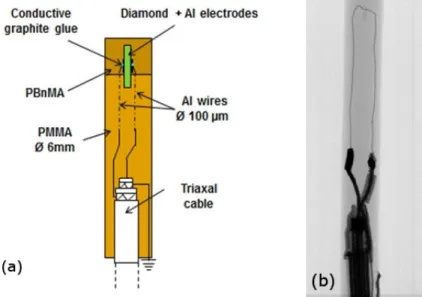
An Element Six electronic grade synthetic single crystal diamond was used to develop water-equivalent SCDDo (Figure 1). The sample dimensions were 1 mm x 1 mm x 165 µm. 100 nm-thick aluminum electrodes were deposited on both sides of the diamond, using an evaporation system. The mounted detector exhibits a small detection volume of about 0.165 mm3, as required for small beam
dosimetry. Materials present in this device were optimized using Monte Carlo simulations in a previous work (Marsolat et al 2013), in order to respect the low-Z requirements for small beam dosimetry and to obtain almost a water-equivalent detector: aluminum electrodes, aluminum wires of 100 µm in diameter, conductive graphite glue, Polybenzylmethacrylat (PBnMA) and Polymethylmethacrylat (PMMA) encapsulation. The triaxial cable was connected at a distance larger than 3 cm in order to avoid perturbation of the deposited dose in the diamond. Finally, conductive colloid graphite covered the device and was connected to ground in order to reduce environmental noise. The position of detection volume in the water-equivalent housing was verified with X-rays radiography. The diamond was located 1.6 mm below the top surface of the housing.
Figure 1: (a): Schema of water-equivalent SCDDo. (b): X-rays radiography of the device.
Dose profiles, depth dose curves and OFs obtained with the SCDDo were compared to those obtained with different commercial active detectors. The unshielded 61017 diode (PTW, Freiburg, Germany) is a p-type silicon diode operating at 0 V, with a disk-shaped sensitive volume perpendicular to the detector axis. Its detection volume has dimensions of 0.6 mm in diameter and 30 µm in thickness. The reference point is located on detector axis, 0.77 mm from detector tip. A good performance of this new unshielded diode and its previous model (PTW 60012) has been observed by many authors (Griessbach et al 2005, Scherf et al 2009, Pantelis et al 2010, Pantelis et al 2012, Dzierma et al 2012) in small beam measurements, compared to shielded diodes. The copper shielded n-type EDGE diode (Sun Nuclear Corp., Florida, USA), polarized at 0 V, with a surface area of 0.64 mm² and thickness of 30 µm has been investigated as well. The PTW 31014 PinPoint ionization chamber is a miniaturized ionization chamber commercially available for small beam and is known as a good reference detector for beam sizes from 3 x 3 cm² to 10 x 10 cm² (Martens et al 2000, Laub and Wong 2003, Scott et al 2008). It operates at the nominal voltage of ±400 V and exhibits a large volume of 15 mm3 (2 mm
diameter by 5 mm length). The PTW 60003 natural diamond detector was polarized at + 100 V and its sensitive volume dimensions range from 1 to 6 mm3. Its active volume is located on detector axis, 1
mm below the top surface of the housing.
2.2. Radiation beams and experimental setup
Clinical environment measurements were performed with the SCDDo at La Pitié Salpêtrière Hospital (Paris, France), under photon beams produced by a Varian Clinac 2100 C medical linear accelerator. A micro multileaf collimator system (µMLC m3, BrainLab) dedicated to stereotactic treatments was attached to this accelerator.
Measurements were performed in a PTW MP3 motorized water phantom, at a source-surface distance (SSD) of 100 cm. The SCDDo was positioned in the water tank with its cable parallel to the beam axis and the smallest dimension of the diamond detection volume (its thickness of 165 µm) in cross-plane direction. All measurements were performed with a 6 MV photon beam, except for the study of energy dependence.
Current-voltage characteristic, repeatability and dose linearity of the SCDDo response were studied with a dose rate of 400 MU.min-1, at 10 cm-depth in water, for a 10 x 10 cm² field. In these conditions,
the absolute dose determined with a calibrated PTW 31003 ionization chamber was 0.6605 cGy.MU-1.
Current-voltage (I-V) characteristic of the device was examined in order to determine the optimal operating voltage for a maximum charge collection, current in the device under radiation is recorded versus bias applied on the device. I-V curve was measured for bias voltages ranging from 0 V to 100 V, in 10 V steps, using a remotely controlled Keithley 6517A electrometer. The repeatability was studied with ten consecutive irradiations with a constant dose of 100 MU and by determining the
coefficient of variation (the percentage ratio of standard deviation to mean charge). The dose dependence of the SCDDo response was measured by irradiating the detector with a dose range from 10 to 800 MU.
The dose rate dependence of the detector response was then investigated by varying both dose per pulse and pulse repetition frequency, for a 10 x 10 cm² field, at 10 cm-depth in water. The first method consists of changing the SSD from 107 cm to 83 cm. The dose rate measured with the reference chamber was varied from 2.34 to 3.64 Gy/min. Measurements were performed by irradiating the SCDDo at each SSD with a constant dose of 1 Gy. To expand the dose rate range, the second method consists of changing the pulse repetition frequency from 80 MU.min-1 to 400 MU.min-1, corresponding
to a dose rate variation from 0.53 to 2.64 Gy.min-1. Measurements were performed by irradiating the
SCDDo at each pulse repetition frequency with a constant dose of 1.32 Gy.
The energy dependence of the detector response was studied by irradiating the SCDDo with a dose of 0.66 Gy, in a 10 x 10 cm² field, at 10 cm-depth in water, for the beam qualities available on the accelerator: 6 MV and 18 MV photon beams.
Repeatability, dose linearity, dose rate and energy dependence of the detector were studied by connecting the SCDDo to a PTW UNIDOS electrometer commonly used in dosimetry.
Lateral dose profiles and depth dose curves were measured with the SCDDo for the smallest field size available with the µMLC m3 (0.6 x 0.6 cm2) and for the 10 x 10 cm² reference field. The dose profiles
measured at 10 cm-depth in water were compared to those obtained with three commercially available active detectors: the silicon diode providing a good spatial resolution (PTW 60017), the PTW 31014 PinPoint ionization chamber and a PTW natural diamond detector for which the precise active volume is unknown. Dose profiles were normalized at 100 per cent on beam axis and the 20 % - 80 % penumbras were evaluated for all detectors. The depth dose curves measured with the SCDDo for 0.6 x 0.6 cm² and 10 x 10 cm² field sizes were compared to those obtained with the PTW 60017 silicon diode and the PinPoint chamber. Depth dose curves were normalized at the depth of maximum dose (dmax). The entrance surface dose (De), the value of dmax and the percentage depth dose (PDD) at 10 cm
in water were analyzed for all detectors. For lateral dose profiles and depth dose curves measurements, all detectors were positioned vertically with the stem and cable aligned with the beam to ensure their uniform irradiation (Scott et al 2008) and they were connected to a PTW Tandem Dual Channel electrometer controlled by Mephysto software.
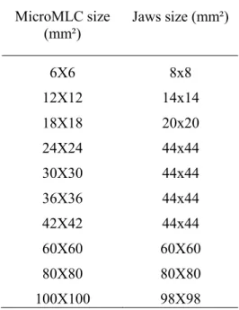
Output factor (OF) measurements were performed with the SCDDo and compared to those obtained with the active commercially detectors presented previously. For the first time, OFs were investigated with a single crystal diamond dosimeter up to 0.6 x 0.6 cm² field size (table 1). Measurements were performed with a SSD of 100 cm and at a depth of 10 g.cm-2 for all fields. As recommended by the
latest Brainlab instructions (BrainLAB Physics 2008) a systematic few millimeters withdrawal of the jaws outside the field defined by the leaves of the MLC enabled uncertainties arising from the jaw’s aperture and centering to be reduced (table 1).The commercial active detectors were connected to a PTW UNIDOS electrometer and positioned vertically in a water tank, except for EDGE diode which was positioned with its stem perpendicular to the beam axis. Precise positioning of detector reference point on beam axis was performed by acquiring lateral dose profiles for 0.6 x 0.6 cm² field size, before OF measurements. For the PinPoint chamber, OF measurements were performed with both positive and negative polarity (± 400 V) and averaged since a polarity effect was observed.
Table 1. Field sizes with microMLC. MicroMLC size (mm²) Jaws size (mm²) 6X6 8x8 12X12 14x14 18X18 20x20 24X24 44x44 30X30 44x44 36X36 44x44 42X42 44x44 60X60 60X60 80X80 80X80 100X100 98X98
Finally, long term stability was studied by irradiating the SCDDo in a 60Co beam during 14 hours, at
the French national metrology laboratory (LNHB, CEA, Gif-sur-Yvette, France). The detector was connected to a Keithley 6517A electrometer and it was positioned in a graphite phantom on beam axis of 60Co beam. After a first irradiation of about 3h30, the beam was switched off during 45 minutes,
and this cycle was repeated 4 times. The stability was analyzed after a pre-irradiation dose of about 5 Gy (30 minutes of irradiation) by determining the maximum current variation in comparison with the first measured value. A deviation less than ± 0.1 % is required by the LNHB (Le Roy et al 2011).
3. Results and discussion
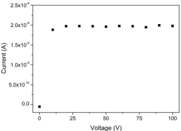
The preliminary I-V curve with 6 MV photon beam obtained for the SCDDo is shown in Figure 2 from 0 V to 100 V. The diamond detector signal saturates for bias voltage higher than 20 V at a current value of 1.95 nA. This saturated current (IR) was compared to the theoretical current value IP
described by equation (1) (Hoban et al 1994, Schirru et al 2010):
(1) The ratio G= IR/IP is defined as the gain factor or the charge collection efficiency. Assuming a dose
rate D = 2.64 Gy.min-1 (measured with the calibrated ionization chamber), the density of diamond
=3.51, the electronic charge e = 1.6·10-19 C, the SCDDo sensitive volume V= 1.65·10-4 cm3 and the
energy required to create an electron-hole pair in diamond w = 13 eV, we obtain IP = 1.96 nA. This
confirms the 100 % charge collection efficiency at bias voltage higher than 20 V, due to the high quality of diamond material and electrical contacts.
eV D IP
Figure 2: I-V characteristic of the SCDDo measured with a 6 MV photon beam.
The following studies were performed with a bias voltage of 50 V which is high enough to gain saturated signal from I(V) curve. After a pre-irradiation of 5 Gy, the coefficient of variation determined for 10 consecutive irradiations of the SCDDo with a constant dose of 0.66 Gy was 0.06 % and confirmed the excellent repeatability of the SCDDo response. A sensitivity of 44.5 nC.Gy-1 was
deduced from these measurements. The dose linearity of the SCDDo response was verified for a 10 x 10 cm² field size, by irradiating the detector with a dose range from 10 to 800 MU. Dose linearity was observed with a linearity coefficient R2 equal to 1 (Figure 3).
Figure 3: Dose linearity of the SCDDo response in 10 x 10 cm² field at a dose rate of 400 MU.min-1. Error
bars are less than the height of data points (■). Linear fit is plotted with solid line.
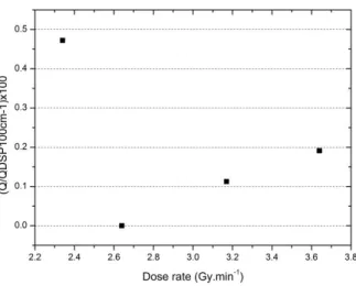
The dose rate dependence of the SCDDo response is shown in Figure 4 and Figure 5. The percentage deviation of the measured charge with respect to the one measured at SSD 100 cm and 400 MU.min-1
is reported in Figure 4.a and Figure 5.a. A deviation lower than 0.5 % is observed in the dose rate range investigated by changing the dose per pulse (dose rate from 2.34 to 3.64 Gy.min-1), and a
maximum deviation of 1 % is obtained by changing the pulse repetition frequency (dose rate from 0.53 to 2.64 Gy.min-1). Thus, the depth dose curve measured with the SCDDo will not require
Figure 4: Dose rate dependence of the SCDDo response in 10 x 10 cm² field, by changing the dose per pulse (SSD modification). Percentage variation of the measured charge is normalized to the value at SSD of 100 cm. Error bars are less than the height of data points (■).
Figure 5: Dose rate dependence of the SCDDo response in 10 x 10 cm² field, by changing the pulse repetition frequency. (a) Percentage variation of the measured charge normalized to the value at 400
MU.min-1. Error bars are less than the height of data points (■).
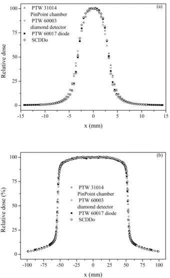
The energy dependence of the detector response was determined for 6 MV and 18 MV photon beams, in a 10 x 10 cm² field, at 10 cm-depth in water. The SCDDo current was measured for a constant dose of 0.66 Gy, for both beam qualities. The variation of the diamond response was only about 1.2 %. The cross-plane dose profiles measured with the SCDDo and three commercially available detectors are displayed in Figure 6, for a 0.6 x 0.6 cm² and a 10 x 10 cm² field. The 20 % - 80 % penumbras are reported in Table 2 for cross-plane and in-plane dose profiles. The SCDDo penumbras are slightly better than those obtained with the PTW 60017 diode which is considered as an excellent spatially resolved commercial detector for small beams. The SCDDo penumbras are much better than those measured with the PTW 31014 ionization chamber and the PTW 60003 diamond detector because of the volume averaging effect. Table 2 confirms also the best spatial resolution of the SCDDo in
cross-plane direction compared to in- cross-plane, due to its small thickness orientation. These penumbra values confirm the excellent spatial resolution of the diamond detector, thanks to its small detection volume.
Figure 6: Cross-plane dose profiles measured with the SCDDo, the PTW 60017 diode, the PTW 31014 PinPoint chamber and a PTW diamond detector, for a 6MV photon beam, with a Varian Clinac 2100 C linac and a µMLC m3. Depth of measurements: 10 cm in water. SSD = 100 cm. Normalization on beam axis. (a) 0.6 x 0.6 cm² beam size. (b) 10 x 10 cm² beam size.
Table 2. 20%-80% penumbras of dose profiles measured with the SCDDo, the PTW 60017 diode, the PTW 31014 PinPoint chamber and a PTW diamond detector at 10 cm-depth in water, for a 6 MV photon beam and two beam sizes: 0.6 x 0.6 cm² and 10 x 10 cm².
Field size (cm²) SCDDo penumbra (mm) PTW 60017 diode penumbra (mm) PTW 31014 PinPoint chamber penumbra (mm) PTW 60003 diamond detector penumbra (mm) In-plane Cross-plane In-plane Cross-plane In-plane Cross-plane In-plane Cross-plane 0.6 x 0.6 1.87 1.64 1.96 1.79 2.39 2.28 2.48 2.34
10 x 10 4,39 4.03 4,80 4.23 5.08 4.86 5.08 4.79
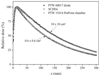
Depth dose profiles measured with the SCDDo, the unshielded silicon diode (PTW 60017) and the PinPoint ionization chamber (PTW 31014) are displayed in Figure 7, for a 0.6 x 0.6 cm² and a 10 x 10 cm² field. The entrance surface dose (De), the depth of dose maximum (dmax) and the percentage depth
dose (PDD) at 10 cm are reported in Table 3 for both investigated field sizes.
All detectors are in good agreement for the 10 x 10 cm² reference field size, except for De values reported in table 3. Since the active volume is located at 0.77 mm and 1.6 mm below the top surface of the housing for the diode and for the SCDDo respectively, the SCDDo build up thickness is more important than the diode one and this explains the difference of entrance surface dose (De) for both detectors. In future diamond detector development, the build-up thickness will be easily reduced in order to improve De measured with the SCDDo. The entrance surface dose obtained with the PinPoint chamber is also higher than the diode one, because the PinPoint chamber was positioned with its cable parallel to beam axis and its active volume has a length of 5 mm in this orientation; the averaging effect influences the entrance surface dose and leads to larger uncertainties in depth dose curve measurements.
For the 0.6 x 0.6 cm² field size, a good agreement is observed between the SCDDo and the diode depth dose curves, except for the entrance surface dose values for the same reasons explained previously. For this small beam, PDD determined at 10 cm with the PinPoint chamber is higher than the SCDDo and diode one. The reason of this last result is the dose underestimation at dmax with the PinPoint
chamber, because its detection volume is too large compared to the beam size at this depth in water and because the presence of air in ionization chamber increases the loss of lateral electronic equilibrium, decreasing the dose measured on the beam axis. But at higher depth in water, the field size increases, the lateral electronic disequilibrium decreases, and the dose measured with the PinPoint chamber is getting closer to the expected value. Since the depth dose curve is normalized at dmax, PDD
at higher depth is slightly overestimated with this ionization chamber.
Figure 7: Depth dose curves measured with the SCDDo, the PTW 60017 diode and the PTW 31014 PinPoint chamber, for a 6MV photon beam, 0.6 x 0.6 cm² and 10 x 10 cm² field sizes, with a Varian Clinac
2100 C linac and a µMLC m3. SSD = 100 cm. Normalization at dmax.
Table 3. Depth of maximum dose (dmax) and percentage depth dose (PDD) at 10 cm-depth in water
measured with the SCDDo, the PTW 60017 diode and the PTW PinPoint chamber, for a 6 MV photon beam and two beam sizes.
Field size (cm²)
SCDDo PTW 60017 Diode PTW 31014 PinPoint
chamber dmax (mm) PDD at 10 cm (%) De (%) dmax (mm) PDD at 10 cm (%) De (%) dmax (mm) PDD at 10 cm (%) De (%) 0.6 x 0.6 11.4 55.8 66.3 11.3 55.3 41.3 11.7 56.3 57,7 10 x 10 13.9 66.6 62.7 14.0 66.4 43.2 15.0 66.3 52,8
OFs normalized at the 10 x 10 cm² field, measured with the SCDDo, the PinPoint chamber, the shielded Sun Nuclear EDGE diode, the unshielded PTW 60017 diode and two PTW diamond detectors are displayed in Figure 8. Moreover, the deviation of OF obtained with commercial detectors with respect to the one measured with the SCDDo is shown in this figure. These OF measurements confirm the increase of interdetector variations with decreasing field size and these variations are considerable below 1.8 x 1.8 cm². For field sizes larger than 1.8 x 1.8 cm2, the SCDDo and the
PinPoint chamber are in good agreement with a deviation below ± 0.5 %. For the smallest beams (less than 1.8 x 1.8 cm2), the PTW 31014 PinPoint chamber underestimates OFs because of its too large
detection volume of air, as explained previously. The results obtained with the SCDDo in small beams are very satisfactory: OFs values are higher than those obtained with the PinPoint detector, with a maximum difference of 7.8 % for the smallest 0.6 x 0.6 cm² field size.
OF values obtained with two different PTW diamond detectors are different for the smallest field sizes with a maximum deviation of 3.2 % for the 0.6 x 0.6 cm² field. The large range of possible sensitive volume for these detectors (from 1 to 6 mm3) explains these discrepancy results. Since their active
volume is larger than the one of SCDDo (0.15mm3), OFs measured with PTW diamond detectors are
lower than those obtained with the SCDDo, with a maximum deviation of - 6.8 %. Moreover, even for large beams (larger than 3 x 3 cm²), both PTW diamond detectors underestimate the OF values compared to the PinPoint chamber which is a reference detector for these field sizes (a deviation of - 1.9 % with respect to the PinPoint chamber for the 4.2 x 4.2 cm² field size).
A good agreement between the EDGE diode, the PinPoint chamber and the SCDDo is observed for field sizes larger than 1.8 x 1.8 cm² whereas a slight deviation of the PTW 60017 diode OFs is observed in this field size range. This difference of behavior is due to the design of the diodes. For the EDGE diode, the metallic shielding allows to selectively absorb the low energy photons which would otherwise lead to an over-response of the diode due to the photoelectric effect in silicon (Griessbach et
al 2005, Eklund and Ahnesjö 2010, Pantelis et al 2010, Cranmer-Sargison et al 2011). The metallic
shielding is replaced by a polymer plastic in the unshielded PTW 60017 diode. The scatter photon contribution increases with the beam size (Wu et al 1993, Heydarian et al 1996) and thus the over-response of the unshielded diode to low-energy photons is more important for larger field leading to a slight underestimation of the OFs.
SCDDo OFs are lower than those measured with the EDGE diode in beam sizes less than 1.8 x 1.8 cm² with a maximum deviation of 2.2 % for the 0.6 x 0.6 cm² beam. This is due to the increase of electron scattering from the metallic shielding into the active volume, which reduces the lateral electronic disequilibrium in small beam and leads to an over-response of the EDGE diode in small beam. This study confirms the better performance of the unshielded PTW 60017 diode in small beam, compared to the EDGE diode (Pantelis et al 2010, Pantelis et al 2012, Francescon et al 2012). A maximum variation of 1.6 % between the SCDDo and the unshielded PTW 60017 diode is observed in the whole investigated field size range.
Figure 8: (a) Output factors measured with the SCDDo, the SunNuclear EDGE diode, the PTW 60017 diode, the PTW 31014 PinPoint chamber and two different PTW diamond detectors, for 6MV photon beam, with a Varian Clinac 2100 C linac and a µMLC m3. Depth of measurements: 10 cm in water. SSD = 100 cm. (b) Relative difference between the commercial detectors OFs and the SCDDo OFs.
Finally, the long-term stability was studied by irradiating the SCDDo in a 60Co beam. The diamond
detector was polarized at 50 V with a Keithley 6517A electrometer and the signal was measured during a succession of long-time irradiations (3h30). After a pre-irradiation of about 5 Gy, the stability of the current over the whole irradiation was analyzed according to the LNHB requirements (Le Roy
et al 2011). The maximum current variation of the SCDDo is lower than ± 0.1 % and respects their
criteria (Figure 9).
Figure 9: Maximum current variation of the SCDDo with respect to the first current value, after a pre-irradiation of 5 Gy, over about 17 hours of measurements.
4. Conclusion
Water-equivalent diamond dosimeter was developed using a commercially available single crystal from Element Six Ltd. Clinical environment measurements were performed to evaluate the suitability of the device for small beam dosimetry. The detector was polarized at 50 V to have a maximum charge
collection. A high sensitivity of 44.5 nC.Gy-1 was obtained by applying this bias voltage to the
SCDDo. An excellent repeatability (0.06 %) was observed with this device. The dose linearity of the SCDDo response was verified with 6 MV photon beam, for a large dose range. A low dose rate dependence of the SCDDo response less than 1% was observed, by changing the dose per pulse or the pulse repetition frequency. Finally, a low energy dependence of 1.2 % of the diamond response was observed between 6 MV and 18 MV beam quality.
Lateral dose profiles measured with the SCDDo, for the smallest field size available with the µMLC-m3 (0.6 cm x 0.6 cm) and for the 10 x 10 cm² reference field size, presents an excellent spatial resolution due its small detection volume (0.15 mm3). The 20 % - 80 % penumbras measured with the
SCDDo are smaller than those measured with the well spatially resolved PTW 60017 diode, PTW 31014 PinPoint chamber and PTW 60003 diamond detector. Depth dose curves measured with the SCDDo are in good agreement to those obtained with the PTW 60017 diode and the PTW 31014 PinPoint chamber for a 10 x 10 cm² field size. For the smallest field size (0.6 x 0.6 cm²), the diode and SCDDo depth dose curves are in good agreement. The PinPoint PDDs are slightly higher than those obtained with the other detectors due to its large detection volume of air. A decrease of the encapsulating material thickness of about 1 mm will be performed to decrease the SCDDo entrance dose.
Output factors measured with the SCDDo for field sizes smaller than 1.8 x 1.8 cm² are higher than those measured with the PTW 31014 PinPoint ionization chamber which is well known to underestimate the OF values in small beam. For larger field sizes, both detectors are in good agreement (better than 0.5 %). SCDDo OFs in small beams are lower than those measured with a Sun Nuclear EDGE diode that overestimates OF values due to its metallic shielding. To our knowledge, this is the first study demonstrating the performance of a diamond dosimeter in beam sizes smaller than 1 cm x 1 cm, in comparison to several active dosimeters. A future work will perform the comparison between SCDDo and passive dosimeters (TLD (Bassinet et al 2010) and EBT films (Huet
et al 2012)) because recently published studies have pointed out the good agreement between different
passive detectors for small beam OF measurements (Huet et al 2011, Pantelis et al 2012, Francescon
et al 2012).
ACKNOWLEDGEMENTS: This work was performed in the framework of project “DIADOMI” which is granted by French National Research Agency.
REFERENCES
Almaviva S et al 2009 Synthetic single crystal diamond dosimeters for Intensity Modulated Radiation Therapy applications Nucl. Instrum. Methods Phys. Res. A, Accel. Spectrom. Detect. Assoc.
Equip. 608 191-4
Angelis C D, Onori S, Pacilio M, Cirrone G A P, Cuttone G, Raffaele L, Bucciolini M and Mazzocchi S 2002 An investigation of the operating characteristics of two PTW diamond detectors in photon and electron beams Med. Phys. 29 248–54.
BrainLAB Physics 2008 Technical Reference Guide Rev 1.0
Baba F, Shibamoto Y, Tomita N, Ikeya-Hashizume C, Oda K, Ayakawa S, Ogino H and Sugie C 2009 Stereotactic body radiotherapy for stage I lung cancer and small lung metastasis: evaluation of an immobilization system for suppression of respiratory tumor movement and preliminary results Radiat. Oncol. 4 15
Bassinet C, Robbes I, Barbier L, Baumann M, Kernisant B, Trompier F 2010 Characterization of 7LiF:Mg,Ti TLD micro-cubes Radiat. Meas. 45 646‑8
Betzel G T, Lansley S P, Baluti F, Reinisch L and Meyer J 2012 Clinical investigations of a CVD diamond detector for radiotherapy dosimetry Phys. Med. 28 144-52
Ciancaglioni I, Marinelli M, Milani E, Prestopino G, Verona C, Verona-Rinati G, Consorti R, Petrucci A and De Notaristefani F 2012 Dosimetric characterization of a synthetic single crystal diamond detector in clinical radiation therapy small photon beams Med. Phys. 39 4493-501
Cranmer-Sargison G, Weston S, Evans J A, Sidhu N P and Thwaites D I. 2011 Implementing a newly proposed Monte Carlo based small field dosimetry formalism for a comprehensive set of diode detectors. Med. Phys. 38 6592‑602
Das I J, Downes M B, Kassaee A and Tochner Z 2000 Choice of Radiation Detector in Dosimetry of Stereotactic Radiosurgery-Radiotherapy J. Radiosurg. 3 177-86
Dzierma Y, Licht N, Nuesken F and Ruebe C 2012 Beam properties and stability of a flattening- filter free 7 MV beam—An overview Med. Phys. 39 2595–602
Eklund K and Ahnesjö A 2010 Spectral perturbations from silicon diode detector encapsulation and shielding in photon fields Med. Phys. 37 6055–60
Fidanzio A, Azario L, Miceli R, Russo A and Piermattei A 2000 PTW-diamond detector: dose rate and particle type dependence Med. Phys. 27 2589-93
Francescon P, Kilby W, Satariano N and Cora S 2012 Monte Carlo simulated correction factors for machine specific reference field dose calibration and output factor measurement using fixed and iris collimators on the CyberKnife system Phys. Med. Biol. 57 3741‑58
Garino Y, Lo Giudice A, Manfredotti C, Marinelli M, Milani E, Tucciarone A and Verona-Rinati G 2006 Performances of homoepitaxial single crystal diamond in diagnostic x-ray dosimetry Appl.
Griessbach I, Lapp M, Bohsung J, Gademann G and Harder D 2005 Dosimetric characteristics of a new unshielded silicon diode and its application in clinical photon and electron beams Med.
Phys. 32 3750–54
Heydarian M, Hoban P W and Beddoe AH 1996 A comparison of dosimetry techniques in stereotactic radiosurgery Phys. Med. Biol. 41 93–110
Hoban P W, Heydarian M, Beckham W A and Beddoe A H 1994 Dose rate dependence of a PTW diamond detector in the dosimetry of a 6 MV photon beam Phys. Med. Biol. 39 1219-29
Huet C, Bassinet C, Derreumaux S, Moignier C, Baumann M, Lacornerie T et al. 2011 Output Factors of a Cyberknife System : Comparison Between Measurements and Monte Carlo Calculations 3525‑26
Huet C, Dagois S, Derreumaux S, Trompier F, Chenaf C, Robbes I 2012 Characterization and optimization of EBT2 radiochromic films dosimetry system for precise measurements of output factors in small fields used in radiotherapy Radiat. Meas. 47 40‑9
Kondziolka D, Lunsford L D and Flickinger J C 1998 Gamma Knife Radiosurgery as the First Surgeryfor Trigeminal Neuralgia Stereotact. Funct. Neurosurg. 70 187-91
Kondziolka D, Lunsford L D and Flickinger J C 2002 Stereotactic radiosurgery for the treatment of trigeminal neuralgia Clin J Pain 18 42-7
Laub W U and Wong T 2003 The volume effect of detectors in the dosimetry of small fields used in IMRT Med. Phys. 30 341-47
Le Roy M, De Carlan L, Delaunay F, Donois M, Fournier P, Ostrowsky A, Vouillaume A and Bordy J M 2011 Assessment of small volume ionization chambers as reference dosimeters in high-energy photon beams Phys. Med. Biol. 56 5637–50
Marsolat F, Tromson D, Tranchant N, Pomorski M, Lazaro-Ponthus D, Bassinet C, Huet C, Derreumaux S, Chea M, Boisserie G, Alvarez J and Bergonzo P 2013 Diamond dosimeter for small beam stereotactic radiotherapy Diamond Relat. Mater. 33 63-70
Martens C, De Wagter C and De Neve W 2000 The value of the PinPoint ion chamber for characterization of small field segments used in intensity-modulated radiotherapy Phys. Med.
Biol. 45 2519-30
Pantelis E et al. 2010 On the implementation of a recently proposed dosimetric formalism to a robotic radiosurgery system Med. Phys. 37 2369‑79
Pantelis E, Moutsatsos A, Zourari K, Petrokokkinos L, Sakelliou L, Kilby W, et al 2012 On the output factor measurements of the CyberKnife iris collimator small fields: Experimental determination of the
k
clin msr msr clin f f Q Q ,, correction factors for microchamber and diode detectors Med. Phys. 39 4875‑85
Scherf C, Peter C, Moog J, Licher J, Kara E, Zink K, Rödel C and Ramm U 2009 Silicon diodes as an alternative to diamond detectors for depth dose curves and profile measurements of photon and electron radiation Strahlenther Onkol. 185 530–6
Schirru F, Kisielewicz K, Nowak T and Marczewska B 2010 Single crystal diamond detector for radiotherapy J. Phys. D: Appl. Phys. 43 265101
Scott A J D, Nahum A E and Fenwick J D 2008 Using a Monte Carlo model to predict dosimetric properties of small radiotherapy photon fields Med. Phys. 35 4671-84
Tranchant N, Tromson D, Descamps C, Isambert A, Hamrita H, Bergonzo P and Nesladek M 2008 High mobility single crystal diamond detectors for dosimetry: Application to radiotherapy Diamond Relat. Mater. 17 1297-301
Tromson D, Descamps C, Tranchant N, Bergonzo P, Nesladek M and Isambert A 2008 Investigations of high mobility single crystal chemical vapor deposition diamond for radiotherapy photon beam monitoring J. Appl. Phys. 103 54512–16
Tromson D, Rebisz-Pomorska M, Tranchant N, Isambert A, Moignau F, Moussier A, Marczewska B and Bergonzo P 2010 Single crystal CVD diamond detector for high resolution dose measurement for IMRT and novel radiation therapy needs Diamond Relat. Mater. 19 1012-16 Wu A, Zwicker R D, Kalend A M and Zheng Z 1993 Comments on dose measurements for a narrow
Wulf J, Hädinger U, Oppitz U, Olshausen B and Flentje M 2000 Stereotactic radiotherapy of extracranial targets: CT-simulation and accuracy of treatment in the stereotactic body frame
Radiother. Oncol. 57 225-36