HAL Id: insu-02369905
https://hal-insu.archives-ouvertes.fr/insu-02369905
Submitted on 26 Aug 2020
HAL is a multi-disciplinary open access
archive for the deposit and dissemination of
sci-entific research documents, whether they are
pub-lished or not. The documents may come from
teaching and research institutions in France or
abroad, or from public or private research centers.
L’archive ouverte pluridisciplinaire HAL, est
destinée au dépôt et à la diffusion de documents
scientifiques de niveau recherche, publiés ou non,
émanant des établissements d’enseignement et de
recherche français ou étrangers, des laboratoires
publics ou privés.
Exceptionally preserved cryptoniscium larvae
-morphological details of rare isopod crustaceans from
French Cretaceous Vendean amber
Mario Schädel, Vincent Perrichot, Joachim Haug
To cite this version:
Mario Schädel, Vincent Perrichot, Joachim Haug. Exceptionally preserved cryptoniscium larvae -
mor-phological details of rare isopod crustaceans from French Cretaceous Vendean amber. Palaeontologia
Electronica, Coquina Press, 2019, 22, pp.22.3.71. �10.26879/977�. �insu-02369905�
Palaeontologia Electronica
palaeo-electronica.orghttp://zoobank.org/30999446-D188-41C9-9643-98CC42CB7FDB
Schädel, Mario, Perrichot, Vincent, and Haug, Joachim T. 2019. Exceptionally preserved cryptoniscium larvae - morphological details of rare isopod crustaceans from French Cretaceous Vendean amber. Palaeontologia Electronica 22.3.71 1-46. https://doi.org/ 10.26879/977
palaeo-electronica.org/content/2019/2757-cretaceous-epicaridea Copyright: November 2019 Palaeontological Association.
This is an open access article distributed under the terms of Attribution-NonCommercial-ShareAlike 4.0 International (CC BY-NC-SA 4.0), which permits users to copy and redistribute the material in any medium or format, provided it is not used for commercial purposes and the original author and source are credited, with indications if any changes are made.
Exceptionally preserved cryptoniscium larvae -
morphological details of rare isopod crustaceans
from French Cretaceous Vendean amber
Mario Schädel, Vincent Perrichot, and Joachim T. Haug
ABSTRACT
Epicaridea is an ingroup of Isopoda that comprises only parasitic crustaceans. Within parasitic isopods, epicarideans represent a special case: throughout their ontogeny they switch from a small intermediate host (copepod) to a final host (various larger crustaceans), and develop through distinct larval phases (epicaridium, micronis-cium and cryptonismicronis-cium). Young males of some species retain a larval morphology. Recent findings of fossil epicarideans in amber from the Miocene of Mexico consisted in the only epicaridean body fossils, until one specimen has been figured from Creta-ceous amber from France. Here we provide a detailed analysis of this specimen and 20 more specimens from the same locality. The presented specimens represent the oldest occurrence of epicaridean body fossils, extending their fossil record by 67 mil-lion years.
The fossils are exceptionally well preserved and, despite their small size of less than 0.5 mm, reveal even fine morphological details. The specimens correspond either to cryptoniscium larvae or males that have retained their larval morphology. There are no morphological features in the fossils that argue against conspecifity of all speci-mens. All character states found in the fossils are also present in extant species. Given the displayed combination of character states and the age difference, it is unlikely that the specimens are conspecific to any extant species nor to much younger fossils from the Miocene of Mexico. The species Vacuotheca dupeorum gen. et sp. nov. is described and interpreted as an epicaridean of uncertain affinities, but that is not part of the epicaridean ingroup Dajidae. Furthermore, multiple aspects of the evolutionary history of parasitic isopods and epicarideans in particular are discussed. This includes possible scenarios for host changes that could have led to the life cycle of modern epi-carideans and the evolution of size within epicaridean larvae.
SCHÄDEL, PERRICHOT, & HAUG: CRETACEOUS EPICARIDEA
Mario Schädel. Ludwig-Maximilians-Universität München, Department of Biology II, Zoomorphologygroup, Großhaderner Straße 2, 82152 Planegg-Martinsried, Germany. [email protected] Vincent Perrichot. Univ. Rennes, CNRS, Géosciences Rennes, UMR 6118, 35000 Rennes, France. [email protected]
Joachim T. Haug. Ludwig-Maximilians-Universität München, Department of Biology II, Zoomorphology group, Großhaderner Straße 2, 82152 Planegg-Martinsried, Germany and Ludwig-Maximilians-Universität München, GeoBio-Center, Richard-Wagner-Str. 10, 80333 München, Germany. [email protected]
Keywords: aquatic amber; Cymothoida; Epicaridea; fluorescence microscopy; fossilised ontogeny; palaeo-parasitism
Submission: 3 March 2019. Acceptance: 17 September 2019.
INTRODUCTION General Background
Isopoda (woodlice and their relatives) is an enormously diverse group of malacostracan crus-taceans. Having a marine origin, isopod species did not only master the transition to a fully terres-trial life (Oniscidea), they also inhabit deep sea and freshwater environments, and some groups even developed parasitic lifestyles (Williams and Bunk-ley-Williams, 2019). Some isopod species have been known as parasites of fishes and crustaceans for a long time (e.g., Müller, 1862). However, the evolution of these groups of parasites is still quite enigmatic to the present day.
Fossils can provide important clues to the early evolution of a group, by combining highly specialised modern-type features with more plesio-morphic aspects of the morphology. Fossils may provide evolutionary “steps-in between”. Yet, in the case of parasitic isopods it is not so simple.
Isopod fossils are not very common in the fos-sil record in general and identifying a parasitic life-style based on fossil morphology is quite challenging (see discussion in Nagler and Haug, 2015). Thus, the record of parasitic isopod body fossil is, so far, highly limited.
Even when fossils are available, a solid understanding of the biology of the suspected extant relatives is required to interpret their signifi-cance. Also, hypotheses on the relationships between animal groups of interest should always be critically evaluated in light of the usually more detailed known extant species.
Whether all parasitic isopods belong to a monophyletic group that excludes non-parasitic species is still a matter of debate. It seems widely accepted that most fish parasites and some preda-tory and scavenging forms (Cirolanidae,
Corallani-dae, Tridentella, AegiCorallani-dae, and Cymothoidae) are closely related and form the monophyletic group Cymothoida (Wägele, 1989; Brusca and Wilson, 1991; Dreyer and Wägele, 2001; Brandt and Poore, 2003). However, the position of Gnathiidae (only larval forms are fish parasites) and Epi-caridea (parasites on crustaceans) is still under debate (Wägele, 1989; Brusca and Wilson, 1991; Dreyer and Wägele, 2001, 2002). Brusca and Wil-son (1991) suggested a sister group relationship between Epicaridea and Gnathiidae (outside of Cymothoida), whereas Dreyer and Wägele (2001, 2002) suggested Epicaridea being the sister group of Cymothoidae (within Cymothoida).
The analysis of Nagler et al. (2017) combines close relationships proposed for Cymothoidae and Epicaridea (Wägele, 1989) and between Gnathii-dae and Epicaridea (Brusca and Wilson, 1991) by interpreting a group including Gnathiidae and Epi-caridea as a sister group to Cymothoidae. Support for this view is currently mainly provided by an exceptionally preserved fossil of the group Urda Münster, 1840, combining characters of Epi-caridea, Gnathiidae and Cymothoidae (Nagler et al., 2017).
Isopoda is an ingroup of the diverse group Peracarida. All peracaridans share a unique spe-cialisation: the adult female develops a brood pouch that is covered with sclerites protruding from the legs (oostegites), providing for prolonged maternal care. As a result, most species do not produce true larval offspring in the strict sense (for difficulties of the term see Haug, in press). The immatures, that leave the brood pouch, largely resemble the adults in morphology and ecology (but see discussion in Lang et al., 2007). This holds also true for the stem species of Isopoda (Ax, 2000).
PALAEO-ELECTRONICA.ORG
In fish-parasitising isopods (mostly species of Aegidae and Cymothoidae) dispersal happens in the so-called manca stage or the subsequent juve-niles. The manca stage lacks a fully developed seventh pereopod (thoracic appendage 8, append-age of post-ocular segment 13) that the adults have (Boyko and Wolff, 2014) but otherwise resembles the adult in the general body organisa-tion. Yet, based on their ecological function (disper-sal) they may be interpreted as functional larvae (if dispersal is considered a larval feature; see Haug, in press). In Cymothoidae subsequently gradual morphological changes in favour of a close para-site-host interaction can happen, which can, for example, lead to the loss of the bilateral symmetry in late stages of the individual development (e.g., van der Wal et al., 2019).
In epicarideans, much smaller offspring is released from the brood pouch and the ontogene-sis can be separated in distinct steps with very dif-ferent ecological functions corresponding to very different morphologies, too (Figure 1). In most epi-carideans the ontogeny can be differentiated into three distinct true larval stages, accepted as such by most authors - epicaridium, microniscium and cryptoniscium - and the subsequent further devel-opment towards adults (Williams and Boyko, 2012; Boyko and Wolff, 2014).
The Post-embryonic Ontogeny of Epicarideans Epicaridium. With only one reported exception
(Miyashita, 1940) epicaridean crustaceans hatch from their eggs as epicaridium larvae. The name of this larval type derives from its discovery on the definitive host, i.e., a caridean shrimp, where lar-vae were released from the brood pouch of the female (Fraisse, 1878). Epicaridium larvae are stout in appearance with short but wide thoracic segments. There is a clear distinction between the free trunk segments (segments that have a dorsal sclerite and not conjoined with others) into pereon-segments (posterior, free thoracic pereon-segments) and the pleon-segments regarding the morphology of the legs. At least the anterior pereopods are well differentiated. The pleopods bear distinct setae (Dale and Anderson, 1982).
After their release they become planktic and infest copepods (Boyko et al., 2013). The small epicaridium larvae grasp the appendages of the copepods and move to the trunk where they will moult and transform to the next distinct larval stage: the microniscium.
Microniscium. Relatively little is known about the
life of microniscia, besides that they are parasitis-ing copepods crustaceans. At some point (maybe still as epicaridia?) they pierce through the integu-ment of the host and from then on feed on the host’s body fluid (Pike pers. comm. in Marshall and Orr, 2013). Due to the relatively large size of the microniscium compared to its host it causes a tre-mendous negative effect on the reproductive rate of the copepods individual (Uye and Murase, 1997). It is also likely (but has not been reported yet) that the microniscium kills its host. In this case, the microniscium would rather correspond to a par-asitoid than a parasite. The mechanism of detach-ment from this intermediate host and when or where moulting towards next stage happens remains to be investigated.
The epicarideans use the phase of attachment to the copepod with a steady income of nutrients to drastically change the overall body morphology. While the epicaridium is rather stout in appearance compared to the subsequent larval stages, it pos-sesses well-developed setae and specialized, fully developed appendages (Anderson and Dale, 1981). The microniscium is more slender. Early microniscia lack fully developed thoracic append-ages, and the seventh pereopod (last thoracic appendage) is missing entirely as well as the seta-tion of the pleopods. The absence of the last tho-racic appendage is reminiscent to the condition in manca stages in other isopod species. During the
FIGURE 1. Sketched and simplified illustration of the
life cycle of epicarideans. 1.1: Planktic epicaridium larva. 1.2: Microniscium larva feeding on a copepod (intermediate host). 1.3: Planktic cryptoniscium larva.
SCHÄDEL, PERRICHOT, & HAUG: CRETACEOUS EPICARIDEA
microniscium larval stage a significant growth and a morphological change (supposedly without moulting) towards the morphology of the cryptonis-cium stage can be observed (Anderson and Dale, 1981).
Cryptoniscium. The cryptoniscium is the last
dis-tinct larval stage that can be observed throughout the ontogeny of species of all epicaridean lineages. The cryptoniscium develops from a microniscium that is attached to a planktic copepod. Cryptoniscia are all still rather small and resemble microniscia in overall body shape (Nielsen and Strömberg, 1973). Similar to epicaridia, cryptoniscium larvae are planktic and in search for a host. They swim actively and at least some cryptoniscia are able to curl up (for protection?) (Fraisse, 1878). The over-all morphology of cryptoniscium larvae is relatively uniform in all epicaridean species known from this stage (Anderson and Dale, 1981). The body is elongated with a convex dorsal and a concave ven-tral surface. The mouthparts form a sucking mouth cone. The seventh pereopod (thoracic appendage eight) is present and the dactyli of the pereopods, i.e., the terminal elements of posterior seven tho-racic appendages, are recurved forming a func-tional subchelae with the proximal appendage elements. The pleopods have long natatory setae; the uropods are rod-shaped with long distal setae.
For the species Entoniscoides okadai Miyash-ita, 1940, it has been reported that the hatching stage has a cryptoniscium-like habitus. This was shown by the examination of the brood pouch of an adult female where pre-hatched and hatched lar-vae, with the appearance of a cryptoniscium, were observed. Furthermore, an embryonic stage resembling microniscium larvae has been described (although this statement is not directly evident from the provided photograph; Miyashita, 1940). Without free swimming epicaridia, it is likely that Entoniscoides okadai is not parasitising cope-pods. With respect to the current phylogenetic hypothesis it seems unlikely that this represents an ancestral feature, but is better understood as a decrease of step numbers during ontogeny along with an intensification in maternal care.
Later development and sex. The stage following
the cryptoniscium has been termed ‘bopyridium’ by some authors (e.g., Oliveira and Masunari, 2006). Boyko and Wolf (2014) critically questioned the value of this term. We support their critical view. The term indicates the presence of a distinct stage of life, yet it refers to a phase of morphological tran-sition between the cryptoniscium and the adult (including several moults) and cannot be properly
outlined using morphological features. In other groups comparable stages of eucrustaceans would have been simply addressed as ‘juveniles’.
The sexual development is highly variable within epicaridean species. It can be (1) strictly protandric, meaning that all individuals are males at first, or (2) depend on an external trigger, such as the presence of on adult female on the same host. Yet, also a direct, externally triggered, devel-opment from the cryptoniscium towards both sexes is possible (summarised in Wägele, 1989). Possi-ble genotypic determination for both sexes has also been reported once (Hiraiwa, 1936). When a female dies on the alive host, adult males can also transform into functional females (Reverberi, 1947).
Hosie (2008) stated that in the epicaridean subgroup Cryptoniscoidea males are often not dis-tinguishable from cryptoniscia. The author uses the term ‘male’ for all non-planktic cryptoniscoideans that show no signs of modification towards a female habitus. We think this practice is critical; it heavily depends on assumptions about the sexual development and behaviour of epicarideans, which has not been studied in detail for most species.
Epicaridean Ecology
The entire larval phase of epicaridean crusta-ceans lasts around 10 to 30 days (Caroli, 1928; Anderson, 1975). The dispersal of the larval stages strongly correlates with the length of the larval phases. A passive transportation of up to 100 km distance has been reported during the time of the larval development (Owens and Rothlisberg, 1991). Further spatial dispersal obviously depends on the mobility of the host.
As their name suggests, adult epicarideans can be found especially on caridean shrimps. How-ever, they are not restricted to them as final hosts, but also infest a variety of other crustaceans including other isopod species (Nielsen and Ström-berg, 1965). Some species are also hyperparasitic on other epicarideans (Rybakov, 1990) or rhizo-cephalans (parasitic barnacles) (Williams and Boyko, 2012). Epicarideans have even be reported infesting cephalopods (Pascual et al., 2002); the authors suggest that similar cases may have sim-ply been overseen in the past due to their small size. Hence, cephalopods could indeed represent additional host species.
Given the planktic dispersal stages in Epi-caridea and the multiplicity of hosts observable in only one species (Bourdon, 1968), it is surprising to note the apparent impacts of geographical
bounda-PALAEO-ELECTRONICA.ORG
ries on the distribution of extant representatives. Markham (1968) mentioned the example of the epicaridean ingroup Orbioninae (Bopyroidea), which occurrence is restricted to the Indo-Pacific although suitable hosts are globally distributed.
Epicarideans are not limited to full marine environments but also occur in brackish estuarine habitats (Anderson and Dale, 1981). There are also records for species living in strict freshwater environments (Chopra, 1923).
Epicaridean Ingroup Relationships
As a result of their complex life cycle in combi-nation with the presence of two planktic larval stages, associating larvae and adults is challeng-ing. Thus, many species are known either as cryp-toniscium larvae only (e.g., Schultz, 1975) or lack the description of this stage (e.g., Williams and An, 2009). This clearly affects the research on the sys-tematics of the group, leading Boyko et al. (2013, p. 496) to the following assertion about the taxon-omy within Epicaridea: “[…] genera are defined by the gross morphology of the females, and species by characters of cryptoniscium larvae.” Future
studies have to overcome the taxonomic bias that has been caused by the arbitrary distinctions between species and higher level characters.
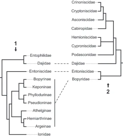
Very few studies have focused on the epi-caridean ingroup relationships. Beside a few older publications (Shino, 1965; Markham, 1968; reviewed in Boyko and Williams, 2009) for which the results are non-replicable (lacking any descrip-tion of methods), there are only three studies focusing on this issue (Wägele, 1989; Boyko et al., 2013; Boyko and Williams, 2015). These phyloge-netic analyses are both limited with respect to the number of included epicaridean species and, as a consequence, are complementary rather than comparable (see Figure 2). Both found Entonisci-dae as the sister group of BopyriEntonisci-dae and DajiEntonisci-dae inside Cryptoniscoidea.
Fossil Record of Epicaridea
Until recently, there was simply no report for epicaridean body fossils. Hitherto, the fossil record of Epicaridea was consisting of swellings observed from the branchial chambers of fossil decapod crustaceans. These swellings were first identified
FIGURE 2. Confronting phylogenetic hypotheses in Epicaridea. Dashed lines represent supported monophyletic
groups. 2.1: Molecular phylogeny from Boyko et al. (2013). 2.2: Phylogeny based on putative apomorphic morpholog-ical characters from Wägele (1989).
SCHÄDEL, PERRICHOT, & HAUG: CRETACEOUS EPICARIDEA
by Bell (1863) and attributed by actualism to the internal colonization of the gill chamber, as nowa-days performed by adult bopyroideans.
These fossil deformations supposedly induced by epicarideans have been listed and reviewed by Markham (1968), Wienberg Rasmus-sen et al. (2008), Klompmaker et al. (2014) and Klompmaker and Boxhall (2015). Their oldest occurrence is reported from a lobster-like crusta-cean (Erymidae) from the Toarcian (Lower Juras-sic) of Western New Guinea (Soergel, 1913). This occurrence is questionable, as the repository of the depicted specimen is unknown, and because there is no record for such swellings in the Middle Juras-sic so far (see also Klompmaker et al., 2014; Klompmaker and Boxshall, 2015). Klompmaker et al. (2014) reported a ‘peak’ in infestation during the Late Jurassic and supposed that this, rather than being a sampling artefact, could be linked to syn-ecological reasons (occurrence of potential host species, biological defence strategies, etc.). Klompmaker et al. (2014) furthermore showed that more different species of true crabs (Brachyura) were infected in comparison to the representatives of its sister group (squat lobster, hermit crabs, false crabs; all together Anomura/Anomala). Yet, ano-muran/anomalan crustaceans seem to have been more frequently infected than brachyuran crabs when considering the number of infected individu-als per taxon for a Cretaceous assemblage.
Klompmaker et al. (2014) also erected the ich-notaxon Kanthyloma crusta for these Epicaridea-caused swellings (see Klompmaker and Boxhall, 2015 for a further discussion regarding this nomen-clatural practice). Attempts have been made to investigate the preservation of isopod body fossils within swellings in fossil crustaceans through com-puted tomography without success (N. Robin, 2019 pers. comm.). In experimental studies on the taphonomy of decapod crustaceans, remains of epicarideans are still present up to 25 days after the death of the infected host (Klompmaker et al., 2017).
A preservation type that could have the chance to preserve epicarideans is amber. That liv-ing arthropods submerged in water can get trapped by resin was experimentally shown (Schmidt and Dilcher, 2007) and can be explained by active or passive collision with the submerged resin (Figure 3). Yet, aquatic and especially marine organisms are relatively rare in amber considering their over-all proportion in amber inclusions. However, there are some aquatic or even marine organisms in many amber localities (Schmidt et al., 2004;
Key-ser and Weitschat, 2005; Girard et al., 2008; Saint Martin et al., 2015; Serrano-Sánchez et al., 2015, 2016; Xing et al., 2018).
More recently there were two reports of epi-caridean body fossils preserved in amber. 1) There is a record from Miocene Mexican amber (Serrano-Sánchez et al., 2016). The fossils, which come from the Campo La Granja site, are clearly recog-nisable as larval epicarideans. These specimens were the first fossil record of epicaridean body fos-sils as well as a rare occurrence of fossil crusta-cean larvae in general. 2) Shortly after this primary description, Néraudeau et al. (2017) reported a second set of epicaridean larvae in the palaeonto-logical content of a new French amber deposit. In this case, the fauna is significantly older than the Mexican epicarideans (about 90 million years) and allows access to better apparent morphological details.
Here, we describe 21 exceptionally well-pre-served epicarideans from Cretaceous amber of Vendée, France. We further discuss the implica-tions of the find for our understanding of the evolu-tionary history of epicaridean crustaceans. The aspect of body size for the known fossil epicarid-ean larvae in comparison to the extant representa-tives is discussed. Also, we critically discuss multiple possible scenarios that could have lead to the complex life cycle of extant epicarideans. The taphonomical implications of the herein presented fossils are discussed with respect to the
circum-FIGURE 3. Illustration of possible entombment
condi-tions suggested for Vendean amber: cryptoniscium lar-vae living in an aquatic environment close to the resin producing tree and getting trapped by making contact with submerged liquid resin.
PALAEO-ELECTRONICA.ORG
stances that could have lead to their preservation in amber.
GEOLOGICAL SETTING
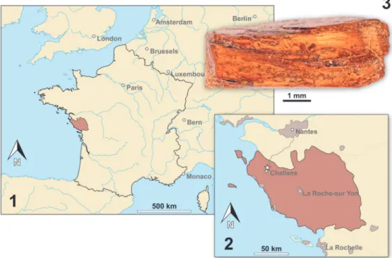
The Vendean amber deposit is located in northwestern France (Pays-de-la-Loire region) at La Robinière, a locality near the village of La Gar-nache (department of Vendée). The amber pieces were sampled with the help of local amateur palae-ontologists at an only temporarily accessible out-crop (road construction works). Amber was only found in lignitic (dark, carbon rich) lenses within grey coloured clay in the initial digging site Gar-nache 1 but not in other nearby outcrops with simi-lar lithology (Néraudeau et al., 2017). The stratigraphic correlation and dating of the sediment that contained the amber yielded severe difficul-ties, namely the inaccessibility of Garnache 1 and the insufficient resolution of the local geological map (Néraudeau et al., 2017). Néraudeau et al. (2017) used palynomorphs from Garnache 1 to date the amber bearing sediment by (relative) biostratigraphy. Based on their results they sug-gested a Turonian (Late Cretaceous) age for the sediment. The Turonian is correlated with an abso-lute age of 93.9 to 89.8 million years (Ogg et al., 2012, International Chronostratigraphic Chart v. 2018/08). Chemical analyses of the amber matrix favoured Cupressaceae related trees as the origin of the now fossilised resin (Nohra et al., 2015; Néraudeau et al., 2017).
The sediment surrounding the Vendean amber pieces was most likely deposited in an estu-arine or lagoonal coastal environment within the Challans-Commequiers Basin (Néraudeau et al., 2017). Charentese amber of Southwest France is slightly older (latest Albian-earliest Cenomanian) and comes from a different geological basin (Aquit-aine Basin) (Perrichot et al., 2010). The slightly older Albian (Early Cretaceous) “Iberian amber” was found in northern and eastern Spain (Penalver and Delclòs, 2010). Despite the spatial proximity today, the Iberian basins and the French basins with (arthropod bearing) Cretaceous amber do not directly correspond as they represent coastal regions of separated landmasses in the Creta-ceous. Iberian amber comes from a series of geo-logical basins (mainly Basque-Cantabrian Basin and Maestrat Basin) roughly portraying the coast-line of the Iberian terrane during the Cretaceous (Penalver and Delclòs, 2010). Charentese amber (Aquitaine Basin) and Vendean amber were depos-ited in basins along the west coast of the European archipelago and are linked to coastal depositional
environments including marine or brackish water-bodies near the amber trees (Girard et al., 2008; Perrichot et al., 2010; Saint Martin et al., 2015; Néraudeau et al., 2017). All Iberian amber locali-ties are, just like Vendean amber, associated with lignitic sediments deposited in deltaic or estuarine environments and, in the case of the Basque-Can-tabrian Basin (El Soplao), also with marine influ-ence (Penalver and Delclòs, 2010).
Vendean amber, although the sample size is very limited, has already yielded a diverse spec-trum of fossil arthropod species. A complete list is given in Néraudeau et al. (2017). Aquatic inclu-sions known in Vendean amber (apart from the herein described epicaridean crustaceans) are a water mite (“Hydracarina”), centric diatoms and one tanaidacean crustacean (Peracarida: Tanaida-cea) (Saint Martin et al., 2015; Sánchez-García et al., 2016; Néraudeau et al., 2017).
MATERIAL AND METHODS Material and Repository
The focus of this study is small epicaridean isopod specimens preserved in amber. The fossils are embedded in 17 pieces of Vendean amber. Vendean amber refers to Cretaceous amber found in the department of Vendée, France (Figure 4), which comprises a small collection of amber pieces found in the outcrop Garnache 1 (coordinates: 46°52.802’ N 1°51.583’ W., elevation 12 m). Ven-dean amber is dated to a Turonian (Late Creta-ceous) age (93.9 to 89.8 million years old) (Néraudeau et al., 2017). The amber pieces stud-ied herein originate from the private collection of Fanny Dupé, which has been donated to the col-lection of the Geological Department and Museum of the University Rennes 1 (IGR.GAR-8.1-1, IGR.GAR-8.1-2, IGR.GAR-8.2, IGR.GAR-28, IGR.GAR-41-1, IGR.GAR-41-2, IGR.GAR-48, IGR.GAR-51, IGR.GAR-53-1, IGR.GAR-53-2, IGR.GAR-64, IGR.GAR-65, IGR.GAR-89, IGR.GAR-90, IGR.GAR-92, IGR.GAR-93, IGR.GAR-94, IGR.GAR-95-1, IGR.GAR-95-2, IGR.GAR-97, IGR.GAR-98). Each piece contained either one or two visible larvae. Altogether the studied amber pieces bear 21 visible inclusions of epicarideans (see Appendix 1 for a more detailed description of the amber pieces).
The pieces were manually polished using a Buehler Metaserv 3000 polisher and Buehler Car-biMet silicon carbide papers to remove the altered, opaque outer surface of the amber samples. Whenever possible, a further polishing was made
SCHÄDEL, PERRICHOT, & HAUG: CRETACEOUS EPICARIDEA
to obtain flat surfaces for optimal observation and imaging of the inclusions. Some pieces that con-tained multiple inclusions were cut using a scalpel blade as a microsaw in order to separate the synin-clusions and facilitate their respective study.
Referencing
To precisely address each specimen (more than one specimen can occur with the same collec-tion number) we amended the colleccollec-tion number with the suffix “-1” or “-2”. A distinction between two neighbouring specimens is warranted by a study of the photographic images (Figures 5-9) and a description of the preservational circumstances of each specimen (Appendix 1).
Documentation Methods
Imaging was performed with a Keyence BZ-9000 epi-fluorescence inverted microscope and a Keyence VHX-6000 digital microscope with a 20-2000x lens. The pieces of amber were photo-graphed fully submerged in water (fluorescence microscopy) or dry or partly wetted with a cover slip on top. For the fluorescence microscopy we experi-enced the best results using incident light with an excitation wavelength centre of 545 nm (generally used for rhodamine-based stains, ‘TRITC’ filter cube).
For some of the images gathered with the Keyence VHX-6000 digital microscope, the imple-mented focus-stacking method was used to create in-focus images. In all other cases, stacks of unprocessed images were saved for later custom-ized image processing.
Image Processing
Using the VHX-6000 digital microscope the internal stacking algorithm was used for focus-stacking for some images. Additionally, single images were separately merged with CombineZP (Alan Hadley, GPL) for better results. Fluorescent microscopy images were also separately merged using CombineZP and Macrofusion/EnfuseGUI (both based on the Enfuse image blending algo-rithm, GPL). Panoramic image compositions were stitched “manually” in GIMP (GNU Image Project) or automatically stitched in Hugins (based on Enfuse and Enblend, GPL). The microphotographs were post-processed in GIMP and arranged and labeled in Inkscape (GPL). Graphs were plotted in R and manually adjusted in Inkscape without actions that could alter the position of data points relative to each other or the axes. Drawings and schemes were created in Inkscape and post pro-cessed in GIMP roughly applying the approach proposed by Coleman (2003).
FIGURE 4. Map of France (4.1) and a detailed map of the Vendée department (reddish) (4.2). The fossil site is
PALAEO-ELECTRONICA.ORG
Phylogenetic trees in Figure 2 were created in R (ape, phytools and paleotree) from a manually written edge matrix, converted to a phylo-object and then both converted to a single “cophylo”-object. The final plot was afterwards adjusted and styled in Inkscape.
A phylogenetic tree, figured below, to illustrate character distributions among epicarideans, is
based on the molecular phylogeny of Boyko et al. (2013) and the assignment to (genus-ranked) higher groups (Boyko et al., 2008). The tree topol-ogy was created with a manually written edge matrix in a spreadsheet file and converted to a phylo-object in R (ape and phylobase). The final plot along with matching character states (Appen-dix 2) was generated using the phylo.heatmap
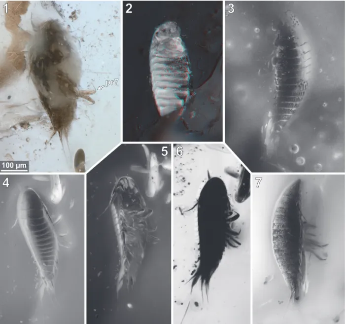
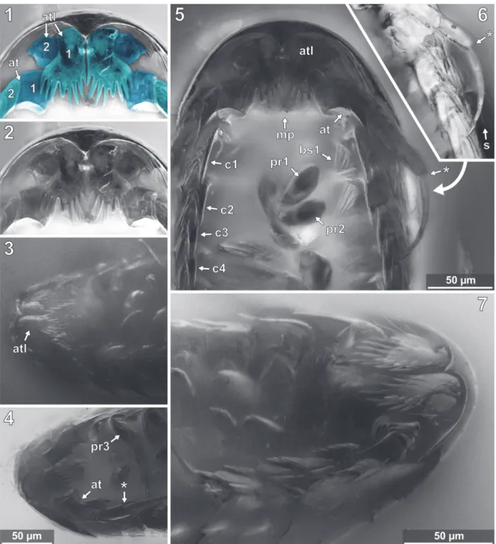
FIGURE 5. Vacuotheca dupeorum sp. nov., comparative overview of the type material sorted by collection number
(same scale). 5.1: Paratype IGR.GAR-8.1-1, lateral view, epifluorescence. 5.2: Paratype IGR.GAR-8.1-2, latero-ven-tral view, reflected light. 5.3-5.6: Paratype IGR.GAR-8.2, lateral view (5.3-5.5) and lateral view of the opposite side (5.6), epifluorescence (5.3, 5.6), reflected light (5.4) and 3D red-cyan anaglyph of reflected light micrograph (5.5).
SCHÄDEL, PERRICHOT, & HAUG: CRETACEOUS EPICARIDEA
function from the phytools package (Revell, 2017) and adjusted in Inkscape.
Measurements
Lengths of cryptoniscium larvae (Appendix 3) were collected from the literature or measured from scaled figures. If not declared otherwise, the body length is measured from the anterior-most point of
the head-shield to the posterior-most point of the pleotelson (posteriormost tergite fused with telson). The correction for the z-depth (three-dimensional orientation of the specimens in the resin) was done by examination of the original stack of unpro-cessed images. The spatial distances between the focal planes of the images are uniform and could be extracted from the microscope. Thus, by
count-FIGURE 6. Vacuotheca dupeorum sp. nov., comparative overview of the type material sorted by collection number
(same scale). 6.1-6.2: Paratype IGR.GAR-41-1, dorsal view, epifluorescence (6.1) and reflected light (6.2). 6.3: Para-type IGR.GAR-41-2, dorsal view, epifluorescence. 6.4-6.5: ParaPara-type IGR.GAR-48, ventrolateral view (6.4) and dorsal view (6.5). 6.6-6.7: Paratype IGR.GAR-51, located at the surface of the amber piece and cracked in roughly frontal plane, ventral view of the dorsal surface (6.6) and dorsal view (6.7), reflected light (6.6) and epifluorescence (6.7).
PALAEO-ELECTRONICA.ORG
ing interjacent images between structures in focus (defined and known pitches) the z-depth could be determined.
Nomenclature
The body of isopod crustaceans is organised into 20 segments, the ocular segment and 19 post-ocular segments, and the non-somitic telson. The
segments form three distinct functional units or tag-mata. The first seven segments form the functional head (cephalothorax) including the ocular ments and six appendage-bearing post-ocular seg-ments (segseg-ments of antennula, antenna, mandible, maxillula, maxilla and maxilliped). The trunk is fur-ther subdivided into two tagmata. The anterior one (pereon) is formed by seven segments (post-ocular
FIGURE 7. Vacuotheca dupeorum sp. nov., comparative overview of the type material sorted by collection number
(same scale). 7.1-7.3: Paratype IGR.GAR-53-2, dorsal view (7.1, 7.2) and ventral view (7.3), reflected light with (7.1) and without (7.2) polarising filter and epifluorescence (7.3). 7.4-7.6: Paratype IGR.GAR-64, dorso-lateral view (7.4) and ventro-lateral view (7.5, 7.6), epifluorescence (7.4, 7.6) and transmitted light (7.5). 7.7: Paratype IGR.GAR-65, lateral view, epifluorescence. 7.8: Paratype IGR.GAR-89, ventro-lateral view, epifluorescence. Dashed lines mark areas with artificially created background.
SCHÄDEL, PERRICHOT, & HAUG: CRETACEOUS EPICARIDEA
segments 7-13); each with a separated free tergite and a pair of uniramous walking appendages (tho-racic appendages, thoracopods, pereopods). The third tagma, pleon, is formed by post-ocular seg-ments 14-19 and the telson. Pleon segseg-ments 1-5 each have a separate free tergite and a pair of biramous swimming appendages (pleopods). Pleon segment six (post-ocular segment 19) is conjoined dorsally with the telson (pleotelson) and bears a pair of biramous appendages (uropods).
We herein use the term microniscium and cryptoniscium instead of microniscus larva and cryptoniscus larva to highlight the interpretation of this morphology as a distinct ontogenetic appear-ance rather than referring to the historical interpre-tation as (genus-ranked) animal groups (e.g., “Microniscidae” in Bonnier, 1900). Our intention hereby is to use terms that have no prior charge and to be more consistent with the term epicarid-ium.
FIGURE 8. Vacuotheca dupeorum sp. nov., comparative overview of the type material sorted by collection number
(same scale). 8.1: Paratype IGR.GAR-89, ventro-lateral view, reflected light; pr7, pereopod 7. 8.2: Paratype IGR.GAR-90, located at the surface of the amber piece and cracked in roughly frontal plane, ventral view of the dorsal surface, 3D red-cyan anaglyph of reflected light micrographs. 8.3: Paratype IGR.GAR-92, lateral view, epifluores-cence. 8.4-8.6: dorsal view (8.4) and ventral view (8.5, 8.6), epifluorescence (8.4, 8.5) and transmitted light (8.6). 8.7: Paratype IGR.GAR-94, lateral view, epifluorescence.
PALAEO-ELECTRONICA.ORG
In most cases where the terms microniscium and cryptoniscium have been used many authors did use incorrect plural forms. The correct plural form of microniscium is microniscia and for crypto-niscium is cryptoniscia (second/o-stem declension in a neuter case).
Taxonomic Practice
The International Code of Zoological Nomen-clature (ICZN) recommends (no strict regulation) to
write genus and species names in italic letters with the intention to separate the (binominal) species name from ‘higher taxa’ (ICZN 2012, App. B, 6.). However, in our view this is problematic because the genus, besides its function as part of the spe-cies name, also ideally represents a natural group (when not monospecific). Therefore we suggest writing generic names in italics when they are used as part of the species name but writing in regular letters when they are used to address natural
FIGURE 9. Vacuotheca dupeorum sp. nov., comparative overview of the type material sorted by collection number
(same scale). 9.1-9.3: Paratype IGR.GAR-95-1, ventral view, reflected light (9.1), transmitted light (9.2) and epifluores-cence (9.3). 9.4: Paratype IGR.GAR-95-2, lateral view, epifluoresepifluores-cence. 9.5: Paratype IGR.GAR-97, ventral view, epi-fluorescence. 9.6-9.7: Paratype IGR.GAR-98, dorsal view, epifluorescence (9.6) and reflected light (9.7). Dashed lines mark areas with artificially created background.
SCHÄDEL, PERRICHOT, & HAUG: CRETACEOUS EPICARIDEA
groups (e.g., the groundpattern of Drosophila). This should help the reader to differentiate between references to species vs. references to groups.
RESULTS Summarizing Description
This description is based on multiple speci-mens (Figures 5-9). To warrant the traceability between characters and specimens, described characters are followed by an abbreviated refer-ence to the specimens in which the described fea-tures were observed: e.g.,“IGR.GAR-8.1 specimen 2” is cited as “[8.1-2]”. We tried to cover all charac-ters that were recommended for future descriptions proposed by Nielsen and Strömberg (1965, 1973) wherever it was possible.
General Body Form
The general body form is strictly bilateral with the anterior-posterior body axis being the longest [all specimens]. The dorsal surface is convex with greatest dorsal-ventral extent at about half of the overall body length (Figure 5.4) [8.1-1, 8.2, 28, 48, 64, 92, 93, 94, 95-2]. The dorsal outline of the com-plete body (without appendages) is ovate to drop-shaped with the broadest point at about the half of the body length and tapering posteriorly (Figures 7.1, 10.1) [41-1, 41-2, 53-2, 95-1, 97, 98]. The ven-tral side of the animal (without appendages) is con-cave, and the resulting space is occupied by the appendages (Figure 5.7) [8.1-2, 28, 48, 53-1, 64, 89, 93, 95-1, 95-2, 97]. The overall size of the main body (excluding anterior and posterior append-ages, i.e., antennula and uropods) ranges from 366 µm [53-1] to 495 µm [8.2] with a mean of 423
FIGURE 10. Vacuotheca dupeorum sp. nov., reconstructions and drawings. 10.1: Reconstruction in dorsal view
(based on multiple specimens) including the striation pattern (based on paratype IGR.GAR-93). 10.2: Reconstruction based on paratype IGR.GAR-95-1 and holotype IGR.GAR-28, head shield in ventral view, numbers refer to the ele-ments of antennula (numbers on the left side) and antenna (numbers on the right side). 10.3: Drawing of the paratype IGR.GAR-41-1, uropod region in dorsal view. bs, basipod of the uropod; en, endopod of the uropod; ex, exopod of the uropod; pl5, pleon segment 5; pt, pleotelson (pleon segment 6 and telson). Drawing of the holotype IGR.GAR-28, coxal plates in ventro-lateral view, mirrored. per3-per7, pereon segments 3-7; pl1, pleon segment 1.
PALAEO-ELECTRONICA.ORG
µm and a corresponding standard deviation of 32 µm.
Dorsal Sclerites
Dorsal areas of the ocular segment and post-ocular segments 1-6 (segments of antennula, antenna, mandibula, maxillula, maxilla and maxil-liped) form a single dorsal sclerite, head shield. The dorsal surfaces of the post-ocular segments 7-18 (trunk segments 1-12) form free tergites. [8.1-1, 8.2, 28, 41-1, 48, 53-2, 64, 92, 93, 94]. The tergite of the post-ocular segment 19 (pleon segment 6, uropod segment) is conjoined with the telson [8.2, 41-1, 48, 53-2, 64, 92, 93, 94, 98] forming a pleo-telson that is roughly triangular in dorsal view (Fig-ures 10.3, 7.1, 11.8). The pleotelson has a rounded posterior corner and a toothed posterior margin
[41-1, 48, 53-2, 64, 93, 98] bearing six straight pos-terior pointed teeth with blunt tips (Figure 11.6-11.8) [41-1, 53].
Head Shield
The anterior margin of the head shield is almost half-circular in dorsal view (Figure 6.1-2) [41-1, 41-2, 53-2, 64, 95-1, 98]. The head shield has a convex dorsal surface, and its ventral mar-gins lie in one plane (Figure 5.4). A median poste-rior-pointed extension protrudes from the anterior margin of the head shield forming a triangular ven-tral plain surface and corresponding to lateral con-cave lateral spaces that are occupied by the antennulae (Figure 12.1) [8.1-2, 28, 48, 53-1, 64, 93, 95-1, 97].
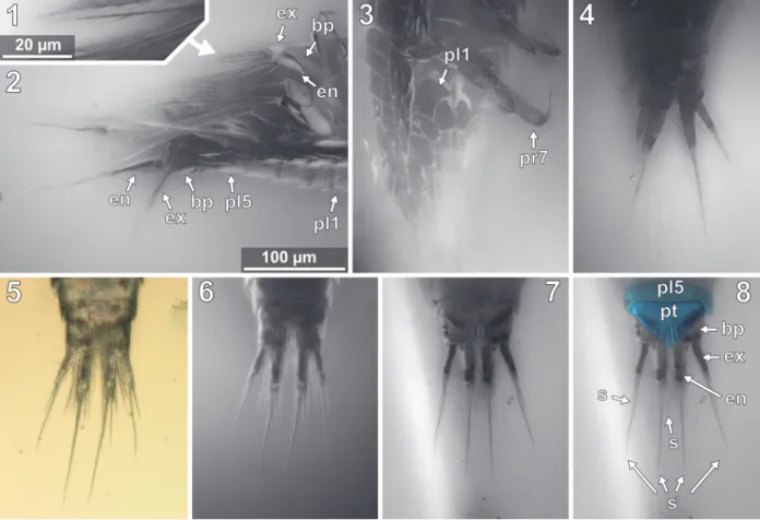
FIGURE 11. Vacuotheca dupeorum sp. nov., detailed images of the pleon and the uropod region (11.2-11.8 with
same scale). 11.1: Holotype IGR.GAR-28, setulose setae on pleopod 1, ventro-lateral view, epifluorescence. 11.2: Holotype IGR.GAR-28, pleon and uropods in ventro-lateral view, epifluorescence. bas, basipod of pleopod 1; en, endopod of the pleopod 1; ex, exopod of the pleopod 1; pl1 and pl5, pleopod segment 1 and 5; up, uropod segment.
11.3: Paratype IGR.GAR-48, pleon region in ventral view, epifluorescence. pr7, propodus of pereopod 7. 11.4:
Para-type IGR.GAR-64, uropod region in dorsal view, epifluorescence. 11.5-11.6: ParaPara-type IGR.GAR-53-2, uropod region in dorsal view, reflected light (11.4) and epifluorescence (11.5). 11.7 - 11.8: Paratype IGR.GAR-41, uropod region in dorsal view, epifluorescence. bas, basipod of the uropod; en, endopod of the uropod; ex, exopod of the uropod; pl5, pleon segment 5; pt, pleotelson (pleon segment 6 and telson); st, setae.
SCHÄDEL, PERRICHOT, & HAUG: CRETACEOUS EPICARIDEA
FIGURE 12. Vacuotheca dupeorum sp. nov., detailed images of the head region. 12.1-12.2: Paratype IGR.GAR-95-1,
head region in ventral view, epifluorescence, numbers refer to the elements of antennula (atl) and antenna (ant), in blue colour (12.1), same scale as 12.4. 12.3: Paratype IGR.GAR-64, head region in ventro-lateral view, epifluores-cence, same scale as 12.4. 12.4: Paratype IGR.GAR-53-1, head region in ventro-lateral view, epifluorescence. pr3, propodus of pereopod 3; *, junction between antennal peduncle and flagellum (element 4 and element 5). 12.5-12.6: Paratype IGR.GAR-95-1, head and pereon region in ventral view (12.5) and distal antenna elements in ventral view (12.6), epifluorescence, same scale. bs1, basipod of pereopod 1; cp1-cp4, coxal plates of pereon segments 1 to 4;
dc1-dc2, dactsyli of pereopods 1 and 2; mp, mouthparts; s, seta. 12.7: Holotype IGR.GAR-28, head region in
PALAEO-ELECTRONICA.ORG
Eye-structures are not apparent [8.1-1, 8.2, 28, 41-1, 48, 53-2, 64, 92, 93, 94, 98]. However, this must not necessarily mean that the living ani-mal possessed no optical sensory organs (see dis-cussion).
Tergites
The tergites have a convex dorsal surface, which anteriorly conforms with the head-shield. The preservation of the tergite surfaces is variable. In some specimens it appears smooth (Figure 5.4) [8.2, 28, 41-1, 48, 93, 98]. In other specimens the smooth surface is disrupted by large extensive or multiple small crater-like gaps (Figure 8.7) [8.1-1, 41-1, 51, 65, 94], which can appear darker or brighter with respect to the fluorescent characteris-tics of the surrounding surface areas. One speci-men shows a fluent transition between the small and large gaps on the dorsal surface (Figure 8.3) [92].
In some of the specimens a striation pattern is visible, which consists of more or less parallel sometimes bifurcating lines, which appear brighter or darker under fluorescent light than the surround-ing surface areas (Figure 8.4, 10.1) [8.2, 41-2, 48, 53-2, 92, 93, 98]. The striation has some variation
between the specimens. Also, the position of the specimens and their accessibility by microscopy preclude further statement about the bilateral sym-metry of the striation. The striation pattern is also preserved in specimens where the organic matter of the specimen is separated from the amber matrix (shrinking). Here, the surface of cavity in the resin bears the morphological information of the (putative) original surface of the animal. The stria-tion pattern is thus depicted by the light refracstria-tion of the amber surface, which has kept it as a coun-terpart (Figure 6.6) [48, 51]. The ventro-lateral mar-gins of the tergites of the pleon segments each have two pointed lobes directing posterior (Figure 13.3) [8.1-2, 28, 48, 53-1, 94].
Antennula
The antennula (appendage of post-ocular segment 1) consists of three peduncle elements and two flagella (Figures 10.2, 12.1-2) [28, 93, 95]. The first element has a large plate-like posterior-oriented extension bearing multiple teeth on its dis-tal margin; the anterior margin is continuous and without a plate-like extension [8.1-2, 28, 48, 53-1, 64, 89, 93, 95-1, 97]. The first antennula element bears three setae antero-laterally and distally, all
FIGURE 13. Vacuotheca dupeorum sp. nov., detailed images of the lateral body side. 13.1: Paratype IGR.GAR-53-1,
ventro-lateral view, epifluorescence. at, antenna; pr3-pr7, pereopod segments 3 to 7, arrows point to the correspond-ing coxal plates. 13.2: Paratype IGR.GAR-48, ventro-lateral view, epifluorescence. 13.3: Holotype IGR.GAR-28, ven-tro-lateral view, epifluorescence. bp, basipod of pleopod 1; en, endopod of pleopod 1; ex, exopod of pleopod 1.
SCHÄDEL, PERRICHOT, & HAUG: CRETACEOUS EPICARIDEA
arising close to each other (Figures 10.2, 12.2, 12.7, Appendix 4) [28, 48, 93, 95]. The functional ventral surface (originally anterior) of the antennula plates has sharp furrows that correspond to the proximal origins of the posterior pointing teeth of the posterior expansion of the first antennular ele-ment (Figure 12.7) [28, 93]. There are eight teeth on the plate-like posterior-oriented extension of the first antennula element in all specimens (where counting was possible) (Figures 10.2, 12.2, 12.3, 12.4, 12.7) [28, 53-1, 64, 95].
Element two is about as long as element one (without extension) and is roughly quadratic in ven-tral view [28, 53-1, 95]. Element two lacks teeth and plate-like extensions (Figures 10.2, 12.1-12.2) [28, 48, 53-1, 93, 95].
A presumably present third antennular ele-ment is not discernible from the microscopic photo-graphs. It is the third element that usually bears two distal flagella, which are also apparent here. Each flagellum consists of a single element [28, 93, 95]. The anterior flagellum bears three delicate setae and the posterior flagellum bears at least one delicate seta (Appendix 4) [28].
Antenna
The antenna (appendage of post-ocular seg-ment 1) is composed of nine eleseg-ments, coxa, basi-pod and seven endobasi-pod elements, functionally organised into four peduncle elements [28, 53, 93, 95-1] and five flagellum elements (Figures 10.2, 12.5, 12.6) [8.1-1, 8.1-2, 8.2, 28, 53-1, 53-2, 89, 93, 94, 95-1]. In dorsal view the first two peduncle elements are always concealed by the body, and the third antennal element protrudes from the pos-tero-lateral corner of the head shield (Figure 7.1) [8.1-1, 8.1-2, 8.2, 28, 64, 41-1, 41-2, 53-1, 53-2, 90, 93, 95-1, 97, 98].
The peduncle elements are distinctly wider than the flagellum elements [8.1-1, 8.1-2, 8.2, 28, 41-1, 41-2, 53-1, 53-2, 48, 93, 94, 95-1, 97]. Ele-ment one and two (coxa and basipod) together form a continuous concave median (functional pos-terior) margin that distally ends in the spine-like prolonged postero-distal corner of the second ele-ment (Figure 10.2, 12.4, 12.5) [28, 53-1, 95]. Ele-ment two bears at least one seta distally at its anterior (functional ventral) side. Element three bears three setae distally on the ventral side. Ele-ment 4 bears at least one seta distally on its ante-rior (functional ventral) side (Figure 10.2, Appendix 4 and 5) [28, 53-1].
The flagellum elements are barrel shaped to slightly conical and decrease in diameter distally
(Figure 12.6) [8.1-1, 8.1-2, 8.2, 28, 53-1, 89, 93, 94, 98]. Fifth antennal element (proximal flagellum article) with two distal setae (Figure 10.2) [8.1-1]; sixth element with at least one distal seta [53-2, 93, 95]; seventh element with two distal setae [8.1-1, 28]; eighth element with one distal seta [8.1-1, 53-2, 28]; ninth (distal-most) element with two distal setae (Figure 10.2, Appendix 4) [28, 53-2].
Mouthparts
The mouthparts (appendages of post-ocular segments 3-6; mandible, maxillula, maxilla, maxil-liped) form a posteroventral-pointing cone (Figure 12.5) [28, 53, 93, 95]. The cone is concealed by an anterior larger sclerite that encompasses about two thirds of the perimeter of the cone and a smaller tri-angular posterior sclerite (only visible in the origi-nal stack of images, Appendix 4) [28, 53]. The cone is apically truncated with a narrow opening (Figure 12.2, Appendix 4) [28, 53, 95-1].
Pereopods (appendages of the pereon segments/post-ocular segments 7-13)
Each of the seven free thoracic segments bears a pair of appendages (pereopods). Each consists of seven elements. Element one, coxa, forms a plate like structure that lies in extension to the lateral margin of its corresponding tergite (coxal plates). Coxal plates bear posterior teeth (Figures 10.4, 13.2) [8.1-2, 8.2, 28, 48, 53-1, 89, 93, 95-1, 95-2, 97]. All coxal plates have four teeth (in specimens where the preservation allowed for counting) [28, 48, 53-1, 64]. Posterior to the coxal plates, in the pleon segments, are lateral exten-sions of the tergites that superficially resemble the coxal plate morphology (see description of tergites, Figure 13.3).
Element two (basipod) is large. Element three (ischium) is slightly shorter. Elements four and five (merus, carpus) are short. Element six (propodus) is large. Element seven (dactylus) is spine-like and slightly curved inwards. The thoracic appendages become progressively longer towards the posterior end of the body. The first two pereopods are both short, the third is longer, the fourth even more. The fifth pereopod is longer than the fourth and about as long or only slightly shorter than pereopods 6 and 7, which are the longest and about the same length (Figure 14.2, 15) [53-1].
Pereopod 1. The basipod is broad and with a
con-cave space at the median side (Figure 12.5, 12.7 14.1) [28]. The propodus is broad and only weakly anterior-posteriorly compressed with an oval out-line in anterior view (Figure 12.5, 14.1) [28, 53-1,
PALAEO-ELECTRONICA.ORG
95-1], the median margin of the propodus is distally with a soft angle. The dactylus is curved inward and with a pointed tip (Figure 14.1).
Pereopod 2. The propodus is weakly compressed
in anterior-posterior axis, with an oval in outline in anterior view (Figure 14.1) [28, 53-1, 95-1], the median margin of the propodus is distally with a soft angle.
Pereopod 3. The basipod is long and slender,
much narrower than in pereopod 1 (Figures 14-15) [53-1]; the propodus is weakly compressed in ante-rior-posterior axis, with an oval outline in anterior view (Figures 14-15) [28, 53-1, 95-1], the median margin of the propodus is distally with a soft angle and a set of two setae distal to the angle (Figure 12.4) [28, 53-1, 95-1]. The dactylus is curved inward and with a pointed tip (Figures 14-15).
Pereopod 4. The basipod is long and slender,
much narrower than in pereopod 1 (Figures 14-15) [53-1]. The propodus is compressed in anterior-posterior axis (Figure 14.1) (resulting in an even
anterior and posterior surface) and longer and nar-rower as that of pereopods 1-3, the median margin of the propodus is distally with a distinct soft angle and a set of two setae distal to the angle (Figures 14-15) [28, 53-1, 95-1]. The dactylus is slightly curved inward and with a pointed tip (Figure 14.1-2).
Pereopod 5. The basipod is long and slender,
much narrower than in pereopod 1 (Figures 14-15) [53-1]. The propodus is compressed in anterior-posterior axis (resulting in an even anterior and posterior surface) and longer and narrower as that of pereopods 1-3 (Figure 14.1), the median margin is distally with a set of two setae [28, 53-1, 95-1]. The dactylus is slightly curved inward and with a pointed tip (Figure 14.4).
Pereopod 6. The basipod is long and slender,
much narrower than in pereopod 1 (Figures 14-15) [53-1]. The ischium is compressed in anterior-pos-terior axis with a convex lateral margin and a straight median margin (Figures 14-15) [53-1]. The
FIGURE 14. Vacuotheca dupeorum sp. nov., detailed images of the pereopods. 14.1: Holotype IGR.GAR-28,
ventro-lateral view, epifluorescence. pr1-2 and pr4-6, pereopods 1-2 and 4-6. 14.2: Paratype IGR.GAR-53-1, pereopods in lateral view, right side of the image is anterior, epifluorescence, for labels see Figure 15 (corresponding drawing with labels). 14.3: Paratype IGR.GAR-8.1-1, posterior pereopods in lateral view, right side of the image is anterior, epifluo-rescence. pr6-pr7, pereopods 6 and 7. 14.4-14.5: Paratype IGR.GAR-95-1, pereopods in ventral view, upper side of the image is anterior, same scale. pr4-pr7, pereopods 4 to 7.
SCHÄDEL, PERRICHOT, & HAUG: CRETACEOUS EPICARIDEA
merus is short and roughly triangular in anterior view (Figures 14-15) [53-1, 89, 95-1]. The carpus is short and roughly triangular in anterior view (Fig-ures 14-15) [53-1, 89, 95-1]. The propodus is antero-posteriorly compressed (resulting in an even anterior and posterior surface) and longer and narrower as that of pereopods 1-3 (Figure 14.1), the median margin is distally with a set of two setae [28, 53-1, 95-1] and with two proximo-distal strings of muscles proximo-distally attaching to the lat-eral and median side of the dactylus joint (Figure 14.5) [89, 95-1]. The dactylus is slightly curved inward and with a pointed tip.
Pereopod 7. The basipod is long and slender,
much narrower than in pereopod 1 (Figures 14-15) [53-1]. The ischium is anterior-posteriorly com-pressed with a convex lateral margin and a straight median margin (Figures 14-15) [53-1]. The merus is short and roughly triangular in anterior view (Fig-ures 14-15) [53-1, 89, 95-1]. The carpus is short and roughly triangular in anterior view (Figures 14-15) [53-1, 89, 95-1]. The propodus is antero-poste-riorly compressed (resulting in an even anterior and posterior surface) and longer and narrower as that of pereopods 1-3, the median margin is distally with a set of two setae (Figure 14.4) [28, 53-1, 95-1] and with two proximo-distal strings of muscles distally attaching to the lateral and median side of
the dactylus joint (Figure 8.1) [89, 95-1]. The dacty-lus is slightly curved and with a pointed tip (Figure 14.3).
Pleopods (appendages of the pleon segments/ post-ocular segments 14-18)
The pleopods consist of a broad basipod which distally bears the median endopod and the lateral exopod (Figures 13.3, 11.2) [28, 48, 53-1, 97]. All elements are strongly compressed in ante-rior-posterior axis and roughly leaf-shaped. The endopods are broader and more massive than the corresponding exopods [28, 48, 53-1, 97].
Endopod and exopod bear long setae distally (Figure 11.2) [8.1-1, 8.1-2, 8.2, 28, 48, 53-1, 53-2, 64, 89, 97]. The setae originate in an obtuse angle from the pleopods and point posteriorly (Figure 11.2) [8.1-1, 28, 64]. Pleopod 1 is with at least five setae on the endopod and four setae on the exo-pod (Figure 11.2) [28]. Pleoexo-pod 2 is with at least five setae on the exopod [28]. Pleopod 3 is with at least three setae on the endopod and four setae on the exopod (Figure 11.2) [28].
At least in pleopods 1, the distal setae are ulose with delicate posterio-laterally protruding set-ulae. The setulae are less than 1 µm in diameter and ca. 15 µm long (Figure 11.1) [28].
FIGURE 15. Vacuotheca dupeorum sp. nov., paratype IGR.GAR-53-1, drawing of pereopods 3 to 7 (pr3–pr7). (r),
right body side; (l), left body side; bs, basipod; is, ischium; mr, merus; cp, carpus; pr, propodus; dc, dactylus. Notice the setae on the propodi of pereopods 3 and 4.
PALAEO-ELECTRONICA.ORG
Uropod (appendage of post-ocular segment 19)
The uropods consist of a basipod which dis-tally bears the median endopod and the lateral exopod. The basipod of the uropods is massive and rectangular in dorso-ventral view [8.1-1, 41-1, 41-2, 53-1, 53-2, 64, 93, 98]. Basipods are appar-ently movable in relation to the trunk as specimens with (parallel) posterior pointed (Figure 11.7) [41-1, 41-2, 53-2] and somewhat spread (laterally diverg-ing) basipods (Figure 11.4) [64, 98] suggest. Endo-pods and exoEndo-pods are truncated cone-shaped (tapering distally). Endopods and exopods are apparently movable in relation to the basipod as the angle between both elements and the angle between each of the elements and the correspond-ing basipod vary in one specimen (Figure 11.4). Endo- and exopods are ovate to rectangular in cross-section (greatest diameter in dorsoventral direction) (Figure 6.3) [41-2]. The endopods are longer and thicker than the exopods (Figure 10.3, 11.4-8) [8.1-1, 8.2, 28, 53-1, 53-2, 64, 89, 98] and distally bear one long and one short seta (Figure 11.5) [53-2]. The exopods are about as long as the basipods (Figure 10.3, 11.4-8) [8.1-1, 53-2, 64, 28] and distally bear one long (about twice as long as the exopod) and one short seta (Figures 10.3, 11.5, 11.7) [53-2].
DISCUSSION Systematic Interpretation
Assuming that the cone shaped feeding appa-ratus consists of appendages of more than one segment (four segments in Epicaridea), the func-tional head comprises at least four appendage-bearing segments, which is apomorphic for Euar-thopoda (sensu Walossek, 1999, e.g., Haug et al., 2013). The trunk is divided in two distinct sets of segments (thorax and pleon), which are consid-ered as an apomorphy of Eumalacostraca (Walossek, 1999).
The body is dorsoventrally flattened, and the tergite of the first thoracic segment (maxilliped) is conjoined with the tergites of the functional head. The lateral flagellum of the antennula is not well developed but consists only of a single short ele-ment. Also, all pereopods (appendages of post-ocular segments 7-13) lack an exopod. This combi-nation of characters is unique and characterizes the group Isopoda (Ax et al., 2000; Wilson, 2009). All pereopods bear lateral plate-like extensions of the coxa (coxal plates), which is an autapomorphy of Scutocoxifera (Dreyer and Wägele, 2002). The mouthparts (mandible, maxillula, maxilla and
maxil-liped) form a cone-like structure, which is only known for parasitic isopods within Cymothoida (if including Gnathiidae).
The combination of the following characters is typical for larvae of the group Epicaridea (Latreille, 1825): body elongated and drop shaped; mouthparts forming a cone like structure; anten-nula with enlarged first element; pereopods with large propodi and thin, spine like and often curved dactyli; truncated cone-shaped uropod rami.
Within Epicaridea, a further determination pro-viding identifications to monophyletic groups is not possible due to the absence of undisputed apo-morphies in most groups. Within Epicaridea, Daji-dae (Sars, 1883) is the only group with a well-accepted apomorphy that can be seen in the cryp-toniscium stage. In Dajidae cryptoniscia have an oral cone with a conspicuous sucking disk (Bres-ciani, 1966; Schultz, 1975; Wägele, 1989).
Thus, the herein presented specimens can be interpreted as epicarideans that are not (latin: nec) part of the epicaridean ingroup Dajidae (Epicaridea nec Dajidae). We demonstrated that the morphol-ogy of the herein presented specimens fits per-fectly with that of the cryptoniscium larvae of Epicaridea. However, the exact ontogenetic phase of the fossils cannot be determined with certainty. In some epicaridean lineages (Cryptoniscoidea) the adult male does (at least superficially) not differ morphologically from the cryptoniscium (Hosie, 2008). Therefore, the studied fossils could not only represent cryptoniscium larvae but also adult males with a paedomorphic morphology. Paedo-morphic, strict-protandric males (as they occur in most cryptoniscoideans) have been recorded to switch between host animals on a regular basis to inseminate females and finally find a host that is not infected by other epicarideans where they transform into a female (Wägele, 1989).
Conspecifity
We assume conspecifity for the herein studied specimens. This is based on the lack of conspicu-ous morphological differences among the individu-als (as lain out in the description). Also, the body size is relatively uniform with a standard deviation of 32 µm (7.5 % of the mean body size). Only the dorsal striation pattern is subject to some variation within the studied specimens (Figures 5.3, 5.4, 6.2, 6.6, 7.2, 8.3, 8.4, 9.7, 10, 14.8). However, without data on the degree of variability of the striation pat-tern in modern species, it is impossible to draw conclusions on the intra- and interspecific variabil-ity of this character in extinct species.
SCHÄDEL, PERRICHOT, & HAUG: CRETACEOUS EPICARIDEA
Striation
The dorsal surface as well as various other body regions of cryptoniscium larvae bears a sur-face pattern that superficially appears as lines (striae/striation). In the first extensive study focus-ing on the surface structure of cryptoniscia usfocus-ing scanning electron microscopy, Nielsen and Ström-berg (1973) categorized striation patterns in two types. They characterized the striae on the dorsal side of the head shield as “rather broad cuticular ridges separated by narrow furrows.” Striae on other parts of the body, like the pleopod basipod, were characterized as “pectinate scales.” They also perfomed transmission electron microscopy to study the structure of the striae. By this, they found the striae to affect only the epicuticle but not the endocuticle. This distinction appears somewhat arbitrary because both types of striae are purely epicuticular, and both the “pectinate scales” and the ridges and furrows are asymmetric, as the transmission electron microscopy images show. It could be possible that both types differ only (gradu-ally) in scale, collocation and manifestation of the fringes (ctenae).
Judging from the scale of the striae it is very likely that especially a dense striation pattern appears like a homogeneous surface in light microscopy and is thus been overlooked possibly partly here, but also more generally in the litera-ture.
The visibility of this pattern in some of the herein studied fossils is highly dependent on the illumination of the specimen (e.g., compare Figure 6.1 vs. 6.2 or 7.1 vs. 7.2). The striation pattern is more or less pronounced in cryptoniscia of different epicaridean groups (Nielsen and Strömberg, 1973; Hosie, 2008). To our knowledge there is no infor-mation about the intraspecific variability of the stri-ation for modern species that would be useful for the interpretation of future fossil findings.
Eyes
Although eyes are not visible in the studied specimens, we cannot conclude their absence. In many modern cryptoniscia the compound eyes are highly reduced so that they are only recognizable as dark spots beneath the dorsal surface of the head shield (Nielsen and Strömberg, 1973). Only one extant species of epicarideans has been recorded to have cryptoniscia with externally visi-ble eyes, as well as a single fossil specimen (Schultz, 1975; Serrano-Sánchez et al., 2016).
Antennula
Wägele (1989) suggested that the toothed posterior projection of the first antennula element (antennular plate) could be an autapomorphy for Cryptoniscoidea. Based on figures and descrip-tions in taxonomic literature (summarized in Figure 16), we cannot support this assumption. Indeed, at least two species with a toothed margin of the antennula plate have been interpreted as repre-sentatives of Bopyroidea (Probopyrus bithynis Richardson, 1905, in Dale and Anderson, 1982) and Leidya distorta Comalia and Panceri, 1858, in Torres Jordá, 2003).
Also, Wägele (1989) suggested that a contin-uous margin of the antennula plate, in contrast to a toothed margin (orange colour compared to beige colour in Figure 16) could be an autapomorphy of monophyletic group combining Asconiscidae, Crin-oniscidae and CryptCrin-oniscidae. This must be seen as distinct from cases in which antennula element one has no distinct posterior projection (red in Fig-ure 16). We support this assumption based on our study of literature. If future phylogenetic analyses support the monophyly of this group, we recom-mend the erection of a proper name for this group, as well as to include the two species abyssorum Bourdon, 1981, and longicaudatus Schultz, 1975, in it, which possess this specific structure (both known from cryptoniscia only and traditionally inter-preted as Cryptoniscoidea incertae sedis).
Given that the distribution of a toothed anten-nula plate in cryptoniscia is not restricted to a sin-gle subgroup of Epicaridea, also a different polarity of the character than proposed by Wägele (1989) has to be considered. The toothed antennula plate could represent an autapomorphy of Epicaridea that has been lost several times independently.
The orientation of the mouthparts (sucking cone) seems to constrain the shape and size of the proximal element of the antennula. This seems to affect whether or not a posterior extension of the antennular plate is developed and also, if there is a posterior expansion it seems to affect the orienta-tion of the antennular plate. Indeed, in species that have mouthparts anteriorly directed (e.g.,
Probopy-rus pandalicola) the median margins of the first
antennula element are not parallel but diverge pos-teriorly.
Little intraspecific variability in epicaridean species has been recorded regarding the number of these teeth. However, a few cases of intraspe-cific but also intra-individual differences have been recorded (Nielsen and Strömberg, 1973).
PALAEO-ELECTRONICA.ORG
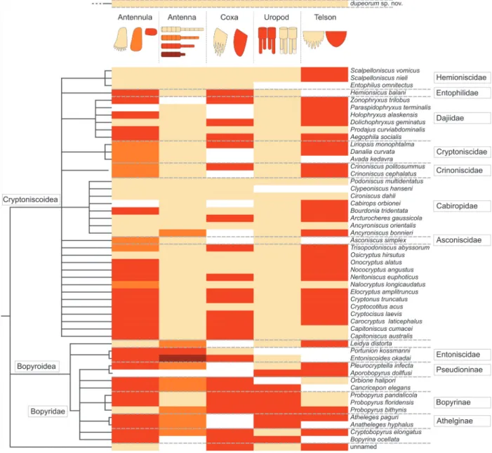
FIGURE 16. Phylogenetic tree of Epicaridea (Boyko et al., 2013) (topology on the left side, species names and
taxo-nomic groups on the right side) mapped with characters gathered from descriptions and illustrations of literature. Characters are coded in colour as depicted in the illustration at the top. Beige is reserved for character states in
Vac-uotheca dupeorum gen. et sp. nov. (at the very top). Antenna: five flagellum elements (beige), four flagellum elements
(orange), three flagellum elements (red), one flagellum elements (orange). Uropod: endopods longer or equal as exo-pods (beige), endoexo-pods shorter than exoexo-pods (red). Antennula: with posterior extension and teeth (beige), with poste-rior extension and without teeth (orange), without posteposte-rior extension (red). Coxa: coxal plates with teeth (beige), coxal plates without teeth (red). Telson: posterior margin with teeth (beige), posterior margin without teeth (red). (Fraisse, 1878; Giard and Bonnier, 1887; Bonnier, 1900; Thompson, 1901; Caullery, 1907; Miyashita, 1940; Nielsen and Strömberg, 1965; Bresciani, 1966; Bourdon, 1972, 1976, 1981; Holdich, 1975; Schultz, 1975, 1980; Kensley, 1979; Bourdon and Bruce, 1980; Anderson and Dale, 1981; Coyle and Mueller, 1981; Dale and Anderson, 1982; Adkinson and Collard, 1990; Rybakov, 1990; Pascual et al., 2002; Torres Jordá, 2003; Shimomura et al., 2005; Hosie, 2008; Boyko, 2015).