HAL Id: hal-02088184
https://hal.univ-lorraine.fr/hal-02088184
Submitted on 2 Apr 2019
HAL is a multi-disciplinary open access
archive for the deposit and dissemination of
sci-entific research documents, whether they are
pub-lished or not. The documents may come from
teaching and research institutions in France or
abroad, or from public or private research centers.
L’archive ouverte pluridisciplinaire HAL, est
destinée au dépôt et à la diffusion de documents
scientifiques de niveau recherche, publiés ou non,
émanant des établissements d’enseignement et de
recherche français ou étrangers, des laboratoires
publics ou privés.
Caspase-2 activation in the absence of PIDDosome
formation
Claudia Manzl, Gerhard Krumschnabel, Florian Bock, Bénédicte Sohm,
Verena Labi, Florian Baumgartner, Emmanuelle Logette, Jürg Tschopp,
Andreas Villunger
To cite this version:
Claudia Manzl, Gerhard Krumschnabel, Florian Bock, Bénédicte Sohm, Verena Labi, et al.. Caspase-2
activation in the absence of PIDDosome formation. Journal of Cell Biology, Rockefeller University
Press, 2009, 185 (2), pp.291-303. �10.1083/jcb.200811105�. �hal-02088184�
JCB:
ARTICLE
The Rockefeller University Press $30.00
Correspondence to Andreas Villunger: [email protected] Abbreviations used in this paper: DD, death domain; GAPDH, glyceraldehyde-3-phosphate dehydrogenase; MEF, mouse embryonic fibroblast; MOMP, mito-chondrial outer membrane permeabilization; mPIDD, mouse PIDD; NF-B, nuclear factor B; PE, phycoerythrin; PI, propidium iodide; Puma, p53 up-regulated modulator of apoptosis; UVR, UV radiation.
Introduction
PIDD (p53-induced protein with a death domain [DD]) was
identified as one of many transcriptional targets that may
medi-ate apoptosis induction by the tumor suppressor p53 (Lin et al.,
2000). PIDD is widely expressed in various organs and cell
types and is characterized by the presence of certain
struc-tural motives, including Leu-rich repeats, ZU5 domains, and a
C-terminal DD (Tinel and Tschopp, 2004). DD-containing
pro-teins play important roles in the formation of multimeric
signal-ing complexes that regulate diverse cellular responses, includsignal-ing
cytokine secretion, nuclear factor B (NF-B) activation, cell
survival, and apoptosis (Reed et al., 2004). RAIDD
(receptor-interacting protein-associated ICH-1/CED-3 homologous
pro-tein with a DD), a bipartite adapter molecule, was identified as
a possible interaction partner of PIDD (Tinel and Tschopp,
2004). RAIDD contains a caspase recruitment domain as well
as a C-terminal DD (Duan and Dixit, 1997). The caspase
re-cruitment domain of RAIDD was previously described to
inter-act with pro–caspase-2 (Duan and Dixit, 1997) and has been
implicated in the activation of this ill-defined initiator caspase
thought to be required for DNA damage–induced apoptosis in
certain tumor cells and the metabolic death of oocytes (Degterev
and Yuan, 2008). A trimolecular complex containing PIDD,
RAIDD, and caspase-2 was subsequently identified as the
long-sought-after activation platform of caspase-2 and dubbed
the PIDDosome (Tinel and Tschopp, 2004). This complex appears
to form spontaneously in cell extracts from different cell lines
upon temperature shift in vitro, but formation in extracts from
cells undergoing apoptosis after DNA damage has not been
de-tected so far (Read et al., 2002). A second, PIDD-containing
multimeric protein complex was described shortly thereafter
that contains the Ser-Thr kinase RIP-1 (receptor-interacting
protein 1) and IKK-/NF-B essential modulator, the
regula-tory subunit of the IB kinase complex, which were both
impli-cated in the activation of NF-B signaling after genotoxic stress
(Janssens et al., 2005). Subsequent analysis revealed that PIDD
possesses autoproteolytic activity, facilitating the generation of
two different active protein fragments, termed PIDD-C and
PIDD-CC. Although PIDD-CC was proposed to be required for
P
IDD (p53-induced protein with a death domain
[DD]), together with the bipartite adapter protein
RAIDD (receptor-interacting protein-associated ICH-1/
CED-3 homologous protein with a DD), is implicated in
the activation of pro–caspase-2 in a high molecular weight
complex called the PIDDosome during apoptosis
induc-tion after DNA damage. To investigate the role of PIDD in
cell death initiation, we generated PIDD-deficient mice.
Processing of caspase-2 is readily detected in the
ab-sence of PIDDosome formation in primary lymphocytes.
Although caspase-2 processing is delayed in simian virus
40–immortalized pidd
/mouse embryonic fibroblasts, it
still depends on loss of mitochondrial integrity and
effec-tor caspase activation. Consistently, apoptosis occurs
nor-mally in all cell types analyzed, suggesting alternative
biological roles for caspase-2 after DNA damage.
Because loss of either PIDD or its adapter molecule RAIDD
did not affect subcellular localization, nuclear translocation,
or caspase-2 activation in high molecular weight complexes,
we suggest that at least one alternative PIDDosome-
independent mechanism of caspase-2 activation exists in
mammals in response to DNA damage.
Caspase-2 activation in the absence of PIDDosome
formation
Claudia Manzl,
1Gerhard Krumschnabel,
1Florian Bock,
1Benedicte Sohm,
1Verena Labi,
1Florian Baumgartner,
1Emmanuelle Logette,
2Jürg Tschopp,
2and Andreas Villunger
11Division of Developmental Immunology, Biocenter, Innsbruck Medical University, A-6020 Innsbruck, Austria 2Department of Biochemistry, University of Lausanne, CH-1066 Epalinges, Switzerland
© 2009 Manzl et al. This article is distributed under the terms of an Attribution– Noncommercial–Share Alike–No Mirror Sites license for the first six months after the publica-tion date (see http://www.jcb.org/misc/terms.shtml). After six months it is available under a Creative Commons License (Attribution–Noncommercial–Share Alike 3.0 Unported license, as described at http://creativecommons.org/licenses/by-nc-sa/3.0/).
THE
JOURNAL
OF
CELL
BIOLOGY
ple modes of regulation of PIDD protein expression (Cuenin
et al., 2008). Although all of these findings support a critical
role for PIDD in cell death and caspase-2 activation, targeting
RAIDD or PIDD expression by siRNA failed to interfere with
caspase-2 processing in response to 5-fluoruracil treatment in
HCT-116 colon carcinoma cells (Vakifahmetoglu et al., 2006).
In addition, thymocytes and fibroblasts from mice lacking
RAIDD were reported to respond normally to cell death
induc-tion by DNA-damaging agents or TNF (Berube et al., 2005).
However, cell death induced by overexpression of PIDD in
fibroblasts strictly depended on the presence of RAIDD but not
caspase-2, suggesting that PIDD can also engage cell death in
a manner independent of this initiator caspase (Berube et al.,
2005). To investigate the relevance of PIDD for caspase-2
acti-vation and cell death under physiological conditions, we have
generated a PIDD-deficient mouse model by homologous
re-combination in embryonic stem cells.
activation of caspase-2 and cell death, PIDD-C was suggested
to activate NF-B, DNA repair, and survival after low grade
DNA damage (Tinel et al., 2007).
Consistent with a proapoptotic role for PIDD in
p53-induced cell death, its overexpression facilitated caspase-2
acti-vation and cell death induction in response to DNA damage in
HeLa cells (Tinel and Tschopp, 2004), and RNA interference or
antisense oligonucleotides targeting PIDD mRNA delayed cell
death induced by overexpression of p53 in H1299 colon cancer
(Baptiste-Okoh et al., 2008) or K562 myelogenous leukemia
cells, respectively (Lin et al., 2000). Interestingly, a correlation
between apoptotic index and PIDD expression was recently
re-ported in oral squamous cell carcinoma patient samples, whereas
no correlation was found regarding the p53 mutation status in
these tumors (Bradley et al., 2007). The latter finding is
consis-tent with the observation that the basal expression of PIDD does
not depend on the presence of functional p53, suggesting
multi-Figure 1. Targeting the pidd locus in mice. (A) Schematic representation of the pidd/lrdd gene locus on mouse chromosome 7 (chr 7). Exons 3–15 of the
pidd gene were replaced by the open reading frame of -galactosidase. The loxP element-flanked neomycin selection marker cassette was removed in vivo by cre-mediated deletion. The locations of the 5 and 3 probes used for Southern blot analysis are indicated as horizontal black bars. (B) Southern blot analysis
on genomic DNA derived from wild-type, pidd+/, and pidd/ mice. (C) Exon-specific PCR using three different primer pairs spanning exons 7–8 or 13–15
confirmed correct targeting of the pidd gene. (D) RT-PCR was performed on cDNA generated from total RNA derived from thymocytes (top two panels) that were left untreated or exposed to 2.5 Gy of irradiation or SV40-immortalized wild-type (wt) and PIDD-deficient MEFs (bottom two panels) cultured in the absence or presence of etoposide to confirm absence of pidd mRNA. (E) Western blot analysis on thymocyte extracts derived from wild-type and pidd/ mice
assessment failed to reveal any gross abnormalities, and pidd
/mice show normal lymphocyte development and subset
distri-bution in the relevant primary and secondary lymphatic organs
(Fig. S2 A and not depicted). Collectively this demonstrates that
PIDD is dispensable for embryonic development and postnatal
life in unstressed animals.
Loss of PIDD fails to protect lymphocytes from apoptosis induction
To investigate the role of PIDD in cell death induction, we
iso-lated primary thymocytes as well as mature T and B cells from
spleen and granulocytes from bone marrow by FACS sorting
from age-matched wild-type, pidd
/, and caspase-2
/mice.
Lymphocytes overexpressing antiapoptotic Bcl-2 in all
hemato-poietic cells and cells lacking p53 or its proapoptotic target gene
puma
/, a BH3-only Bcl-2 family member (Villunger et al.,
2003), were included in some of our assays as internal controls.
Thymocytes were put in culture without further treatment
(spontaneous death) or exposed to DNA damage triggered by
topoisomerase II inhibitor etoposide, the DNA-intercalating
drug doxorubicin, or or UV irradiation. In parallel, cells were
also exposed to heat stress or were treated with the broad-spectrum
kinase inhibitor staurosporine, the ER stressors tunicamycin,
brefeldin A, or thapsigargin as well as cytochalasin D or taxol,
both of which disrupted cytoskeletal structures, or to death
receptor agonists Fas ligand or TNF- (Table I). Cell
viabil-ity was monitored over time using Annexin V/propidium iodide
(PI) staining and flow cytometric analysis. Loss of PIDD failed
to protect thymocytes (Fig. 2), mature T and B cells, or
granu-locytes (not depicted) from any of the tested cell death stimuli.
In contrast, overexpression of Bcl-2 or loss of p53 or puma
each efficiently protected thymocytes and mature T and B
cells from DNA damage–induced apoptosis (Fig. 2 and not
de-picted), which is consistent with previous results (Villunger et al.,
2003; Erlacher et al., 2006). To test whether the proposed
Results
Generation of PIDD-deficient mice
Our targeting strategy aimed to replace exons 3–15 of the PIDD
locus, replacing all relevant protein domains with the open
read-ing frame of -galactosidase followed by a loxP element-flanked
neomycin resistance cassette (Fig. 1 A). Correct targeting of the
pidd
locus was confirmed by Southern blot analysis and
exon-specific PCR on genomic DNA derived from embryonic stem
cells and, subsequently, tail tissue, mouse embryonic fibroblasts
(MEFs), and liver derived from wild-type, pidd
+/, and pidd
/mice (Fig. 1, B and C; and not depicted). RT-PCR analysis was
performed to confirm the absence of pidd mRNA in thymocytes
and SV40-immortalized MEFs (Fig. 1 D). Finally, to ascertain
lack of protein, a polyclonal antiserum was generated using the
DD of PIDD (aa 770–910) as immunogen. Reactivity of the
affinity-purified IgG fraction was confirmed in MEFs that
over-express a Flag-tagged version of mouse PIDD (mPIDD). Three
protein fragments corresponding to the full-length protein,
pro-cessed PIDD-C, and PIDD-CC were detected by the anti-Flag
antibody and the purified anti-PIDD IgG fraction (Fig. S1 and
not depicted). Transient cotransfection of 293T cells with an
expression construct encoding mPIDD together with two
differ-ent ldiffer-entiviral vectors encoding alternative short hairpin RNAs
targeting mPIDD confirmed specificity of the purified
anti-serum (Fig. S1 and not depicted). Western blot analysis performed
on thymocyte extracts derived from wild-type and pidd
/mice
revealed that PIDD-CC appears to be the only detectable form
(Fig. 1 E). Efforts to identify endogenous full-length protein or
PIDD-C by immunoblotting or immunoprecipitation were
re-peatedly unsuccessful, suggesting that PIDD may be rapidly
autoprocessed in thymocytes (unpublished data).
Mice lacking both alleles of PIDD were born in normal
Mendelian frequency and did not show fertility problems,
gen-der bias, or any other overt phenotype. Furthermore, histological
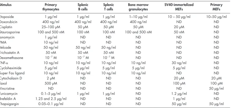
Table I. Summary of stimuli and final concentrations used to induce cell death
Stimulus Primary thymocytes Splenic B cells Splenic T cells Bone marrow granulocytes SV40-immortalized MEFs Primary MEFs Etoposide 1 µg/ml 1 µg/ml 1 µg/ml 1–10 µg/ml 10 – 50 µg/ml 10–50 µg/ml Doxorubicin 400 ng/ml 400 ng/ml 400 ng/ml 400 ng/ml ND ND Cisplatin 25–100 µM 50 µM 50 µM 50 µM 20 µM ND
Staurosporine 100 and 500 nM 100 nM 100 nM 100 and 500 nM 50 nM ND
Ionomycin 1 µg/ml ND ND ND ND ND PMA 10 ng/ml ND ND ND ND ND Velcade 50 ng/ml 50 ng/ml 50 ng/ml ND ND ND Trichostatin A 50 nM 50 nM 50 nM ND ND ND Dexamethasone 107 M 107 M 107 M ND ND ND TNF- 10 ng/ml 10 ng/ml 10 ng/ml 10 ng/ml 30 ng/ml ND Cycloheximide 5 µg/ml 5 µg/ml 5 µg/ml 5 µg/ml 5 µg/ml ND
Super Fas ligand 10 ng/ml 10 ng/ml 10 ng/ml 10 ng/ml ND ND
Cytochalasin D 2 µM ND ND ND 20 µM 20 µM Taxol 500 µM ND ND ND 100 µM 100 µM Vincristine ND ND ND ND ND 50 µg/ml Tunicamycin 0.1–5 µg/ml 1 µg/ml 1 µg/ml ND 1.2 µg/ml ND Brefeldin A 1.25 and 2.5 µg/ml ND ND ND 1 µg/ml ND Thapsigargin 0.05–0.1 µg/ml ND ND ND 50 µg/ml 50 µg/ml
Higher doses of UVR, etoposide treatment, or transient
expo-sure to 45°C did not allow clonal survival of primary (Fig. S3)
or immortalized cells (Fig. S4) independent of their genotypes.
To address the possible relevance of PIDD for transient,
short-term survival, primary as well as SV40-immortalized
MEFs were analyzed for their initial cell death responses. MEFs
from wild-type, pidd
/, caspase-2
/, or puma
/mice (Fig. 3 A)
as well as primary low passage wild-type and pidd
/or
cas-pase-2
/MEFs (Fig. 3 B) were exposed to DNA damage
in-duced by etoposide, UV irradiation, serum deprivation, heat
stress, ER stress, cytochalasin D, taxol, or staurosporine (Table I),
but, again, neither loss of PIDD nor of caspase-2 protected these
cells significantly from any of these apoptotic stimuli. In
con-trast, loss of puma transiently protected MEFs from the effects
of DNA damage (Fig. 3 A and not depicted).
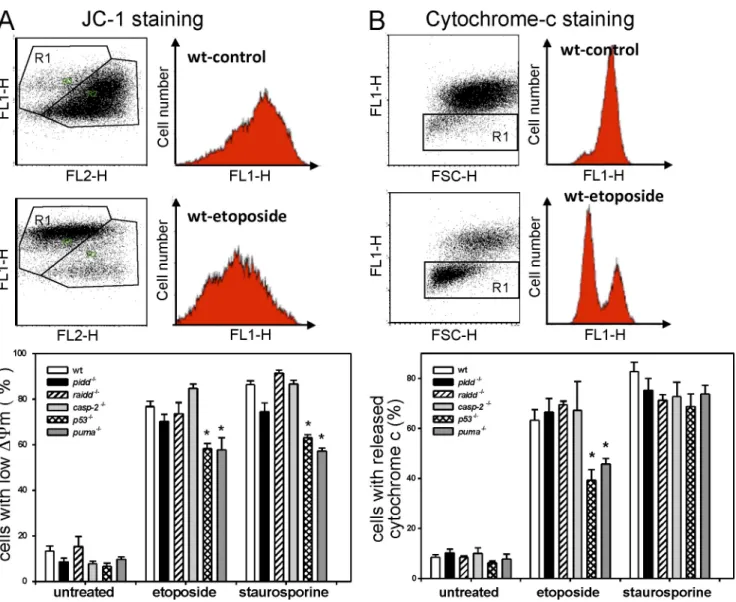
To gain insight into possible minor mechanistic
differ-ences in apoptosis induction too subtle to be captured by
An-nexin V/PI staining, we also investigated the impact of PIDD
deficiency on mitochondrial membrane depolarization using
JC-1 staining (Fig. 4 A) and by monitoring cytochrome c release
(Fig. 4 B). In SV40-immortalized fibroblasts (Fig. 4) or
thymo-cytes (not depicted) exposed to DNA damage, none of these
parameters was significantly altered as a result of the absence
of PIDD or caspase-2, but, again, loss of p53 or Puma
par-tially blocked these events upon DNA damage. To backup our
PIDD–RAIDD–caspase-2 apoptosis pathway could act in a
re-dundant manner with the Bcl-2–regulated one, we also assessed
survival in thymocytes lacking PIDD and p53 up-regulated
modulator of apoptosis (Puma) simultaneously. However, the
degree of protection from apoptosis induced upon DNA
dam-age was comparable between puma
/and pidd
/puma
/double-deficient thymocytes (Fig. 2), arguing against such
re-dundancy. To exclude the possibility that loss of PIDD may
have an impact on the survival of lymphocytes in vivo, masked
by the lack of coregulatory factors in our in vitro analysis, we
also exposed wild-type and pidd
/mice to graded doses of
whole body irradiation (2.5 and 5.0 Gy) and evaluated
lympho-cyte cellularity as well as subset distribution of immature and
mature T and B cells in thymus, bone marrow, and spleen 20 h
after irradiation. Consistent with our findings in vitro, loss of
PIDD did not enhance the survival of lymphocytes after
irradia-tion in vivo (Fig. S2 A and not depicted).
Because PIDD has been implicated in caspase-2–mediated
apoptosis as well as NF-B activation, enabling DNA repair
(Janssens et al., 2005), we investigated the impact of loss of
PIDD on clonal survival after DNA damage in primary as well
as SV40-immortalized MEFs transiently exposed to heat shock,
etoposide, UV radiation (UVR) or irradiation. Loss of PIDD
or caspase-2 did not influence residual survival of MEFs
ob-served upon low dose of UVR or transient exposure to 43°C.
Figure 2. PIDD is dispensable for cell death induction in lymphocytes. Thymocytes derived from mice of the indicated genotypes were put in culture with-out further treatment (spontaneous cell death) or exposed to apoptosis inducers as indicated over time. Cell death was monitored by Annexin V–FITC/PI staining and flow cytometric analysis at the indicated time points. Data are means ± SD from four to eight independent experiments per genotype, with the exception of pidd/puma/ (n = 2). The extent of apoptosis induced specifically by different stimuli was calculated by the equation (induced apoptosis
spontaneous cell death)/(100 spontaneous cell death). No significant differences in cell death induction were observed between wild-type and PIDD- and caspase-2–deficient cells treated with any of the indicated stimuli by analysis of variance.
of cells to etoposide treatment or irradiation. However,
pro-cessing occurred with similar efficiency in wild-type and
PIDD-deficient cells and coincided with the appearance of the active
form of caspase-3, indicating that the classical mitochondrial
outer membrane permeabilization
(MOMP)–apoptosome-mediated apoptosis pathway was active either preceding caspase-2
cleavage or caspase-2 may have been activated in a
dimerization-dependent manner in a parallel pathway (Fig. 5 A). However,
because processing of both caspases was efficiently blocked in
thymocytes overexpressing Bcl-2 (Fig. 5, B and C), we
con-clude that caspase-2 processing requires MOMP and
cyto-chrome c release, at least under the conditions tested in this
study. Thymocytes lacking Puma showed a significant delay
but, in contrast to Bcl-2–overexpressing cells, not a complete
block in caspase-2 processing (Fig. 5, B and C), indicating that
additional effector proteins such as Noxa or Bim contribute to
DNA damage–induced thymocyte apoptosis, as shown
previ-ously (Erlacher et al., 2006; Michalak et al., 2008). Importantly,
caspase-2 processing occurred with similar decreased efficiency
in thymocytes lacking Puma or PIDD and Puma simultaneously,
demonstrating that caspase-2 cleavage during DNA damage–
induced thymocyte apoptosis triggered by irradiation (Fig. 5 D)
or etoposide treatment (not depicted) requires MOMP and that
PIDD does not contribute to caspase-2 processing under these
conditions, not even in a redundant manner with the Bcl-2–
regulated activation pathway.
Somewhat surprisingly, in contrast to our observations
in primary lymphocytes, caspase-2 processing was delayed in
SV40-immortalized MEFs lacking PIDD. This coincided with
slightly reduced levels of activated caspase-3 (Fig. 6 A) and
could be interpreted in a manner that caspase-2 may be able
to act upstream of MOMP or in a parallel PIDD-dependent
observations, excluding a role for the PIDDosome in cell death
induction, we also included raidd
/MEFs in our analysis.
Consistent with our results using PIDD-deficient MEFs, raidd
/MEFs also responded like wild-type cells to cell death induction
by etoposide or staurosporine treatment (Fig. 4, A and B).
Caspase-2 processing is normal in primary lymphocytes but delayed in immortalized MEFs lacking PIDD
To investigate the potential role of PIDD and RAIDD in caspase-2
processing, which can occur independent of
dimerization-induced activation, we first analyzed caspase-2 cleavage
(pro-cessing) in protein lysates extracted from primary thymocytes
and SV40-immortalized MEFs derived from wild-type,
PIDD-deficient, and RAIDD-deficient mice upon DNA damage.
Again, cells overexpressing Bcl-2 and deficient for p53 or puma
were included in our analysis as controls. By using a
monoclo-nal antibody (clone 11B4) that recognizes the large subunit of
caspase-2, we were able to detect its pro-form (50 kD) as well
as an 32-kD fragment that arises from intrasubunit cleavage
between the p20 and the p10 subunit during autoprocessing,
after prior removal of the caspase recruitment domain, or
pro-cessing and the large subunit only (p20), which is generated by
subsequent autoproteolysis or proteolysis of the pro-domain
(O’Reilly et al., 2002). Because the generation of these
frag-ments can also be caused by caspase-3–mediated cleavage of
caspase-2, it has to be pointed out in this study that the
appear-ance of processed caspase-2 fragments in Western blotting
analysis does not necessarily reflect its true
dimerization-induced activation.
In primary thymocytes (Fig. 5) and splenocytes (not
de-picted), cleaved caspase-2 was readily detectable after exposure
Figure 3. Normal cell death responses in pri-mary and SV40-immortalized MEFs. (A and B) SV40-immortalized MEFs (A) and primary MEFs (B) from mice of the indicated genotypes were put in culture without further treatment (spontaneous cell death) or exposed as indi-cated over time. Cell death was monitored by Annexin V–FITC/PI staining and flow cyto-metric analysis at the indicated time points. Data are means ± SD from three independent exper-iments performed in duplicate using SV40- immortalized MEFs and two to three indepen-dent experiments performed in duplicate using primary MEFs. The extent of apoptosis induced specifically by different stimuli was calculated by the equation (induced apoptosis spon-taneous cell death)/(100 sponspon-taneous cell death). wt, wild type.
and wild-type MEFs or thymocytes (unpublished data).
There-fore, we limited our efforts to investigate the proteolytic activity
of caspase-3/7 during cell death induction in extracts derived from
wild-type, pidd
/, caspase-2
/, or p53
/SV40-immortalized
MEFs after treatment with etoposide, serum deprivation, or
exposure to UV irradiation. Caspase-3/7 activity
(Ac-DEVD-AMC cleavage) was comparable in wild-type and caspase-2– and
PIDD-deficient cell lysates, whereas lysates from p53-deficient
cells showed reduced activity in response to etoposide treatment
at the time of analysis (Fig. S5 A). A time course comparing
caspase-3/7–like activities in etoposide-treated thymocytes
expressing or lacking PIDD also failed to reveal significant
dif-ferences between genotypes (Fig. S5 B).
Collectively, our findings suggest that PIDD is not required
to trigger caspase-2 processing in response to DNA damage
in primary lymphocytes and that caspase-2 cleavage is
effec-tively prevented by the maintenance of mitochondrial integrity
pathway in immortalized MEFs, as previously reported for
certain tumor cell lines by others (Robertson et al., 2002,
2004). To investigate whether caspase-2 processing can occur
independent of effector caspase activation in SV40-immortalized
MEFs and, thus, MOMP, we investigated processing of caspase-2
in response to DNA damage in MEFs lacking both caspase-3
and -7. However, in these cells, caspase-2 cleavage did not occur
in response to etoposide treatment or UV irradiation (Fig. 6 B).
This demonstrates that processing of caspase-2, usually
ob-served during DNA damage–induced apoptosis, depends on the
presence of caspase-3 and/or -7 and is a secondary event
down-stream of effector caspase activation.
As previously pointed out by others, fluorescence-based
caspase-substrate cleavage assays cannot be used to quantify
caspase-2 activity (McStay et al., 2008), and, in fact, we
ob-served that the supposedly caspase-2–specific substrate
Ac-VD-VAD-AMC was equally well processed in caspase-2–deficient
Figure 4. Impact of PIDDosome loss on mitochondrial function and cytochrome c release. (A) Mitochondrial membrane potential was determined by JC-1 staining, and representative FACS blots of control and etoposide-treated SV40-immortalized MEFs are depicted. The bar graph shows quantification of FACS analysis using JC-1 staining of SV40-immortalized MEFs of the indicated genotypes 24 h after treatment. (B) Cytochrome c release was monitored in untreated or etoposide- or staurosporine-treated SV40-immortalized MEFs after 24 h by flow cytometric analysis. Representative FACS blots of control and etoposide-treated SV40-immortalized MEFs are depicted. The bars represent the percentage of cells that released cytochrome c. (A and B) The bar graphs represent the means ± SEM of n ≥ 3 independent experiments per genotype and treatment. *, P < 0.05 compared with wild type (wt).
spontaneously upon cell rupture followed by a temperature
shift in HeLa or 293T cell extracts (Tinel and Tschopp, 2004).
To investigate whether caspase-2 can still be recruited into this
high molecular weight fraction in the absence of components
of the PIDDosome, we performed gel filtration analysis on
extracts derived from wild-type, pidd
/, or raidd
/SV40-
immortalized MEFs before and after temperature shift.
Immuno-blotting of eluted fractions after size-exclusion chromatography
revealed that a high molecular weight complex of ≥670 kD
containing caspase-2 is formed in all genotypes analyzed upon
incubation of cell extracts at 37°C (Fig. 7 A). To monitor for
quantitative differences in recruitment, we also directly
com-pared caspase-2 levels in the high molecular weight fractions
of all genotypes investigated, but comparable amounts were
detected in wild-type, RAIDD- and PIDD-deficient extracts
(Fig. 7 B). Together, this suggests that the formation of this
complex containing multimerized caspase-2 can occur
inde-pendently of PIDD or RAIDD.
Finally, we also investigated subcellular localization of
caspase-2 in MEFs lacking PIDD or RAIDD before and after
DNA damage by immunostaining and laser-scanning
micros-copy. Endogenous caspase-2 was found mainly in the cytoplasm
in untreated MEFs, but a significant portion appeared to translocate
into the nucleus after etoposide treatment, which is consistent
by Bcl-2 family proteins. However, in SV40-immortalized MEFs
lacking PIDD, caspase-2 processing is less effective, perhaps
as the result of impaired p53 function, albeit still depends on
effector caspase activation upon DNA damage. Regardless,
reduced caspase-2 activation in the absence of PIDD does not
translate into increased short-term (Figs. 2 and 3) or long-term
clonal survival in response to the stimuli tested in this study
(Figs. S3 and S4).
Caspase-2 multimerization and activation can occur in the absence of the PIDDosome
As mentioned in the previous section, monitoring cleavage of
initiator caspases is not indicative for their activation in
re-sponse to external signals because this can also be mediated by
activated effector caspases (Riedl and Salvesen, 2007).
Dimer-ization and subsequent autoprocessing in high molecular weight
complexes such as the Apaf-1 containing apoptosome, the
death-inducing signaling complex, or the PIDDosome are considered the
initial activating events for pro–caspase-9 or -8 or pro–caspase-2
(Riedl and Salvesen, 2007).
Recruitment of pro–caspase-2 into a high molecular
weight complex of ≥670 kD, together with the components of
the PIDDosome, RAIDD and PIDD, has been reported to occur
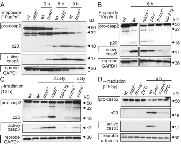
Figure 5. Caspase-2 processing occurs in thymocytes lacking the PIDDosome. (A) Thymocytes isolated from wild-type (wt) and pidd/ mice were cultured
without further treatment or stimulated with etoposide. Caspase-2 processing and cleavage of effector caspase-3 was monitored over time by Western blot analysis. The asterisk indicates a residual caspase-2 signal remaining upon reprobing with anti-GAPDH antibody. (B and C) Caspase-2 and -3 processing in etoposide-treated (B) or -irradiated thymocytes (C) of the indicated genotypes. tg, transgenic. (D) Thymocytes isolated from wild-type, pidd/, puma/,
and pidd/puma/ mice were cultured without further treatment or after exposure to irradiation for 6 h. Caspase-2 processing and cleavage of effector
caspase-3 were monitored by Western blot analysis. After detection of caspase-2, membranes were first stripped and reprobed with anti–active caspase-3 antibody and finally probed without stripping with an antibody recognizing GAPDH or -tubulin to compare protein loading. DKO, double knockout.
transformed human cancer cell lines (Tinel and Tschopp, 2004;
Baptiste-Okoh et al., 2008). Our study clearly indicates that PIDD
and caspase-2 are dispensable for apoptosis induction in
re-sponse to DNA damage, growth factor deprivation, ER stress,
disruption of cytoskeletal architecture, and pan-kinase inhibition
as well as Fas ligand– or TNF-–induced cell death in
lympho-cytes and SV40-transformed MEFs (Figs. 2 and 3). Loss of
mito-chondrial membrane potential or release of cytochrome c was
also not delayed in cells lacking caspase-2, RAIDD, or PIDD
(Fig. 4). Collectively, this demonstrates that the PIDDosome is
not required for efficient apoptosis induction in response to a wide
range of stimuli, including DNA damage (Table I). It is unclear
at present whether apoptosis induced by overexpression of p53
is impaired in PIDD-deficient cells, as suggested by knockdown
experiments in H1299 carcinoma cells (Baptiste-Okoh et al.,
2008), but stabilization of p53 by adding the Mdm2 inhibitor
Nutlin-3a killed PIDD-deficient MEFs as effectively as wild-type
cells while sparing p53
/MEFs, arguing against a prominent
role for PIDD in p53-induced apoptosis (unpublished data).
Caspase-2 processing occurred with comparable kinetics
in wild-type, raidd
/, and pidd
/thymocytes after DNA
dam-age and was always observed concomitantly with the appearance
of the activated form of caspase-3 (Fig. 5). Similar observations
have been made in HCT116 cells treated with PIDD- or
RAIDD-specific siRNA, but the cleavage of caspase-2 observed in
re-sponse to 5-fluoruracil treatment in these cells was attributed
to incomplete knockdown of the PIDDosome components
(Vakifahmetoglu et al., 2006). However, our observations
re-ported in this study suggest that caspase-2 cleavage occurs
sec-ondary to MOMP and activation of the classical apoptosome during
apoptosis induced by DNA damage. Consistently, all of these
events were blocked by preserving mitochondrial integrity,
either by overexpression of Bcl-2 or loss of Puma (Fig. 5).
Impor-tantly, PIDD also does not act in a redundant manner with the
Bcl-2–regulated pathway of caspase-2 processing, as its
cleav-age, as well as cell death, is comparable in puma
/and pidd
/puma
/cells (Figs. 2 and 5). Our data on caspase-2 processing
are in line with the observation that its cleavage depends on
the presence of Apaf-1 and caspase-9 in primary lymphocytes
(O’Reilly et al., 2002; Marsden et al., 2004) and cannot be used
as a readout for its activation during apoptosis induction and
contradict reports where activation of caspase-2 was positioned
upstream of MOMP and cytochrome c release in response to
DNA damage in tumor cell lines such as Jurkat or HeLa cells or
when recombinant caspase-2 was added directly to isolated
mito-chondria (Guo et al., 2002; Robertson et al., 2002, 2004).
Pri-mary MEFs lacking caspase-2 were also reported to resist cell
death induced by disruption of the cytoskeleton (Ho et al., 2008),
which we failed to observe in SV40-immortalized MEFs (Fig. 3 A).
All of these differences may be inherent to the cell types
ana-lyzed and/or the mode of immortalization/transformation, which
can affect the apoptotic response of cells in a highly diverse
manner. In line with the latter, we also observed that caspase-2
processing was delayed in pidd
/SV40-immortalized MEFs
(Fig. 6 A). This phenomenon was observed in independent clones
derived from individual mouse embryos (unpublished data).
Nevertheless, MEFs of both genotypes were equally sensitive to
with previous results (Colussi et al., 1998). However, loss of
raidd
or pidd did not influence the subcellular distribution or
translocation of caspase-2 in response to DNA damage, as
as-sessed by immunofluorescence analysis (Fig. 8). In addition, we
monitored cytoplasmic to nuclear translocation of caspase-2 in
wild-type and PIDD-deficient MEFs after exposure to
irradia-tion by Western blot analysis but again failed to observe
signifi-cant differences between genotypes regarding caspase-2 nuclear
translocation (Fig. 9). In summary, these results indicate that an
additional platform for caspase-2 activation exists in
mamma-lian cells or that caspase-2 can multimerize and autoprocess in
the absence of such a platform.
Discussion
We have generated mice that lack the p53-induced protein PIDD
and found that its absence does not interfere with normal mouse
development, caspase-2 activation, or cell death induction by
several different apoptotic stimuli. Formation of the PIDDosome
has previously been suggested to be rate limiting for
activa-tion of caspase-2 and p53-induced cell death in several
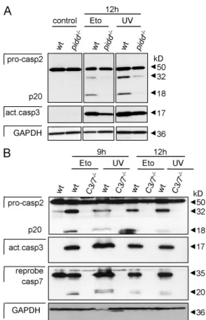
Figure 6. Caspase-2 processing is delayed in PIDD-deficient MEFs but still requires effector caspase activation. (A) Immortalized wild-type (wt) and pidd/ SV40-MEFs were exposed to 50 µg/ml etoposide (Eto) or
5 mJ/cm2 UVR, and lysates were immunoblotted for caspase-2 processing,
stripped, and reprobed with anti–active caspase-3 antibody followed by a reprobing with anti-GAPDH. (B) Wild-type and caspase-3/7 double-deficient SV40-MEFs were exposed to DNA damage as in A, and lysates were immunoblotted for caspase-2 processing. After detection of caspase-2, membranes were first stripped and reprobed with anti–active caspase-3 antibody followed by an antibody recognizing GAPDH to compare protein loading. Finally, membranes were stripped before detection of caspase-7.
can also form in the absence of the PIDDosome upon
tempera-ture shift (Fig. 7, A and B) but contain the active form of the
protease (Read et al., 2002). It will be important to demonstrate
that such a high molecular weight complex containing caspase-2
actually forms in whole cells in vivo upon apoptosis induction
or may only be triggered in vitro upon temperature shift.
Finally, subcellular localization of caspase-2 before and after
exposure of cells to DNA damage was not affected by loss of
PIDD or RAIDD, but, interestingly, caspase-2 accumulated in
the nucleus of SV40-MEFs upon etoposide treatment before
de-tectable signs of apoptosis and the activation of effector
cas-pases (Figs. 8 and 9).
Collectively, our findings suggest that at least one
alterna-tive PIDDosome-independent mechanism of caspase-2
activa-tion exists in mammals. Furthermore, caspase-2 dimerizaactiva-tion
and activation may even occur in the absence of any activating
platform, as observed upon overexpression in bacteria. A local
and transient increase in protein concentration either alone or in
combination with one of the many described posttranslational
modifications (Krumschnabel et al., 2009) may suffice to
pro-mote its full activation. Finally, we postulate that caspase-2
ac-tivation by PIDD does not contribute to apoptosis induction but
regulates events triggered in response to DNA damage such as
cell cycle arrest or repair.
apoptosis in response to all stimuli tested (Fig. 3, A and B).
Similar observations have been previously reported using MEFs
lacking RAIDD (Berube et al., 2005). Nevertheless, cleavage of
caspase-2 upon DNA damage strictly depended on the presence
of activated effector caspase-3 and/or -7 (Fig. 6 B). All together,
this suggests that the PIDDosome may contribute to caspase-2
processing after DNA damage in an effector caspase-dependent
manner. Although counterintuitive at first sight, effector
cas-pases appear to possess the potential to influence events
consid-ered to occur upstream of mitochondria such as the activation
and translocation of Bax (Lakhani et al., 2006). Therefore, we
propose that modulation of caspase-2 activity via the PIDDosome
does not contribute to apoptosis but may rather influence other
events triggered by DNA damage such as NF-B activation, cell
cycle arrest, and/or DNA repair. Consistently, a role for PIDD
and caspase-2 in G2/M checkpoint control and nonhomologous
end joining of double-strand breaks was reported recently (Shi
et al., 2009). However, our experiments investigating the role of
PIDD in NF-B activation upon DNA damage are currently too
preliminary to draw firm conclusions.
Finally, we also excluded that multimerization-induced
activation of caspase-2 in high molecular weight complexes
de-pends on the presence of the PIDDosome because gel filtration
analysis revealed that such caspase-2–containing complexes
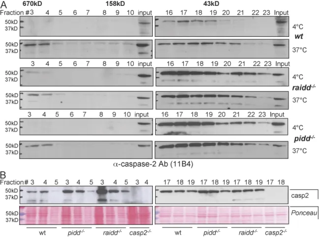
Figure 7. The PIDDosome is not required for the formation of high molecular weight complexes containing caspase-2. (A) Lysates from wild-type (wt),
raidd/, and pidd/ SV40-immortalized MEFs were incubated at 4°C or 37°C for 1 h and subjected to size-exclusion chromatography, and fractions
were precipitated, separated by SDS-PAGE, and immunoblotted with an anti–caspase-2 antibody (Ab). (B) Fractions 3–5, containing complexes ≥600 kD, and fractions 17–19, containing proteins of a molecular weight range of 75–40 kD, of all indicated genotypes were analyzed for the relative caspase-2 expression level. Ponceau staining of the membranes was used to compare protein loading (bottom).
T.W. Mak (University of Toronto, Toronto, Canada) and obtained on a mixed genetic background (72% C57BL/6 background). Caspase-3/7 double-deficient MEFs were provided by R. Flavell (Yale School of Medi-cine, New Haven, CT).
Materials and methods
Generation of pidd/ mice and other mouse strains used
All animal experiments were performed in accordance with the Austrian Tierversuchsgesetz (in accordance with Austrian legislation BGB1 Nr. 501/1988 i.d.F. 162/2005) and have been granted by the Bundesminis-terium für Bildung, Wissenschaft und Kultur. To generate a pidd-targeting construct, C57BL/6 genomic DNA was isolated and subcloned from the Bac clone RPCI-23:141F19 (Roswell Park Cancer Institute), harboring the entire pidd locus (Fig. 1). Exons 3–15 in pidd were replaced with the open reading frame encoding -galactosidase followed by a loxP element-flanked neomycin cassette. Two correctly targeted embryonic stem cell clones were identified by Southern blot analysis and subsequently injected into C57BL/6 blastocysts and delivered by CD1 surrogate mothers to gen-erate chimeric mice. Chimaeras derived from both independent embryonic stem cell clones were mated with wild-type C57BL/6 mice. Germline trans-mission was only obtained from one of these clones, and heterozygous offspring were used for the generation of pidd/ mice. The neomycin
resistance marker was subsequently removed by crossing to female C57BL/6 cre deleter mice (Schwenk et al., 1995).
With the exception of the in vivo irradiation experiments conducted with mice on mixed genetic background (B6/129SV; 1:1), all experi-ments shown were performed on pidd/ cells from mice that were
back-crossed onto C57BL/6 genetic background for eight generations. The generation of caspase-2/ (provided by D.L. Vaux, LaTrobe University,
Melbourne, Australia; O’Reilly et al., 2002), puma/ (provided by A.
Strasser, Walter and Eliza Hall Institute of Medical Research, Melbourne, Australia; Villunger et al., 2003), and vav-bcl-2 transgenic mice (pro-vided by J. Adams, Walter and Eliza Hall Institute of Medical Research, Melbourne, Australia; Ogilvy et al., 1999), all maintained on C57BL/6 genetic background, has been previously described (Villunger et al., 2003). p53-deficient mice on C57BL/6 background were provided by M. Serrano (Centro Nacional de Investigaciones Oncológicas, Madrid, Spain). Mice deficient for raidd (Berube et al., 2005) were provided by
Figure 8. Subcellular localization of caspase-2 in the absence of PIDD or RAIDD. Localization of endogenous caspase-2 (green) was detected in wild-type SV40-immortalized MEFs and MEFs lacking pidd or raidd before (control) or after drug treatment (50 µg/ml etoposide). Cells were costained for p53 (red) and the nucleus detecting fluorochrome DAPI (blue). Overlays of all staining are depicted (merge).
Figure 9. Nuclear translocation of caspase-2 upon DNA damage. Cyto-solic and nuclear extracts from wild-type (wt) and PIDD-deficient MEFs were generated from untreated cultures or 4 h after exposure to 4 Gy of irradiation. Extracts were separated by SDS-PAGE, and caspase-2 was de-tected by immunoblotting. Membranes were reprobed with antilamin- and antitubulin-specific antibodies to assess cross-contamination of fractions as well as anti–active caspase-3 to monitor for effector caspase activation. The asterisk indicates nonspecific (n.s.) bands.
cells were costained with FITC-conjugated Annexin V and 5 µg/ml PI and viability was assessed by flow cytometric analysis. SV40-immortalized MEFs were cultured at a density of 40,000 cells in 12-well plates. To deter-mine cellular viability, cells were stained with FITC-conjugated Annexin V and 5 µg/ml PI and analyzed by flow cytometry.
Quantification of mitochondrial membrane potential
SV40-immortalized MEFs were seeded into 6-well plates at 120,000 cells per well and grown overnight before being stimulated with relevant cell death inducers for up to 24 h. Cells were harvested by trypsinization, washed once with excess PBS, resuspended in DME medium containing 4 µM JC-1 (5,5,6,6-tetrachloro-1,1,3,3-tetraethylbenzimidazolcarbocya-nine iodide; Sigma-Aldrich), and incubated for 20 min at 37°C in the dark. Cells were washed once again with PBS and resuspended in 200 µl PBS. Alterations in mitochondrial membrane potential were then estimated from changes in cell populations with high red and low green fluorescence (high mitochondrial potential) to cells with low red and high green fluorescence (low mitochondrial potential) as determined by FACS analysis.
Quantification of cytochrome c release
SV40-immortalized MEFs were grown to subconfluence on 6-cm dishes, stimulated as indicated, trypsinized, washed in PBS, and permeabilized by the addition of digitonin in 50 µg/ml PBS for 5 min at 4°C. After removing digitonin by centrifugation, cells were fixed with 4% PFA for 20 min at RT. Thereafter, cells were washed three times with PBS, and nonspecific bind-ing was blocked usbind-ing PBS containbind-ing 3% BSA and 0.05% saponin. Sub-sequently, cells were incubated with anti–cytochrome c antibody (diluted to 1:200 in blocking buffer; clone 7H8.2C12; BD) at 4°C overnight. Cells were washed three times in PBS, and Alexa Fluor 488–labeled anti–mouse (Invitrogen) was used as secondary antibody at a dilution of 1:100 in blocking solution and incubated for 1 h at RT. After three further washing steps, cells were resuspended in PBS, and cytochrome c release was deter-mined by flow cytometric analysis.
Biochemical fractionation and immunoblot analysis
Cells were harvested and lysed for 1 h on ice in lysis buffer (1% CHAPS, 20 mM Tris-HCl, pH 7.5, 5 mM MgCl2, 137 mM KCl, 1 mM EDTA, 1 mM
EGTA, 0.5 mM PMSF, 5 mM NaF, and 2 mM Na3VO4; Roche), and the
protein concentration was determined using Bradford reagent (Bio-Rad Laboratories). Protein was separated by SDS-PAGE, transferred to a nitro-cellulose membrane, and incubated with the relevant antibodies (caspase-2 [11B4; Enzo Biochem, Inc.]; cleaved caspase-3 [Asp175; 5A1], caspase-3 [8G10], and caspase-7 [cs-9492; all from Cell Signaling Technology]; glyceraldehyde-3-phosphate dehydrogenase [GAPDH; 71.1; Sigma-Aldrich]; and -tubulin [DM1A; Santa Cruz Biotechnology, Inc.]). Blots were visual-ized by enhanced chemiluminescence (GE Healthcare).
Size chromatography and gel filtration
SV40-immortalized MEFs were washed in PBS and resuspended in a hypo-tonic buffer containing 20 mM Hepes-KOH, 10 mM KCl, 1 mM MgCl2, 1 mM
EDTA, 1 mM EGTA, pH 7.5, and Complete Protease Inhibitor Cocktail (Roche). Resuspended cells were subjected to three rounds of freeze thawing using liquid nitrogen. Cellular debris was removed by centrifugation at 10,000 g for 20 min at 4°C followed by filtration through a 0.5-µm mesh. Cell extracts were incubated at 37°C or 4°C for 60 min and then subjected to size exclusion chromatography. Treated lysates were fractionated using a fast protein liquid chromatography protein purification system on a sorbent (Ultrogel AcA 34; Pall Life Sciences) in a column (Tricorn10/600; GE Healthcare) at 4°C. The column was equilibrated with hypotonic buffer, 1.5–2 mg protein lysates was applied and eluted from the column with the same buffer at a constant flow rate of 0.5 ml/min, and 1-ml fractions were collected. The column was cali-brated with GE Healthcare gel filtration standards containing bovine thyro-globulin (670 kD), aldolase (158 kD), conalbumin (75 kD), and ovalbumin (43 kD). Each fraction from the gel filtration was precipitated with TCA at a final concentration of 13% (vol/vol), and samples were incubated for 5 min at 20°C followed by 15 min at 4°C. After centrifugation at 10,000 g for 20 min at 4°C, pellets were dissolved in 50 µl of 2× Laemmli buffer and 10 µl of 1 M Tris-HCl, pH 8.8. 20-µl samples were resolved on a 4–20% SDS– polyacrylamide gel and transferred to a nitrocellulose membrane. Western blot analysis was conducted according to a standard protocol using the aforementioned caspase-2 antibody provided by D.C. Huang and L. O’Reilly (Walter and Eliza Hall Institute of Medical Research, Melbourne, Australia). Localization of endogenous caspase-2
SV40-immortalized MEFs were plated onto glass cover slides overnight, exposed to the desired stimulus, washed once with PBS, and then fixed Southern blotting and PCR analysis
To confirm correct targeting of embryonic stem cells and deletion of the
pidd gene in tissues, 20 µg total genomic DNA was digested with the
ap-propriate restriction enzymes (DraIII [New England Biolabs, Inc.] for the 3 probe and MluI and EcoRV [Promega] for the 5 probe) overnight. Samples of DNA were size fractioned in 0.7% agarose gels in Tris-acetate-EDTA buffer, depurinated in 0.25 M HCl, denatured in 0.4 M NaOH, and trans-ferred in the same buffer onto Hybond N+ nylon membranes. Filters were
probed with 5 and 3 external probes or a neomycin cassette–specific probe in Church buffer at 65°C overnight. Membranes were washed in 40 mM Na-phosphate buffer containing 1% SDS at 65°C and exposed in a phosphoimager (Typhoon; GE Healthcare) for up to 2 d.
Littermates from heterozygous (pidd+/) intercrosses were genotyped
by PCR using primer pairs specific for the wild-type or the targeted pidd allele and the following cycle conditions: 4 min at 94°C; 40 s at 94°C, 30 s at 55°C, and 60 s at 72°C for 30 cycles; and 5 min at 72°C. Wild-type allele primers used were forward, 5-ACTTCTCCTGGTACTGGCTCTG-3; and reverse, 5-AAGGCTGCAAAGAACTTCTCAC-3. Primers used for the targeted allele (LacZ) were forward, 5-TGCCACTCGCTTTAATGATG-3; and reverse, 5-CTCCACAGTTTCGGGTTTTC-3. The wild-type and targeted
pidd alleles were amplified in separate reactions yielding PCR products of
259 bp and 420 bp in size, respectively.
Primers for RT-PCR were designed exon specific for exon 7–8 with forward, 5-TCGCTGTCGTGAGGTAGTTG-3, and reverse, 5-GAGAAGT-GCTCCCTCTGGTG-3; and for exon 13–15 with forward, 5-GGAAGCT-CAGGATGTTCG-3, and reverse 5-GGTTCAGAGGCATCAAGGAG-3. RT-PCR was performed using the following cycle conditions: 5 min at 94°C; 30 s at 94°C, 30 s at 55°C, and 30 s at 72°C for 30 cycles; and 7 min at 72°C.
Cell culture and reagents
MEFs were isolated from embryonic day 13.5 embryos after trypsin diges-tion and cultured in DME containing 250 µM L-Gln (Invitrogen), penicillin/ streptomycin (Sigma-Aldrich), and 10% FCS. All MEFs used were derived from mouse mutants maintained on C57BL/6 genetic background with the exception of raidd/ MEFs, which were derived from mice maintained on
mixed genetic background (72% B6). Primary MEFs used for experi-ments were always of low passage (less than five). Alternatively, MEFs were immortalized by standard procedures using retroviruses encoding the SV40 large T antigen.
Primary hemopoietic cells were cultured in RPMI-1640 medium (PAA), 250 µM L-Gln, 50 µM 2-mercaptoethanol, nonessential amino acids (Invitrogen), penicillin/streptomycin, and 10% FCS (PAA). For the induction of cell death, the following reagents were used: Super Fas ligand (Enzo Bio-chem, Inc.) at 10 ng/ml, staurosporine (Sigma-Aldrich) at 100 and 500 nM, ionomycin (Sigma-Aldrich) at 1 µg/ml, PMA (Sigma-Aldrich) at 10 ng/ml, VP16 (Sigma-Aldrich) at 1, 10, or 50 µg/ml, doxorubicin (Sigma-Aldrich) at 400 ng/ml, cisplatin (Sigma-Aldrich) at 25, 50, and 100 µM, the ER stressors tunicamycin (Sigma-Aldrich) at 0.1–5 µg/ml, brefeldin A at 1.25 and 2.5 µg/ml, and thapsigargin (Sigma-Aldrich) at 0.05–50 µg/ml, taxol (Sigma-Aldrich) at 100–500 µM, cytochalasin D (Sigma-Aldrich) at 2 µM, vincristine (EMD) at 50 µg/ml, glucocorticoid dexamethasone (Sigma- Aldrich) at 107 M, trichostatin A (Sigma-Aldrich) at 50 nM, bortezomib
(Vel-cade; Millennium Pharmaceuticals, Inc.) at 50 ng/ml, and TNF (PeproTech) at 10 ng/ml in the absence and presence of cycloheximide (Sigma-Aldrich) at 5 µg/ml. irradiation was performed at 1.25, 2.5, and 5 Gy, and UV-C radiation was performed at 5, 10, and 25 mJ/cm2.
Generation of an mPIDD-reactive antiserum
A 6× His-tagged murine PIDD-DD fragment (aa 770–910) was produced in Escherichia coli (strain BL21) from an IPTG-inducible pET21 expression vector and purified by column chromatography using Ni–nitrilotriacetic acid agarose beads. New Zealand white rabbits were immunized three times with 0.5 mg recombinant protein in Freund’s adjuvants. After con-firming serum reactivity with overexpressed mPIDD, the serum was affinity purified using a Sepharose column (GE Healthcare) coupled with PIDD-DD. Sera were mixed with PBS in a ratio of 10:1 and loaded onto the column, washed with 5 bed volumes of buffer A (20 mM Tris, 5 mM EDTA, 500 mM NaCl, and 0.1% NP-40, pH 8.0) followed by 5 bed volumes of buffer B (20 mM Tris, 5 mM EDTA, and 150 mM NaCl, pH 8.0). Next, 0.1 M Gly, pH 2.8, was added to the column to elute the IgG fraction, which was col-lected and neutralized by adding 0.1 vol of 1 M Tris buffer, pH 8.8. Apoptosis assay
Primary thymocytes were cultured at a density of 7.5 × 10⁵/ml in 96-well
Caspase activity assay
Cells were lysed in extraction buffer (10 mM Hepes, 1.5 mM MgCl2, 300
mM sucrose, 10 mM KCl, 10 mM DTT, 0.5% NP-40, and Complete Prote-ase Inhibitor Cocktail, pH adjusted to 7.0) for 30 min on ice and spun down for 10 min at 10.000 g, and the supernatant was stored at 20°C until use. 30 µl of the supernatant (containing 15 µg protein) was trans-ferred into the well of a 96-well plate, and 100 µl of capsase-3 assay buf-fer (50 mM Hepes-KOH, pH 7.4, 2 mM EDTA, 10% sucrose (wt/vol), 0.1% CHAPS (wt/vol), pH adjusted to 7.4, and 5 mM DTT added freshly from a 1-M stock), including 100 µM freshly added caspase-3 substrate (Ac-DEVD-AMC; Enzo Biochem, Inc.), was added. Substrate cleavage activity was then measured in a fluorescence microplate reader (Ascent Fluoroscan; Titertek) by following fluorescence changes over time with excitation and emission wavelengths set at 355 nm and 460 nm, respectively. Enzyme activities were normalized to sample protein contents determined by a Bradford assay.
Online supplemental material
Fig. S1 shows the specific reactivity of the anti-PIDD polyclonal antiserum. Fig. S2 shows that loss of PIDD does not impair lymphocyte development or survival in vivo. Fig. S3 shows the colony formation of primary MEFs in the absence of PIDD. Fig. S4 shows the colony formation of SV40-MEFs in the absence of PIDD. Fig. S5 shows the quantification of caspase activity in the absence of PIDD. Online supplemental material is available at http://www.jcb.org/cgi/content/full/jcb.200811105/DC1.
We thank K. Rossi and M. Saurwein for animal husbandry as well as C. Soratroi and I. Gaggl for excellent technical assistance. We also thank R. Flavell, T.W. Mak, M. Serrano, A. Strasser, D.C. Huang, J. Adams, D.L. Vaux, and L. O’Reilly for providing mice, MEFs, or reagents and P. Lukas for enabling irradiation ex-periments as well as R. Fässler for enabling blastocyst injections.
This work was supported by grants and fellowships from the Austrian Science Fund (FWF; START Y212-B13 and SFB021 to A. Villunger) and the Tiroler Wissenschaftsfond (to C. Manzl). F. Bock is funded by the Marie Curie Research Training Network ApopTrain.
Submitted: 20 November 2008 Accepted: 20 March 2009
References
Baptiste-Okoh, N., A.M. Barsotti, and C. Prives. 2008. A role for caspase 2 and PIDD in the process of p53-mediated apoptosis. Proc. Natl. Acad. Sci.
USA. 105:1937–1942.
Berube, C., L.M. Boucher, W. Ma, A. Wakeham, L. Salmena, R. Hakem, W.C. Yeh, T.W. Mak, and S. Benchimol. 2005. Apoptosis caused by p53-induced protein with death domain (PIDD) depends on the death adapter protein RAIDD. Proc. Natl. Acad. Sci. USA. 102:14314–14320. Bradley, G., S. Tremblay, J. Irish, C. MacMillan, G. Baker, P. Gullane, and S.
Benchimol. 2007. The expression of p53-induced protein with death do-main (Pidd) and apoptosis in oral squamous cell carcinoma. Br. J. Cancer. 96:1425–1432.
Colussi, P.A., N.L. Harvey, and S. Kumar. 1998. Prodomain-dependent nuclear localization of the caspase-2 (Nedd2) precursor. A novel function for a caspase prodomain. J. Biol. Chem. 273:24535–24542.
Cuenin, S., A. Tinel, S. Janssens, and J. Tschopp. 2008. p53-induced protein with a death domain (PIDD) isoforms differentially activate nuclear factor-kappaB and caspase-2 in response to genotoxic stress. Oncogene. 27:387–396.
Degterev, A., and J. Yuan. 2008. Expansion and evolution of cell death pro-grammes. Nat. Rev. Mol. Cell Biol. 9:378–390.
Duan, H., and V.M. Dixit. 1997. RAIDD is a new ‘death’ adaptor molecule.
Nature. 385:86–89.
Erlacher, M., V. Labi, C. Manzl, G. Bock, A. Tzankov, G. Hacker, E. Michalak, A. Strasser, and A. Villunger. 2006. Puma cooperates with Bim, the rate-limiting BH3-only protein in cell death during lymphocyte development, in apoptosis induction. J. Exp. Med. 203:2939–2951.
Guo, Y., S.M. Srinivasula, A. Druilhe, T. Fernandes-Alnemri, and E.S. Alnemri. 2002. Caspase-2 induces apoptosis by releasing proapoptotic proteins from mitochondria. J. Biol. Chem. 277:13430–13437.
Ho, L.H., S.H. Read, L. Dorstyn, L. Lambrusco, and S. Kumar. 2008. Caspase-2 is required for cell death induced by cytoskeletal disruption. Oncogene. 27:3393–3404.
Janssens, S., A. Tinel, S. Lippens, and J. Tschopp. 2005. PIDD mediates NF-kappaB activation in response to DNA damage. Cell. 123:1079–1092.
with 2% PFA at RT for 20 min. To permeabilize the cells, PFA was replaced with 70% prechilled ethanol for 5 min at RT. After three washes with PBS, unspecific binding sites were blocked by incubation with 8% BSA (in PBS) for 1 h at RT. Incubation with primary antibodies (1:200 anti–caspase-2 [clone 11B4] and 1:100 anti-p53 [Pab246; Santa Cruz Biotechnology, Inc.], both in blocking solution) was performed at 4°C overnight followed by three washings with PBS for 10 min each. After incubation with second-ary antibodies (1:100 in blocking solution; Alexa Fluor 488–labeled anti– rat and Alexa Fluor 546–labeled anti–mouse; Invitrogen) for 1 h at RT, cells were again washed with PBS, counterstained with 2 µg/ml DAPI, and washed and fixed with Vectashield antifade mounting medium (Vector Lab-oratories). Images were taken at RT with a confocal laser-scanning micro-scope (Axiovert 100M; Carl Zeiss, Inc.) equipped with LSM 510 acquisition software version 2.8 (Carl Zeiss, Inc.) and a 63× NA 1.4 oil immersion lens at a resolution of 1024 × 1024 pixels, with pinholes set to acquire im-ages <1 µm thick. The adjustment of brightness and contrast to enhance visibility of details was performed using the LSM Image Browser software version 4.2 (Carl Zeiss, Inc.), and the assembly of images, labeling, and export to tiff format were performed in Draw X4 (Corel).
Nuclear extracts
To separate cytoplasmatic and nuclear fractions, SV40-MEFs were har-vested after 4 Gy of radiation at the indicated time points. Cells were lysed in a cytosolic lysis buffer containing 10 mM Hepes, pH 7.9, 10 mM KCl, 0.1 mM EDTA, 0.1 mM EGTA, 1 mM DTT, and 0.5 mM PMSF. Nuclei were pelleted by centrifugation and were lysed in a nuclear buffer contain-ing 20 mM Hepes, pH 7.9, 0.4 M NaCl, 1 mM EDTA, 1 mM EGTA, 1 mM DTT, and 1 mM PMSF during a 30-min incubation at 4°C. Nuclear extracts were centrifuged to obtain the solubilized nuclear fraction.
Statistical analysis
Statistical analysis was performed using the unpaired Student’s t test or analysis of variance, where indicated, and applying the Stat-view 4.1 soft-ware program (Abacus Concepts). P-values of <0.05 were considered to indicate statistically significant differences.
Transient transfection of 293T cells
Specificity of the affinity-purified antiserum was confirmed on extracts from 293T cells overexpressing mPIDD alone or together with an mPIDD-specific short hairpin RNA construct (Tinel and Tschopp, 2004) using Lipofectamine 2000 (Invitrogen), according to the manufacturer’s instruction. Lysates were generated 24 h after transfection (see Biochemical fractionation and immuno-blot analysis) and subjected to SDS-PAGE separation and immunoimmuno-blotting. Ly-sates from MEFs stably transfected with mPIDD served as a positive control. Whole body irradiation experiments
PIDD-deficient mice and littermate controls, both on a mixed genetic back-ground (B6/SV129; 1:1), were exposed to graded doses of irradiation (2.5–5 Gy) at the age of 8–10 wk. Tissues were collected 20 h after irradi-ation to prepare single-cell suspensions. Cells were counted in a chamber hemocytometer (Neubauer), and aliquots were stained using fluorochrome-conjugated cell surface marker–specific antibodies followed by flow cyto-metric analysis (see next section).
Immunofluorescence staining, flow cytometric analysis, and cell sorting Single-cell suspensions from peripheral blood, bone marrow, lymph nodes, spleen, and thymus were surface stained with monoclonal antibodies con-jugated with FITC, R-phycoerythrin (PE), allophycocyanin, or biotin (Invitro-gen). The monoclonal antibodies used and their specificities are RA3-6B2, anti-B220; GK1.5, anti-CD4; YTS169, anti-CD8; RB6-8C5, anti–Gr-1; R2/60, anti-CD43; 5.1, anti-IgM; 11/26C, anti-IgD; MI/70, anti–Mac-1; Ter119, anti-erythroid cell surface marker; T24.31.2, anti–Thy-1; IM7, anti-CD44; H57-59, anti–TCR- (all from eBioscience); 7G6, anti-CD21; and B3B4, anti-CD23 (both from BD). Biotinylated antibodies were de-tected using streptavidin-R-PE (Dako) or streptavidin-vPE-Cy7 (BD). Flow cyto-metric analysis was performed using a cell analyzer (FACScalibur; BD). The sorting of cells was performed using a cell sorter (FACSvantage; BD). Clonal survival of MEFs
Colony formation of primary or SV40-immortalized MEFs was assessed by seeding an increasing numbers of cells per well (6,000, 12,000, and 18,000 cells) 24 h before DNA damage or exposure to heat shock. To in-duce heat shock, cells were incubated for 60 min in a tissue culture CO2
incubator (CB150; Binder) set to 43°C or 45°C and then grown under standard conditions. Etoposide was removed after 24 h of incubation. Sur-viving cells were stained 2 wk later using crystal violet.
Krumschnabel, G., B. Sohm, F. Bock, C. Manzl, and A. Villunger. 2009. The enigma of caspase-2: the laymen’s view. Cell Death Differ. 16:195–207. Lakhani, S.A., A. Masud, K. Kuida, G.A. Porter Jr., C.J. Booth, W.Z. Mehal, I.
Inayat, and R.A. Flavell. 2006. Caspases 3 and 7: key mediators of mito-chondrial events of apoptosis. Science. 311:847–851.
Lin, Y., W. Ma, and S. Benchimol. 2000. Pidd, a new death-domain-containing pro-tein, is induced by p53 and promotes apoptosis. Nat. Genet. 26:122–127. Marsden, V.S., P.G. Ekert, M. Van Delft, D.L. Vaux, J.M. Adams, and A. Strasser.
2004. Bcl-2-regulated apoptosis and cytochrome c release can occur in-dependently of both caspase-2 and caspase-9. J. Cell Biol. 165:775–780. McStay, G.P., G.S. Salvesen, and D.R. Green. 2008. Overlapping cleavage motif selectivity of caspases: implications for analysis of apoptotic pathways.
Cell Death Differ. 15:322–331.
Michalak, E.M., A. Villunger, J.M. Adams, and A. Strasser. 2008. In several cell types tumour suppressor p53 induces apoptosis largely via Puma but Noxa can contribute. Cell Death Differ. 15:1019–1029.
O’Reilly, L.A., P. Ekert, N. Harvey, V. Marsden, L. Cullen, D.L. Vaux, G. Hacker, C. Magnusson, M. Pakusch, F. Cecconi, et al. 2002. Caspase-2 is not required for thymocyte or neuronal apoptosis even though cleav-age of caspase-2 is dependent on both Apaf-1 and caspase-9. Cell Death
Differ. 9:832–841.
Ogilvy, S., D. Metcalf, C.G. Print, M.L. Bath, A.W. Harris, and J.M. Adams. 1999. Constitutive bcl-2 expression throughout the hematopoietic com-partment affects multiple lineages and enhances progenitor cell survival.
Proc. Natl. Acad. Sci. USA. 96:14943–14948.
Read, S.H., B.C. Baliga, P.G. Ekert, D.L. Vaux, and S. Kumar. 2002. A novel Apaf-1-independent putative caspase-2 activation complex. J. Cell Biol. 159:739–745.
Reed, J.C., K.S. Doctor, and A. Godzik. 2004. The domains of apoptosis: a ge-nomics perspective. Sci. STKE. doi:10.1126/stke.2392004re9.
Riedl, S.J., and G.S. Salvesen. 2007. The apoptosome: signalling platform of cell death. Nat. Rev. Mol. Cell Biol. 8:405–413.
Robertson, J.D., M. Enoksson, M. Suomela, B. Zhivotovsky, and S. Orrenius. 2002. Caspase-2 acts upstream of mitochondria to promote cytochrome c release during etoposide-induced apoptosis. J. Biol. Chem. 277:29803–29809. Robertson, J.D., V. Gogvadze, A. Kropotov, H. Vakifahmetoglu, B. Zhivotovsky,
and S. Orrenius. 2004. Processed caspase-2 can induce mitochondria-mediated apoptosis independently of its enzymatic activity. EMBO Rep. 5:643–648.
Schwenk, F., U. Baron, and K. Rajewsky. 1995. A cre-transgenic mouse strain for the ubiquitous deletion of loxP-flanked gene segments including dele-tion in germ cells. Nucleic Acids Res. 23:5080–5081.
Shi, M., C.J. Vivian, K.J. Lee, C. Ge, K. Morotomi-Yano, C. Manzl, F. Bock, S. Sato, C. Tomomori-Sato, R. Zhu, et al. 2009. DNA-PKcs-PIDDosome: a nuclear caspase-2-activating complex with role in G2/M checkpoint maintenance. Cell. 136:508–520.
Tinel, A., and J. Tschopp. 2004. The PIDDosome, a protein complex impli-cated in activation of caspase-2 in response to genotoxic stress. Science. 304:843–846.
Tinel, A., S. Janssens, S. Lippens, S. Cuenin, E. Logette, B. Jaccard, M. Quadroni, and J. Tschopp. 2007. Autoproteolysis of PIDD marks the bifurcation be-tween pro-death caspase-2 and pro-survival NF-kappaB pathway. EMBO
J. 26:197–208.
Vakifahmetoglu, H., M. Olsson, S. Orrenius, and B. Zhivotovsky. 2006. Functional connection between p53 and caspase-2 is essential for apopto-sis induced by DNA damage. Oncogene. 25:5683–5692.
Villunger, A., E.M. Michalak, L. Coultas, F. Mullauer, G. Bock, M.J. Ausserlechner, J.M. Adams, and A. Strasser. 2003. p53- and drug-induced apoptotic responses mediated by BH3-only proteins puma and noxa. Science. 302:1036–1038.