DOI 10.1007/s00439-014-1436-2 OrIGInal InvestIGatIOn
Expanding the clinical and mutational spectrum of Kaufman
oculocerebrofacial syndrome with biallelic UBE3B mutations
Lina Basel‑Vanagaite · Rüstem Yilmaz · Sha Tang · Miriam S. Reuter · Nils Rahner · Dorothy K. Grange · Megan Mortenson · Patrick Koty · Heather Feenstra · Kelly D. Farwell Gonzalez · Heinrich Sticht ·Nathalie Boddaert · Julie Désir · Kwame Anyane‑Yeboa · Christiane Zweier · André Reis · Christian Kubisch · Tamison Jewett · Wenqi Zeng · Guntram Borck
received: 16 December 2013 / accepted: 25 February 2014 / Published online: 11 March 2014 © springer-verlag Berlin Heidelberg 2014
additional patients from five unrelated families using either targeted UBE3B sequencing in individuals with sugges-tive facial dysmorphic features, or exome sequencing. Our results expand the clinical and mutational spectrum of the UBE3B-related disorder in several ways. First, we have identified UBE3B mutations in individuals who previously received distinct clinical diagnoses: two sibs with toriello– Carey syndrome as well as the patient reported to have a “new” syndrome by Buntinx and Majewski in 1990. sec-ond, we describe the adult phenotype and clinical variabil-ity of the syndrome. third, we report on the first instance of homozygous missense alterations outside the HeCt domain of UBe3B, observed in a patient with mildly dys-morphic facial features. We conclude that UBE3B muta-tions cause a clinically recognizable and possibly under-diagnosed syndrome characterized by distinct craniofacial
Abstract Biallelic mutations of UBE3B have recently
been shown to cause Kaufman oculocerebrofacial syn-drome (also reported as blepharophimosis–ptosis–intellec-tual disability syndrome), an autosomal recessive condition characterized by hypotonia, developmental delay, intellec-tual disability, congenital anomalies, characteristic facial dysmorphic features, and low cholesterol levels. to date, six patients with either missense mutations affecting the UBe3B HeCt domain or truncating mutations have been described. Here, we report on the identification of homozy-gous or compound heterozyhomozy-gous UBE3B mutations in six
l. Basel-vanagaite and r. Yilmaz contributed equally.
Electronic supplementary material the online version of this
article (doi:10.1007/s00439-014-1436-2) contains supplementary material, which is available to authorized users.
l. Basel-vanagaite (*)
raphael recanati Genetic Institute and Felsenstein Medical research Center, rabin Medical Center, Beilinson Campus, 49100 Petah tikva, Israel
e-mail: [email protected] l. Basel-vanagaite
sackler Faculty of Medicine, tel aviv University, 69978 tel aviv, Israel
l. Basel-vanagaite
Pediatric Genetics, schneider Children’s Medical Center of Israel, 49202 Petah tikva, Israel
r. Yilmaz · C. Kubisch · G. Borck (*) Institute of Human Genetics, University of Ulm, 89081 Ulm, Germany
e-mail: [email protected] s. tang · K. D. Farwell Gonzalez · W. Zeng ambry Genetics, aliso viejo, Ca 92656, Usa
M. s. reuter · C. Zweier · a. reis
Institute of Human Genetics, Friedrich-alexander-Universität erlangen-nürnberg, 91054 erlangen, Germany
n. rahner
Medical Faculty, Institute of Human Genetics, University of Dusseldorf, 40225 Dusseldorf, Germany
D. K. Grange
Division of Genetics and Genomic Medicine, Department of Pediatrics, Washington University school of Medicine, st. louis Children’s Hospital, st. louis, MO 63110, Usa M. Mortenson · P. Koty · t. Jewett
Department of Pediatrics, section on Medical Genetics, Wake Forest school of Medicine, Winston-salem, nC 27157, Usa H. Feenstra
features, hypotonia, failure to thrive, eye abnormalities, other congenital malformations, low cholesterol levels, and severe intellectual disability. We review the UBE3B-asso-ciated phenotypes, including forms that can mimick tori-ello–Carey syndrome, and suggest the single designation “Kaufman oculocerebrofacial syndrome”.
Introduction
Kaufman oculocerebrofacial syndrome (KOs; MIM 244450) is an autosomal recessive disorder character-ized by developmental delay and intellectual disability (ID), microcephaly, hypotonia, structural eye anoma-lies and other organ malformations, as well as distinctive facial dysmorphic features. Fewer than 10 affected indi-viduals had been reported from the first description in 1971 to 2012 (Kaufman et al. 1971; Figuera et al. 1993; Garcia-Cruz et al. 1988; Dentici et al. 2011) when we redescribed the syndrome (under the acronym BPID syn-drome, blepharophimosis–ptosis–intellectual disability syndrome; MIM 615057) and identified biallelic UBE3B mutations as the underlying cause (Basel-vanagaite et al. 2012). In that report, patients with BPID syndrome were thought to have a distinctive condition because they lacked the eye abnormalities characteristic to patients with KOs, such as microphthalmia and optic nerve anomalies. there is a phenotypic overlap between KOs, toriello–Carey syn-drome (tCs; agenesis of the corpus callosum with facial anomalies and Pierre-robin sequence; MIM 217980; tori-ello et al. 2003) and the phenotype reported by Buntinx and Majewski as a “new” syndrome in 1990 (Buntinx and
Majewski 1990). the differential diagnosis of KOs and
related syndromes extends to other blepharophimosis– intellectual disability syndromes (or Ohdo-like syndromes) such as Ohdo syndrome (MIM 249620), KAT6B-associated say-Barber/Biesecker/Young-simpson syndrome (MIM 603736; Clayton-smith et al. 2011), MED12-associated Maat-Kievit-Brunner syndrome (MIM 300895; vulto-van
silfhout et al. 2013), and Dubowitz syndrome (MIM 223370) (reviewed by verloes et al. 2006).
Only six patients with KOs/BPID syndrome and bial-lelic UBE3B mutations have been described previously (Basel-vanagaite et al. 2012; Flex et al. 2013). Here, we describe six additional patients and review the clinical and molecular spectrum of this recognizable entity.
Methods
Patient ascertainment and clinical evaluations
the study was approved by the ethics Committees of the Universities of Ulm and erlangen-nürnberg, and written informed consent was obtained from the patients’ parents or legal guardian. We report on six individuals from five unrelated families. the affected individuals were evaluated by routine medical history interviews and a physical exami-nation, and clinical records were reviewed. all six patients had undergone brain imaging [magnetic resonance imaging (MrI) in patients 1-5 and computed tomography in patient 6], and the MrI scans were reviewed by one radiologist (n.B.). a basic endocrinological evaluation had been per-formed in all affected individuals.
In patients 1–3, KOs was not regarded as a differential diagnosis and mutations were identified following clinical diagnostic or research exome sequencing. after the reports on UBE3B mutations as the cause of KOs (Basel-vanagaite et al. 2012; Flex et al. 2013) the attending geneticists of patients 1–3 contacted l.B.-v. and G.B. UBE3B mutations were suspected in patients 4–6 based mainly on facial dys-morphic features following a literature review performed by one of the authors (l.B.-v.) although these three chil-dren had previously received clinical diagnoses different from KOs/BPID. Patients 4 and 5 are siblings and have been reported with a diagnosis of typical tCs (toriello et al. 2003); and patient 6 was reported by Buntinx and Majewski (1990).
exome and sanger sequencing
UBE3B mutations were identified by whole exome sequencing in patients 1–3 and by sanger sequencing of the coding exons and exon–intron boundaries in patients 4–6. after the identification of a mutation in a sibling pair with a previous diagnosis of tCs, we sequenced UBE3B in four additional unrelated patients with tCs, two of whom have been reported previously (toriello et al. 2003). exome sequencing was performed using the sureselect Human all exon 50 Mb Kit (agilent) and a paired-end proto-col on an Illumina Hiseq 2000 sequencer (patients 1 and 3) or a sOliD 5500xl instrument (patient 2). reads were H. sticht
Institute of Biochemistry, Friedrich-alexander-Universität erlangen-nürnberg, 91054 erlangen, Germany
n. Boddaert
Department of Pediatric radiology, Hôpital necker, enfants Malades and Medical Faculty, Université Paris Descartes, 75015 Paris, France
J. Désir
Institut de Pathologie et de Génétique, 6041 Gosselies, Belgium K. anyane-Yeboa
Columbia University Medical Center, new York, nY 10010, Usa
aligned to the human genome assembly hg19 (GrCh37) and variants were called and annotated using software inte-grated in the respective in-house exome analysis pipeline as described previously [Gandomi et al. 2013, for patients 1 and 3; Hansen et al. 2013 (patient Mr043), for patient 2]. this resulted in a mean coverage of 136, 97, and 92× in patients 1–3, respectively; and 91, 80, and 89 % of the target bases were covered 10× or more. In individuals born to consanguineous parents, we additionally performed a search for large regions of homozygosity, either by iden-tifying runs of consecutive homozygous snPs in exome sequencing data essentially as described previously (Basel-vanagaite et al. 2012) or by genotyping patient Dna with affymetrix Genome-wide Human snP 6.0 arrays. PCr and sanger sequencing were performed for the validation of variants of interest and for cosegregation studies.
In patients 4–6, the 26 UBE3B coding exons and respec-tive exon–intron boundaries were sequenced on genomic Dna as described previously (Basel-vanagaite et al. 2012). Potentially disease-causing variants were analyzed for their frequency by querying the nHlBI exome sequenc-ing Project database (exome variant server; http://evs.gs. washington.edu/evs). In case of patient 2, we addition-ally sequenced the UBE3B exons of interest in 188 turk-ish control individuals. Polyphen-2 (http://genetics.bwh. harvard.edu/pph2/index.shtml) and sIFt (http://sift.jcvi.org) were used to predict the impact of missense alterations on protein function.
Results
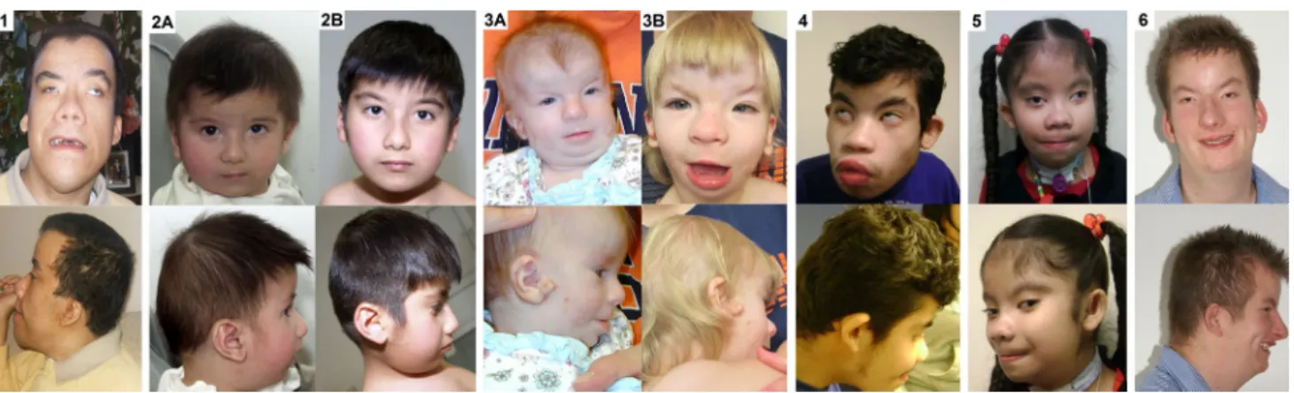
Clinical reports and identification of UBE3B mutations Facial features of patients 1–6 are shown in Fig. 1, clini-cal details are summarized in table 1, and brain MrIs are shown in supplementary Fig. 1.
Patient 1 (Fig. 1, picture 1; table 1) is a 33-year-old man with profound ID and no speech. He is the second son (of three) born to unaffected parents of Hispanic ancestry who are second cousins once removed. He presented at birth with muscular hypotonia and hearing impairment. Unlike the other patients in whom microcephaly or an occipito-frontal circumference (OFC) <10th centile was observed, his OFC was at the 75th percentile. relative macrocephaly was probably of familial origin as his father was reported to be macrocephalic. Brain MrI showed moderately dilated lateral ventricles in patient 1 (supplementary Fig. 1) and diffuse anomalies of the white matter frontally. He has an abnormal sleep pattern and occasional violent behavior.
Using clinical diagnostic whole-exome sequencing, we detected a homozygous nonsense mutation in UBE3B, c.61G>t (p.Glu21*) (table 2; supplementary Fig. 2). the patient’s mother and unaffected brother were heterozygous carriers of the mutation, as shown by sanger sequencing; no Dna from the deceased father was available for genetic studies. In addition, alterations in two other ID-related genes were identified by exome sequencing and confirmed by sanger sequencing; namely, a rare homozygous mis-sense alteration affecting a highly conserved amino acid, c.878G>a (p.arg293Gln; rs35267264), in KIAA1033, and a heterozygous missense alteration of a highly conserved amino acid, c.1307C>t (p.Pro436leu), in EHMT1. a sin-gle homozygous KIAA1033 mutation has previously been suggested as the cause of moderate to severe ID, poor lan-guage and adaptive skills, delayed fine motor development, and short stature in a family from Oman (ropers et al. 2011). Heterozygous alterations in EHMT1 cause Kleefstra syndrome, which is characterized by severe ID, hypotonia, brachycephaly, flat face with hypertelorism, upslanting pal-pebral fissures and a thick everted lower lip (Kleefstra et al. 2006). the following evidence supports the homozygous UBE3B nonsense mutation, rather than one of the variants in KIAA1033 or EHMT1, as the major contributor to the
Fig. 1 Facial features of the individuals described in this study. the numbers correspond to the numbers in table 1. ages of the patients are as follows: 1, 33 years; 2A, 8 months; 2B, 5 years 6 months; 3A, 4 months; 3B, 2 years; 4, 16 years; 5, 10 years 8 months; 6, 25 years
Table
1
Clinical findings in 12 indi
viduals with biallelic
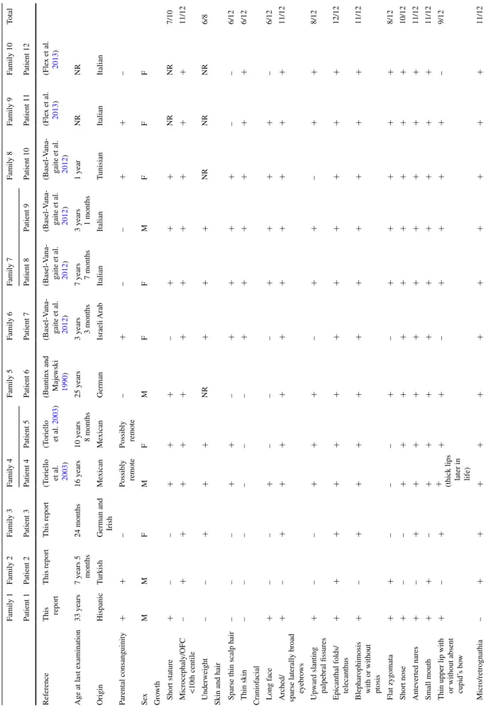
UBE3B mutations Family 1 Family 2 Family 3 Family 4 Family 5 Family 6 Family 7 Family 8 Family 9 Family 10 total Patient 1 Patient 2 Patient 3 Patient 4 Patient 5 Patient 6 Patient 7 Patient 8 Patient 9 Patient 10 Patient 11 Patient 12 r eference t his report t his report t his report (t oriello et al. 2003 ) (t oriello et al. 2003 )
(Buntinx and Maje
wski 1990 ) (Basel-v ana -gaite et al. 2012 ) (Basel-v ana -gaite et al. 2012 ) (Basel-v ana -gaite et al. 2012 ) (Basel-v ana -gaite et al. 2012 ) (Fle x et al. 2013 ) (Fle x et al. 2013 ) a ge at last e xamination 33 years 7 years 5 months 24 months 16 years 10 years 8 months 25 years 3 years 3 months 7 years 7 months 3 years 1 months 1 year nr nr Origin Hispanic t urkish
German and Irish
Me xican Me xican German Israeli a rab Italian Italian t unisian Italian Italian Parental consanguinity + + – Possibly remote Possibly remote – + – – + + – sex M M F M F M F F M F F F Gro wth s hort stature + – – + + + – + + + nr nr 7/10 Microcephaly/OFC <10th centile – + + + + + + + + + + + 11/12 Underweight – – + + + nr + + + nr nr nr 6/8
skin and hair sparse thin scalp hair
– – – + + – + + + + – – 6/12 t hin skin – – – – – – + + + + + + 6/12 Craniof acial l ong f ace + – – + – – – + + + + – 6/12 a rched/
sparse laterally broad eyebro
ws + – + + + + + + + + + + 11/12 Upw ard slanting palpebral fissures + – – + + + – + + – + + 8/12 e
picanthal folds/ telecanthus
+ + + + + + + + + + + + 12/12
Blepharophimosis with or without ptosis
+ – + + + + + + + + + + 11/12 Flat zygomata + + – – – + – + + + + + 8/12 s hort nose + – – + + + + + + + + + 10/12 a nte verted nares + – + + + + + + + + + + 11/12 s mall mouth + + – + + + + + + + + + 11/12 t
hin upper lip with or without absent cupid’
s bo
w
+
–
+
+ (thick lips later in life)
+ + – + + + + – 9/12 Micro/retrognathia – + + + + + + + + + + + 11/12
Table 1 continued Family 1 Family 2 Family 3 Family 4 Family 5 Family 6 Family 7 Family 8 Family 9 Family 10 total Patient 1 Patient 2 Patient 3 Patient 4 Patient 5 Patient 6 Patient 7 Patient 8 Patient 9 Patient 10 Patient 11 Patient 12 Dysplastic ears + + + (with preau -ricular tags) + + + + + + + + (with preau -ricular tags) + (with preau -ricular tags) 12/12 P alatal anomalies – + – + – + + + – – + + 7/12 r
espiratory system tracheomalacia/laryn
-gomalacia/stridor – – – + + – + – – + + + 6/12 Cardio vascular system
Congenital heart disease
–
–
–
–
+ (mild distal right pulmonary artery stenosis and septal hypertro
-ph
y)
–
+ (atrial septal defect, ventricu
-lar septal defect, aortic coarctation)
–
–
+ (atrial septal defect)
–
–
3/12
Gastrointestinal system Feeding difficulties in
inf anc y/ childhood nr + + + + + + + + + + + 11/11 Constipation – – – + – – – + + – + + 5/12
Urogenital system Genital abnormalities
– – – + (small penis) + (hypoplas
-tic labia majora)
– – – – + (hypoplas
-tic labia majora)
+ (Clitoro -me galy) + (Clitoro -me galy) 5/12 r enal abnormalities – –
+ (abnormal shape of one kidne
y
without functional problem)
– – + (grade v vesicoureteral reflux) + (mild left p ye -lectasis)
+ (double right kidne
y, right p yelec -tasis) – – – – 4/12 sk eletal system s
tructural finger/toe abnormalities
– – – + + + – + + – – – 5/12 Pes talus v arus/v algus – – + (metatarsus adductus) – + – – – – – + + 4/12
Congenital dislocation of the hip/coxa valg
a – – – – – – + – – + + – 3/12
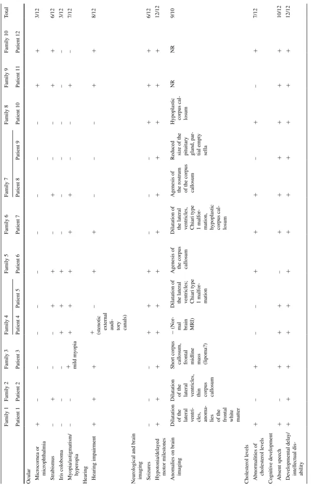
Table 1 continued Family 1 Family 2 Family 3 Family 4 Family 5 Family 6 Family 7 Family 8 Family 9 Family 10 total Patient 1 Patient 2 Patient 3 Patient 4 Patient 5 Patient 6 Patient 7 Patient 8 Patient 9 Patient 10 Patient 11 Patient 12 Ocular Microcornea or microphthalmia + – – – – – – – – – + + 3/12 s trabismus – + – – + + – + – – + + 6/12 Iris coloboma – – – + + + – – – – – – 3/12 Myopia/astigmatism/ hyperopia – – + mild myopia + + + + + – – + – 7/12
Hearing Hearing impairment
+
+
+
+ (stenotic external audi
-tory canals) – + + – – – + + 8/12 n
eurological and brain imaging seizures
– – – + + + – – – + + + 6/12
Hypotonia/delayed motor milestones
+ + + + + + + + + + + + 12/12 a
nomalies on brain imaging Dilatation of the lateral ventri
-cles, anoma
-lies of the frontal white matter Dilatation of the lateral ventricles, thin corpus callosum
short corpus callosum, frontal midline mass (lipoma?)
− ( n or -mal brain Mr I)
Dilatation of the lateral ventricles; Chiari type 1 malfor
-mation
a
genesis of the corpus callosum Dilatation of the lateral ventricles, Chiari type 1 malfor
-mation, hypoplastic corpus cal
-losum
a
genesis of the rostrum of the
corpus
callosum
r
educed size of the pituitary gland, par
-tial empty sella Hypoplastic corpus cal
-losum nr nr 9/10 Cholesterol le vels a bnormalities of cholesterol le vels + – + – – + + + – + – + 7/12 Cogniti ve de velopment a bsent speech + – + + + – + + + + + + 10/12 De
velopmental delay/ intellectual dis
-ability + + + + + + + + + + + + 12/12 NR not reported
patient’s phenotype: (1) His clinical presentation and facial dysmorphism are similar to those observed in other patients with KOs. (2) the KIAA1033 variant has a minor allele fre-quency of 0.14 % (nHlBI exome variant server), suggest-ing that it may be a rare polymorphism. notably, KIAA1033 is located on chromosome 12q23.3, approximately 4.5 Mb away from UBE3B, and both genes are within a 46.6 Mb region of homozygosity on chromosome 12 in patient 1, as determined by homozygosity analysis in the exome sequence. (3) Patient 1 has no facial features suggestive of Kleefstra syndrome. thus, although more than one of the three variants may contribute to the phenotype, including to clinical presentations that are atypical for KOs, the truncat-ing UBE3B mutation is likely the major causative allele.
Patient 2 is a 7-year 5-month-old boy who presented with muscular hypotonia, congenital microcephaly and variable other anomalies (Fig. 1, pictures 2a and 2B; table 1). He developed minimal speech (5–10 single words at 7 years of age) and has an estimated IQ of 20–49. His unaffected parents are first cousins originating from tur-key. Craniofacial dysmorphisms are mild, and he is the only patient in this study without blepharophimosis.
Whole-exome sequencing revealed two homozygous UBE3B missense mutations, c.1445t>a (p.leu482His) and c.1616t>C (p.leu539Pro; table 2, supplementary Fig. 2), with both parents being heterozygous for both mutations. UBE3B is located in a 21.75 Mb region of homozygosity in this patient’s exome. Both mutations affect conserved amino acid residues located outside the IQ and HeCt domains (Gong et al. 2003). therefore, the relatively mild craniofa-cial phenotype in this patient might be related to the atypi-cal location of the mutations compared to other patients, in whom missense alterations within the HeCt domain are predicted to have severe consequences. In silico analysis revealed that leu482 is located in a leucine-rich region span-ning residues 472–500. the regular spacing of hydropho-bic residues (every 7th amino acid position) resembles that of a leucine zipper (Hirst et al. 1996). such leucine zippers form dimers, suggesting that this UBe3B region might be involved in intermolecular interactions. a similar function can be assigned to the region around leu539, which is pre-dicted to exhibit a coiled-coil structure (residues 512–560). For both mutation sites, the type of amino acid exchange will drastically affect the biophysical properties and is therefore likely to disrupt the respective protein-interaction motif.
Five other rare homozygous coding variants were con-firmed by sanger sequencing (none affecting a known dis-ease-associated gene; data not shown).
Patient 3 (Fig. 1, pictures 3a and 3B; table 1) is a 24-month-old girl. she has congenital microcephaly (cur-rently her OFC is at −4 sD), bilateral large complex preau-ricular tags and stenotic external ear canals associated with moderate-to-severe conductive hearing impairment. she
has significant feeding problems with gastroesophageal reflux and vomiting, requiring the placement of a gastros-tomy tube (G-tube). she had two patches of hair on each cheek and hypertrichosis of the back. MrI scan showed partial agenesis of the corpus callosum and a frontal mid-line mass, thought to be a lipoma (supplementary Fig. 1).
Clinical diagnostic whole exome sequencing revealed a homozygous missense mutation, c.2990G>C (p.arg997Pro), in UBE3B (table 2), affecting the HeCt domain. the mutation was confirmed by sanger sequenc-ing (supplementary Fig. 2), and the parents were heterozy-gous carriers of the mutation.
Patients 4 and 5 are siblings of Hispanic american origin with a clinical diagnosis of tCs (Fig. 3 and Fig. 2, respec-tively, in toriello et al. 2003; table 1). recent pictures show-ing their facial dysmorphic features are displayed in Fig. 1, pictures 4 and 5. the presence of UBE3B mutations was suspected after reviewing relevant scientific publications and noticing suggestive facial features (toriello et al. 2003).
Patient 4 is the older sibling and is currently 16 years old. From birth he has had severe respiratory and feeding problems with abnormal swallowing, which necessitated a nissen fundoplication with G-tube placement at 2 months. at age 16, he is still G-tube dependent. He required a tra-cheostomy because of laryngomalacia and subglottic steno-sis and underwent laryngotracheal reconstruction at 2 years 6 months of age. at the age of 13 years, he was found to have scoliosis and focal progressive kyphosis requiring sur-gical intervention.
Patient 5 is the 11-year-old younger sister of patient 4. she had a similar clinical presentation to that of her brother, including poor suck and severe respiratory problems. she also needed a tracheostomy because of tracheomalacia and is still tracheostomy dependent at age 11. she had signifi-cant aspiration and obstructive sleep apnea. she also under-went nissen fundoplication and was fed through a G-tube. In addition, she underwent bilateral tarsometatarsal release and left cuboid closing wedge osteotomy. Both sibs showed general hypertrichosis in their first year of life.
although their parents were not aware of consanguinity, snP-array genotyping detected two large (>10 Mb) blocks of homozygosity in each of the siblings, possibly indicat-ing remote parental consanguinity. the sindicat-ingle region of shared homozygosity was a 11.4 Mb segment on chromo-some 12q23.3–q24.22 that contained UBE3B. sequencing of UBE3B revealed a homozygous c.2335G>a missense alteration in both affected sibs (p.Gly779arg; table 2, sup-plementary Fig. 2); the parents were heterozygous carriers of the mutation. Gly779 is a highly conserved amino acid of the HeCt domain.
We next sequenced UBE3B in four additional patients with tCs, none of whom had the characteristic facial appear-ance associated with KOs. We did not detect any mutations.
Patient 6. this man (Fig. 1, picture 6; table 1) is cur-rently 25 years old and the presence of a UBE3B mutation was suspected because of the recognition of characteristic facial features from a previously published report (Buntinx and Majewski 1990). the neonatal period was complicated by poor suck and hypotonia. Postaxial polydactyly of both hands was noticed at birth and treated by surgery. Cur-rently, he has about 100 words and can speak in 3–4 word sentences.
sequencing of UBE3B revealed a compound heterozy-gous mutation with a c.2098C>t (p.Gln700*) nonsense mutation inherited from his mother and a c.2990G>C (p.arg997Pro) mutation (table 2, supplementary Fig. 2), which was also present in his unaffected brother and likely inherited from their deceased father.
review of the clinical and laboratory features of patients with biallelic UBE3B mutations
table 1 summarizes clinical findings on the patients with biallelic UBE3B mutations reported here and previously (Basel-vanagaite et al. 2012; Flex et al. 2013).
Demographic data
Of the 12 patients with UBE3B mutations, seven were female. Consanguinity was reported in five of the families. In two families, there were two affected sibs.
Pregnancy and birth
no abnormal findings were reported following prenatal ultrasound except for polyhydramnion in one case and oli-gohydramnion in one case. all the patients for whom data were available were born at term.
Growth
Birth weight was normal in most of the patients. the majority of patients had an OFC below the 10th centile at birth. Most affected children had failure to thrive and post-natally decreasing age-matched OFC centiles. all but three affected individuals had short stature, and all but one had microcephaly or an OFC below the 10th centile.
Ectodermal abnormalities
sparse scalp hair was described in infancy in 6/12 patients. the appearance of the scalp hair improved with age. the skin was described as thin in six patients. three affected individuals had hypertrichosis, and two had eczema. Craniofacial abnormalities
several patients were described as having a flat occiput or brachycephaly. Characteristic craniofacial features included arched, sparse and laterally flared eyebrows,
Table 2 UBE3B mutations
Patient Origin Parental con-sanguinity
Mutation (Dna)
Predicted effect on protein
Protein domain Control alleles (evs) Polyphen-2 sIFt 1 Hispanic american second cousins once removed c.61G>t homozygous p.(Glu21*) truncation n-terminal to IQ and HeCt domains 0/13,006 n.a. n.a.
2 turkish First cousins c.1445t>a homozygous p.(leu482His) leucine-rich region 0/13,006 0/376 turk-ish control alleles Probably damaging not tolerated c.1616t>C homozygous p.(leu539Pro) Predicted coiled-coil domain 0/13,006 0/376 turk-ish control alleles Probably damaging not tolerated 3 German and Irish no c.2990G>C homozygous
p.(arg997Pro) HeCt domain 0/13,006 Probably damaging
not tolerated
4 and 5 Mexican no c.2335G>a
homozygous
p.(Gly779arg) HeCt domain 0/13,006 Probably damaging not tolerated 6 German no c.2098C>t heterozygous p.(Gln700*) truncation n-terminal to HeCt domain 0/13,006 n.a. n.a. c.2990G>C heterozygous
p.(arg997Pro) HeCt domain 0/13,006 Probably damaging
blepharophimosis with epicanthal folds with or without ptosis, flat zygomata, a characteristic nasal shape with a relatively narrow nasal bridge and broad nasal base, a flat philtrum (long or short), and thin lips, especially the upper lip, with absent cupid’s bow, as well as a small mouth. the ears were often apparently low-set with over-folded helices or cupping, small earlobes and, in some instances, underde-veloped ear crus. three patients had preauricular tags and two had stenotic external ear canals. With advancing age, the shape of the face became more elongated, the palpebral fissures became up-slanting and, in some patients the lip vermilion became more everted. One adult had a long chin and very thick alae nasi with narrow nares.
Respiratory tract abnormalities
six patients had breathing problems including tracheoma-lacia, subglottic stenosis, laryngomalacia or stridor. two children needed a tracheostomy. two patients had a history of apnea either shortly after birth or obstructive sleep apnea later in life.
Cardiovascular system
Congenital heart malformations are not a common find-ing in the UBE3B-related syndrome (3/12 patients). Heart defects included pulmonary artery stenosis, asD, vsD, and aortic coarctation.
Gastrointestinal tract abnormalities
eleven patients had feeding difficulties including poor suck and gastroesophageal reflux. three patients required G-tube feeding. Constipation was present in five individuals. Urogenital abnormalities
Genital abnormalities were more frequent in females (although the difference is not statistically significant in this small series) and included hypoplastic labia majora and/or minora or clitoromegaly (4/6 females). Micropenis was described in 1/5 males. renal abnormalities included vesicoureteral reflux up to grade v and duplicated renal pelvis.
Skeletal abnormalities
skeletal abnormalities were observed in 10/12 individu-als and included abnormal chest shape (bell-shaped tho-rax, pectus carinatum) and long and slender fingers and toes. Clinodactyly of the fifth finger and hypoplastic distal phalanges were also reported. Congenital dysplasia of the hip was diagnosed in two newborns. Uncommon skeletal
findings included polydactyly, coxa valga, scoliosis, con-genital arthrogryposis, and metatarsus adductus.
Ocular abnormalities
three of the 12 patients had microcornea or microphthal-mia, 6/12 strabismus, three coloboma of the iris, five myo-pia with or without astigmatism, one hyperomyo-pia, three a pale optic disc, and two entropion.
Hearing loss
eight patients had hearing impairment, mostly conductive, but three individuals had sensorineural or mixed conduc-tive–sensorineural hearing loss; and two had cholesteatoma. Neurological abnormalities
Hypotonia and delayed motor milestones were observed in all affected individuals. six patients had seizures, often fever related. Independent walking was mostly achieved by 4–5 years of age; the gait was frequently described as unsteady. ID was present in all patients and was described as severe to profound. Only two patients developed speech, which in both cases was limited. structural Cns abnormal-ities included absent or hypoplastic corpus callosum in six patients and a Chiari 1 malformation in two (supplemen-tary Fig. 1).
Endocrine and cholesterol abnormalities
elevated tsH and reduced thyroid gland volume were diagnosed in one and low GH and aCtH in another patient. seven individuals had low cholesterol levels including low total cholesterol, low HDl, low lDl levels, or combina-tions thereof.
Discussion
recognition of KOs should be possible on clinical grounds in many cases. a consistent and recognizable pattern of typical craniofacial features combined with hypotonia, fail-ure to thrive, eye abnormalities, other congenital malforma-tions, low cholesterol levels and severe ID should indeed facilitate early diagnosis of patients with UBE3B muta-tions. We note, however, that neither ocular nor cerebral malformations are mandatory findings in this “oculocer-ebrofacial” syndrome. evaluation of individuals with KOs should involve all body systems including assessment of the respiratory and gastrointestinal tracts, cardiologic and ophthalmologic evaluation, hearing tests, renal ultrasound, investigation of thyroid function and cholesterol levels,
developmental assessment and orthopedic evaluation. this series also illustrates the facial dysmorphism in adults with KOs and underscores the phenotypic variability, e.g., with respect to speech or limb involvement. although mild limb abnormalities such as long and slender fingers, clinodactyly and camptodactyly have been described in patients with UBE3B mutations, polydactyly has not been reported pre-viously. While polydactyly may be a coincidental finding, a patient with facial features reminiscent of UBE3B-related features and polydactyly has been described by Gabrielli et al. (1994) as having a new form of oro-facio-digital syn-drome. Other cases with suggestive clinical features—but no UBE3B sequence analysis yet—have been reported, such as the patient described by al Frayh and Haque (1987).
It is of interest that three patients, two described in this series and one in our previous study (patient 4 in Basel-vanagaite et al. 2012, who is listed as patient 10 in our table 1), were diagnosed as having tCs. although patients with UBE3B mutations and patients originally described by toriello and Carey (1988) share several overlapping clinical features (short palpebral fissures, small nose with anteverted nares, abnormal ears, laryngeal anomalies, car-diac defects, agenesis of the corpus callosum, microceph-aly, micrognathia and cleft palate), the facial gestalt still is not completely similar. We note that the facial dysmor-phic features of children with UBE3B mutations previously thought to have tCs are more similar to KOs than to the disorder originally described by toriello and Carey (1988). thus, UBE3B mutations define KOs which includes BPID syndrome, the phenotype described by Buntinx and Majew-ski and a subset of patients previously diagnosed with tCs. Because Kaufman first described the typical clinical picture in 1971, we suggest—to avoid confusion—designating the syndrome with biallelic UBE3B mutations as “Kaufman oculocerebrofacial syndrome”.
apart from providing data expanding the clinical phenotype of KOs, our report illustrates some of the challenges and opportunities of exome sequencing in a clinical context. In patient 1, clinical exome sequencing detected rare variants in three ID-related genes. Whereas the clinical similarities with KOs highlighted the UBE3B mutation as the most relevant variant, we cannot exclude a contribution of the variants in KIAA1033 and EHMT1 to the phenotype. large-scale studies have highlighted the difficulties in assigning a single variant to a patient’s phenotype, and in rare instances exome sequencing might even confirm two genetic diseases in one individual (Yang et al. 2013), and such situations probably make it difficult in some cases to assign a diagnosis on clinical grounds only. Furthermore, our data also illustrate how exome sequencing can help to establish a diagnosis in patients with a mild or atypical disease presentation and thereby
contribute to the delineation of the phenotypic spectrum of rare genetic disorders. In patients 2 and 3, respectively, the mild and atypical facial dysmorphism had not raised the suspicion of a UBE3B-related disorder, but exome sequencing revealed a presumably causative homozygous missense variant in each case. the identification of mis-sense alterations outside the HeCt domain in patient 2 who presented with mild craniofacial dysmorphism with no blepharophimosis suggests a possible genotype–phe-notype correlation. Future studies will establish whether missense changes outside the HeCt domain generally lead to a milder phenotype.
In conclusion, we report on six individuals with bial-lelic UBE3B mutations, thereby doubling the number of reported patients. Identification of UBE3B mutations has merged phenotypes that were previously suspected to rep-resent distinct syndromes into a single, possibly underdiag-nosed disorder which can be clinically recognized.
Acknowledgments We wish to thank the family members for their
participation, Gabrielle J. Halpern for help with editing the manu-script, and H. toriello, M. C. addor, r. Hordijk, n. Dikow and K. van Berkel for contributing Dna from patients with tCs. this work was supported by the Israeli Ministry of Health Chief scientist foundation (grant no. 3-4963) and Israeli science Foundation (grant no. 09/558) (l.B.-v.). G.B. was supported by the Deutsche Forschungsgemein-schaft. the authors declare that they have no conflict of interest.
References
al Frayh ar, Haque Kn (1987) anophthalmia, microcephaly, hypo-tonia, hypogonadism, failure to thrive and developmental delay. Dysmorph Clin Genet 1:64–66
Basel-vanagaite l, Dallapiccola B, ramirez-solis r, segref a, thiele H, edwards a, arends MJ, Miró X, White JK, Désir J, abramow-icz M, Dentici Ml, lepri F, Hofmann K, Har-Zahav a, ryder e, Karp na, estabel J, Gerdin aK, Podrini C, Ingham nJ, altmül-ler J, nürnberg G, Frommolt P, abdelhak s, Pasmanik-Chor M, Konen O, Kelley rI, shohat M, nürnberg P, Flint J, steel KP, Hoppe t, Kubisch C, adams DJ, Borck G (2012) Deficiency for the ubiquitin ligase UBe3B in a blepharophimosis–ptosis–intel-lectual-disability syndrome. am J Hum Genet 91(6):998–1010. doi:10.1016/j.ajhg.2012.10.011
Buntinx I, Majewski F (1990) Blepharophimosis, iris coloboma, microgenia, hearing loss, postaxial polydactyly, aplasia of cor-pus callosum, hydroureter, and developmental delay. am J Med Genet 36(3):273–274. doi:10.1002/ajmg.1320360304
Clayton-smith J, O’sullivan J, Daly s, Bhaskar s, Day r, anderson B, voss aK, thomas t, Biesecker lG, smith P, Fryer a, Chan-dler Ke, Kerr B, tassabehji M, lynch sa, Krajewska-Walasek M, McKee s, smith J, sweeney e, Mansour s, Mohammed s, Donnai D, Black G (2011) Whole-exome-sequencing identifies mutations in histone acetyltransferase gene Kat6B in individuals with the say-Barber-Biesecker variant of Ohdo syndrome. am J Hum Genet 89(5):675–681. doi:10.1016/j.ajhg.2011.10.008
Dentici Ml, Mingarelli r, Dallapiccola B (2011) the difficult nosol-ogy of blepharophimosis–mental retardation syndromes: report on two siblings. am J Med Genet a 155a(3):459–465. doi:10. 1002/ajmg.a.33642
Figuera le, García-Cruz D, ramírez-Dueñas Ml, rivera-robles v, Cantù JM (1993) Kaufman oculocerebrofacial syndrome: report of two new cases and further delineation. Clin Genet 44(2):98– 101. doi:10.1111/j.1399-0004.1993.tb03855.x
Flex e, Ciolfi a, Caputo v, Fodale v, leoni C, Melis D, Bedeschi MF, Mazzanti l, Pizzuti a, tartaglia M, Zampino G (2013) loss of function of the e3 ubiquitin-protein ligase UBe3B causes Kauf-man oculocerebrofacial syndrome. J Med Genet 50(8):493–499. doi:10.1136/jmedgenet-2012-101405
Gabrielli O, Ficcadenti a, Fabrizzi G, Perri P, Mercuri a, Coppa Gv, Giorgi P (1994) Child with oral, facial, digital, and skeletal anomalies and psychomotor delay: a new OFDs form? am J Med Genet 53(3):290–293. doi:10.1002/ajmg.1320530315
Gandomi sK, Farwell Gonzalez KD, Parra M, shahmirzadi l, Man-cuso J, Pichurin P, temme r, Dugan s, Zeng W, tang s (2013) Diagnostic exome sequencing identifies two novel IQseC2 muta-tions associated with X-linked intellectual disability with sei-zures: implications for genetic counseling and clinical diagnosis. J Genet Couns
Garcia-Cruz D, arreola r, sanchez-Corona J, Garcia-Cruz O, rente-ria r, villar v, Gonzalez Me, vargas-Moyeda e, Cantu JM (1988) Kaufman oculocerebrofacial syndrome: a corroborative report. Dysmorph Clin Genet 1:152–154
Gong tW, Huang l, Warner sJ, lomax MI (2003) Characterization of the human UBe3B gene: structure, expression, evolution, and alternative splicing. Genomics 82(2):143–152. doi:10.1016/ s0888-7543(03)00111-3
Hansen l, tawamie H, Murakami Y, Mang Y, rehman s, Buchert r, schaffer s, Muhammad s, Bak M, nöthen MM, Bennett eP, Maeda Y, aigner M, reis a, Kinoshita t, tommerup n, Baig sM, abou Jamra r (2013) Hypomorphic mutations in PGaP2, encoding a GPI-anchor-remodeling protein, cause autosomal-recessive intellectual disability. am J Hum Genet 92(4):575–583. doi:10.1016/j.ajhg.2013.03.008
Hirst JD, vieth M, skolnick J, Brooks Cl 3rd (1996) Predicting leu-cine zipper structures from sequence. Protein eng 9(8):657–662. doi:10.1093/protein/9.8.657
Kaufman rl, rimoin Dl, Prensky al, sly Ws (1971) an oculocer-ebrofacial syndrome. Birth Defects Orig artic ser 7(1):135–138 Kleefstra t, Brunner HG, amiel J, Oudakker ar, nillesen WM,
Magee a, Geneviève D, Cormier-Daire v, van esch H, Fryns JP, Hamel BC, sistermans ea, de vries BB, van Bokhoven H (2006) loss-of-function mutations in euchromatin histone methyl
transferase 1 (eHMt1) cause the 9q34 subtelomeric deletion syndrome. am J Hum Genet 79(2):370–377
ropers F, Derivery e, Hu H, Garshasbi M, Karbasiyan M, Herold M, nürnberg G, Ullmann r, Gautreau a, sperling K, varon r, rajab a (2011) Identification of a novel candidate gene for non-syndromic autosomal recessive intellectual disability: the WasH complex member sWIP. Hum Mol Genet 20(13):2585–2590. doi:10.1093/hmg/ddr158
toriello Hv, Carey JC (1988) Corpus callosum agenesis, facial anom-alies, robin sequence, and other anomalies: a new autosomal recessive syndrome? am J Med Genet 31(1):17–23. doi:10.100 2/ajmg.1320310105
toriello Hv, Carey JC, addor MC, allen W, Burke l, Chun n, Dobyns W, elias e, Gallagher r, Hordijk r, Hoyme G, Irons M, Jewett t, leMerrer M, lubinsky M, Martin r, McDonald-McGinn D, neumann l, newman W, Pauli r, seaver l, tsai a, Wargowsky D, Williams M, Zackai e (2003) toriello–Carey syn-drome: delineation and review. am J Med Genet a 123a(1):84– 90. doi:10.1002/ajmg.a.20493
verloes a, Bremond-Grignac D, Isidor B, David a, Baumann C, leroy M-a, stevens r, Gillerot Y, Héron D, Héron B, Benzacken B, lacombe D, Brunner H, Bitoun P (2006) Blepharophimosis– mental retardation (BMr) syndromes: a proposed clinical clas-sification of the so-called Ohdo syndrome, and delineation of two new BMr syndromes, one X-linked and one autosomal recessive. am J Med Genet a 140(12):1285–1296. doi:10.1002/ ajmg.a.31270
vulto-van silfhout at, de vries BB, van Bon BW, Hoischen a, ruiterkamp-versteeg M, Gilissen C, Gao F, van Zwam M, Harteveld Cl, van essen aJ, Hamel BC, Kleefstra t, Wil-lemsen Ma, Yntema HG, van Bokhoven H, Brunner HG, Boyer tG, de Brouwer aP (2013) Mutations in MeD12 cause X-linked Ohdo syndrome. am J Hum Genet 92(3):401–406. doi:10.1016/j.ajhg.2013.01.007
Yang Y, Muzny DM, reid JG, Bainbridge Mn, Willis a, Ward Pa, Braxton a, Beuten J, Xia F, niu Z, Hardison M, Person r, Bekheirnia Mr, leduc Ms, Kirby a, Pham P, scull J, Wang M, Ding Y, Plon se, lupski Jr, Beaudet al, Gibbs ra, eng CM (2013) Clinical whole-exome sequencing for the diagno-sis of mendelian disorders. n engl J Med 369(16):1502–1511. doi:10.1056/neJMoa1306555