HAL Id: cea-03245827
https://hal-cea.archives-ouvertes.fr/cea-03245827
Submitted on 2 Jun 2021HAL is a multi-disciplinary open access
archive for the deposit and dissemination of sci-entific research documents, whether they are pub-lished or not. The documents may come from teaching and research institutions in France or abroad, or from public or private research centers.
L’archive ouverte pluridisciplinaire HAL, est destinée au dépôt et à la diffusion de documents scientifiques de niveau recherche, publiés ou non, émanant des établissements d’enseignement et de recherche français ou étrangers, des laboratoires publics ou privés.
Strategies for surface immobilization of whole
bacteriophages: A review
Larry O’connell, Pierre Marcoux, Yoann Roupioz
To cite this version:
Larry O’connell, Pierre Marcoux, Yoann Roupioz. Strategies for surface immobilization of whole bacteriophages: A review. ACS Biomaterials Science and Engineering, ACS, 2021, �10.1021/acsbio-materials.1c00013�. �cea-03245827�
1
Strategies for Surface Immobilization of Whole
1
Bacteriophages: A Review
2
Larry O’Connell1,2, Pierre R. Marcoux1, Yoann Roupioz2,*
3
1 Univ. Grenoble Alpes, CEA, LETI, F38054 Grenoble, France
4
2 Univ. Grenoble Alpes, CNRS, CEA, IRIG, SyMMES, 38000 Grenoble, France
5
*Correspondence: yoann.roupioz@cea.fr; Tel.:+33-4-38-78-98-79
6
Keywords: Biomaterials, Biosensing, Functional surfaces, Surface functionalization, 7 Bacteriophage 8 9 Abstract 10
Bacteriophage immobilization is a key unit operation in emerging biotechnologies, enabling 11
many new possibilities for biodetection of pathogenic microbes at low concentration, 12
production of materials with novel antimicrobial properties, and fundamental research on 13
bacteriophages themselves. 14
Wild type bacteriophages exhibit extreme binding specificity for a single species – and often 15
for a particular subspecies – of bacteria. Since their specificity originates in epitope recognition 16
by capsid proteins, which can be altered by chemical or genetic modification, their binding 17
specificity may also be redirected towards arbitrary substrates or a variety of analytes in 18
2
addition to bacteria. The immobilization of bacteriophages on planar and particulate substrates 1
is thus an area of active and increasing scientific interest. 2
This review assembles the knowledge gained so far in the immobilization of whole phage 3
particles, summarizing the main chemistries and presenting the current state-of-the-art both for 4
an audience well-versed in bioconjugation methods as well as for those who are new to the 5 field. 6 7 Introduction 8
Bacteriophages, obligate intracellular parasitic viruses that replicate only in their host 9
bacterium, are the most numerous replicating biological entity on Earth, with an estimated 1031
10
phage particles contained within the biosphere, compared to an estimated 1030 bacterial cells
11
1,2. Indeed, the estimated daily turnover of 15% of all bacterial cells due to phage lysis is a
12
testament to their crucial role in microbiological ecology 3.
3 1
2
Figure 1. Illustrations of the bacteriophage morphologies that appear most often in the phage-3
functionalization literature. The Inoviridae family comprises filamentous phages and includes 4
fd and M13. The Myoviridae family features long contractile-tailed phages and includes the 5
well-known T4 coliphage. The Siphoviridae family features long non-contractile-tailed phages 6
and includes D29. Meanwhile, the Podoviridae family are short-tailed and include P22. 7
Relative scales are approximate. 8
9
Phage structure exhibits large variation which falls within a few stereotyped forms (Erreur ! 10
Source du renvoi introuvable.). A small number of phage morphologies are over-represented 11
4
in the phage immobilization literature and include: long contractile-tailed phages (Myoviridae 1
e.g. T4), long non-contractile-tailed phages (Siphoviridae), short-tailed phages (Podoviridae 2
e.g. T7 or P68), filamentous phages (Inoviridae e.g. M13 and fd) 4. Less frequently seen in the
3
phage immobilization literature are tailless phages families Tectiviridae (comprising non-tailed 4
icosahedonal phages, such as PRD1) and Cystoviridae (featuring an outer lipid membrane and 5
no tail, such as phage phi6). Phages are also described in terms of their replication cycle. Lytic 6
phages terminate their replicative cycle with the biochemical lysis of their host, rupturing the 7
cell membrane and releasing up to several hundred progeny virions in one burst. In contrast, 8
upon infection of a suitable host, temperate phages incorporate their genetic material into that 9
of the bacterial cell and may lay dormant before shifting to a lytic cycle, or may instead 10
continually produce a smaller number of phages which are shed from the host on a continuous 11
basis 5.
12
History 13
Bacteriophages were independently discovered by microbiologists Frederick Twort in 14
London in 1915 and by Felix d’Herelle in Paris in 1917 6.It should be noted that extensive
15
research on phage phenomena was also carried out in Poland during the interwar period7, as
16
well as in Brazil. Indeed, the oft-cited pioneering 1919 work by d’Hérelle in the use of phage 17
to treat dysentary in French soldiers was followed relatively soon afterwards by similar trials 18
in 1923 in both Poland7 and Brazil8.
19
Such research eventually led to the widespread use of phages in the Soviet Union for the 20
treatment of routine bacterial infections, with phage therapy being extensively mobilized to 21
meet the needs of the Soviet military beginning in 1939 with the Winter War with Finland, 22
and later in World War II 9.
5
Meanwhile in the West, the discovery by Alexander Fleming in 1928 of penicillin, a broad 1
spectrum antibiotic, led to a paradigm shift in medicine that relied heavily on the widespread 2
administration of what was seen at the time as a “magic bullet”. 3
The geopolitical paranoia of the Cold War and a lack of scientific rigor in reporting of early 4
soviet phage therapy studies resulted in a progressive dismissal of phage therapy in Western 5
medicine 10. In the 1970s, up to 70 patients per year underwent phage therapy to treat bone and
6
joint infections (BJI) in Croix Rousse Hospital, Lyon 11. However, non soviet-aligned states
7
instead pursued a policy of unfettered use of antibiotics 12, the profligate administration of
8
which has now led to widespread antimicrobial resistance (AMR), threatening to return 9
medicine to the “dark ages” before widespread availability of microbial control 13. With the
10
World Health Organization (WHO) announcement in 2014 that AMR was no longer a looming 11
threat but a contemporary crisis 14, the problem of antibiotic resistance is proving increasingly
12
salient 15.
13
In a global context where common pathogenic bacterial strains are rapidly gaining new 14
resistance mechanisms, the pharmaceutical sector is largely withdrawing from the antibiotic 15
discovery field, which has led to a failure to discover any new classes of antimicrobial agents 16
in over three decades 16,17.
17
A growing appreciation for the importance of antibiotic stewardship and the urgency of 18
identifying novel therapies has led to renewed interest in phage therapy as a plausible 19
replacement for antibiotics 18. In the field of biodetection, too, researchers have leveraged the
20
high specificity of bacteriophages for their hosts to create biosensors with single strain 21
specificity for a variety of common human pathogens and with extremely low detection limits. 22
New use cases are also being explored, including the use of phages for antimicrobial, bioactive 23
packaging 19,20 or as nanostructural scaffolds for various supramolecular structures 21.
6
Within this context, biomedical, agricultural, and environmental monitoring applications of 1
bacteriophages would be aided by well-characterized, repeatable, versatile, and (ideally) 2
morphology-agnostic immobilization methods. 3
State of the art 4
The literature concerning phage-functionalization of substrates falls into three main 5
categories, with some overlap: 6
Coupling to a transducer for the specific detection of bacteria or other analytes. 7
Starting in earnest in 2005 22, the last 15 years have seen bacteriophages
8
immobilized on biosensors using a variety of transduction mechanisms, towards the 9
detection of a large variety of target analytes including bacteria and their spores 23–
10
28, antibodies 29–32, prostate specific antigen 32–34, enzymes 35,36, cancer biomarkers
11
37, and glucose 38.
12
As a biosorbent layer for species-targeted biocontrol and anti-fouling 13
The incorporation of bacteriophages into materials (e.g. food packaging and wound 14
dressing) can confer them targeted anti-bacterial properties which can help reduce 15
the proliferation of bacteria in foodstuffs or surrounding wounds. Such bioactive 16
materials have been demonstrated in food packaging that targets Listeria 20,39,
17
Escherichia coli 20,39,40, and Salmonella 19. Bioactive fabrics have been
18
demonstrated with specific antimicrobial action against Pseudomonas aeruginosa 19
41 and E. coli 42.
20
As a structural scaffold for supramolecular nanostructure fabrication 21
The tessellated and highly redundant structure of bacteriophage capsids, together 22
with their chemical uniformity and physical monodispersity, also opens up the 23
possibility of bottom-up fabrication of highly ordered supramolecular structures 24
21,43. Liquid crystalline bacteriophage films 21,43 have been proposed for use as tissue
25
regenerating scaffolds 44, piezoelectric energy harvesting 45, and colorimetric
26
sensors 46.
27
Across all applications, the vast majority of papers reviewed demonstrate immobilization of 28
classic phages such as the lytic T4 coliphage, or the temperate M13 and fd phages. However, 29
examples are also to be found of successful immobilization of various other phages of the 30
Autographiviridae 29,47–49, Tectiviridae 50–52, Herelleviridae 53, Leviviridae 50,53, Myoviridae
31
39,50–64, Siphoviridae 39,41,53,60,65,66 and Podoviridae 50,53,57,58,67–72 families.
32
Viral properties involved in immobilization 33
7
When considering the immobilization of bacteriophages on surfaces or particles, we should 1
first consider what properties of the phage, which moieties and/or surface charges, can be 2
modified or leveraged for chemical and/or physical interaction with the surface or intermediate 3
linker molecules. The vast majority of bacteriophages exhibit an outermost protein layer which 4
encapsulates genetic material on the interior (with the exception of Cystoviridae, which 5
encapsulated the capsid in a lipid envelope). Bacteriophage capsids are composed of proteins 6
which are in turn composed of long chains of amino acid subunits, which display primary 7
amine (−NH2) groups on their N-terminus, and carboxyl groups (-COOH) on their C-terminus
8
(Erreur ! Source du renvoi introuvable.). Both the amine and carboxyl groups of polypeptide 9
amino-termini are frequent targets for immobilization chemistry, but in the specific case of 10
bacteriophages, the termini available for conjugation will be greatly outnumbered by amino 11
acid side chains. 12
Amino acids vary in the composition of their side chains, with some side chains endowing a 13
polarity, charge, or hydrophobic/hydrophilic character, each of which may be leveraged in 14
isolation or in combination for the purposes of conjugation. For example, two amino acids in 15
particular, cysteine and methionine, contain sulfhydryl side chains which interact strongly with 16
gold, a common substrate for bio-functionalization. The amino acids lysine, arginine, 17
asparagine, and glutamine feature a second primary amine group on the side chain in addition 18
to their N-terminus. 19
Each amino acid also has a characteristic isoelectric point which determines the pH below 20
which the side chain is protonated in aqueous solutions. The local hydrophobic character and 21
charge of a phage is the result of an aggregate sum of the local constituent amino acids. The 22
isoelectric point of a bacteriophage dictates the charge it presents to the solution and thus its 23
stability and resilience against aggregation73–75. There is significant evidence that tailed
24
bacteriophages – which make up over 95% of described phages 76 – have a net dipole moment,
8
with a negatively charged head and positively charged tail and tail fibers at physiological 1
pH39,77,78.
2
Genes coding amino acids on the outer bacteriophage surface can also be targeted for site-3
specific genetic engineering and mutagenesis, altering them in order to introduce new 4
functional groups in order to optimize their chemical and/or physical characteristics for the 5
purposes of immobilization 38,48,79–89.
6
To summarize, when developing immobilization strategies, the researcher has at their disposal: 7
Primary amine groups (−NH2), present at the N amino-termini of proteins and in the side chains
8
of lysine, arginine, asparagine, and glutamine
9
Carboxylic groups (−COOH), present at the C amino-termini of proteins and in the side chains
10
of aspartic and glutamic acid
11
Sulfhydryl side chains (−SH) of the amino acids cysteine and methionine
12
The local and overall charge of the phage
13
The dipole moment of the phage
14
Site-directed genetic engineering of the phage coat
15
It is starting from this basis that the full suite of immobilization strategies can be derived. 16
Immobilizing biological entities on surface or nanoparticle substrates can lead to 17
conformational and other changes that impede their original function. When choosing an 18
immobilization strategy, it is important to consider what purpose the phage immobilization is 19
serving. Is the goal to maximize analyte capture itself? Or is it rather to maximize the limit of 20
detection (LOD) of a biosensor? Is the surface to be used for the purpose of biocontrol (e.g. 21
wound dressing or food packaging)? What kind of environment will the phage-functionalized 22
surface be used in? Is it important that the phage maintains the same infectivity as its non-23
immobilized form? Is it important that the phage be oriented in a given direction relative to the 24
substrate? 25
The answers to each of these questions places different requirements on the surface chemistry 26
and optimal phage density. For example, Naidoo et al. have found that bacterial capture 27
efficiency only correlates with increasing phage surface density up to a certain threshold, 28
beyond which the capture efficiency actually decreases 58. However, in a biocontrol context
9
where shedding of phage particles from a substrate may be desirable, this effect may not be 1
problematic and we may indeed aim to achieve a maximal surface density limited only by 2
geometric constraints. 3
Consideration of the end result is needed in order to optimize the immobilization strategy. 4
Phage density may represent only a proximal measure for a different performance metric of the 5
phage-functionalized substrate. The researcher may indeed aim for lower phage surface 6
densities depending on the application, and one should not necessarily aim to maximize surface 7
density for its own sake. 8
Scope and organization of the review 9
This review groups the literature based on immobilization technique, rather than grouping 10
by substrate, phage, or detection scheme (in the case of biosensors). This arrangement makes 11
the particularities and commonalities between immobilization strategies more apparent. 12
This review does not treat the immobilization of phage-derived proteins (e.g. endolysins or 13
recombinant coat proteins), since this has been treated elsewhere and overlaps with general 14
strategies for protein immobilization, a topic too large for a single review. For an excellent 15
treatment of protein immobilization, see Hermanson 90. Likewise, this review does not treat
16
phage immobilization on chromatographic columns or microtiter plates, since this falls in the 17
category of phage display literature, thoroughly reviewed in Hust and Lim 91.
18
The most common substrates are briefly described, as well as the scientific interest in phage-19
functionalization in each case. Thereafter, the most common and successful immobilization 20
strategies are outlined with reference to their application in the literature, grouped into covalent 21
chemistries, non-covalent and physical methods (e.g. physisorption, electrostatic adsorption, 22
avidin-biotin linkage etc.), and finally genetic modification techniques, which are treated 23
separately since they exist as a modification of the bacteriophage itself but facilitate the 24
preceding chemisorptive and physisorptive immobilization strategies. 25
10 Oriented immobilization
1
A phage’s host range and ability to replicate is largely based on its ability to specifically 2
recognize cells of its host bacterial strain via the phage’s receptor-binding domains (RBDs) – 3
epitope-recognizing regions on the phage capsid. This initiating event is a sina qua non and 4
relies on serendipitous encounters between the RBDs and a bacterial cell surface. 5
Over 95% of described phages are tailed 76. and thus have an inherent asymmetric, bilateral
6
geometry; having a preferred orientation with regards to attachment to their host cell. For a 7
conjugated phage to retain its binding and replicative capacity, it must be oriented on the 8
surface such that its RBDs are exposed to the environment, and hence any host cells that may 9
be present 92. Such a tail-upward orientation will hereafter be referred to simply as “oriented”
10
immobilization. Most authors simply accept the lower bacterial capture associated with random 11
phage orientation, relying on immobilization of a sufficiently large number of phages that 12
includes a small subpopulation of serendipitously tail-upward oriented phages. Nevertheless, 13
Nogueira et al. recognize that maximizing tail exposure is a key criterion for maintaining 14
immobilized phage infectivity 41. and Hosseinidoust et al. conclude in an influential review
15
paper that the main hurdle to designing efficient phage-based biosensors appears to be 16
controlling the orientation of the immobilized phages 92.
17
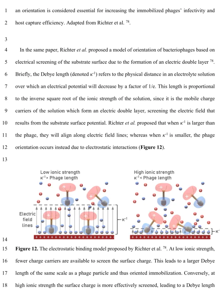
The bacteriophage head presents a net negative charge, while the tail fibers present a net 18
positive charge 77. Thus, the dipole moment of a phage particle causes the head to be oriented
19
towards a positively charged surface 78. In addition to local charge distribution, many phages
20
such as T7, present an overall net negative charge 93,94.
21
In most circumstances there is a strong entropic preference towards horizontal alignment of 22
tailed and filamentous phages. However, as we shall see, a significant minority of the studies 23
reviewed have taken advantage of the local charge or other properties of bacteriophage capsids 24
to claim oriented immobilization as a result of their protocols (Figure 2). Frequently, however, 25
11
insufficient evidence and/or no clear mechanisms are presented to prove oriented 1
immobilization has been achieved. This is partly due to the difficulty in determining the 2
orientation of nanoscale objects, but post-functionalization characterization techniques do exist 3
that can help justify such claims. 4
5
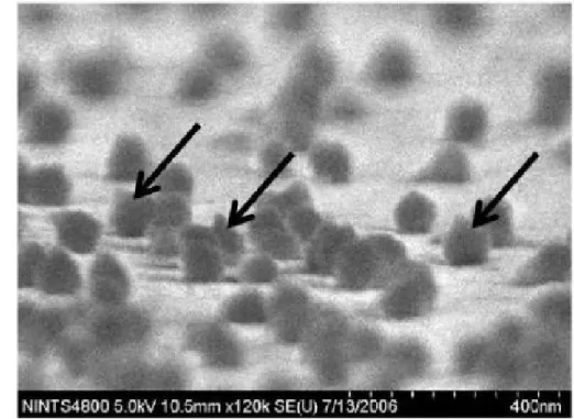
Figure 2. Scanning electron micrograph of in vivo biotinylated T4 phage, immobilized on a 6
streptavidin-coated gold surface. Arrows indicate phage particles with tail-up orientation. 7
Reproduced from Tolba et al. 83.
8
A note on surface density 9
A high phage surface density is not the sole criterion for high phage infectivity and/or 10
bacterial capture. For example, Tawil et al. found that phages were immobilized on gold with 11
double the density when cross-linked via L-cysteine compared to glutaraldehyde, but that no 12
significant difference was found in the bound phages’ lytic behavior 72. Similarly, Leppänen et
13
al. rigorously compared infectivity and phage surface density after surface treatments with 14
different combinations of 11-MUA and (3-aminopropyl)triethoxysilane (APTES), concluding 15
12
that a higher phage density did not always result in a higher infectivity 95. Indeed, Naidoo et al.
1
achieved an impressive surface density of 199±2 phages/µm2 but found that – above a value
2
of a 18.9±0.8 phages/µm2 – higher phage density led to a reduction in total bacterial capture
3
58.
4
These results suggest that careful consideration and design of the surface functionalization 5
can lead to dramatically improved activity and/or bacterial capture of phage-functionalized 6 surfaces. 7 Common substrates 8 Gold 9
Gold surfaces are by far the most popular surface for phage functionalization, serving as the 10
substrate in just over half of all papers reviewed on this topic. Gold exhibits ideal properties 11
for functionalization with many biological entities 90, and for bacteriophages the case is no
12
different. Gold is biocompatible, non-oxidizable, readily available, easily cleaned, and its 13
deposition on a variety of substrates is a mature technology 96–98. Gold also exhibits a very high
14
binding affinity for sulfhydryl (R-SH, aka thiol) groups of around 200 kJ/mol, which also 15
permits easy formation of self-assembled monolayers 98. Thiol groups are frequently found –
16
or easily introduced – in most target ligands for the purpose of immobilization 98.
17
Facilitating bioconjugation is the principal motivation behind the deposition of a gold layer 18
on transducers that do not involve the intrinsic properties of gold itself (e.g. magnetoelastic or 19
quartz crystal microbalance (QCM) biosensors, which instead rely on the resonance of an 20
underlying amorphous ferromagnetic material 99 or quartz crystal 100, respectively). However,
21
gold also exhibits ideal properties where its biocompatibility and ease of conjugation is a happy 22
coincidence. Gold features a high density of easily polarizable free electrons – a prerequisite 23
for strong interaction with electromagnetic fields 101 – making it an ideal material for use in
13
surface plasmon resonance (SPR). For SPR, there are effectively only two metals with 1
appropriate properties: gold and silver 102. While silver actually yields a superior SPR effect, it
2
is less biocompatible and less chemically stable – particularly towards oxidation – compared 3
to gold, and so the latter has become the de facto default for SPR biosensors 101,102.
4
Magnetic beads 5
Magnetic bead surfaces are a frequent substrate for phage conjugation since, once 6
functionalized, they can be used for immunomagnetic separation (also sometimes referred to 7
as phagomagnetic separation) and concentration of low-titer analytes in order to boost the 8
capture efficiency of biosensor surfaces (Figure 4) 47,48,56,68,69,89,103,104. Briefly, phages specific
9
to a given bacterial strain are immobilized on the beads, which are then mixed with a sample 10
and bind to the target analyte (Figure 5). Application of a magnetic field can then be used to 11
concentrate and confine the captured analyte to a given region, for example on the surface of a 12
biosensor. Phage immobilization has been shown for magnetic beads with a variety of surface 13
chemistries including tosyl 68,69, carboxyl 47,49,69,103, streptavidin 48,83,89,105, azide 81, and
14
isothiocyanate-terminated coatings 104.
15
Carbon allotropes 16
Carbon allotropes (e.g. graphene, nanotubes) and glassy/vitreous carbon are common 17
components of inks used to fabricate printed electrodes (SPEs). Electrode screen-18
printing enables cheap, versatile electrodes for use in clinical assays106, food processing107, and
19
environmental monitoring 108. Such electrodes can also be composed of gold, silver, or
20
platinum; but carbon is more typical due to its relatively low cost 109. The versatility and easy
21
manufacture of SPEs has led to carbon allotropes becoming a target for phage immobilization 22
for the fabrication of biosensors specific to various bacterial strains 54,55,65,110,111, and in one
23
paper a biosensor for West Nile virus-specific IgG 29. Carbon nanotubes can also be used as an
14
intermediate structure for the immobilization of phages on different substrates, as was 1
demonstrated by Farooq et al. for bacterial cellulose matrices for ultra-sensitive and selective 2
electrochemical detection of Staphylococcus aureus 112. Carbon allotrope-specific binding
3
peptides have been discovered that may enable recombinant phage immobilization on such 4
substrates 113–116 (see Polymer and other binding domains below).
5
Polymers 6
Phage-functionalization of polymers is of interest since such materials can be formed into 7
antimicrobial wound dressing 41,117, food packaging 20, and biocontrol surfaces resistant to
8
biofilm formation 118. They also form the coating of many commercially available magnetic
9
beads 47,49,68,69,103. Polymers that have been successfully conjugated or co-polymerized with
10
phages include polycaprolactone 41, polymethyl methacrylate (PMMA) 86, polyethersulfone 118,
11
polystyrene 119, polyhydroxyalkanoates 40, polyethylene 120, and
poly(3,4-12
ethylenedioxythiophene) (PEDOT) 33,121,122.
13
It is interesting to note that a large number of peptides have been discovered that have high 14
binding affinities for specific polymers 123–129, and that such proteins have been expressed on
15
the capsids of recombinant phages to facilitate immobilization 83,86,89.
16
Cellulose-based materials 17
A particularly important subcategory of polymers is cellulose and cellulose-derived 18
materials. As the most abundant natural polymer on Earth 130, cellulose features in the
phage-19
functionalization literature because it forms the principal component of paper. With its high 20
porosity, hydrophilicity, chemically inert character, and slight negative charge at neutral pH 21
42,53, paper lends itself to the fabrication of cheap, mass-produced devices for healthcare 131 and
22
environmental monitoring 132.
15
Phage-functionalization of paper and cellulose-derived polymers is motivated by the 1
development of antimicrobial food packaging materials 19,20,39, and low cost biosensors
2
53,133,134. Phage-containing bio-inks can be printed onto paper substrates, and it has been shown
3
that T4 phages are capable of resisting the shear stress and drying processes involved in 4
industrial printing 135. It has also been demonstrated that careful consideration of the bio-ink
5
constituents can enable production of bioactive paper that is stable for several days after 6
printing 136. Anany et al. have demonstrated a dipstick assay based on inkjet-printed phages
7
that efficiently captured and infected E. coli and Salmonella Newport in broth and food 8
matrices 53. Farooq et al. demonstrated phage-functionalization of bacteria-derived cellulose
9
fibers, but via intermediate multi-walled carbon nanotubes 112.
10
There is ample precedent for lateral flow devices that rely on capillary action along capillary 11
beds to channel fluid for the purposes of biochemical analysis, the most well-known of which 12
is probably modern pregnancy tests 137 and covid-19 antigen tests. Such microfluidic
paper-13
based devices may also be combined with electrode screen printing for more elaborate 14
microfluidic electrochemical sensors 137,138. Phage-derived proteins 139 have been incorporated
15
into lateral flow assays, but to our knowledge in all cases have been immobilized onto 16
intermediate supports (e.g. nanoparticles) rather than the polymer substrate of the device itself. 17
The ability to reliably immobilize phages directly on paper substrates would open new avenues 18
for the development of low-cost, mass-produced bioassays 134.
19
Some evidence suggests immobilization is more successful if paper has been precoated with 20
polydiallyldimethylammonium chloride (polyDADMAC), a cationic polyelectrolyte which 21
imparts a positive charge to the cellulose fibers and thus electrostatically binding the phage 22
heads 134. Phage resistance to dry environments can be improved by incorporating gelatin into
23
the deposition ink 136
16
When considering cellulose-based substrates, of note is the existence of cellulose binding 1
modules (CBMs) – polypeptides that bind strongly to cellulose 140 and which can be expressed
2
by genetically-engineered phages to facilitate their immobilization on such substrates 83,89.
3
Silicon-based materials 4
In this review, phage-functionalization of several materials including silicon nitride 37,
5
optical fibers 141–143, atomic force microscopy probes 144, silica nanoparticles 60,62, and glass
6
50,51,67,79,80 are all grouped into the category of silicon-based substrates. Such surfaces are
7
typically rendered reactive for immobilization chemistry through amino-silanization 8
37,46,50,51,60,62,67,79,80,95,141,142,144–148.
9
Optical fiber biosensing is of particular interest since it can exploit the properties of optical 10
biosensing techniques (e.g. SPR), while avoiding drawbacks associated with bulky equipment 11
141. Using a smaller sensing element and sample volume can be advantageous in the analysis
12
of small amounts of precious analytes, and can accommodate multiple parallel measurements 13
on adjacent bundled optical fibers 149.
14
Since the fiber-optic material itself is cheap, this may offer the possibility of disposable, 15
single-use sensors 150,151. Finally, since the nature of optical fiber geometry means that signal
16
transduction can take place at a physically remote location relative to the optical setup, this 17
form of biosensing enables remote detecting and monitoring capabilities in potentially harsh 18
environments 149, allowing one to bring the sensor to the sample rather than bring the sample
19
to the sensor. Phage-functionalized optical fibers have been demonstrated for the specific 20
detection of E. coli 141–143.
21
Covalent methods 22
17 1
18
Figure 3. Schematic representation of a selection of the most popular bacteriophage 1
immobilization strategies, broadly grouped into covalent bonding, physisorption, and genetic 2
modification. At left, the substrates that have been demonstrated in the literature with the 3
adjacent immobilization method. Note: bacteriophages are represented by a generic 4
Podoviridae but in most cases represent other phage morphologies 5
EDC/NHS Chemistry 6
For substrates presenting carboxyl (−COOH) groups – be they endogenous or introduced – 7
a common covalent conjugation method involves the use of the carbodiimide EDC (1-Ethyl-3-8
(3-dimethylaminopropyl)carbodiimide) to activate the carboxyl groups, creating an unstable 9
ester intermediate 152. Typically, a separate molecule containing a succinimidal group in the
10
form of NHS (N-hydroxysuccinimide) is then introduced to the surface and supplants the 11
carbodiimide, which is released into the solution. This stabilizes the activated carboxyl group 12
and forms a second NHS-ester intermediate which is more vulnerable to nucleophilic attack 13
from amino groups, in turn priming the NHS to be supplanted by the primary amine of an 14
amino acid 67,152.
15
When a suspension of amine-containing ligands – such as phages or proteins – is introduced 16
to the sensor surface, a primary amine of the ligand reacts with the NHS (the leaving group) 17
and is then covalently bonded to the surface while the succinimidal group is released into the 18
solution. After ligand binding is complete, the surface may be washed with ethanolamine 19
whose amine groups will react with the remaining activated carboxyl “sites”, which blocks 20
further binding.152. In this way, the EDC/NHS solution facilitates amide bonding between
21
carboxyl groups of the surface and amine groups of the ligand 67.
22
Such EDC/NHS coupling is extremely popular for surface functionalization of gold 23
following introduction of exogenous carboxyl groups (for example by formation of an 24
19
alkanethiol self-assembled monolayer ) 34,57,153–158, but has also been demonstrated for a variety
1
of carboxylated substrates including glassy carbon electrodes 55, glass 50, magnetic beads 103,
2
and polymers 40,159.
3
The creation of the second NHS-ester intermediate is not obligatory and in cases where a 4
high yield of conjugated ligands is not crucial, the use of NHS may be omitted entirely. Such 5
EDC-mediated binding has been used to covalently immobilize phages on glassy carbon 6
electrodes 110,111, magnetic microbeads 103, and carboxyl-polystyrene latex beads 119. However,
7
the addition of even small amount of NHS to an EDC coupling reaction can boost the yield of 8
conjugated ligand by a factor of 20 160. NHS activation decreases the water-solubility of the
9
activated carboxylate molecule and for this reason is often instead sold as sulfo-NHS, wherein 10
the charged sulfonate group preserves or increases water-solubility. 11
One paper made a rigorous comparison of the surface density of immobilized phage BP14 12
on gold that resulted from different combinations of EDC/NHS with L-cysteine, 11-MUA, and 13
glutaraldehyde cross-linking; finding that cysteine – when combined with 11-MUA and 14
ECD/NHS – gives an incredible 103 improvement in phage activity (measured by bacterial
15
lysis) compared to simple physisorption 72.
16
EDC/NHS can also be used in the other direction: activating the surface carboxyl groups of 17
the phages themselves in order to facilitate grafting to substrates featuring primary amines – 18
be they endogenous or introduced (e.g. via aminosilanization) 67,144,146,161,162. However, this
19
approach is believed by some researchers to lead to increased blocking of the phage receptors 20
responsible for bacterial capture 50, which seems to have been confirmed in experiment 95,146.
21
One feature of amide binding is the non-uniform nature of the ligand orientation. Since a 22
ligand typically displays several primary amines, any one of which may react with the activated 23
substrate carboxyl group, this allows a variety of ligand orientations during immobilization. 24
This issue is compounded for very large molecules. On the scale of a bacteriophage, the large 25
20
number of available primary amines means orientation may be effectively random unless 1
effective mitigation strategies are employed, as discussed below. 2
Some substrates will already feature carboxyl groups, without the need to introduce a 3
carboxyl intermediate. Such is the case for polyhydroxyalkanoate (PHA), a bacterially 4
produced biopolymer 163 that is currently under investigation as a biodegradable
food-5
packaging. Wang et al. have demonstrated that plasma treatment of PHA films results in 6
surface carboxyl groups which can be activated and bound to phages using EDC/NHS, for the 7
purposes of selective bioburden reduction in foodstuffs 40.
8
Carboxyl-activated magnetic beads are commercially available, for example Dynabeads 9
MyOne™ Carboxylic Acid magnetic beads from Invitrogen. Although of proprietary 10
formulation, the manufacturer describes these beads as having a coating of glycidyl ether and 11
a core of highly cross-linked polystyrene with ferromagnetic inclusions. These beads have been 12
employed for the detection of E. coli with an LOD as low as 103 colony-forming units/milliliter
13
(CFU/mL) by impedimetric 49,103 and linear sweep voltammetry assays 49, as well as a
14
colorimetric scheme based on release of endemic β-galactosidase from lysed analyte cells 47.
21 1
22
Figure 4. Schematic representation of a typical magnetic bead and its use for phagomagnetic 1
separation. (A) A carboxyl-terminated magnetic bead is activated with EDC and NHS, priming 2
it for conjugation. (B) A ligand bearing primary amines, in this case a phage, is mixed with the 3
beads and is conjugated to the bead surface through amide bonding. (C) Mixing the 4
functionalized beads with bacteria causes the beads to bind to the surface of any bacteria 5
present. (D) and (E) The bacteria can now be retained in a standard tube while the supernatant 6
is removed, or concentrated near a biosensor transducer surface, by the application of a 7
magnetic field. 8
23 1
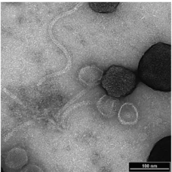
Figure 5. Scanning electron micrograph of P22 phages immobilized on carboxyl-activated 2
magnetic beads following magnetic capture of a Salmonella bacterium. Reproduced from 3
Laube et al. (2014) 69.
4
A similar product is that of Ademtech SA, in the form of Carboxyl-Adembeads which feature 5
a superparamagnetic core of magnetite, covered with a proprietary styrene-based copolymer 6
which presents carboxyl groups to the solution.† Regardless of the magnetic bead product used,
7
24
in all cases in the literature reviewed, the particles’ carboxyl groups are activated by EDC/NHS, 1
exposed to the phage for immobilization, and used for the magneto-separation and subsequent 2
detection of bacteria (e.g. Salmonella 69 or E. coli 47,49,103) by varying detection mechanisms
3
(Scheme 1). 4
Alternatively, carboxyl groups may be grafted to the substrate. This has been shown for 5
graphene which can be electrochemically oxidized to produce carboxyl sites, allowing phage-6
functionalization of glassy 110 and screen-printed carbon electrodes 111. A similar process has
7
been demonstrated for carboxylated multi-walled carbon nanotubes for the phage-mediated 8
detection of S. aureus 112.There are many advantages of EDC/NHS chemistry that have led to
9
it becoming a staple in the bioconjugation literature. Both EDC and the resultant isourea formed 10
after binding are water soluble, meaning one can avoid organic solvents which could otherwise 11
harm the ligand. The carbodiimide reaction occurs with high yield up to pH 7.5, allowing 12
conjugation at physiological pH which is well-tolerated by most biological ligands. Since 13
carbodiimides such as EDC are what is known as zero-length crosslinkers, no additional 14
chemical structure is introduced between the conjugated molecules after cross-linking 90.
15 16 17
Self-assembled monolayers of thiolated molecules 18
No discussion of surface immobilization techniques would be complete without mention of 19
thiol-gold bonding, a workhorse of surface chemistry. Also known as sulfhydryl groups, thiol 20
moieties are widely exploited for the formation of self-assembled monolayers (SAMs) on soft 21
metal substrates 98, and are regularly employed in the functionalization of planar and
22
nanoparticulate metals – gold in particular – with proteins, antibodies, and DNA. The most 23
popular thiol-containing molecules are the alkanethiols which feature a sulfhydryl headgroup, 24
an alkane chain of specified length, and a terminal functional group which can be used to 25
25
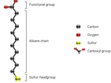
introduce carboxyl sites to the substrate, for example (Figure 6). This stereotypical structure 1
serves as a good model for a brief discussion of SAM formation. 2
In a classical SAM formation model, a soft metal surface is incubated with a solution of 3
alkanethiol molecules. The strong affinity of the sulfur headgroup for the metal surface leads 4
to an initial “lying-down” configuration of the alkanethiol molecules, chemisorbed to the 5
surface strongly by the metal-sulfur bond of the headgroup but also lightly physisorbed via van 6
der Waals interactions between the surface and the alkane chain 164.
7
8
Figure 6. Structure of a typical alkanethiol, in this case 11-mercaptoundecanoic acid (11-9
MUA). Hydrogens have been omitted for simplicity. 10
Since the sulfur headgroups of arriving conjugate molecules have a higher affinity for the 11
metal surface compared to the hydrocarbon chains, over time the latter are displaced from the 12
surface as more alkanethiol molecules diffuse to and bind with the substrate (Figure 7). This 13
gives rise to a phase transition to a “standing up” configuration with a strong entropic 14
preference for upright orientation of the conjugated molecules relative to the surface. Mutual 15
intramolecular van der Waals interactions between the alkane chains stabilize the SAM, 16
resulting in the formation of a closely packed (surface coverage ≈1/3), crystalline monolayer 17
of alkanethiolate molecules 164.
26 1
Figure 7. Progressive formation of an alkanethiol self-assembled monolayer (SAM) on a gold 2
substrate. Diffusion of alkanethiol from the solution to the surface is followed by binding of 3
the molecule to the gold surface through a strong gold-sulfur bond and also by interactions with 4
the alkane chain, leading to a “lying down” configuration. With increasing incubation duration, 5
more of the molecule diffuses to the surface. As surface density increases, arriving sulfur 6
headgroups compete with and displace the alkane chains on the surface, leading to a standing-7
up configuration. Finally, above a threshold density, intra-molecular forces between the alkane 8
chains stabilize the SAM with the functional groups presented to the solution above 9
10
While thiols exhibit high binding affinity for a variety of transition metals (Au, Ag, Cu, Pt, 11
Pd, Ni)98 and metal alloys, gold’s other advantageous properties make it the de facto standard
12
substrate for SAM formation and bioconjugation, for reasons discussed above 98.
13
An advantage of thiol-based immobilization is that the thiol groups readily displace 14
biological contaminants and interferents during adsorption 98. Thiol-based SAMs are known to
15
be stable for periods from days to weeks even in the presence of complex liquid media, as is 16
often the case in biological experiments 98.
17
Cross-linking molecules containing thiol moieties have been used to immobilize phages of 18
various morphologies on gold surfaces 72,95,153, nanoparticles 158, and plasmonic quasicrystals
19 70. 20 Carbon Oxygen Sulfur Carboxyl group
27
Both cysteamine and the amino acid L-cysteine bind to gold through a strong thiol linkage 1
while presenting an amine group which can be cross-linked to primary amines via 2
glutaraldehyde (Erreur ! Source du renvoi introuvable.) 66,72,165. In the case of L-cysteine, a
3
carboxyl group is also present and is available for activation, for example by EDC/NHS 57,66.
4
11-mercapto-undecanoic acid (11-MUA) is an example of an alkanethiol featuring a long 5
alkane chain terminated at one end by a thiol moiety and on the other end by a carboxyl group 6
(Erreur ! Source du renvoi introuvable.). Mutual van der Waals interactions between the 7
alkane chains and the high affinity of the sulfur headgroup for the gold surface leads 11-MUA 8
to readily form SAMs on gold surfaces, presenting abundant carboxyl groups to the solution 9
which may be activated by EDC/NHS 164.
10
11-MUA has been used as a tether molecule to functionalize gold substrates with phages 11
such as M13 to detect peptides by SPR 153; T4 and BP14 to detect E. coli and S. aureus by SPR
12
and impedance measurements 57; T4 to detect E. coli by differential pulse voltammetry (DPV)
13
157; and various phages for the detection of S. aureus by SPR, and Bacillus cereus by
14
ferromagnetoelasticity 155.
15
As mentioned above, by combining 11-MUA with L-cysteine, Tawil et al. were able to 16
achieve a 103-fold increase in phage activity relative to simple physisorption 72.
17
The authors attributed this improvement to the creation of uniform, regularly interspaced 18
depressions in the L-cysteine / 11-MUA monolayer, with a length scale similar to that of the 19
phage head of 36 ± 2 nm, permitting uniform and oriented immobilization of the phages. 20
21
Another example of a thiol-based linker is 3-mercaptopropionic acid (3-MPA) (Erreur ! 22
Source du renvoi introuvable.). 3-MPA has a similar structure to 11-MUA, featuring terminal 23
carboxyl and thiol moieties, but with a much shorter carbon chain linking them. Sedki et al. 24
functionalized glassy carbon electrodes by coating them with 3-MPA-functionalized gold 25
28
nanoparticles which were then activated with EDC/NHS and conjugated to M13 phages, 1
forming an electrochemical impedance-based bacterial biosensor which achieved an 2
impressive LOD of just 14 CFU/mL 158. Han et al. used a SAM of 3-MPA to immobilize fd
3
phage for DPV-based detection of prostate specific antigen at concentrations as low as 3 pg/mL 4
34.
5
While 11-MUA and 3-MPA are both examples of monothiols, there also exist dithiol linkers 6
such as dithiobis(succinimidyl propionate), also known as Lomant’s reagent, DTSP, or 7
sometimes DSP 90. DTSP is a homobifunctional NHS ester crosslinking agent, in the form of
8
a homodimer exhibiting both thiol and succinimide moieties, linked by a disulfide bridge 9
allowing SAM-formation on clean gold substrates (Erreur ! Source du renvoi introuvable.). 10
Upon introduction to a gold surface, DTSP cleaves at the disulfide bridge, resulting in two 11
thiolate moieties bonded to the surface, each bound via a carboxyl group to a terminal 12
succinimidyl group that is exposed to the solution. This succinimidyl group is thus available as 13
a binding site for primary amines of a target ligand, as with the EDC/NHS method 166.
14
Following conjugation, rinsing with ethanolamine blocks further binding of primary amines. 15
29 1
30
Figure 8. Schematic of a generalized protocol for DTSP functionalization and phage-1
conjugation, followed by surface blocking with bovine serum albumin (BSA). (A) A bare gold 2
substrate is cleaned by plasma or otherwise. (B) After incubation on the gold surface, DTSP 3
cleaves at the disulfide bridge and forms a monolayer on the substrate. (C) Sequential 4
introduction of the ligand (phage) followed by ethanolamine results in conjugation of the phage 5
to the surface and blocking of the remaining activated carboxyl sites by ethanolamine. (D) The 6
surface is then incubated with BSA in order to reduce non-specific binding. Not to scale. Note 7
that immobilization will not always result in a tail-upward orientation as shown. 8
9
Arya et al. first proposed DTSP for the immobilization of phages in 2011 166. By 2012,
10
Naidoo et al. were able to leverage DTSP to reach an incredible surface density of 199±2 11
phages/µm2. However, they found a threshold surface coverage of 18.9±0.8 phages/µm2,
12
beyond which higher phage density led to a reduction in total bacterial capture 58. This result
13
was influential and is widely cited in the phage immobilization literature, with many 14
subsequent papers quoting their results with reference to this “jamming effect”. Notable 15
subsequent research by Richter et al. combined DTSP with application of an alternating electric 16
field during immobilization to “bake in” a tail-upward orientation of T4 phage as a surface 17
density of 13.64 phages/µm2 167. DTSP has also been used to immobilize T4 166 and P22 58
18
bacteriophages on gold surfaces for detection of E. coli 58,166,167 and Salmonella Typhimurium
19
58.
20
4-amino-thiophenol (4-ATP) is an aromatic thiol featuring a primary amine moiety (Erreur ! 21
Source du renvoi introuvable.). 4-ATP has been used to immobilize T4 phages on nano-22
sculptured thin films of silver for detection of E. coli by surface-enhanced Raman scattering 23
(SERS) with an LOD of 1.5×10² CFU/mL 168. In this method, glutaraldehyde is used to
cross-24
link primary amines present both on 4-ATP and on the phage capsid. 25
31
In a different approach, Rippa et al. deposited and diazotized an SAM of 4-ATP on plasmonic 1
quasicrystals of gold 70. The diazonium moieties were then available for covalent linkage to
2
the histidyl groups of bacteriophage Tbilisi. This SERS-based sensor was capable of detecting 3
femtomolar concentration of immobilized phages, but this work did not extend the method for 4
the detection of an analyte. The choice of plasmonic quasicrystals is motivated, among other 5
considerations, by the higher spatial density and larger field strengths of electromagnetic hot-6
spots, enhancing SPR and SERS effect and thus the sensitivity of the sensor. 7
Horikawa et al. made a comparison between carboxy-terminated, aldehyde-terminated, and 8
methyl-terminated SAMs for immobilization of filamentous fd phages on the gold surface of 9
magnetoelastic sensors 169. The resultant phage surface coverage was found to be 46.8%,
10
49.4%, 4.2%, and 5.2% for bare gold, carboxy-, aldehyde-, and methyl-functionalized 11
resonators, respectively. This paper is notable in its findings that physisorption can be as 12
performant as activated carboxyl-mediated immobilization. The authors propose that the 13
aldehyde and methyl surface treatments were so effective in reducing phage adsorption that 14
they may be employed as “anti-phage surfaces”, which may provide the possibility of negative 15
surface patterning using these surface treatments as a form of negative resist. 16
Glutaraldehyde 17
Glutaraldehyde – a symmetrical, bireactive compound with an aldehyde (−CHO) at each end 18
– is frequently used as a cross-linker (Erreur ! Source du renvoi introuvable.). The aldehydes 19
can react with an amine to form an imine – a group with a carbon-nitrogen double bond. In this 20
way, glutaraldehyde cross-links amine groups. 21
Singh et al. immobilized L-cysteine and cysteamine on a gold surface via their thiol side-22
chains, and then cross-linked the amine groups of these immobilized species to those exposed 23
on the capsids of wild type T4 phages. The surfaces were then used for the specific capture of 24
E. coli EC12 bacteria, as confirmed by scanning electron microscopy (SEM) and X-ray 25
32
photoelectron spectroscopy (XPS) 165. Compared to simple physisorption, they found a 37-fold
1
improvement of phage immobilization at 18±0.15 phages/µm2, resulting in a 9-fold higher
2
bacterial capture density of 11.9±0.2 bacteria/100µm2.
3
Similarly, He et al. coated interdigitated gold electrodes with L-cysteine before crosslinking 4
via glutaraldehyde to phage D29, to form an impedimetric sensor of Mycobacterium smegmatis 5
and Mycobacterium tuberculosis 66.
6
Richter et al. further developed this technique to achieve some of the most convincing 7
evidence for oriented immobilization in the literature. By combining an alternating electric 8
field with glutaraldehyde cross-linking to a gold substrate, the authors leveraged the permanent 9
dipole moment of T4 phages to yield a tail-outward orientation that is “baked in” at the time of 10
immobilization 167. This allowed the authors to achieve immobilized phage densities on gold
11
of 13.64 phage/µm2 and 17.32 phage/µm2 for oriented and non-oriented layers, respectively.
12
The same paper demonstrated a very low LOD of 102 CFU/mL E. coli without any
pre-13
enrichment step. 14
Immobilization chemistries featuring glutaraldehyde have been demonstrated for phage-15
functionalization of silica-based materials such as optical fibers 141, silicon nitride 37, and silica
16
nanoparticles 62 for the purposes of biodetection and biocontrol, respectively. A widely-cited
17
paper demonstrated glutaraldehyde cross-linking of T4 phages onto long-period gratings 18
etched into optical fiber for the specific detection of E. coli with an LOD of 103 CFU/mL 141.
19
Glutaraldehyde has also been used for the creation of bacteriophage-functionalized metal-20
organic frameworks (MOFs). MOFs – coordination networks with organic ligands 170 – are
21
characterized by an easily tunable size (nano- to micron scale depending on synthesis 22
parameters) 171 and potentially large surface area 63. An area of active research is the use of
23
MOFs as fluorescent probes for molecular sensing. Bharwaj et al. have used glutaraldehyde to 24
create bacteriophage-functionalized MOFs for the detection of Staphylococcus arlettae 63 by
33
means of fluorescence and S. aureus 64 by photoluminescence-based biodetection. Very low
1
LODs were demonstrated of 102 CFU/mL for S. arlettae and 31 CFU/mL for S. aureus.
2
In a different approach, Yoo et al. demonstrated a variation of glutaraldehyde cross-linking 3
whereby a solution of M13 phages was simply drop cast and allowed to dry on a cysteamine 4
monolayer on gold, then exposed to a glutaraldehyde vapor for three days in order to cross-link 5
the phages 147. This allowed the study of mouse fibroblast proliferation under the influence of
6
growth factors immobilized to the phages themselves, taking advantage of the phages’ self-7
assembled nanofibrous matrix structure. 8
In all cases, cross-linking with glutaraldehyde relies on the presence of amine groups on the 9
substrate, which is typically achieved through amino-silanization of the surface, deposition of 10
an intermediate molecule presenting both thiol and amine moieties (e.g. L-cysteine and 11
cysteamine), or through electropolymerisation, for example of polytyramine 172.
12
Silane-based self-assembled monolayers 13
For immobilization on silicate-based materials, a preferred method is silanization of the 14
substrate using alkoxysilanes which act as hetero-bifunctional crosslinkers between inorganic 15
mineral surfaces and organic ligands 37,50,51,60,62,67,79,80,95,141,142,144–146,173,174.
16
Typically, the inorganic surface is treated with a strong oxidizer (e.g. piranha solution or 17
aqua regia) or oxygen plasma to form surface silanol groups which feature dangling hydroxyl 18
(-OH) groups. These hydroxyl groups will then condensate with the three alkoxy groups on the 19
alkoxysilane*, leaving a primary amine bonded to the surface. One can then perform standard
20
amine-immobilization chemistry with these amines, as above (Figure 3). 21
* In reality the hydroxy groups will replace between zero and three alkoxy groups in each
alkoxysilane. One can even have the terminal amino (−NH2) group bond with the surface
34 1
Figure 9. Transmission electron micrograph of T4 myovirus covalently immobilized on 2
APTES-functionalized 1 µm diameter silica particles 146. Reproduced with the permission.
3
The most popular alkoxysilanes for phage immobilization are (3-4
aminopropyl)triethoxysilane (APTES or APTS) and (3-aminopropyl)trimethoxysilane 5
(APTMS) 90. Alkoxysilanes can be used either for electrostatic or covalent immobilization of
6
proteinaceous ligands (Figure 3). In covalent bonding, the amine groups of the alkoxysilane 7
react with aldehyde either present on a cross-linker molecule such as glutaraldehyde 37,141,142,
8
or on the phage itself after enzymatic modification to yield a reactive aldehyde as demonstrated 9
by Kwak et al. 79. Alternatively, the phage's endogenous carboxyl groups can be activated by
10
EDC/NHS to facilitate amide bonding with the substrate aminosilane layer 67,144,146 (Figure 3),
11
although – as explained above – activation of the phage by EDC/NHS can be detrimental to 12
phage infectivity 50,95,146.
13
highly cross-linked via these remaining alkoxy groups, with only occasional bonds to the surface 258.
35
In contrast, electrostatic binding with alkoxysilanes relies on the differing isoelectric points 1
of the ligand and the silanized surface, as discussed later in this work in the ‘Electrostatic 2
binding’ subsection. 3
4
Hosseinidoust et al. demonstrated covalent immobilization of a variety of phage families 5
through silanization of glass with APTES, achieving a consistent surface density of 4.5 +/- 0.7 6
phages/μm2, as calculated from SEM imagery of the surface 50.
7
A modification of this strategy is to instead use EDC/NHS to activate the carboxyl groups of 8
the phages themselves, which are then bound to the primary amines of an amino-silanized 9
surface. Handa et al. used this technique to functionalize atomic force microscopy probes 144,
10
and in a separate paper achieved 67% surface coverage of phage P22 on glass substrates 67.
11
However, EDC/NHS activation of the phage carboxyl groups is believed by some researchers 12
to lead to increased blocking of the phage receptors 50,146.
13
An interesting application of organosilane grafting has been demonstrated for the phage-14
functionalization of indium tin oxide (ITO) 173,174. ITO is the most well-known of all
15
transparent conductors – exhibiting exceptional optical transmissivity combined with low 16
electrical resistance 175 – and is seen as a promising material in biosensing technology 176. In a
17
pair of papers, Liana et al. investigate phage adsorption onto bare, amine, methyl, and carboxyl-18
functionalized planar and particulate ITO 173,174. These comparative studies found divergent
19
results between planar and particulate ITO, but a consistent drop in performance was observed 20
with the introduction of amine groups on the substrate compared to carboxylic and hydroxyl 21
groups, suggesting the latter as more promising routes for phage immobilization on ITO. 22
Miscellaneous covalent techniques 23
Isothiocyanate 24
36
Isothiocyanate compounds feature a terminal sulfur atom and a central electrophilic carbon 1
which is susceptible to nucleophilic attack by the primary amines of amino acids, yielding a 2
thiourea linkage with the latter, with no leaving group in the reaction 90. Isothiocyanates react
3
best at alkaline pH, and it has been demonstrated that by carefully controlling pH during 4
conjugation, it is possible to modify only the N-terminal α-amines while leaving side-chain 5
amines unmodified 177. Zhang et al. functionalized isothiocyanate-terminated magnetic beads
6
for the purposes of phagomagnetic separation in an enzymatic assay for the presence of E. coli 7
O157:H7, with a comparatively poor LOD of 4.9×10⁴ CFU/mL 104.
8
Electro-deposited polytyramine 9
Niyomdecha et al. demonstrated immobilization of M13 phages using glutaraldehyde cross-10
linking between primary amines on the phage capsid and the activated primary amine groups 11
of an electrodeposited polytyramine layer on a gold surface, for use as a capacitive biosensor 12
for Salmonella 172. Rinsing with ethanolamine blocks any unoccupied aldehyde groups and
13
reduces non-specific binding following functionalization. The use of electrodeposited 14
polytyramine for immobilization of phages has some precedent, having been demonstrated for 15
enzymes 178, myoglobin 179, and oligonucleotides 180.
16
However, a potential complication in the use of electropolymerisation for phage 17
functionalization may lie in the necessity of an electric field during immobilization. Richter et 18
al. have already demonstrated that application of an electric field during immobilization can 19
influence the final orientation of the phages 78,167. It is the opinion of the author that this effect
20
may prove detrimental to phage infectivity since an anodic substrate may attract the positively 21
charged phage tail fibers and result in head-outward immobilization. 22
Tosyl 23
37
A toluenesulfonyl (tosyl) is a molecule or group with the formula CH3C6H4SO2 (Erreur !
1
Source du renvoi introuvable.). Tosyl chloride is used to activate hydroxyl groups on a 2
substrate in nonaqueous conditions, creating a tosyl ester. When subsequently placed in 3
aqueous conditions, this ester can then react with sulfhydryls, amines, and hydroxyls to form 4
thioether, secondary amine, and ether linkages, respectively. This allows the immobilization of 5
proteins either through the primary amino group or sulfhydryl side-groups 90,181. The tosyl
6
group is an effective “leaving group”, since it is easily released during the conjugation reaction. 7
Dynabeads M-280 from Invitrogen are tosyl-activated, 2.8 µm diameter polystyrene beads 8
which feature superparamagnetic inclusions and an outer polyurethane coating. They have been 9
demonstrated to be capable of immobilizing P22 Podoviridae for the purposes of 10
phagomagnetic separation and detection of Salmonella in two papers 68,69. Such methods were
11
capable of reaching an impressive LOD of only 3 CFU/mL 68 and as low as 0.06 CFU/mL if
12
combined with a pre-enrichment step 69.
13
Non-covalent & physical methods 14
Physisorption 15
Many papers make use of simple “physisorption” which is generally not considered to 16
involve covalent bonding between the phage and the substrate. The simplicity of these methods 17
is appealing since the functionalization operation can be as trivial as cleaning the substrate and 18
then incubating it with a purified phage suspension 35,143,182–204. However, physisorption yields
19
a functionalization that is more variable and less robust since the physisorbed phage can detach 20
following changes in ionic strength, temperature, pH, or even high fluid velocities at the 21
substrate surface 92.
22
There appears to be no clear consensus on the physical mechanism behind physisorption, 23
with many papers proposing several factors which could play a role such as van der Waals 24