HAL Id: tel-02918254
https://tel.archives-ouvertes.fr/tel-02918254
Submitted on 20 Aug 2020HAL is a multi-disciplinary open access
archive for the deposit and dissemination of sci-entific research documents, whether they are pub-lished or not. The documents may come from
L’archive ouverte pluridisciplinaire HAL, est destinée au dépôt et à la diffusion de documents scientifiques de niveau recherche, publiés ou non, émanant des établissements d’enseignement et de
Impact of OGT on late steps of the hepatitis C virus
replication cycle
Katharina Herzog
To cite this version:
Katharina Herzog. Impact of OGT on late steps of the hepatitis C virus replication cycle. Virology. Université de Strasbourg, 2019. English. �NNT : 2019STRAJ085�. �tel-02918254�
UNIVERSITÉ DE STRASBOURG
ÉCOLE DOCTORALE 414
UMR_S 1110 Institut de Recherche sur les maladies virales et hépatiques
Thèse
présentée par
Katharina HERZOG
soutenue le 29 Octobre 2019 Strasbourg, France
Pour obtenir le grade de :
Docteur de l’Université de Strasbourg
Discipline/Spécialité : Sciences de la Vie et de la Santé Aspects moléculaires et cellulaires de la Biologie
Impact of OGT on late steps of the
hepatitis C virus replication cycle
THÈSE dirigée par :Dr. Mirjam B. ZEISEL Prof. Thomas F. BAUMERT
RAPPORTEURS :
Prof. Philippe ROINGEARD Dr. Annette MARTIN
AUTRE MEMBRE DU JURY : Prof. Philippe GEORGEL
UNIVERSITÉ DE STRASBOURG
ÉCOLE DOCTORALE 414
UMR_S 1110 Institut de Recherche sur les maladies virales et hépatiques
Thèse
présentée par
Katharina HERZOG
soutenue le 29 Octobre 2019 Strasbourg, France
Pour obtenir le grade de :
Docteur de l’Université de Strasbourg
Discipline/Spécialité : Sciences de la Vie et de la Santé Aspects moléculaires et cellulaires de la Biologie
Impact of OGT on late steps of the
hepatitis C virus replication cycle
THÈSE dirigée par : Dr. Mirjam B. ZEISEL Prof. Thomas F. BAUMERT
RAPPORTEURS :
Prof. Philippe ROINGEARD Dr. Annette MARTIN
This thesis was supported by the international PhD program of the University of Strasbourg with an initiative of excellence (IdEX) scholarship.
Success
He has achieved successWho has lived well, laughed often, and loved much;
Who has enjoyed the trust of pure women, the respect of intelligent men and the love of little children;
Who has filled his niche and accomplished his task;
Who has left the world better then he found it whether by an improved poppy, a perfect poem or a rescued soul;
Who has never lacked appreciation of Earth’s beauty or failed to express it; Who has always looked for the best in others and given them the best he had;
Whose life was an inspiration; Whose memory a benediction.
ACKNOWLEDGEMENTS
A big THANK YOU to my thesis committee, Prof. Philippe Roingeard, Dr. Annette Martin and Prof. Philippe Georgel for taking your limited time to read my thesis and participate in my thesis defense! I would like to thank, Prof. Thomas Baumert, for giving me the opportunity being a PhD student in your lab and for being my teacher in many situations.
I would like to express my heartfelt thanks to Dr. Mirjam Zeisel. Words cannot express how thankful I am being your PhD student: I have learnt so many things from you from basic technical issues, preparing good presentation to at least scientific writing. You always gave me the feeling that I can ask you all kind of questions and taught me how to handle even difficult situations!
I give my special thanks to Dr. Simonetta Bandiera. Thank you for your help when I arrived in Strasbourg, for your practical tips and your way of explaining even a difficult context!
I express my gratitude to all members of the unit, past and present. Thank you for helping me learning all the experimental skills, for your help in administrative and technical issues, for supporting me during these three years and for always helping me when I had questions or when I needed help! Mum, du wundervoller Mensch, was haben wir schon alles durchgestanden, ich bin so dankbar dich an meiner Seite zu wissen. Du bist nicht nur meine Mum, sondern viel mehr meine engste Vertraute, meine Freundin und meine erste Anlaufstelle, wenn ich Rat brauche, diejenige, die mich in den Arm nimmt, Trost spendet und mir zur richtigen Zeit einen Schubs in die richtige Richtung gibt. Das war immer so und wird auch immer so bleiben! Danke für deine Unterstützung und deine Liebe.
Dad, du allwissendes Lexikon, so gerne hätte ich manche Fähigkeiten von dir. Du hast mich unterstützt, immer und bedingungslos. In meinen tiefsten Tiefs hast du mit deiner ehrlichen und direkten Art und deiner Fähigkeit, manche Dinge auf ihre Fakten herunterzubrechen, geholfen Dinge aus einer anderen Perspektive zu betrachten, aber genauso hast du dich in meinen höchsten Hochs mit mir gefreut. Weil ich es vielleicht noch nie so gesagt habe: du bist ein unverzichtbarer Bestandteil in meinem Leben!
Philipp, mein kleiner wunderbarer Bruder und Partner in crime. Deine Geburt hat nicht nur meine Kindheit, sondern mein ganzes Leben bereichert. Ohne dich, wäre ich nur halb ich! Worte können an dieser Stelle nicht beschreiben, wie wichtig und unverzichtbar du für mich bist!
Last but not least, Manu, my love. Danke für dein offenes Ohr, deine tröstenden Worte, deine Freude, deine Fähigkeit selbst in größter Dunkelheit Licht zu sehen, dafür das wir über unsere kleinen Insider so herzlich lachen können und für deine bedingungslose Liebe!
Table of Contents
I. HEPATITIS C – An Introduction ... 2
1.1 Pathogenesis of HCV infection ... 3
1.2 HCV – Basic virology ... 4
1.2.1 Genomic organization of HCV RNA ... 4
1.2.2 Genetic diversity of HCV – One challenge for vaccine design ... 5
1.2.3 HCV virion structure: The lipoviral particle ... 5
1.2.4 The HCV life cycle ... 7
1.3 Model systems to study HCV ... 17
1.3.1 In vitro model systems ... 17
1.3.2 In vivo model systems ... 21
1.4 HCV treatment ... 23
II. PROTEIN GLYCOSYLATION ... 28
2.1 Post-translation modifications of HCV proteins ... 28
2.2 O-linked N-acetylglucosaminylation (O-GlcNAcylation) ... 31
2.3 OGT and O-GlcNAcylation in disease and cancer ... 34
III. OBJECTIVES ... 38
IV. RESULTS ... 40
V. DISCUSSION ... 46
VI. RÉSUMÉ DE LA THÈSE DE DOCTORAT ... 50
VII. REFERENCES... 56
VIII. ANNEX ... 78
List of Figures
Figure 1. Estimated global HCV prevalence in 2015. ... 2
Figure 2. Acute (resolving) HCV infection versus chronic HCV infection. ... 3
Figure 3. Overview of (A) HCV genome organization and IRES-dependent translation into polyprotein and (B) viral proteins and their function. ... 4
Figure 4. Infectious LVP versus VLDL. ... 7
Figure 5. HCV attachment and entry into hepatocytes. ... 9
Figure 6. HCV replication and formation of the membranous web ... 12
Figure 7. Potential function of the cytosolic nuclear pore complexes in HCV replication and assembly ... 13
Figure 8. Model of the formation of LVPs and comparison to cell culture-derived HCV (HCVcc). 16 Figure 9 In vitro HCV cell culture models. ... 19
Figure 10. Host-targeting agents (HTAs) at different steps of the HCV replication cycle ... 26
Figure 11. Basic principle of N-and O-linked glycosylation, and phosphorylation. ... 29
Figure 12. Schematic representation of N-glycosylation sites of E1 and E2. ... 30
Figure 13. O-GlcNAcylation versus phosphorylation. ... 32
Figure 14. The three isoforms of OGT. ... 32
Figure 15. Functional distribution of identified O-GlcNAcylated proteins ... 34
Figure 16. miRNAs involved in the HCV life cycle and expression in Huh7.5.1 cells. ... 38
List of Tables
Table 1. Combinational DAA treatment. ... 24ABBREVIATIONS aa Amino acids ApoB Apolipoprotein B ApoC Apolipoprotein C ApoE Apolipoprotein E CD81 Cluster of differentiation 81 CLDN1 Claudin-1
DAA Direct-acting antiviral
DMSO Dimethyl sulfoxide
DNA Deoxyribonucleic acid
EGF Epidermal growth factor
EGFR Epidermal growth factor receptor
ER Endoplasmic reticulum gt Genotype HCC Hepatocellular carcinoma HCV Hepatitis C virus HCVcc Cell culture-derived HCV HCVpp HCV pseudoparticles
HDL High density lipoprotein
HIV Human Immunodeficiency Virus
Huh7 Human hepatoma 7
INF-α Interferon-alpha
INF-γ Interferon-gamma
IRES Internal ribosome entry site
JFH1 Japanese Fulminant Hepatitis I
kb Kilobase
kDa Kilodalton
LD Lipid droplet
LDL Low-density lipoprotein
miR micro-RNA
MLV Murine leukemia virus
mRNA messenger RNA
MTP Microsomal triglyceride transfer protein
NAFLD Non-alcoholic fatty liver disease
NASH Non-alcoholic steatohepatitis
ncRNA non-coding RNA
OGA β-N-acetylglucosaminidase
OGT O-linked N-acetylglucosamine transferase
OCLN Occludin
PHH Primary human hepatocytes
RIG-I Retinoic acid-inducible gene I
RNA Ribonucleic acid
RNAi RNA interference
SCID severe combined immunodeficiency
siRNA small interfering RNA
SR-BI Scavenger receptor class B type I
TCID50 50% tissue culture infectious dose
TJ Tight junction
TLR Triglyceride-rich lipoprotein
uPA urokinase plasminogen activator
UTR Untranslated region
I. HEPATITIS C – An Introduction
Hepatitis C is an infectious liver disease caused by the hepatitis C virus (HCV) that in most subjects chronically infects the human liver. HCV infection is one of the major etiologies, besides hepatitis B virus (HBV) infection, alcoholic liver disease (ALD) and non-alcoholic fatty liver disease, resulting in chronic hepatitis and progressive liver disease and thereby leading to development of lethal complications, i.e. cirrhosis and hepatocellular carcinoma (HCC), the second leading cause of cancer mortality worldwide (El-Serag and Davila 2011; Ryerson, Eheman et al. 2016).
HCV itself is mainly transmitted through direct blood-to-blood contact occurring through medical treatment with unsterilized needles, blood transfusions and more recently through needle-sharing drug abuse, also through sexual contact and mother-to-child transmission. Upon its discovery in 1989 (Choo, Kuo et al. 1989), enormous progress has been made in the field to develop diagnostic and therapeutic strategies for HCV treatment. These efforts resulted in the development of highly efficient direct-acting antivirals (DAAs) targeting specific HCV proteins and enabling cure in more than 95% of infected individuals by achieving sustained virologic response (SVR) since 2013. Indeed, the combination of different DAAs resulted in cure rates over 96% regardless of the HCV genotype of patients and with a relative short duration of treatment (Bourliere, Pietri et al. 2018). Nevertheless, major clinical and scientific challenges still remain: DAA therapy is cost-intensive and still only available to a fraction of HCV-infected patients (Chung and Baumert 2014), some infected individual developed resistance to DAAs, and an urgently needed HCV vaccine to prevent the global spread of infection is still lacking due to the high diversity of the virus, limited models for testing vaccines and a partial knowledge of protective immune response (Bailey, Barnes et al. 2019). Of note, HCC risk remains high for decades even after SVR (Morgan, Baack et al. 2013; Kanwal, Kramer et al. 2019). An estimated 71 million individuals are persistently infected with HCV with big geographical differences worldwide (WHO Global hepatitis report, 2017). The prevalence of chronic HCV infection is high, including developed countries as shown in Figure 1.
1.1
Pathogenesis of HCV infection
Hepatitis C is caused by HCV infection of hepatocytes resulting in acute (20-30% of infected patients) or more often in chronic HCV infection (70-80% of infected patients) (reviewed in Zeisel, Cosset et al. 2008). The difference between both outcomes of infection is in principle pretty simple: Acute infections consequently lead to clearance of virus through an effective immune response mediated by HCV-specific neutralizing antibodies and T-cell responses, contrarily in chronic infections the immune system is not able to clear the virus resulting in high levels of viral replication and in dysfunctionality of the immune system (see Figure 2) (reviewed in Zeisel, Cosset et al. 2008; Park and Rehermann 2014).
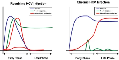
Figure 2. Acute (resolving) HCV infection versus chronic HCV infection.
While in resolving HCV infection the HCV RNA levels decrease due to effective immune responses through rapid induction of neutralizing antibodies and HCV-specific T-cell responses (left), while the contrary result can be observed during chronic HCV infection (right). (Zeisel, Cosset et al. 2008).
Obviously, chronic HCV predisposes to progressive fibrosis, cirrhosis and HCC either directly through changes of the cellular metabolism or indirectly as a result of persistent chronic inflammation. Approximately 1-4% of patients with HCV-related cirrhosis develop HCC (NIH 2002) which is, per basic definition, a randomly occurring genomic and epigenomic change leading to an alteration of cellular gene expression and an abnormal proteome. Epigenetic and gene expression alterations observed in hepatitis C patients can be associated with high risk to HCC, even after the virus could be eradicated by treatment with DAAs (Hamdane, Juhling et al. 2019; Perez, Kaspi et al. 2019). Additionally to epigenetic changes , four pathways and biological processes can be involved in the progression of hepatocarcinogenesis: (i) oxidative stress pathways, (ii) p53 cell cycle pathways, (iii) PI3K/Akt/mTOR and MAPK pathways as well as (iv) WNT/β-catenin pathways (Zucman-Rossi, Villanueva et al. 2015).
1.2
HCV – Basic virology
1.2.1 Genomic organization of HCV RNA
HCV, a member of the Flaviviridae family, is an enveloped, single-stranded positive-sense RNA virus that is mainly restricted to hepatocytes. Its genome encompasses approximately 9.6 kb encoding a single polyprotein precursor of around 3000 amino acids (aa) and is flanked by 5’-and 3’-untranslated regions (UTRs). The internal ribosome entry site (IRES), located at the 5’UTR of the genome, mediates cap-independent translation of the polyprotein contributing to an efficient translation (Tsukiyama-Kohara, Iizuka et al. 1992; Wang, Sarnow et al. 1993). Moreover, miR-122, a crucial liver-enriched host factor, binds to two target sites close to the 5’UTR promoting HCV translation (Henke, Goergen et al. 2008) and replication (Jopling, Yi et al. 2005) by stabilizing HCV RNA (Shimakami, Yamane et al. 2012) (see Figure 3).
The synthesized polyprotein is co-and post-translationally processed by host and viral proteases into at least ten viral proteins: The three structural proteins Core, and the glycoproteins E1 and E2 building up the virion as well as the seven nonstructural (NS) protein mainly involved in replication and assembly through distinct enzymatic activities, namely p7, NS2, NS3, NS4A, NS4B, NS5A and NS5B (see Figure 3).
Figure 3. Overview of (A) HCV genome organization and IRES-dependent translation into polyprotein and (B) viral proteins and their function.
1.2.2 Genetic diversity of HCV – One challenge for vaccine design
HCV can be classified into seven genotypes and several subtypes demonstrating its high degree of genetic heterogeneity and thereby the challenges for research regarding efficient vaccine design: (i) HCV is able to rapidly mutate and (ii) to closely interact with the host lipid metabolism, both leading to escape from protective immune responses.
The genotypes (gt) are numbered from 1 to 7, whereas a variable number of sub-genotypes is designated with a lower case letter forming the basis of classifying HCV into genotypes such as 1a, 1b etc. (Simmonds, Bukh et al. 2005; Smith, Bukh et al. 2014). There is a designated distribution of all HCV strains worldwide: While genotype 1, the (from global perspective) most common genotype (almost half of all infections), dominates in Europe and the Americas, genotype 3, the second most common genotype (around 20-30% of infections), is mostly found in Asia and Northern Europe. Genotype 2 and 4 are less widespread but often found in North Africa and Middle East (around 10% of infections) (Gower, Estes et al. 2014).
1.2.3 HCV virion structure: The lipoviral particle
HCV is an enveloped virus that contains core proteins forming a nucleocapsid around the viral RNA genome which is surrounded by an endoplasmic reticulum (ER)-derived envelope with incorporated viral glycoproteins E1 and E2 which are involved in binding and entry into host cells (reviewed in Lindenbach 2013).
One outstanding characteristic of HCV particles is their tight link with the host cell lipid metabolism; even more their close association with host lipoproteins (Felmlee, Hafirassou et al. 2013; Lavie and Dubuisson 2017). To understand why one speaks of an important hallmark of the virus, it is absolutely essential to understand the process underlying the lipid homeostasis taking place in the liver: Lipids (e.g. triacylglycerides) are synthesized within the liver and their transport through aqueous medium (blood) is enabled through binding with proteins. A process in which triacylglycerides are pooled together with cholesterol and a variable number of proteins into hydrophilic lipoprotein complexes which can be subdivided upon their density into five groups: Chylomicrons (mostly generated in the intestine), very low density lipoproteins (VLDL), intermediate density lipoproteins (IDL), low density lipoproteins (LDL) and high density lipoproteins (HDL). The association of lipids with these so-called apolipoproteins (Apo) is not only important for the transport of lipids through the organism to their organs of need but also for the uptake process into cells through specific membrane receptors.
In the process of VLDL assembly, ApoB and microsomal triglyceride transfer protein (MTP) are required: After synthesis and translocation into the rough ER lumen (rER), ApoB is charged with phospholipids and cholesterol via MTP leading to formation of a neutral lipid core that is transformed into a sphere-shaped particle. This so called VLDL2 contains exchangeable ApoE and ApoC and can be released after movement to a distal compartment in the secretory pathway. Alternatively, luminal lipid droplets (luLDs) are formed as second precursors at the smooth ER (sER) following triglyceride enrichment via MTP and close association with ApoE but not with ApoB. Fusion of VLDL2 with this second precursors is leading to a triglyceride-rich lipoprotein (TLR), namely VLDL1 (reviewed in Olofsson, Bostrom et al. 2009; Shelness and Sellers 2001. Due to the differences in density, lipoproteins can be separated via ultracentrifugation.
To come back to HCV, the virus itself hijacks the host cell lipid metabolism, more particularly, parts of the VLDL and LDL secretion pathway for production of infectious virions. In fact, infectious viral particles could be found in patient-derived serum associated with VLDLs or LDLs forming a so-called lipoviral particle (LVP) (Thomssen, Bonk et al. 1992; Andre, Komurian-Pradel et al. 2002; Nielsen, Bassendine et al. 2006). As a consequence, LVPs show distinct biophysical properties than VLDLs or LDLs: (i) obviously, LVPs are rich in cholesterol and triacylglycerides displaying (very) low density, and (ii) are containing apolipoproteins (e.g. ApoB, ApoA, ApoE and ApoC) and more interestingly, (iii) patient-derived HCV particles differ in density between 1.25g/ml to below 1.06g/ml with infectivity inversely correlated to density meaning that low-density viruses are more infectious. In fact, infectious LVPs have a density between 1.03 to 1.10g/ml and thus can be separated as well as lipoproteins via density gradient ultracentrifugation (Gastaminza, Cheng et al. 2008; Piver, Boyer et al. 2017; Catanese, Uryu et al. 2013; Meunier, Russell et al. 2008) (see Figure 4). Findings that could be confirmed using in vivo (Nielsen, Bassendine et al. 2006; Thomssen, Bonk et al. 1992; Andre, Komurian-Pradel et al. 2002) as well as in vitro (Gastaminza, Kapadia et al. 2006; Lindenbach, Meuleman et al. 2006; Merz, Long et al. 2011) model systems. Interestingly, recent evidence suggests that HCV LVPs contain several exchangeable ApoE and one non-exchangeable ApoB molecule (Piver, Boyer et al. 2017).
Figure 4. Infectious LVP versus VLDL.
Infectious HCV particles (LVP, right) are composed of ApoE and ApoB, which are also part of very-low-density lipoproteins (VLDL, left), and viral components. Consequently LVPs share common features with VLDLs (Felmlee, Hafirassou et al. 2013).
Consequently, through the association with VLDLs or LDLs and different kinds and amounts of apolipoproteins, HCV particles are heterogeneous and thus vary in shape and size ranging from 40 to 80 nm in diameter.
In fact, the association with ApoB and ApoE does not only lead to the masking of viral epitopes preventing the virus to be neutralized by HCV-specific antibodies (Catanese, Uryu et al. 2013; Merz, Long et al. 2011; Fauvelle, Felmlee et al. 2016) but even more this association helps to facilitate HCV entry into hepatocytes (reviewed in Zeisel, Felmlee et al. 2013).
1.2.4 The HCV life cycle
a. Virion attachment and entry
The initial step of the HCV life cycle is its attachment and entry into hepatocytes, a complex, multistep process involving both viral envelope glycoproteins E1/E2 and lipoprotein components which makes it difficult to entirely clear the exact sum of events and details that occur during this first step of the life cycle. The process itself can be subdivided into three key steps: (i) viral attachment to the hepatocyte, (ii) receptor-mediated endocytosis, and (iii) endosomal fusion.
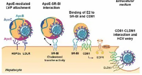
Since LVPs are transported via bloodstream, initial attachment occurs at the basolateral membrane of the hepatocytes through binding of virion-associated ApoE to cell surface heparan sulfate proteoglycan (HSPGs) syndecan-1 or syndecan-4 and low-density lipoproteins receptor (LDLR) (Jiang, Cun et al. 2012; Shi, Jiang et al. 2013; Lefevre, Felmlee et al. 2014; Xu, Martinez et al. 2015; Grigorov, Reungoat et al. 2017).
Four main host-derived entry factors have been described, namely tetraspanin CD81 (Pileri, Uematsu et al. 1998), scavenger receptor BI (SR-BI) (Scarselli, Ansuini et al. 2002), and the tight junction proteins Claudin 1 (CLDN1) (Evans, von Hahn et al. 2007) and Occludin (OCLN) (Ploss, Evans et al. 2009), as well as numerous cofactors, especially two receptor tyrosine kinases, epidermal growth factor receptor (EGFR) and ephrin receptor A2 (EphA2) (Lupberger, Zeisel et al. 2011). Interactions of virion-associated ApoB-100 with SR-BI is proposed to induce lipoprotein-HCV dissociation (Scarselli, Ansuini et al. 2002; Dreux, Dao Thi et al. 2009; Maillard, Huby et al. 2006) leading to direct interaction of HCV glycoprotein E2 with SR-BI and CD81 (Dao Thi, Granier et al. 2012; Scarselli, Ansuini et al. 2002; Pileri, Uematsu et al. 1998; Bartosch, Vitelli et al. 2003). At this stage of HCV entry, additional entry factors are required: CLDN1 and OCLN (Evans, von Hahn et al. 2007; Ploss, Evans et al. 2009; Liu, Yang, Shen et al. 2009). Two models have been proposed. Recently, imaging-based studies in a three-dimensional polarized hepatoma system reported that an initial co-localization of HCV with SR-BI, CD81 and EGFR at the basolateral membrane occurs, leading to trafficking and accumulation of HCV virions at the tight junctions, where the interaction with CLDN1 and OCLN takes place (Baktash, Madhav et al. 2018). Furthermore, HCV may also interact with CLDN1 on the basolateral membrane of hepatocytes. Indeed, although CLDN1 and OCLN are classified as tight junction proteins, a minority of these proteins can be found on the basolateral membrane (reviewed in Zeisel, Dhawan et al. 2018). Using a CLDN1-targeting monoclonal antibody (mAb) and confocal microscopy, Mailly et al could observe a minority pool of CLDN1 found on basolateral membrane of hepatocytes in chimeric mouse liver as well as in normal liver tissue (Mailly, Xiao et al. 2015). Interestingly, fluorescence resonance energy transfer-based studies could show that using CLDN1-sprecific mAb leads to the perturbation of CD81-CLDN1 co-receptor formation at hepatocyte basolateral membrane and inhibition of HCV entry (Mailly, Xiao et al. 2015; Fofana, Krieger et al. 2010).
Host cell kinases have been shown to contribute to the regulation of viral entry by promoting co-receptor association between CD81 and CLDN1 which is essential for HCV entry (Harris, Farquhar et al. 2008; Harris, Davis et al. 2010; Farquhar, Harris et al. 2008; Lupberger, Zeisel et al. 2011). Indeed, inhibition of EGFR and EphA2 via the protein kinase inhibitors erlotinib and dasatinib, respectively, led to disruption of CD81-CLDN1 co-receptor formation resulting in inhibition of HCV entry (Lupberger, Zeisel et al. 2011). These results propose the direct contribution of EGFR and EphA2 in CD81-CLDN1 co-receptor complex (Lupberger, Zeisel et al. 2011) and moreover, the identification of HRas, a GTPase acting downstream of EGFR signaling, and its association with CD81 and CLDN1 supports the model that kinase signaling pathways play a role in this formation
consequently to an impaired viral entry (Farquhar, Harris et al. 2008). It could also be shown that binding to CD81 triggers the autophosphorylation of EGFR (Diao, Pantua et al. 2012), resulting in basolateral diffusion of CD81; which in turn leads to an association with CLDN1 and the formation of the CD81-CLDN1 co-receptor complex (Harris, Farquhar et al. 2008; Harris, Davis et al. 2010). In contrast to CD81, SR-BI and CLDN1, the role of OCLN in the viral entry process has been less well studied. Nonetheless, one group was able to create mutants of OCLN proteins blocking HCV cell entry via specific antibodies. It could be demonstrated that OCLN is required in late steps of HCV entry and may be directly interacting with HCV virions in vitro (Sourisseau, Michta et al. 2013). Indeed, Shimizu et al. were able to develop functional mAbs directed against extracellular domains of OCLN confirming that OCLN is required in late steps of HCV entry, and inhibition of OCLN via mAbs resulted in inhibition of HCV infection in vitro and in vivo. Moreover, by using these mAbs HCV cell-free and cell-to-cell transmission was efficiently blocked (Shimizu, Shirasago et al. 2018). CD81-CLDN1 bound HCV virions are internalized via clathrin- and dynamin-dependent endocytosis (Blanchard, Belouzard et al. 2006; Farquhar, Hu et al. 2012), a process in which EGFR-signaling appears to be required (Baktash, Madhav et al. 2018).
These processes describe the complex mechanism of cell-free HCV entry, while an alternative route of HCV entry is cell-cell transmission (Timpe, Stamataki et al. 2008; Brimacombe, Grove et al. 2011). In contrast to cell-free HCV entry, HCV cell-cell transmission is insensitive to most neutralizing antibodies and thus represents the main mode of viral spread. There is an overlap of host factors required for both entry routes; however, while cell-free HCV entry is strictly dependent on CD81 interactions, cell-cell transmission can also occur in a CD81-independent mode of action (Witteveldt, Evans et al. 2009; Jones, Catanese et al. 2010). Consequently, HCV-infected hepatocytes are found in discrete clusters inside the liver in an abundance of one to fifty-four percentage of all hepatocytes (Wieland, Makowska et al. 2014).
Membrane fusion is mediated by viral glycoproteins E1 and E2, after acidification of HCV-bearing endosomes leading to uncoating and cytoplasmic release of the genome (Douam, Dao Thi et al. 2014; Lavillette, Bartosch et al. 2006; Sharma, Mateu et al. 2011).
b. Genome translation and co-translational processing
Upon its release into the cytoplasm, the positive-strand HCV RNA genome directly serves as a template for viral polyprotein synthesis at the rough ER. Basically, a polyprotein precursor of 3000 aa in length is synthesized and co-and post-translationally cleaved by cellular (e.g. signal peptidases) and viral proteases (NS2, NS3-4A heterodimer) into 10 mature viral proteins.
In contrast to eukaryotic messenger RNA (mRNA), HCV RNA genome lacks a 5’-terminal cap and a 3’-terminal poly(A) tail. To overcome this genetic differences, the 5’UTR shows some special structural finesse: (i) the presence of a functional IRES within the 5’UTR of the viral genome allows to anchor in the ribosome and ensure cap-independent translation initiation (Lukavsky 2009), and (ii) the direct targeting of liver-enriched miR-122, on two target sites within the 5’UTR as well as three additional target sites in the coding region and the 3’UTR, stabilizes HCV RNA and contributes to HCV translation and replication (Niepmann, Shalamova et al. 2018; Jopling, Yi et al. 2005); even more, this interaction prevents degradation of the viral genome by host degradation machinery through exo -and endonucleases (reviewed in Li, Yamane et al. 2015). miRNAs are a class of small non-coding RNA molecules that normally target specific mRNA by base-pairing with a complementary site typically located at the 3’UTR, thus post-transcriptionally regulating gene expression (Saliminejad, Khorram Khorshid et al. 2019). In fact, miR-122 acts completely contrary to normal destabilizing actions of miRNAs on host mRNAs by the binding and stabilization of the 5’UTR of HCV RNA genome and thus promoting viral replication and persistence (reviewed in Li, Yamane et al. 2015).
After initiation of translation, protein synthesis is blocked by a signal sequence between core and E1 targeting the ribosome to the translocon complex of the ER where translation proceeds. The nascent polypeptide is cleaved by the signal peptidases of the ER and the 191 aa core precursor is released into the cytosolic side of the ER (Santolini, Migliaccio et al. 1994) and further processed into the mature core protein of 21 kDa size by an ER-residing cellular protease (McLauchlan, Lemberg et al. 2002). E1 and E2 are the next viral proteins that are released from the polyprotein precursor after cleavage by host signal peptidases, both containing a large N-terminal ectodomain and a C-terminal
and anchored into the ER membrane resulting in protruding of their ectodomains into the ER lumen (Cocquerel, Op de Beeck et al. 2002). NS2 is able to release itself from the polyprotein by its autocatalytic activity (Grakoui, McCourt et al. 1993), while the remaining nonstructural proteins are cleaved by NS3-4A protease (Tomei, Failla et al. 1993). All nonstructural viral proteins are anchored within the ER and oriented towards the cytosolic side.
Of note, one could see miR-122 as well as the presence and expression rates of the HCV entry factors (see p.7) and the close association with host-derived lipoproteins (see p.5) as possible reasons explaining the strict hepatotropism of HCV (Dubuisson and Cosset 2014).
c. Genome replication
Importantly, the HCV genome does not integrate into the host genome and therefore continuous replication of the viral genome is required for the maintenance of chronic infection. After processing of the viral proteins, the nonstructural proteins NS3 to NS5B induce distinct membrane alterations that contain the sites of viral RNA replication.
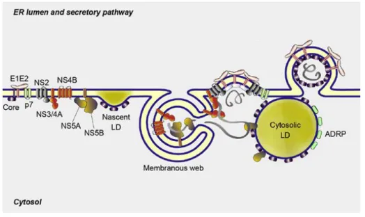
Through this enormous rearrangements of intracellular ER membranes, the HCV replication complex, also known as membranous web, is formed by activity of viral NS4B (Gouttenoire, Penin et al. 2010) and of the cellular lipid kinase phosphatidylinositol-4-kinase (PI4Kα), whose lipid kinase activity is initiated by interaction with NS5A and NS5B (Paul, Hoppe et al. 2013; Ferraris, Beaumont et al. 2013; Reiss, Rebhan et al. 2011). Additionally, the activated replication machinery also requires NS4A acting as a co-factor forming a heterodimer with NS3 and triggering NS3 protease function (Bartenschlager, Lohmann et al. 1995), and host factor cyclophilin A (CyPA) (Liu, Yang, Robotham et al. 2009; Kaul, Stauffer et al. 2009). Of note, the viral RNA-dependent RNA polymerase, NS5B, plays the key role in HCV RNA synthesis (Lohmann, Korner et al. 1997; Behrens, Tomei et al. 1996). The membranous web is a typical morphological feature of positive-strand RNA viruses (Miller and Krijnse-Locker 2008), and can be visualized via electron microscopy of HCV-infected hepatocytes, often in close proximity with lipid droplets (LDs) indicating the tight link of HCV with host cell lipid metabolism (see Figure 6); indeed HCV core (McLauchlan, Lemberg et al. 2002) and NS5A (Shi, Polyak et al. 2002) could be found in association with LDs implicating a possibly key role in coordination of viral replication and virion morphogenesis (Bartenschlager, Penin et al. 2011) (see p.13).
Figure 6. HCV replication and formation of the membranous web (adapted from Dubuisson and Cosset 2014).
In the primer-independent initiation step of RNA replication, a negative-strand of the genome is generated serving as template for progeny positive-strand viral genomes. After formation of a dinucleotide that acts as a primer, elongation of the nascent negative-strand RNA chain occurs mediated by NS5B proceeding in a 3’-to-5’ direction resulting in a positive-strand RNA molecule without the help of other factors (Lohmann 2013). About termination only little is known, but one can imagine that the polymerase dissociates from the template after reaching its end. Of note, the polymerase NS5B does not work properly, but rather error-prone resulting in high genetic variability of HCV isolates. The newly generated positive-strand RNA copies either serve as template for continuing viral protein synthesis or move to LDs resulting in assembly of progeny virions or they remain within the membranous web undergoing negative-strand RNA synthesis for replication (Lohmann 2013). HCV is able to create an environment conducive to its replication and assembly: the formation of the membranous web is one example. Moreover, the ratio of neutral to membrane lipids is reduced upon HCV infection and membrane lipids as cholesterol and phospholipids were gathered in microsomal fractions of HCV-infected cells (Hofmann, Krajewski et al. 2018). Additionally, HCV seems to recruit various cytoplasmic nuclear pore complexes (Nups) to site of replication where they could be found in increased numbers and accumulated at the membranous web and even more co-localized with core or NS5A (Levin, Neufeldt et al. 2014; Neufeldt, Joyce et al. 2013). It is proposed that cytoplasmic Nups form channels across the double membrane structures of the membranous web to serve as gatekeepers by facilitating movement of several HCV proteins and host proteins without being recognized by the pattern recognition receptors (PRRs) of the innate immune system (see Figure 7) (Levin, Neufeldt et al. 2014; Neufeldt, Joyce et al. 2013). Upon Dengue
implicating a possible conserved role of cytoplasmic Nups upon positive-strand RNA virus infections (Neufeldt, Joyce et al. 2013).
Figure 7. Potential function of the cytosolic nuclear pore complexes in HCV replication and assembly (Neufeldt, Joyce et al. 2013).
d. Viral assembly and egress
After replication of the viral genome, RNA progeny can be used for encapsulation into new virions which can be released using a noncytolytic pathway related to the VLDL secretory pathway (Chang, Jiang et al. 2007; Jiang and Luo 2009; Gastaminza, Cheng et al. 2008; Huang, Sun et al. 2007). Several viral and host proteins are involved in the viral assembly process that can be divided into an early and late phase of assembly, whereupon detailed information on different steps is still lacking. During the early phase, the nucleocapsid is formed by the involvement of different recruitment processes with the aim to localize in close proximity to LDs and to the assembly site in the ER lumen: (i) Shuttle between HCV core protein and LDs , (ii) movement of the replication complex through action of NS5A, (iii) recruitment of glycoproteins E1 and E2 by interaction with NS2 (reviewed in Popescu, Riva et al. 2014).
HCV core protein attaches to LDs through its hydrophobic domain, replacing adipose differentiation-related protein (ADRP, Figure 6) leading to an accumulation of LDs in perinuclear regions (Boulant, Douglas et al. 2008; Boulant, Montserret et al. 2006). In terms of assembly efficiency, core-LD association is a crucial step resulting in downstream recruitment of viral proteins (Miyanari, Atsuzawa et al. 2007). However, an inverse correlation of core-LD motility and assembly efficacy of
HCV was determined implicating that core is see-sawing from LD-ER interface to the ER or
vice-versa (Shavinskaya, Boulant et al. 2007). Diacylglycerol acyltransferase 1 (DGAT1) is the key host
factor involved in this step of assembly by facilitating recruitment of core onto the LD by direct core-DGAT1 interaction (Herker, Harris et al. 2010).
Since HCV RNA replication takes plays within the membranous web, two possible models are proposed where assembly starts at ER membrane or at the surface of cytosolic LDs (cLDs): (1) Transfer of HCV core protein onto the surface of cLDs and re-recruitment to ER membrane at assembly sites where interaction of core with NS5A occurs concluding that cLDs might serve as trafficking vehicles to transport core from sites of translation and replication to sites of viral assembly; or (2) Initiation of nucleocapsid formation occurs on the surface of cLDs, while distributions of HCV RNA to core protein is mediated by NS5A that is colocalized onto surface of cLDs. Currently, discrimination between both hypotheses is not easy by considering the low assembly efficiency of HCV per cell (reviewed in Bartenschlager, Penin et al. 2011).
NS5A as well as DGAT1 seem to play a central role in the early stage of viral assembly. Indeed, an interaction between the hyperphosphorylated form of NS5A and core could be observed leading to the relocation of NS5A to LDs (Masaki, Matsunaga et al. 2014), while the hypophosphorylated form of NS5A correlates with genomic replication (Evans, Rice et al. 2004). Given the direct interaction of core-DGAT1, also NS5A directly interacts with DGAT1 resulting in its recruitment to LDs (Camus, Herker et al. 2013). Additionally, an interaction between NS5A and ApoE is also suggested to contribute to the recruitment of this apolipoprotein to assembly sites (Benga, Krieger et al. 2010). Besides, NS5A was also reported to interact with Rab18, a small G protein, to facilitate its recruitment to LD and HCV assembly site (Salloum, Wang et al. 2013). The transition from replication to assembly is represented by the recruitment of NS5A together with the replication complex to LDs (reviewed in Lindenbach and Rice 2013).
The last step of the early phase, and in fact the first step of late phase, of viral assembly is the recruitment of envelope proteins at assembly sites. Several groups reported that the viral envelope glycoproteins E1 and E2 are bound in complexes composed of NS2, p7 and NS3 (Jirasko, Montserret et al. 2010; Popescu, Callens et al. 2011; Stapleford and Lindenbach 2011; Ma, Anantpadma et al. 2011) resulting in an accumulation of these complexes in close proximity of NS5A, core and LDs (Jirasko, Montserret et al. 2010; Popescu, Callens et al. 2011). However, the concrete nature and composition of this complex is not yet fully understood, however, it could be determined that NS2 and HCV E2 are associated with detergent-resistant membranes (DRMs) which are required for
(Aizaki, Morikawa et al. 2008; Aizaki, Lee et al. 2004; Paul, Hoppe et al. 2013; Shi, Polyak et al. 2002). Recently, Boyer et al had a deeper look on the nature of this NS2 complex and found a direct protein-protein interaction of NS2 and E1E2 using immunoprecipitation assays revealing an association of NS2 with NS3 via DRMs. Contrarily, NS5A and E1E2 do not associate but rather interact separately with NS2-E1-E2-NS3 complex via unstable DMRs (Boyer, Dreneau et al. 2019). Interestingly, core was found to interact with NS2 and E1E2 through unstable DMRs suggesting a crucial role of DMRs transporting the NS2-E1-E2-NS3-NS5A-core complex for HCV assembly initiation (Boyer, Dreneau et al. 2019).
The late phase of viral assembly is the maturation and release of HCV particles in tight association with the host VLDL pathway within the ER lumen (Chang, Jiang et al. 2007; Jiang and Luo 2009; Gastaminza, Cheng et al. 2008; Huang, Sun et al. 2007). Indeed, it could be shown that HCV core accumulates in lipid fractions which contain high amounts of cholesterol and sphingolipids (Matto, Rice et al. 2004); these two lipids are observed to be enriched in viral particles and in turn involved in their infectivity (Aizaki, Morikawa et al. 2008). The nucleocapsid could be integrated into the core of luminal LDs (luLDs); however, how E1 and E2 are integrated into virions remains unclear. It is proposed that there is an interaction between NS3, E1 and E2 by either NS2 alone or together with p7 (Yi, Ma et al. 2009; Phan, Beran et al. 2009). Indeed, two studies observed that NS2 brings together E1, E2, p7, NS3 and NS5A in close proximity of LD by protein-protein interactions between all involved components (Jirasko, Montserret et al. 2010; Ma, Anantpadma et al. 2011). Since HCV E2 is the well-studied HCV glycoprotein that plays the major role for neutralization of HCV-specific antibodies and the binding capacity to CD81, the involvement of HCV E1 in HCV replication cycle still remains to be elucidated. By studying the role of E1 for viral morphogenesis, Haddad et al used mutants of E1 observing first a shift in the receptor usage from CLDN1 to CLDN6 for two mutants, and second virus carrying one mutant (D263A) was assembled and released but devoid of viral RNA revealing a crucial role for E1 in incorporating HCV RNA into the nucleocapsid (Haddad, Rouille et al. 2017). The step in which the apolipoproteins incorporate into mature infectious particles is also not fully clear. It is assumed that ApoB-positive precursors, formed in the rough ER (rER), fuse together with ApoB-negative precursor which incorporate the viral nucleocapsid resulting in LVPs associated with nonexchangeable ApoB (see p.5). ApoE and ApoC, both exchangeable apolipoproteins, could be associated to the LVPs within the ER lumen. However, there are differences between HCV particles depending in which cell type they are produced (detailed comparison is found on p.20).
Figure 8. Model of the formation of LVPs and comparison to cell culture-derived HCV (HCVcc).
Precursors are formed: (1) VLDL2 resulted from the translocation of nascent ApoB into the ER lumen and the association with phospholipids, triglycerides and ApoE/C which are not shown for clarity (left), (2) luminal LD (luLD) is generated at the smooth ER (sER) or the membranous web (m.w.) and enriched with triglycerides via MTP (right). E1 and E2 associate with luLD after release from the ER membrane and the nucleocapsid is proposed to insert within the hydrophobic core of the luLD. In primary human hepatocytes the nucleocapsid-loaded luLD fuses with the VLDL2 resulting in a LVP, while in the cell culture model VLDL1 formation is lacking, cell culture-derived (HCVcc) is released without ApoB association (more details of in vitro models see on p.20). For clarity reasons, VLDL1 is generated as a fusion of VLDL with the triglyceride-riche luLD (Bartenschlager, Penin et al. 2011.
LVPs are transported along the VLDL secretory pathway (reviewed in Bartenschlager, Penin et al. 2011) to the Golgi, where the HCV glycoproteins E1 and E2 are post-translationally modified (reviewed in Vieyres, Dubuisson et al. 2014). The exact mechanism of HCV budding at the plasma membrane is still lacking. Immunoprecipitation and electron microscopy (EM) analysis revealed that HCV LVPs assemble in the ER and are transported to Golgi through vesicular transport mediated by COPII vesicles to enter the Golgi secretory pathway (Syed, Khan et al. 2017). In line with these findings, Takacs et al could observe that the secretion of ApoE and ApoB is differentially controlled by Rab1b which is a major regulator of transport from the ER to Golgi, and thus regulates secretion of HCV as well (Takacs, Andreo et al. 2017). Golgi protein 73 (GP73) has been demonstrated to be involved in HCV secretion: normally as a Golgi membrane resident protein and upregulated upon HCV infection, GP73 is colocalized with the HCV replication complex in Huh7.5.1 cells and is able to directly mediate an interaction of NS5A and ApoE and thus promoting the secretion of HCV
HCV is hijacking proteins of the endosomal sorting complex required for transport (ESCRT), e.g. hepatocyte growth factor-regulated tyrosine kinase substrate (HRS), for budding at the plasma membrane. Indeed, HRS is able to directly interact with NS2 and NS5A in HCV-infected cells supporting efficient viral assembly as well as viral budding (Barouch-Bentov, Neveu et al. 2016). In line with this, another group found viral particles and structural proteins in endosomal compartments but not in compartments of the Golgi assuming that release of HCV occurs through noncanonical secretory route which is different from the canonical Golgi-mediated secretion (Bayer, Banning et al. 2016).
In sum, the above described processes followed the hypothesis that virions exist as hybrid lipoviral particles protecting from neutralization with HCV-specific antibodies (Andre, Komurian-Pradel et al. 2002). However, in a proposed two-particle model, the virus and serum lipoproteins stably interact as separate particles via protein-protein interaction. Indeed, when cholesterol is chemically removed from HCVcc particles their infectivity is lost, while adding back exogeneous cholesterol led to restorage (Aizaki, Morikawa et al. 2008). Additionally, another group could observe a rapid shift of buoyant density of viral particles in serum of fasting HCV patients concluding that virions and serum lipoproteins associate in a transient and exchangeable way (Felmlee, Sheridan et al. 2010). Both hypotheses could be imaginable, but they still need to be confirmed.
1.3
Model systems to study HCV
1.3.1 In vitro model systemsAfter its discovery in 1989 (Choo, Kuo et al. 1989), several efforts to culture HCV in vitro failed through lacking robustness, inefficient replication and thus failure for detailed molecular studies of the viral life cycle (reviewed in Bartenschlager and Lohmann 2000). After ten years of research, several breakthrough developments led to improvement to study the virus in vitro: the replicon system in 1999 followed by retroviral pseudoparticles in 2003 and finally the cell culture system recapitulating the entire viral lifecycle in 2005 (see Figure 9).
a. HCV replicon system
The establishment of sub-genomic replicons that autonomously amplify in cultured human hepatoma cells was a first major breakthrough to study viral replication: the genotype 1b HCV genome that was derived from a chronically infected patient was trimmed to those components essential for replication (NS3 to NS5B), while the viral structural genes (core, E1, E2) as well as p7 and NS2 were deleted
(see Figure 9). Practically, this shortened HCV genome was cloned into a plasmid resulting in a replicon encoding the 5’UTR of HCV, the first 12 codons of the core protein fused with a G418 selection cassette, the IRES from encephalomyocarditis virus (EMCV) allowing translation of the non-structural proteins, as well as viral NS3, NS4A, NS4B, NS5A, NS5B, and 3’UTR. After undergoing in vitro transfection, the construct is transfected into human hepatoma 7 (Huh7) cells (see p.20) resulting in direct translation of viral RNA and G418 selection of those Huh7 cells displaying high levels of HCV RNA replication (Lohmann, Korner et al. 1999). Subsequently, full length HCV replicons were developed, ultimately leading to the establishment of recombinant full-length infectious HCV (see HCVcc below).
b. HCV Pseudoparticles (HCVpp)
Four years after developing the replicon system, the next important achievement was the generation of retroviral pseudoparticles displaying functional HCV glycoproteins, E1 and E2, for dissection of HCV entry process. In basic principal, pseudoparticles are retroviral capsids with incorporated viral glycoproteins in their envelopes. Consequently, these pseudoparticles allow to study the relevance and need of those glycoproteins in viral attachment and entry processes; the latter can be easily quantified using a reporter gene located inside the pseudoparticles. HCVpp could be successfully developed by integrating HCV glycoproteins in retroviral particles: A system in which 293T cells are co-transfected with expression vectors encoding HCV E1 and E2, the gag-pol proteins of either murine leukemia virus (MLV) or human immunodeficiency virus (HIV), and a retroviral genome encoding a reporter gene such as green fluorescent protein (GFP) or luciferase (Bartosch, Dubuisson et al. 2003; Hsu, Zhang et al. 2003) (see Figure 9). Using HCVpp is an elegant way to study HCV entry independently of the entire viral life cycle which in turn highlights the limitation of HCVpp to the entry process: they are produced in a non-liver cell line and thus assemble as retroviruses on plasma membranes without getting associated with lipoproteins, contrarily liver-generated HCV particles assemble in the ER in close association with lipoproteins (see p.5 and 13).
c. Cell culture-derived HCV (HCVcc)
After successful development of sub-genomic replicons and the HCVpp system, the challenge to establish an HCV permissive cell culture system remained.
The initial production of cell culture-derived HCV particles is based on the genotype 2a HCV strain, namely JFH1 (Japanese Fulminant Hepatitis 1) that was isolated from a Japanese patient with acute
genomes, three groups proposed a robust in vitro system recapitulating the entire HCV life cycle in Huh7 cells (Lindenbach, Evans et al. 2005; Wakita, Pietschmann et al. 2005; Zhong, Gastaminza et al. 2005) which since then is the laboratory standard for in vitro HCV studies. These produced particles, well-known as cell culture-derived HCV (HCVcc), are able to infect new target cells, thereby completing the entire HCV life cycle (see Figure 9). This achievement led to the development of several chimeras that consist of JFH1 replicase genes NS3 to NS5B and core to NS2 of alternative HCV genomes allowing to study HCV entry, neutralization and assembly of all seven known HCV genotypes. Among all these chimeras, Jc1, consisting of the JFH1 nonstructural proteins (NS3 to NS5B) and core-E1-E2-p7 and partially NS2 of J6 (HCV gt 2a), produces infectious virus titers that are around 100 to 1000-fold higher than the original JFH1 strain (Pietschmann, Kaul et al. 2006). Over the years, chimeras of all seven known genotypes of HCV based on JFH1 have been developed giving the possibility to study the entire viral life cycle and additionally, neutralization via HCV-specific antibodies (Gottwein, Jensen et al. 2011; Gottwein, Scheel et al. 2009; Scheel, Gottwein et al. 2008; Scheel, Gottwein et al. 2011; Jensen, Gottwein et al. 2008; Gottwein, Scheel et al. 2007), however, with differences concerning infectivity. In order to rapidly and efficiently detect viral replication and infection, reporter genomes of different HCV chimeric genomes containing luciferase or GFP were generated (reviewed in Vieyres and Pietschmann 2013). Furthermore, non JFH1-based HCVcc enabling the production of recombinant HCV of different genotypes have been established but JFH1-based HCVcc remain the most widely used and efficient models.
Figure 9 In vitro HCV cell culture models.
In vitro HCV cell culture models to investigate different steps of the viral life cycle: HCV pseudoparticle system (left) to study viral entry, the HCV replicon (middle) system to study viral replication, and full-length recombinant cell culture-derived HCV (right) system to investigate the entire viral replication cycle. (Adapted from Steinmann and Pietschmann 2013).
d. HCV-permissive host cells
Since HCV primarily replicates in human hepatocytes, cultured primary human hepatocytes (PHH) from liver resection of patients represent the most physiological in vitro model closest to the natural host (Ploss, Khetani et al. 2010). However, PHH display several disadvantages that make them not easy to handle in laboratories: difficult to obtain, high donor-dependent variability and limited time of culture (time-dependent dedifferentiation and approximately two weeks before apoptosis occurs); even worse, once infected with HCV PHH only show low-level replication (Fournier, Sureau et al. 1998; Molina, Castet et al. 2008; Rumin, Berthillon et al. 1999), hence making it difficult to conduct complex experiments.
Thus, the most HCV-permissive and widely used cell line are Huh7 human hepatoma cells that were originally isolated from HCC of a Japanese patient (Nakabayashi, Taketa et al. 1982). Several Huh7 subclones with increased HCV permissiveness could be obtained, by selection of replicon-containing Huh7 cells. The subclones Huh7.5 (Blight, McKeating et al. 2002), Huh7.5.1 (Zhong, Gastaminza et al. 2005), and Huh7-Lunet (Friebe, Boudet et al. 2005) cells were obtained after cure of the HCV-transfected Huh7 cells from the replicon via treatment with INF-α, INF-γ or selective inhibitors. It is not entirely clear why these Huh7 subclones yield higher levels of HCV RNA replication. However, in case of Huh7.5 cells mutations in the retinoic acid-inducible gene I (RIG-I) were shown to be involved in increased viral replication (Sumpter, Loo et al. 2005).
However, Huh7-derived cells are dedifferentiated and asynchronously dividing cancer cells, in contrast to primary hepatocytes which are differentiated and quiescent (Michalopoulos and DeFrances 1997). However, they show hepatocyte-specific gene expression and arrested cell growth by adding 1% of dimethyl sulfoxide (DMSO) to the culture medium, while permissiveness to HCV is maintained (Sainz and Chisari 2006); and thus are closer to hepatocytes.
Importantly, cultured Huh7-derived cells and in vivo hepatocytes show differences in their capability in producing lipoproteins, and thus HCV particles differ in their properties in dependency of the cells in which they are produced. In infected patients, the heterogenous density of circulating HCV particles and their infectivity is negatively correlated meaning low-density viruses are more infectious (Bradley, McCaustland et al. 1991; Hijikata, Shimizu et al. 1993). Those HCV particles can be immunoprecipitated via ApoB-specific antibodies confirming the association of HCV with triglyceride-rich lipoproteins (TRLs) within hepatocytes and their circulation via the bloodstream as LVPs (Andre, Perlemuter et al. 2005; Nielsen, Bassendine et al. 2004). In comparison to patient-derived LVPs, the negative correlation for density and infectivity could also been shown for HCVcc
al. 2011) - and not ApoB-specific antibodies (Merz, Long et al. 2011; Huang, Sun et al. 2007) highlighting the tight association of HCVcc with ApoE and ApoC and the inefficiency of Huh7-derived cells in producing authentic VLDL (reviewed in Bartenschlager, Penin et al. 2011; Vieyres and Pietschmann 2019). However, Piver et al described that at least a small fraction of HCVcc derived from Huh7.5 cells displayed ApoB and ApoE as well as E1E2 on the surface after immunogold staining following immunocapture (Piver, Boyer et al. 2017). Recently, it was confirmed that Huh7.5 cells growing in serum-free medium produced immature HCV particles, however, when incubating those cells/particles with physiological serum conditions and concentrations of lipoproteins resulted in the maturation of HCV particles to fully lipidated and notably, ApoB-containing infectious virions displaying low density very similar to patient-derived LVPs (Denolly, Granier et al. 2019). In addition to HCV maturation through the cell secretory pathway, also extracellular lipidation of particles may occur through serum itself properly after egress (Denolly, Granier et al. 2019). In contrast, HCV nucleocapsids derived from patients and cell culture appeared to be similar in size and structure and interestingly those nucleocapsids are found to be surrounded by an irregular, detergent-sensitive crescent which may be consistent with lipids (Piver, Boyer et al. 2017).
1.3.2 In vivo model systems
Cell culture model systems to study HCV are of high importance, however, the complex immunological host responses and liver disease progression cannot be fully answered in vitro. Since lacking control parameters such as time and dose of infection strongly limited clinical research in patients, attention of research was directed to animal models such as chimpanzees, tree shrews and rodents by keeping in mind that ideally, similarly to HCV-infected patients, HCV-infected animals should develop chronicity followed by liver disease progression towards cirrhosis and HCC.
Chimpanzees (Pan troglodytes) are with more than 98% genetic identity the closest living relatives to human and susceptible to HCV infection (Abe, Kurata et al. 1993), and therefore represented an ideal model to study HCV in vivo for over 20 years. After HCV infection, viremia as well as antiviral innate and adaptive immune responses are detectable following a mild acute hepatitis. In contrast to humans, chimpanzees rarely develop chronicity (Bassett, Brasky et al. 1998; Major, Dahari et al. 2004), and thus do not develop progressive liver disease (Walker 1997). High costs and increased ethical concerns limited the use of this animal model (National Research Council Committee on the Use of Chimpanzees in and Behavioral 2011), although it is the only well-studied model to study
protective immunity against HCV, probably leading to vaccine development (Cooper, Erickson et al. 1999).
The tree shrew (Tupaia belangeri), a squirrel-like mammal originally found in South-East Asia, has also been shown to be susceptible to HCV infection (Xu, Chen et al. 2007; Xie, Riezu-Boj et al. 1998) followed by chronicity and development of liver disease in some tree shrews (Amako, Tsukiyama-Kohara et al. 2010). Moreover, ethical concern and lack of tool to analyze HCV-host interactions in these animals preclude a wide usage of this model.
Rodent models represent a cheap and easy to breed model to study HCV. In general, mice and rats are not susceptible for HCV infection and need to undergo experimental modifications. The most commonly used rodent models can be divided into (i) the human liver-chimeric mouse models and (ii) the transgenic mouse models:
(i) In human liver-chimeric models, mouse livers are humanized by xenotransplantation of primary human hepatocytes or hepatoma cell lines resulting in a subsequent susceptibility to HCV infection. To successfully xenograft human hepatocytes into mouse livers, the xenograft recipients require specific genetic defects leading to death of the original mouse hepatocyte and to dysfunctionality of the mouse immune system. Currently, two major types of mice are used: first, mice overexpressing the urokinase plasminogen activator (uPA) gene under albumin promotor control were crossed with mice that suffer from a severe combined immunodeficiency syndrome (SCID) resulting in uPA-SCID mice; once xenografted with human hepatocytes, these mice are efficiently infected with HCV followed by chronic infection (Mercer, Schiller et al. 2001) and led to achievements concerning preclinical studies of antiviral compounds (Mailly, Xiao et al. 2015) and understanding of steps in the viral lifecycle (e.g. composition of LVPs, role of HCV-specific antibodies or viral entry) in vivo (Lindenbach, Meuleman et al. 2006; Vanwolleghem, Bukh et al. 2008; Lacek, Vercauteren et al. 2012; Meuleman, Catanese et al. 2012).
Second, mice that have fumarylacetoacetate hydrolase (Fah-/-) deficiency accumulate hepatotoxic metabolites consequently leading to liver failure (Grompe, al-Dhalimy et al. 1993). These Fah-/- mice were crossed with mice suffering from severe immune system dysfunctionality (Rag2-/- IL2Rγnull) resulting in FRG mice. The liver degeneration resulting from depletion of Fah can be prevented by a drug, namely NTBC (2-(2-nitro-4-trifluoromethylbenzoyl)1,3-cyclohexanedione)). Hence, this model provides an advantage over uPA-SCID mice: the timing of human hepatocyte transplantation can be anticipated by removing NTBC (Azuma, Paulk et al. 2007; Bissig, Le et al. 2007).
immunoevasion mechanisms. The mouse livers in these model systems are provided with all human host factors required to enable (parts of) the HCV replication cycle in mouse hepatocytes. Expression of SR-BI, OCLN, CLDN1 and CD81 under an albumin promotor enables to detect HCV entry in mouse livers (see p.7); of note, in line with results from in vitro experiments (see p.7) expression of human OCLN and CD81 is the minimum requirement enabling the study of viral entry in mouse livers (Dorner, Horwitz et al. 2011). Additionally, knockout of STAT1, IRF1, IRF7 and IFNAR1, all important players in innate immunity, results in low-level HCV RNA replication and production of infectious virus in mice (Dorner, Horwitz et al. 2013).
1.4
HCV treatment
As we now deep dived into the HCV molecular biology to which knowledge has markedly advanced, molecular mechanisms of disease progression and vaccine development still needs to be figured out. By thinking about viral infection, the aim of every viral therapy is eradication of virus, known in hepatitis C clinical context as sustained virologic response (SVR) which is defined as undetectable HCV RNA levels over 12 to 24 weeks after end of therapy.
Throwback to the 80’s and 90’s, the only available therapy for HCV infection was based on interferon (INF). INF-α exerts its antiviral effect by inducing IFN-stimulated genes that in turn inhibit HCV replication. Until 1998, three INF-α injections a week for up to 48 weeks were the therapy of choice, unfortunately resulting in cure of only one-fifth of infected patients (Carithers and Emerson 1997; Hoofnagle and di Bisceglie 1997). This application was improved by injection of INF-α combined with orally taken ribavirin up to 24 to 48 weeks, an antiviral medication which could increase the antiviral effect of INF-α resulting in around 35-45% of cure in infected patients (McHutchison, Gordon et al. 1998). The addition of polyethylene glycol to INF-α (PEG-INF-α) gave rise to increased half-life of the drug and in combination with ribavirin an increased effectivity (Manns, McHutchison et al. 2001) was achieved replacing the standard INF-α for more than ten years. Of note, HCV-infected patients responded differently to the PEG-INF-α/ribavirin therapy depending on their HCV genotype: while a SVR of around 50% was achieved for patients infected with HCV genotype 1, up to 80% of SVR was shown for patients infected with genotypes 2, 3, 5 and 6 (Antaki, Craxi et al. 2010). Nevertheless, the INF-based therapy had many side-effects including flu-like symptoms, gastritis or even worse depression (Manns, Wedemeyer et al. 2006).
The achievement of uncovering key viral proteins involved in the HCV replication cycle led to the development of direct-acting antivirals (DAAs) which are specifically targeting nonstructural proteins of HCV. DAAs can be classified into four main groups: NS3/NS4A protease inhibitors,
NS5B nucleoside and non-nucleoside polymerase inhibitors and NS5A inhibitors. In 2011, a NS3/4A protease inhibitor was combined with the PEG-INF-α/ribavirin increasing SVRs for genotype 1. Nevertheless, this triple therapy was only available for genotype 1-infected patients combined with severe side-effects (Bacon, Gordon et al. 2011; Jacobson, McHutchison et al. 2011; Zeuzem, Andreone et al. 2011). In 2014-2015, new interferon-free DAA regimen, including sofosbuvir (NS5B inhibitor) and daclatasvir or ledipasvir (NS5A inhibitor), became much more efficient (SVR around 90%) with the big plus of only little side-effects (Afdhal, Zeuzem et al. 2014; Afdhal, Reddy et al. 2014; Sulkowski, Gardiner et al. 2014). The combination of different DAAs - that are now the current standard of care - lead to highly improved SVR rates and shortened therapy duration; moreover, defined DAA combinations are efficient for all HCV genotypes resulting in SVR rates of 95% (reviewed in (Asselah, Boyer et al. 2016; Li and De Clercq 2017). Currently recommended DAAs are: dasabuvir (DSV), elbasvir (EBR), glecaprevir (GLE), grazoprevir (GZR), ledipasvir (LDV), ombitasvir (OBV), pibrentasvir (PIB), paritaprevir (PTV), ritonavir (r), sofosbuvir (SOF), velpatasvir (VEL) and voxilaprevir (VOX). Among all these recommended DAAs, the indication is depended on the HCV genotype, the severity of liver disease as well as prior therapy. Combinations of DAAs are very efficient, even if combinations of 2 regimens is preferred to triple combination in order to avoid drug-drug interactions and severe side-effects (see Table 1) (EASL 2018).
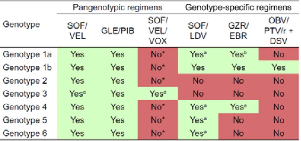
Table 1. Combinational DAA treatment.
Combination of DAA treatment. Recommendation for treated and non-treated patients without or with compensated cirrhosis (a-e). *Triple combination. DSV: dasabuvir, EBR: elbasvir, GLE: glecprevir, GZR: grazoprevir, LDV: ledipasvir, OBV: ombitasvir, PIB: pibrentasvir, PTV: paritaprevir, r: ritonavir, SOF: sofosbuvir, VEL: velpatasvir, VOX: voxilaprevir (EASL 2018).
Even if may appear that with the achievement of DAAs HCV research could be disregarded, there are still some important challenges that remain (reviewed in Bartenschlager, Baumert et al. 2018). First, DAAs are only available to around 10% of infected patients (Edlin 2016) and the high costs of
more in the presence of advanced liver disease progression. These patients are less able to clear the virus suggesting that there is still need to investigate the fine tuning of optimal DAA combination, treatment duration and dosages for difficult patient populations (Hezode, Fontaine et al. 2013; Ferenci 2015). Third and in line with the second limitation, the risk to develop HCC can be reduced after SVR, but HCC can occur after years of viral clearance suggesting that there is a “point-of-no-return” when virus-induced liver disease came to the point where HCC will develop even in the absence of HCV (Baumert, Juhling et al. 2017). Fourth, a minority of patients can develop resistance to DAA due to the error-prone HCV RNA polymerase which creates a pool of genetic variants in each patient leading to viral polymorphism in DAA-targeted regions resulting in resistance to DAAs (Esposito, Trinks et al. 2016; Pawlotsky 2016).
In addition to DAAs, the standard of care for chronic hepatitis C, alternative strategies have been developed. When reminiscing about the HCV replication cycle, several host factors play a crucial role in different steps. When reminiscing about the HCV replication cycle, several host factors play a crucial role in different steps.
Indeed, host-targeting agents (HTAs) appeared to be of therapeutic interest. Monoclonal antibodies specifically targeting CD81 (Fofana, Xiao et al. 2013), CLDN1 (Mailly, Xiao et al. 2015; Fofana, Krieger et al. 2010) or SR-BI (Meuleman, Catanese et al. 2012; Zahid, Turek et al. 2013) were developed and could be shown to efficiently inhibit viral entry in vitro and in vivo in liver chimeric mice. Additionally, small molecule inhibitors of SR-BI, ITX 5061 and ITX 7650, could efficiently block HCVcc and HCVpp infection of PHH or human hepatoma cell lines (Syder, Lee et al. 2011). ITX 5061 has been evaluated in phase 1 clinical trials (ClinicalTrials.gov identifier: NCT01165359 and NCT01292824). Moreover, a monoclonal CLDN1-specific antibody was shown to cure chronic HCV infection in the uPA-SCID chimeric mouse model (Mailly, Xiao et al. 2015; Colpitts, Tawar et al. 2018). Of note, synergy between host-targeting entry inhibitors combined with DAA-based treatment could be demonstrated (Xiao, Fofana et al. 2015; Paciello, Urbanowicz et al. 2016). Another important host factor for the HCV life cycle is liver-enriched miR-122 that has been shown to play a crucial role in the stabilization of HCV RNA and consequently in HCV replication and translation (Jopling, Yi et al. 2005). miR-122 antagonists significantly decrease HCV replication (Jopling, Yi et al. 2005; Jopling, Schutz et al. 2008) and miravirsen and RG-101 demonstrated efficacy against HCV in clinical trials (van der Ree, de Vree et al. 2017; Stelma, van der Ree et al. 2017). In fact, these inhibitors can be regarded as pan-genotypic antivirals due to the conserved miR-122 binding sites across HCV genotypes (Li, Gottwein et al. 2011; van der Ree, de Vree et al. 2017). Cyclophilin A (CypA) inhibitors, namely alisporivir or DEB025, could efficiently block CypA interaction with NS5A leading to inhibition of HCV replication and additionally, these inhibitors are able to rehabilitate the innate immune response against HCV (Daito, Watashi et al. 2014; Hopkins,
Bobardt et al. 2012; Naoumov 2014). Phase 3 clinical trials that examined a triple therapy with alisporivir (DEB025), PEG-INF and RBV have been completed (ClinicalTrials.gov identifier: NCT01446250, NCT01318694).
In fact, the given examples represent only a small overview of HTAs that have been studied for their ability to improve antiviral treatment, several other host factors involved in the viral life cycle are at different stages of preclinical or clinical development (reviewed in Crouchet, Wrensch et al. 2018; Zeisel, Crouchet et al. 2015; Zeisel, Lupberger et al. 2013 (see Figure 10).
Figure 10. Host-targeting agents (HTAs) at different steps of the HCV replication cycle (Crouchet, Wrensch et al. 2018).
Taken together, HCV infection can now be regarded as curable disease, although some limitations and challenges still exist. Since HCC can develop even after successful eradication of HCV, a current question is what kind of molecular imprinting is left by HCV in the host genome that drives carcinogenesis even after SVR in patients (see HEPATITIS C – An Introduction).