HAL Id: hal-02631425
https://hal.inrae.fr/hal-02631425
Submitted on 27 May 2020
HAL is a multi-disciplinary open access
archive for the deposit and dissemination of
sci-entific research documents, whether they are
pub-lished or not. The documents may come from
teaching and research institutions in France or
abroad, or from public or private research centers.
L’archive ouverte pluridisciplinaire HAL, est
destinée au dépôt et à la diffusion de documents
scientifiques de niveau recherche, publiés ou non,
émanant des établissements d’enseignement et de
recherche français ou étrangers, des laboratoires
publics ou privés.
Copyright
pv. campestris: A toolbox for sequential plant n-glycan
processing
Stéphanie Dupoiron, Claudine Zischek, Laetitia Ligat, Julien Carbonne, Alice
Boulanger, Thomas Duge de Bernonville, Martine Lautier, Pauline Rival,
Matthieu Arlat, Elisabeth Jamet, et al.
To cite this version:
Stéphanie Dupoiron, Claudine Zischek, Laetitia Ligat, Julien Carbonne, Alice Boulanger, et al.. The
N-Glycan Cluster from Xanthomonas campestris pv. campestris: A toolbox for sequential plant
n-glycan processing. Journal of Biological Chemistry, American Society for Biochemistry and Molecular
Biology, 2015, 290 (10), pp.6022-36. �10.1074/jbc.M114.624593�. �hal-02631425�
The N-Glycan Cluster from Xanthomonas campestris pv.
campestris
A TOOLBOX FOR SEQUENTIAL PLANT N-GLYCAN PROCESSING
*Received for publication, November 13, 2014, and in revised form, January 13, 2015Published, JBC Papers in Press, January 13, 2015, DOI 10.1074/jbc.M114.624593 Stéphanie Dupoiron‡§1,2, Claudine Zischek¶储1, Laetitia Ligat‡§3, Julien Carbonne‡§, Alice Boulanger¶储4,
Thomas Dugé de Bernonville¶储5, Martine Lautier¶储**, Pauline Rival‡§¶储6, Matthieu Arlat¶储**, Elisabeth Jamet‡§,
Emmanuelle Lauber¶储7, and Cécile Albenne‡§
From the‡Université de Toulouse and§CNRS, Laboratoire de Recherches en Sciences Végétales, UMR 5546, BP 42617, F-31326
Castanet-Tolosan, France,¶INRA and储CNRS, Laboratoire des Interactions Plantes-Microorganismes, UMR 2594, F-31326 Castanet-Tolosan, France, and the **Université de Toulouse, UPS, F-31062 Toulouse, France
Background:Eight glycoside hydrolases (GHs) encoded by genes belonging to one operon have been identified in the
phytopathogenic bacterium Xanthomonas campestris.
Results:Five of these GHs are involved in the sequential degradation of a plant N-glycan in vitro.
Conclusion:This is the first evidence of N-glycan degradation by plant pathogen enzymes.
Significance:Our results suggest that N-glycans may be metabolized by phytopathogenic bacteria.
N-Glycans are widely distributed in living organisms but
rep-resent only a small fraction of the carbohydrates found in plants. This probably explains why they have not previously been con-sidered as substrates exploited by phytopathogenic bacteria during plant infection. Xanthomonas campestris pv. campestris, the causal agent of black rot disease of Brassica plants, possesses a specific system for GlcNAc utilization expressed during host plant infection. This system encompasses a cluster of eight genes (nixE to nixL) encoding glycoside hydrolases (GHs). In this paper, we have characterized the enzymatic activities of these GHs and demonstrated their involvement in sequential degradation of a plant N-glycan using a N-glycopeptide contain-ing two GlcNAcs, three mannoses, one fucose, and one xylose
(N2M3FX) as a substrate. The removal of the␣-1,3-mannose by
the␣-mannosidase NixK (GH92) is a prerequisite for the
subse-quent action of the-xylosidase NixI (GH3), which is involved
in the cleavage of the-1,2-xylose, followed by the␣
-mannosi-dase NixJ (GH125), which removes the␣-1,6-mannose. These
data, combined to the subcellular localization of the enzymes, allowed us to propose a model of N-glycopeptide processing by
X. campestris pv. campestris. This study constitutes the first
evi-dence suggesting N-glycan degradation by a plant pathogen, a feature shared with human pathogenic bacteria. Plant N-glycans should therefore be included in the repertoire of molecules putatively metabolized by phytopathogenic bacteria during their life cycle.
In the context of host-bacteria interactions, i.e. symbiosis or pathogenesis, the question of the degradation of host N-glyco-proteins by microorganisms is relevant, and only a few studies demonstrating orchestrated processes involving catabolic enzymes have been reported. Glycosylation is the most wide-spread post-translational modification of proteins found in nature (1). It concerns not only eukaryotes but also some bac-teria and archaea for which several examples of sugar attach-ment on proteins have been reported (2, 3). In eukaryotes, two types of glycosylation are described, N- and O-glycosylation. Whereas O-glycosylation can display a huge diversity, in terms of structure and of the sequence surrounding the amino acid carrier, N-glycosylation is more conserved (4). N-Glycosylation occurs in the secretory pathway, by adding a N-glycan to the amide group of asparagine in the Asn-Xaa-Ser/Thr context, where Xaa can be any amino acid except Pro. It starts in the endoplasmic reticulum by the co-translational transfer of a pre-formed lipid-linked oligosaccharide onto the nascent polypep-tide. Then maturation steps involving glycosidases and glyco-syltransferases take place in the Golgi apparatus through secretion of proteins to their final destination. Resulting N-gly-cans can be of three types, either high mannose-, hybrid-, or complex-type (4). This global mechanism is common to most eukaryotic systems, but the action of specific maturation enzymes leads to different complex N-glycan structures. For instance, in plants, the first GlcNAc of the core can be substi-tuted with an ␣-1,3-fucose, instead of an ␣-1,6-fucose for *This work was supported by French Agence Nationale de la Recherche
Grant ANR-08-BLAN-0193-01, the local Fédération de Recherche Agrobio-sciences, Interactions et Biodiversité, the Université Paul Sabatier, the Cen-tre National de la Recherche Scientifique, the Institut National de la Recher-che Agronomique, and the French Ministry of Research and Technology (to A.B.).
1Both authors contributed equally to this work.
2Present address: AgroParisTech, Chaire Agro-Biotechnologies Industrielles,
247 Rue Paul Vaillant Couturier, F-51100 Reims, France.
3Present address: INSERM UMR1037, Plateforme Protéomique, Pôle
Tech-nologique du Centre de Recherches en Cancérologie de Toulouse, 2 Ave-nue Hubert Curien, F-31100 Toulouse, France.
4Present address: Laboratoire de Chimie Bactérienne, CNRS UMR 7283,
F-13009 Marseille, France.
5Present address: EA2106, Biomolécules et Biotechnologies Végétales, UFR
Sciences et Techniques, Université François Rabelais de Tours, 37000 Tours, France.
6Present address: Dept. of Genome Sciences, University of Washington,
Seat-tle, WA 98195.
7To whom correspondence should be addressed: Laboratoire des
Interac-tions Plantes-Microorganismes, UMR 2594/441, Chemin de Borde Rouge, CS52627, F-31326 Castanet-Tolosan, France, Tel.: 33-5-6128-5047; Fax: 33-5-6128-5061; E-mail: [email protected].
at INRA Institut National de la Recherche Agronomique on May 6, 2019
http://www.jbc.org/
human N-glycans. In addition, the-mannose of the core can be decorated with a -1,2-xylose in plants, whereas a -1,4-GlcNAc can be found at this position in human glycans. From a functional point of view, N-glycans can modify the fold-ing, the activity, or the stability of proteins and subsequently play key roles in a number of physiological processes, such as signal transduction, targeting, cell-cell recognition, infection, and immunity (5–7).
Investigation into the alteration of N-glycan structure, because of either aberrant N-glycosylation build-up or degra-dation, has focused on a better understanding of the molecular basis of these biological processes. N-Glycan degradation involves carbohydrate-active enzymes, in particular glycoside hydrolases (GHs)8presently classified into 133 families (8). In
addition, synergic processes may also occur, in particular involving glycoside phosphorylases (9, 10). To date, the most documented model of glycoconjugate breakdown concerns the gut bacterium Bacteroides thetaiotaomicron, which displays a broad repertoire of endo- and exo-glycosidases designed to hydrolyze plant polysaccharides, as well as human N-glycans (11–14). The biological role of GHs involved in N-glycan deg-radation is unclear, but it may consist of mannose foraging for basic metabolic inputs. In Bacteroides fragilis, the don locus, which confers a selective advantage during extraintestinal infections, is involved in transferrin deglycosylation (15).
Enterococcus faecalis, a nosocomial human pathogen, produces an endo--N-acetylglucosaminidase able to release high man-nose-type N-glycans from glycoproteins. This activity, associ-ated with mannosidase action, may play a role in the survival and persistence of the pathogen in vivo (16). Similarly,
Strepto-coccus oralis, responsible for infection of immunocompro-mised patients, was shown to deglycosylate complex-type
N-glycans through a sequential mechanism. Released mono-saccharides are assumed to be used by the bacteria to sustain growth (17). However, S. oralis enzymes responsible for N-glycan processing have not been identified. More recently, enzymes from Streptococcus pyogenes and Streptococcus
pneu-moniaeclassified in the GH38 (␣-1,3-mannosidase) and GH125 (␣-1,6-mannosidase) families, respectively, have been identi-fied. These enzymes are active on N-glycans, highlighting the processing of N-glycans by Streptococcus bacteria (18, 19). Finally, it was shown that the human pathogen
Capnocy-tophaga canimorsusdeglycosylates surface glycoproteins from the host and supports its growth on the released glycan moiety (20). A large enzymatic complex has been identified, and a func-tional model of deglycosylation processing has been proposed (21).
Despite fast growing advances in understanding N-glycan processing by human pathogenic bacteria, nothing is known about the capacity of plant pathogens to achieve such
mecha-nisms. In the plant pathogenic bacterium Xanthomonas
camp-estrispv. campestris (Xcc) strain ATCC33913, we have recently identified an operon coding for eight GHs (NixE to NixL;
N-acetylglucosamine-induced in Xcc) that belongs to the GlcNAc exploitation system. At the transcriptional level, the expression of nixE to nixL is induced in the presence of GlcNAc, and nixE to nixH are under the control of the LacI family NagR repressor, the repressive effect of which is relieved in the presence of GlcNAc (22). This operon was shown to be involved in the exploitation of GlcNAc-containing molecules derived from the plant during infection (22). Based on their predicted functions, Nix GHs were proposed to be involved in the cleavage of glycosidic bonds found in plant N-glycans, and the nixE-nixL cluster was called N-glycan cluster (22).
The objective of the present work was to experimentally demonstrate the involvement of Nix enzymes in plant N-glycan degradation. The predicted activities were first confirmed on pNP synthetic substrates, and the kinetic parameters were determined. Then Nix enzymes were shown to sequentially process an N-glycopeptide. Our data provide the first evidence of N-glycan degradation by a plant pathogenic bacterium in
vitroand broaden the source of substrates putatively exploited by phytopathogens like Xanthomonas strains during infection. This study constitutes a new example of N-glycan processing by bacteria and provides new insights into the deglycosylation mechanism carried out by GHs.
EXPERIMENTAL PROCEDURES
Bacterial Strains, Plasmids, and Growth Conditions—The
Xccstrains and plasmids used in this study are listed in Table 1.
Xcccells were grown at 28 °C in MOKA-rich medium (23) or in minimal medium for hrp gene expression (MME) (24).
Esche-richia colicells were grown on LB medium at 37 °C. For solid medium, agar was added to 1.5% (w/v) final concentration. Antibiotics were used at the following concentrations for Xcc: 50g䡠ml⫺1rifampicin, 50g䡠ml⫺1kanamycin, and 5g䡠ml⫺1 tetracycline. Antibiotics were used at the following concentra-tions for E. coli: 50g䡠ml⫺1ampicillin, 50g䡠ml⫺1kanamycin, and 10g䡠ml⫺1tetracycline.
Plasmid Constructions—DNA manipulations followed the procedures described by Sambrook et al. (25). All constructs were verified by sequencing.
nixEto nixL single genes were amplified by PCR using appro-priate oligonucleotide primers. In Xcc ATCC33913 strain, insertion of one nucleotide five codons upstream of the stop codon of nixG has led to a frameshift with an extension of 281 amino acids as compared with NixG of all Xcc strains sequenced to date including the most related one, Xcc strain 8004 (26). There-fore, for this gene, we constructed plasmids expressing NixG8004 (named NixG under “Results”).
For complementations, PCR products were cloned into the multiple cloning site of the complementation plasmid pCZ1016 (27) (or pCZ917 (28) for nixH because we were not able to introduce pCZ1016-nixH in Xcc). Plasmids were named
pC-nixEto pC-nixL, respectively (Table 1). The nixE-nixL cluster was subcloned into the multiple cloning site of the pCZ1016 plasmid from the pCZ1034-nixE-nixL (22).
8The abbreviations used are: GH, glycoside hydrolase; Xcc, Xanthomonas
campestris pv. campestris; MME, minimal medium for hrp gene expression;
MBP, maltose-binding protein; pNP, para-nitrophenyl; pNP-␣-Fuc,
pNP-␣-L-fucopyranoside; pNP--GlcNAc, pNP-N-acetyl--D-glucosaminide; pNP--GalNAc, pNP-N-acetyl--D-galactosaminide; pNP--Man, pNP--D -mannopyranoside; pNP--Xyl, pNP--D-xylopyranoside; ␣-Man,
pNP-␣-D-mannopyranoside; pNP--Gal, pNP--D-galactopyranoside; CE, crude extract; ConA, concanavalin A.
at INRA Institut National de la Recherche Agronomique on May 6, 2019
http://www.jbc.org/
For protein expression, we cloned nixE to nixL without the sequence encoding the N-terminal signal sequence into pMAL-c2 in frame with malE, which encodes the maltose-binding protein (MBP, translational fusion at the N-terminal end of Nix proteins; New England Biolabs, Inc.). The plasmids obtained were named pMAL-nixE to pMAL-nixL (Table 1). No activity could be detected for MBP-fused NixH, NixI, and NixK expressed in E. coli. Therefore, in a second set of experiments, we cloned nixE to nixL genes into pCZ1016 (or pCZ917 for
nixH; see above) with a sequence coding for eight histidines at the 3⬘ end of the gene upstream of the stop codon. Plasmids were named pC-nixE-8His to pC-nixL-8His, respectively (Table 1), and proteins were expressed in Xcc. Plasmids were
introduced into E. coli by electroporation and into Xcc strains by triparental conjugation as described by Turner et al. (29).
Construction of Xcc Deletion Mutants—Deletion mutants were constructed using the sacB system (30). PCR amplicons of 700 bp corresponding to regions located upstream and downstream of the region to delete were cloned into the pK18mobsacB plasmid. The deleted regions are indicated in Table 1.
Enzymatic Activity Analyses Using para-Nitrophenyl (pNP) Glycosides on Xcc Total Extracts—pNP-␣-L-fucopyranoside (pNP-
␣-Fuc), pNP-N-acetyl--D-glucosaminide (pNP--GlcNAc),
pNP-N-acetyl--D-galactosaminide (pNP--GalNAc), pNP--D
-mannopyranoside (pNP--Man), pNP--D-xylopyranoside
TABLE 1
Plasmids and strains used or generated in this study
Plasmids and strains Descriptiona
Location of mutations or Xcc cloned sequences
relative to the putative start codon Reference
X. campestris pv. campestris
WT Wild-type strain; rifampicin-resistant strain derivative of Xcc LMG568/ATCC33913
Ref. 54
⌬nagR ⌬XCC3412; RifR From start to stop Ref. 28
⌬nixE-nixL ⌬XCC2888–2895; RifR From start of XCC2888 to stop of XCC2895 Ref. 22
⌬xpsD ⌬XCC0670 From start to stop This study
⌬xcsD ⌬XCC3425 From start to stop This study
⌬xpsD-xcsD ⌬XCC0670 ⌬XCC3425 From start to stop This study
E. coli
TG1 K-12 F⬘ [traD36 proAB⫹lacIq
lacZ⌬M15]supE thi-1⌬ (lac-proAB) ⌬
(mcrB-hsdSM)5, (rK⫺mK⫺)
Lucigen威
Plasmids
pK18mobsacB Mobilizable pBR322 derivative containing a genetically modified sacB gene; KmR
Ref. 30 pCZ917 pFAJ1700 derivative, containing 2094 bp of
pSC150 with lacI, Ptac promoter and T7 terminator; TetRAmpR
Ref. 28
p0; pCZ1016 pCZ917 derivative, containing Ptac and T7 terminator, and without LacI gene; TetR
AmpR
Ref. 27
pC-nixE-nixL pCZ1016-XCC2888-XCC2895; TetRAmpR From⫺ 20 of XCC2888 to stop of XCC2895 This study
pC-nixE pCZ1016-XCC2888; TetRAmpR From⫺ 20 to stop This study
pC-nixF pCZ1016-XCC2889; TetRAmpR From⫺ 30 to stop Ref. 22
pC-nixG pCZ1016-XCC28908004; Tet
RAmpR From⫺ 30 to stop Ref. 22
pC-nixH pCZ917-XCC2891; TetRAmpR From⫺ 30 to stop This study
pC-nixI pCZ1016-XCC2892; TetRAmpR From⫺ 30 to stop This study
pC-nixJ pCZ1016-XCC2893; TetRAmpR From⫺ 25 to stop This study
pC-nixK pCZ1016-XCC2894; TetRAmpR From⫺ 30 to stop This study
pC-nixL pCZ1016-XCC2895; TetRAmpR From⫺ 26 to stop This study
pC-nixE-8His pCZ1016-XCC2888-8His; TetRAmpR From⫺ 20 to stop with sequence coding for
eight histidines upstream of the stop codon
This study pC-nixF-8His pCZ1016-XCC2889-8His; TetRAmpR From⫺ 30 to stop with sequence coding for
eight histidines upstream of the stop codon
This study pC-nixG-8His pCZ1016-XCC28908004-8His; Tet
RAmpR From⫺ 30 to stop with sequence coding for
eight histidines upstream of the stop codon
This study pC-nixH-8His pCZ917-XCC2891-8His; TetRAmpR From⫺ 30 to stop with sequence coding for
eight histidines upstream of the stop codon
This study pC-nixI-8His pCZ1016-XCC2892-8His; TetRAmpR From⫺ 30 to stop with sequence coding for
eight histidines upstream of the stop codon
This study pC-nixJ-8His pCZ1016-XCC2893-8His; TetRAmpR From⫺ 25 to stop with sequence coding for
eight histidines upstream of the stop codon
This study pC-nixK-8His pCZ1016-XCC2894-8His; TetRAmpR From⫺ 30 to stop with sequence coding for
eight histidines upstream of the stop codon
This study pC-nixL-8His pCZ1016-XCC2895-8His TetRAmpR From⫺ 26 to stop with sequence coding for
eight histidines upstream of the stop codon
This study pC-pghAxc-8His pCZ1016-XCC3459-8His; TetRAmpR From⫺ 30 to stop with sequence coding for
eight histidines upstream of the stop codon
This study pMAL-nixE pMAL-c2-XCC2888; AmpR From⫹ 117 to stop This study
pMAL-nixF pMAL-c2-XCC2889; AmpR From⫹ 69 to stop This study
pMAL-nixG pMAL-c2-XCC28908004; Amp
R From⫹ 102 to stop This study
pMAL-nixH pMAL-c2-XCC2891; AmpR From⫹ 84 to stop This study
pMAL-nixI pMAL-c2-XCC2892; AmpR From⫹ 84 to stop This study
pMAL-nixJ pMAL-c2-XCC2893; AmpR From⫹ 153 to stop This study
pMAL-nixK pMAL-c2-XCC2894; AmpR From⫹ 69 to stop This study
pMAL-nixL pMAL-c2-XCC2895; AmpR From⫹ 69 to stop This study
a
AmpR
, KanR
, RifR
, and TetR
indicate resistance to ampicillin, kanamycin, rifampicin, and tetracyclin, respectively.
at INRA Institut National de la Recherche Agronomique on May 6, 2019
http://www.jbc.org/
(pNP--Xyl), pNP-␣-D-mannopyranoside (pNP-␣-Man), and
pNP--D-galactopyranoside (pNP--Gal) were obtained from
Sigma-Aldrich. All these substrates were diluted in dimethyl-formamide at 100 mg䡠ml⫺1except pNP--GalNAc, which was diluted at 25 mg䡠ml⫺1.
Enzymatic activities in Xcc were analyzed as previously described for-glucuronidase assays (31). Briefly, after over-night precultures in MOKA-rich medium, bacterial cells were harvested by centrifugation, washed, and resuspended in MME minimal medium. 5 ml of MME minimal medium were inocu-lated at an A600of 0.2, and 5 ml of MME supplemented with 10
mMGlcNAc or MOKA media were inoculated at an A600of 0.1.
After 6 h of growth at 28 °C, cells were centrifuged 5 min at 9000⫻ g and resuspended in water, and A600was measured. For all the substrates except pNP-␣-Man, 40 l of 5⫻ -glucuron-idase extraction buffer (250 mMNaPi, pH 7, 50 mM
-mercap-toethanol, 50 mMEDTA, pH 8, 0.5% Triton X-100, 0.5% sodium
lauryl sarcosine (32)) were added to 160l of bacterial cells. For pNP-␣-Man substrate, 160 l of cells were centrifuged and resuspended in 200l of BugBuster Master Mix (Novagen威; EMD Chemicals Inc.). After 15 min at 37 °C, pNP substrates were added at a final concentration of 1 mg䡠ml⫺1. When the coloration turned to yellow, the reaction was stopped by addi-tion of 100l of 1MNa2CO3and incubated 10 min on ice. After
centrifugation for 5 min at 11,000⫻ g, A415and A550were measured using the supernatant. Activities were given as Miller units [1000⫻ (A415⫺ 1.75 ⫻ A550)/(timemin⫻ volumeml⫻ A600)] (31).
Subcellular Localization of Nix Enzymes—The localization of Nix enzymes was analyzed in Xcc strains carrying plasmids expressing His-tagged proteins. Cell lysate fractions, periplas-mic-enriched fractions, and supernatant fractions were pre-pared from the same overnight culture. Cell lysate fractions were prepared using MagneHisTM purification system
(Pro-mega) from the pellet of 4 ml of culture. Proteins were eluted in 70l, and 10 l were loaded on the gel.
Periplasmic-enriched fractions were prepared from the pel-let of 2 ml of culture using chloroform extraction as described (33). His-tagged proteins were further purified as described above. Proteins were eluted in 50l, and 20 l were loaded on the gel.
Supernatant fractions were prepared from the supernatant of 6 ml of culture. Remaining cells were removed by filtration on 0.45-m filters (Millipore). Proteins were concentrated using Amicon威Ultracel-30K(Millipore)bycentrifugationat4000⫻gto obtain a final volume of 100l. Ten l were loaded on the gel. His-tagged proteins were detected by Western blot using anti-His antibody (GE Healthcare) after separation on 10% SDS-polyacryl-amide gels and transfer to Immobilon-P (Millipore).
Recombinant Protein Production and Purification—For each NixX-8His enzymes, 100 ml of MOKA-rich medium were inoc-ulated at an A600of 0.15 from overnight precultures of Xcc WT
strains containing pC-nixX-8His plasmids (Table 1). After about 6 h at 28 °C (i.e. to an A600of 0.6), cultures were centri-fuged at 4000 ⫻ g for 20 min at 4 °C. Pellets were stored at ⫺20 °C prior to enzyme purification. Pellets (corresponding to 25 ml of cultures) were resuspended in 2 ml of 1⫻ -glucuron-idase extraction buffer (see above) and incubated for 15 min at
37 °C. Supernatants obtained after centrifugation at 9000⫻ g for 15 min at 4 °C corresponded to crude extracts (CEs). These CEs were dialyzed against 50 mMsodium acetate buffer (pH 5) using MidiGeBAflex tubes (Gene Bio-Application) and used for deglycosylation tests of N-glycopeptides. Alternatively, CEs were used for affinity purification. Enzymes were purified either one by one or in mixture (from independent cultures), with a maximum of three proteins co-purified. Depending on the level of expression and on the concentration required for each purified protein, 1– 4 ml of CE were incubated with 100 – 500l of MagneHisTMresin (Promega). Proteins purification was performed following supplier’s instructions. The first elu-tion fracelu-tion (200l to 1 ml) was dialyzed against optimal buffer (Table 2) using MidiGeBAflex tubes (Gene Bio-Application). Dialyzed fractions were used for enzyme assays, both for kinetic parameter determination and for N-glycopeptide degradation. MBP-fused Nix proteins were expressed in E. coli and puri-fied by affinity chromatography (New England Biolabs, Inc). Elution fractions (50l) were dialyzed against optimal buffer (Table 2) using MidiGeBAflex tubes and used for enzymatic assays. Protein content was estimated at each step of the purification using the Bradford method (34), and proteins were analyzed on 12.5% SDS-polyacrylamide gels (35) stained with colloïdal blue using PageBlueTM Protein Staining Solution
(Fermentas).
Determination of Enzymatic Parameters—Enzymatic param-eters of NixX-8His proteins purified from Xcc and/or MBP-Nix proteins purified from E. coli were determined using pNP-gly-cosides corresponding to each enzymatic activity. To deter-mine optimal conditions, assays were first performed from 4 to 60 °C and from pH 3.5 to 7 (sodium citrate, sodium acetate, sodium succinate, and MES buffers were tested) using sub-strates at a final concentration of 1 mg䡠ml⫺1(i.e. from 3.3 to 3.7 mM) and purified enzymes. Enzymatic assays were carried out
in triplicate at 37 °C in optimal buffer (50 mMsodium acetate, pH 5, or 50 mMMES, pH 6; Table 2). Kinetic parameters were
determined using 3.7 to 12.9g䡠ml⫺1(final concentrations) of NixX-8His purified enzyme by quantifying pNP release rate from pNP-glycosides at final concentrations ranging from 0.1 to 40 mM. Assays were achieved in microplates in 200l of final
volume, and the A405was measured at regular time intervals using TriStar LB 941 microplate reader (Berthold Technolo-gies) for up to 16 h. The extinction coefficients of pNP were determined experimentally in 50 mMsodium acetate buffer (pH
5; 137.3M⫺1䡠cm⫺1) and in 50 mMMES (pH 6; 739.9M⫺1䡠cm⫺1),
and were used to calculate pNP concentrations. The apparent kinetic parameters were determined by fitting the initial rates of pNP release to the Michaelis-Menten equation.
Thin Layer Chromatography—E. coli recombinant strains harboring pMAL-nixF and pMAL-nixG8004constructs were
harvested from overnight precultures and used to inoculate 100 ml of fresh LB medium at a final A600of 0.1. Growth was
sub-sequently allowed until A600reached 0.5. Isopropyl-D
-thio-galactopyranoside was added at 200Mfinal concentration,
and cultures were continued for 2 h. Bacterial cells were recov-ered by centrifugation and resuspended in 25 ml of lysis buffer (20 mMTris-HCl, pH 6, 200 mMNaCl, 1 mMEDTA, 10 mM
-mercaptoethanol). Crude enzymatic preparations were
at INRA Institut National de la Recherche Agronomique on May 6, 2019
http://www.jbc.org/
obtained after applying several cycles of French press and after centrifugation at 5000⫻ g for 5 min to eliminate the remaining debris. Total soluble protein concentration was estimated as described above. Chitooligosaccharides (Megazyme) were incubated at a final concentration of 10 mMwith 1g of crude
enzymatic preparation in reaction buffer (0.1 M phosphate
buffer, pH 6, optimum pH). Reactions were performed at 37 °C for 24 h in 20l. Degradation products were separated on a TLC plate (SilicaGel 60; Merck) with a mobile phase consisting of a mix of butanol/water/acetic acid (2/2/1). Chitooligosaccha-rides and GlcNAc were revealed with the aniline/diphenyl-amine/o-phosphoric acid reagent used as a dipping solution (2 g of diphenylamine, 2 ml of aniline, 15 ml of o-phosphoric acid, 85 ml of acetone). After immersion in dipping solution for 2 s, the plate was heated at 100 °C for at least 10 min.
N-Glycopeptide Preparation—An N-glycopeptide was pre-pared from a commercial recombinant avidine from egg white produced in corn (Sigma-Aldrich). After solubilization in water, 2.5 mg of avidine were separated by SDS-PAGE. Bands of interest were excised from the gel following a slight coloration using Coomassie Blue R-250 at 0.003% (Sigma-Aldrich). Gel pieces were washed in 0.1Macetonitrile/NH4HCO3(50/50 v/v)
and incubated for 45 min in 0.1MDTT (Sigma-Aldrich) in 0.1M
NH4HCO3at 56 °C. After DTT removal, proteins were treated
with 55 mMiodoacetamine (Sigma-Aldrich) in 0.1MNH4HCO3
for 20 min under gentle shaking at room temperature. Proteins were then digested in gel using trypsin (Promega; 1g for 40 g of avidine) overnight at 37 °C. After trypsin denaturation for 10 min at 95 °C and supernatant removal, peptides were extracted from the gel by three successive washes using acetonitrile, 0.1M
NH4HCO3, and acetonitrile. The three supernatants were
pooled and concentrated using SPD SpeedVac威 (Thermo Sci-entific). The resulting sample was resuspended in 1.5 ml of con-canavalin A (ConA) binding buffer (20 mMTris-HCl, pH 7.4,
150 mMNaCl, 1 mMeach MgCl2/MnCl2/CaCl2) for subsequent
purification of N-glycopeptide by affinity chromatography on ConA lectin. ConA chromatography was carried out using 0.5 ml of ConA-agarose matrix (Sigma-Aldrich) in batch mode by decantation. Before equilibration, resin was conditioned with 5 ml of prewash buffer (20 mMTris-HCl, pH 7.4, 1MNaCl, 3.33
mMeach of MgCl2, MnCl2, and CaCl2) to remove unbound
lectins. After resin equilibration with 5 ml of ConA binding buffer, avidine tryptic digest was put in contact with the matrix for 1 h at room temperature. After flow-through removal, the resin was washed three times by 0.5 ml of binding buffer. Gly-copeptides were eluted with 3⫻ 0.25 ml of binding buffer sup-plemented with 1 M methyl-␣-D-glucopyranose
(Sigma-Al-drich). Elution fractions were concentrated three times using SPD SpeedVac威 and stored at ⫺20 °C before use. Elution frac-tion 2 was used for enzymatic assays.
N-Glycopeptide Degradation—NixX-8His purified enzymes were tested using N-glycopeptide as substrate, either alone or in mixture. For mixture assays, enzymes were introduced either all together at t0 or sequentially during kinetics. Substrate (ConA elution fraction 2, diluted 5⫻) was incubated with enzyme preparations (final concentration, 6 –72 g䡠ml⫺1) at 37 °C in 50 mMsodium acetate buffer (pH 5) supplemented with
2 mMCaCl2. Reactions were carried out in low volumes (10l)
TABLE 2 Enzymatic behavior of Xcc NixX-8His recombinant enzymes Optimal conditions and apparent kinetic parameters were determined for hydrolysis of pNP synthetic substrates. Kinetic parameters were measured from three independent experiments. They were not measurable for NixF and NixJ enzymes because appropriated substrates are not available. Activity on N -glycopeptide observed in this study is reported. nd, not determined; ND, not detected. Protein NixE-8His NixF-8His NixG-8His NixH-8His NixI-8His NixJ-8His NixK-8His NixL-8His Activity ␣ -1,3-Fucosidase Endo- -N -acetyl-glucosaminidase  -N -Acetyl-glucosaminidase  -1,4-Mannosidase  -1,2-Xylosidase ␣ -1,6-Mannosidase ␣ -1,3-Mannosidase  -Galactosidase GH family a GH29 GH18 GH20 GH2 GH3 GH125 GH92 GH35 Synthetic substrate used pNP-␣ -Fuc Not available pNP- -GlcNAc pNP- -Man pNP- -Xyl Not available pNP-␣ -Man pNP- -Gal Optimal conditions b pH 5, 37 °C pH 6, 37 °C c pH 6, 37 °C pH 5, 37 °C pH 5, 37 °C ND pH 5, 37 °C pH 6, 37 °C Km app (m M ) 0.7 ⫾ 0.1 nd 0.4 ⫾ 0.1 7.4 ⫾ 2.6 0.47 ⫾ 0.16 ND 1.2 ⫾ 0.2 0.24 ⫾ 0.01 kcat app (s ⫺ 1) 6.1 ⫾ 2.0 nd 4.2 ⫾ 2.2 1552.3 ⫾ 267.7 60.5 ⫾ 17.2 ND 96.0 ⫾ 12.9 42.0 ⫾ 7.4 kcat app /K m app (s ⫺ 1.m M ⫺ 1) 8.71 nd 10.5 209.77 128.72 ND 80.0 175.0 Activity on N -glycopeptide d Yes (4 or 5) ND ND Yes (4 or 5) Yes (2) Yes (3) Yes (1) ND aAccording to CAZy. bBuffers used were 50 m M sodium acetate and 50 m M MES for pH 5 and 6, respectively. cDetermined on chitooligosaccharides using MBP-NixF. dThe activity on N -glycopeptide is indicated, as well as the order (in parentheses) in which each GH is assumed to be involved.
at INRA Institut National de la Recherche Agronomique on May 6, 2019
http://www.jbc.org/
for up to 72 h. Samples (2l) were taken regularly during the kinetics, with highest frequency during the first 4 h following enzyme addition. Reactions were stopped by heating at 95 °C for 5 min. Samples were stored at⫺20 °C till their analysis by MALDI-TOF MS.
MALDI-TOF MS Analysis of N-Glycopeptides— MALDI-TOF MS analyses were performed using a Voyager-DE STR mass spectrometer (Applied Biosystems/MD SCIEX). An ali-quot of 1l of each sample was spotted on the mass spectrom-eter plate (MALDI sample plate SS; Applied Biosystems) with 1l of the matrix solution (6 g䡠l⫺1␣-cyano-4-hydroxycinnamic acid in 50% acetonitrile, 0.1% trifluoroacetic acid). Spectra were acquired in positive reflectron mode with the following param-eters: accelerating voltage, 20 kV; grid voltage, 68%; extraction delay time, 200 ns; and shoot number, 1000. Acquisition m/z range was between 750 and 3000. Mass calibration was per-formed using standard peptide mixture (Applied Biosystems). Spectra were treated using the Data Explorer software (Applied Biosystems).
RESULTS
The N-Glycan Cluster of Xcc Is Involved in N-Glycan Degra-dation—MALDI-TOF analysis of the enriched N-glycopeptide from avidine expressed in corn reveals a major peak of 3006.31
m/z corresponding to an N2M3FX N-glycan (two GlcNacs (N), three mannoses (M), one fucose (F), and one xylose (X); Fig. 1, A and B) attached to an asparagine residue (N underlined in the peptide sequence hereafter) on the WTNDLGSNMTI-GAVNSR tryptic peptide. Minor peaks observable on the mass spectrum (Fig. 1B) may correspond to other glycopeptides and/or peptides present as trace in the N-glycopeptide prepa-ration. The N2M3FX motif, which corresponds to one of the
most widespread N-glycan in plants, should allow observation of ␣- and -mannosidases, -xylosidase, ␣-fucosidase, and -N-acetylglucosaminidase activities.
Enzymatic assays with Xcc total extracts were followed by MALDI-TOF MS analyses of the N-glycopeptide substrate and products. Three Xcc strains were used as enzyme sources: (i) the wild-type strain (WT pC-0), (ii) the strain deleted of the N-gly-can cluster (⌬nixE-nixL pC-0), and (iii) the complemented strain (⌬nixE-nixL pC-nixE-nixL). Regarding the nontreated
N-glycopeptide substrate profile (Fig. 1B), two products corre-sponding to peaks at 2820.10 and 2658.04 m/z were observed after 24 h of reaction in presence of dialyzed CEs of WT strain (Fig. 1C), indicating a disappearance of the substrate. We assume that the peak at 2820.10 m/z corresponds to the loss of the N-terminal tryptophan residue (⫺186.21 Da) of the peptide and the peak at 2658.04 m/z to the additional loss of a hexose residue (⫺162.06 Da). Degradation of the peptide indicates aminopeptidase activity, whereas the loss of a carbohydrate moiety attests of a mannosidase activity because the substrate only contains mannose hexoses. However, this mannosidase activity was not observed when using the deleted strain because we only observed the 2820.10 m/z peak (Fig. 1D), suggesting that the N-glycan cluster is responsible for mannose removal. Interest-ingly, the reaction carried out with CE of the complemented strain overexpressing NixE to NixL resulted in a more advanced degra-dation of the N-glycopeptide. A peak at 2363.94 m/z
correspond-ing to the loss of a tryptophan (⫺186.21 Da), two mannoses (⫺2 ⫻ 162.06 Da), and one xylose (⫺132.04 Da) was observed after 24 h (Fig. 1E). This reveals two additional enzymatic activities, i.e. another mannosidase and a xylosidase, overexpressed in the com-plemented strain. These results indicate that at least three enzymes encoded by the N-glycan cluster of Xcc are involved in
N-glycan degradation in vitro.
Validation of Predicted Nix Enzymatic Activities in Xcc—To study the enzymatic activities of Nix GHs encoded by the N-glycan cluster, we first analyzed the ability of Xcc strains to
B
A
C
D
E
3006.31 In te ns it y (%) 0 50 100 2820.10 2658.04 In te n s it y ( % ) 0 50 100 Int ens it y (%) 0 50 100 2820.10 2363.94 In tens it y (%) 0 50 100 2200 2400 2600 m/z 2800 3000 3200 -W -W -W -W α-1,3 α-1,6 β-1,4 β-1,4 β-1,2 α-1,3 M M M N N F XFIGURE 1. Nix enzymes encoded by Xcc are involved in the cleavage of
the glycosyl moiety of a N-glycopeptide. A, schematic representation of
the N-glycopeptide used in this study. The blue squares represent GlcNAc, the
green circles represent mannose (with different color tones according to
dif-ferent glycosidic linkages), the red triangles represent fucose, and the orange
stars represent xylose. B–E, N-glycopeptides were analyzed by MALDI-TOF
MS. B, nontreated N-glycopeptide. C, N-glycopeptide incubated for 24 h in the presence of total extracts of Xcc wild-type strain (WT pC-0). For the 2658.04
m/z N-glycopeptide, the two half-circles represent the remaining mannose,
which can be either an␣-1,3-mannose (upper half-circle, light green) or an ␣-1,6-mannose (lower half-circle, dark green). D, N-glycopeptide incubated for 24 h in the presence of total extracts of Xcc⌬nixE-nixL pC-0 strain. E, N-glyco-peptide incubated for 24 h in the presence of total extracts of the comple-mented⌬nixE-nixL pC-nixE-nixL strain. The peptide moiety of the N-glyco-peptide (WTNDLGSNMTIGAVNSR, B–E) is represented by a gray line. -W, loss of the N-terminal tryptophan residue on the peptide moiety.
at INRA Institut National de la Recherche Agronomique on May 6, 2019
http://www.jbc.org/
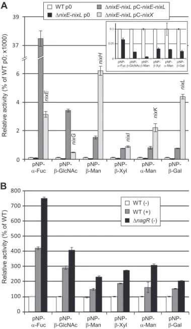
degrade synthetic substrates. Using synthetic pNP-glycoside substrates,␣-fucosidase, -N-acetylglucosaminidase, -man-nosidase, -xylosidase, ␣-mannosidase, and -galactosidase activities were detected in the Xcc WT strain and were reduced in the⌬nixE-nixL deleted strain (Fig. 2A). These activities were still detected in the deleted strain, suggesting that Xcc codes for other enzymes active on these pNP substrates that are not encoded by the N-glycan cluster. All the tested activities were considerably enhanced in the complemented⌬nixE-nixL
pC-nixE-nixLstrain, confirming that these enzymatic activities are encoded by the N-glycan cluster.
Using plasmids expressing each Nix enzyme in the
⌬nixE-nixL deleted strain, we confirmed the predicted activities of NixE (␣-fucosidase, GH29), NixG (-N-acetylglucosaminidase, GH20), NixH (-mannosidase, GH2), NixK (␣-mannosidase, GH92), and NixL (-galactosidase, GH35) (Fig. 2A). Introduc-tion of the plasmid pC-nixI complemented the enzymatic activ-ity of the⌬nixE-nixL strain on pNP--Xyl, suggesting that NixI (annotated as a-glucosidase, GH3) displays a -xylosidase activity. The plasmid expressing nixF (predicted to code for an endo--N-acetylglucosaminidase, GH18) did not complement the activity of the⌬nixE-nixL strain on pNP--GlcNAc, con-sistent with the endo-activity predicted for this enzyme (data not shown). Similarly and as reported for enzymes belonging to the GH125 family (19), the plasmid expressing nixJ (predicted to code for an␣-1,6-mannosidase, GH125) was not active on pNP-␣-Man (data not shown). Activity on pNP-Fuc was 11-fold lower when NixE was expressed from pC-nixE as compared with expression from pC-nixE-nixL (Fig. 2A), although the 5⬘ ends of fragments cloned in both plasmids were identical. Because a similar profile was observed for NixG, this could either be due to a difference in stability of transcripts or to the fact that enzymes of the cluster form a complex that stabilizes these proteins. Conversely, activities observed for NixH, NixK, and NixL were higher when their translation start is located at the 5⬘ end of the transcript (expression from pC-nixX) as com-pared with its location in a polycistronic transcript (expression from pC-nixE-nixL).
Enzymatic activities encoded by the N-glycan cluster were all induced both when cells were cultured in presence of GlcNAc and in the⌬nagR strain as compared with medium without GlcNAc and WT strain, respectively (Fig. 2B). These results confirm that the entire N-glycan cluster is under the control of GlcNAc via NagR, even if we did not observe transcriptional regulation by NagR for nixI to nixL (22).
It should be noted that although NixG was active on pNP- -GalNAc, the level of activity was at least 15-fold lower than when observed using pNP--GlcNAc (3 ⫾ 0 compared with 57⫾ 3 Miller units, respectively, in the WT strain, and 33 ⫾ 2 compared with 550⫾ 25 in the ⌬nixE-nixL pC-nixG strain), suggesting that its preferred substrate is-GlcNAc. This is con-sistent with a common feature of enzymes belonging to the EC 3.2.1.52 class (-N-acetyl-hexosaminidase), which are active on both pNP--GlcNAc and pNP--GalNAc with substrate pref-erence for pNP-GlcNAc (36).
Determination of Enzymatic Parameters of NixX-8His on pNP Synthetic Substrates—Kinetic parameters of NixX-8His recombinant proteins were determined on pNP synthetic sub-strates. The presence of the His-tag did not affect enzymatic activities of the corresponding proteins because activities on pNP synthetic substrates with total extracts of bacterial strains expressing either tagged and nontagged proteins were not sig-nificantly different (data not shown). NixX-8His proteins were purified by affinity chromatography using paramagnetic pre-charged nickel particles. For each of them, colloïdal blue staining of elution fractions after SDS-PAGE separation showed a unique band at the expected molecular mass (data not shown). Protein content of purified preparations was estimated between 6 and 36g䡠ml⫺1. pNP-α-Fuc pNP-β-GlcNAc pNP-β-Man pNP-β-Xyl pNP-α-Man pNP-β-Gal
pNP-α-Fuc β-GlcNAcpNP- β-ManpNP- pNP-β-Xyl α-ManpNP- β-Gal
pNP-Relative activity (% of WT p0; x1000) Relative activity (% of WT) nixE nixG nixH nixI nixK nixL 0 2 4 6 39 37 WT p0 ΔnixE-nixL p0 ΔnixE-nixL pC-nixE-nixL ΔnixE-nixL pC-nixX 0 0.05 0.1
pNP-α-Fucβ-GlcNAcpNP- β-ManpNP- pNP-β-Xyl α-ManpNP- pNP-β-Gal
0 100 200 300 400 500 600 700 800 WT (-) WT (+) ΔnagR (-)
FIGURE 2. Xcc nix genes encode GHs active on synthetic pNP substrates. Enzymatic activities were analyzed on total cell lysates of different Xcc strains, and activities were calculated as Miller units. A, strains were cultivated for 6 h in MOKA-rich medium, and activities were represented as percentages of the wild-type strain (WT p0). For complemented strains with plasmids expressing one nix gene (⌬nixE-nixL pC-nixX strains), the gene expressed is indicated above the histogram bar. The inset corresponds to an enlargement of the histogram for WT p0 and⌬nixE-nixL p0 strains. B, strains were cultivated for 6 h in MME minimal medium supplemented (⫹) or not (⫺) with GlcNAc at 10 mM. Activities were represented as percentages of the WT strain. The error
bars indicate the standard deviations obtained from three independent
experiments.
at INRA Institut National de la Recherche Agronomique on May 6, 2019
http://www.jbc.org/
After the optimal buffer, pH and temperature were deter-mined (Table 2), kinetic parameters were measured in the optimal conditions for each NixX-8His protein. Note that enzy-matic parameters could not be determined for NixF- and NixJ-8His enzymes because no synthetic substrates were available for them. All the tested enzymes obey Michaelis-Menten kinet-ics. Vmaxand Kmwere determined from Lineweaver and Burk linearization by plotting 1/Vmaxversus1/[substrate] at various
concentrations of pNP-carbohydrates (Table 2). Kmvalues are all in the mMorder of magnitude (0.24 –7.4 mM), whereas the
kcat values are more disparate (6.1–1552 s⫺1). Catalytic
effi-ciency (kcat/Km) ranged from 8.7 to 209.7 s⫺1䡠mM⫺1, with NixH and NixE as the most and the least efficient enzymes toward pNP-carbohydrates, respectively (Table 2).
NixF Is an Endo--N-acetylglucosaminidase and NixG Is an Exo--N-acetylglucosaminidase—NixF is annotated as an
endo--N-acetylglucosaminidase. We therefore analyzed its ability to hydrolyze glycosidic bonds between two GlcNAc res-idues. For that purpose, chitooligosaccharides from polymeri-zation degrees 3– 6 (chitotriose to chitohexaose, respectively) were treated with total extracts of E. coli overexpressing NixF from plasmid pMAL and subsequently analyzed by TLC (Fig. 3). Chitohexaose and chitopentaose treated with total extracts of E. coli overexpressing MBP-NixF were degraded into chito-tetraose⫹ chitobiose and chitotriose ⫹ chitobiose, respec-tively. These results confirm that NixF is an endo- -N-acetyl-glucosaminidase. Almost no degradation of chitotetraose or chitotriose by MBP-NixF was observed, suggesting that the minimal size of the substrate for this enzyme is a pentamer. Extracts of E. coli containing an empty pMAL were used as negative controls, and no degradation of chitooligosaccharides was observed. Similar reactions carried out using MBP-NixG led to the total degradation of all tested chitooligosaccharides into GlcNAc (Fig. 3), demonstrating that NixG is an exo- -N-acetylglucosaminidase. This result corroborates the activity on pNP-GlcNAc demonstrated for NixG-8His (see above). This is also in agreement with its involvement in chitooligosaccharides utilization in Xcc (22) and with results obtained for its ortholog
in the Xanthomonadaceae Stenotrophomonas maltophilia (Hex, 45.8% identity with NixG at the amino acid level) (37).
Recombinant NixX-8His Enzymes Are Active on an N-Glyco-peptide Substrate—Following the enzymatic characterization of NixX-8His enzymes using synthetic substrates, we wanted to demonstrate their involvement into N-glycopeptide degrada-tion. First, we performed assays in which the purified N-glyco-peptide was incubated with a mixture of NixE-8His to NixK-8His enzymes purified individually. NixL, predicted as a -galactosidase, was not included because the N-glycopeptide substrate does not contain a terminal galactose residue (Fig. 1A). Kinetic analyses of this reaction by MALDI-TOF MS allowed the detection of different degradation products of the
N-glycopeptide (Fig. 4). It should be noted that the tryptophan moiety removal observed with crude Xcc extracts (see above, Fig.
CHI-1 CHI-2 CHI-3 CHI-4 CHI-5 CHI-6
Standards pMAL pMAL-nixF pMAL-nixG pMAL pMAL-nixF pMAL-nixG pMAL pMAL-nixF pMAL-nixG pMAL pMAL-nixF pMAL-nixG
CHI-6 CHI-5 CHI-4 CHI-3
FIGURE 3. MBP-NixF protein of Xcc is an endo-
-N-acetylglucosamini-dase, and MBP-NixG is an exo--N-acetylglucosaminidase.
Chitooligo-saccharides were analyzed by TLC after incubation during 24 h in presence of crude extracts of E. coli containing empty plasmid pMAL (pMAL) or pMAL expressing MBP-NixF (pMAL-nixF) or MBP-NixG (pMAL-nixG). CHI-1, N-acetyl-glucosamine; CHI-2, chitobiose; CHI-3, chitotriose; CHI-4, chitotetraose; CHI-5, chitopentaose; CHI-6, chitohexaose.
0 50 100 In te n s ity ( % ) 2844.25 2712.21 3006.31 0 50 100 In te n s ity ( % ) 2844.25 2712.21 2550.15 2404.09 0 50 100 2200 2400 2600 2800 3000 3200 Int e ns it y (%) 2550.15 2242.03 Int ens it y (%) 3006.31 0 50 100 2404.09
A
B
C
D
2252.2 2312.4 2372.6 2432.8 2493.0 2242.03 2404.09 0 2 4 2192.0FIGURE 4. NixX-8His enzymes purified from Xcc are involved in
N-glyco-peptide degradation. N-GlycoN-glyco-peptides were analyzed by MALDI-TOF MS at
different time points after addition of a mixture of purified His-tagged NixE-, NixF-, NixG-, NixH-, NixI-, NixJ-, and NixK-8His enzymes. A, nontreated N-gly-copeptide. B, 30 min after addition of NixX-8His enzymes. C, 4 h after addition of NixX-8His enzymes. D, 48 h after addition of NixX-8His enzymes. The inset corresponds to an enlargement of the mass spectra showing peaks corre-sponding to N2and N2M N-glycopeptides. The blue squares represent GlcNAc,
the green circles represent mannose (with different color tones according to different glycosidic linkages), the red triangles represent fucose, and the
orange stars represent xylose. The peptide moiety is represented by a gray line. For the 2844.25 and 2712.21 m/z N-glycopeptides, the two half-circles
represent the remaining mannose, which can be either an␣-1,3-mannose (upper half-circle, light green) or an ␣-1,6-mannose (lower half-circle, dark
green).
at INRA Institut National de la Recherche Agronomique on May 6, 2019
http://www.jbc.org/
1) was not observed here, indicating that NixX-8His enzymes were only involved in glycan degradation. After 30 min of reaction, the peak corresponding to the substrate (3006.31 m/z) almost disap-peared, whereas two peaks at 2844.25 and 2712.21 m/z were detected (Fig. 4B). The first product corresponded to the loss of a hexose (⫺162.06 Da), and the second one corresponded to that of a pentose (⫺132.04 Da), namely mannose and xylose residues, respectively, according to the N-glycan structure (Fig. 1A). After 4 h, two additional products were detected, indicat-ing that the substrate has been completely consumed. The peak detected at 2550.15 m/z corresponded to the loss of an addi-tional mannose (⫺162.06 Da) and the peak at 2404.09 m/z to the additional loss of the fucose residue (⫺146.06 Da) (Fig. 4C). The major reaction product observed at the end of the reaction (peak at 2550.15 m/z described above) corresponds to the N2MF-glycopeptide. Shorter compounds (2242.03 and 2404.09 m/z) were only detected as traces. The smaller form corre-sponds to a glycopeptide with two GlcNAc residues (Fig. 4D). Altogether, our results demonstrate five enzymatic activities, i.e. two ␣-mannosidases (␣-1,3 and ␣-1,6), a -1,4-mannosidase, a -1,2-xylosidase, and an ␣-1,3-fucosidase involved in the degrada-tion of N-glycan (Fig. 1A). Considering results previously obtained with pNP substrates, these activities could be assigned to NixK (␣-mannosidase), NixH (-mannosidase), NixI (-xylosidase), and NixE (␣-fucosidase). In addition, the kinetic of formation of products suggested a sequential degradation of the glycopeptide.
The Degradation of N-Glycopeptide Is Sequential—To con-firm the role of each enzyme and to go further into the mecha-nism of deglycosylation, we carried out assays by introducing each of the seven NixX-8His enzymes of the cluster (i.e. all except NixL-8His; see above) individually. NixK-8His was the only enzyme active on the N-glycopeptide substrate used in this study, indicating that NixK-8His initiates the deglycosylation, generating a product at 2844.25 m/z (Fig. 5A). Considering a possible sequential mechanism, we subsequently introduced in the reaction assay the NixI-8His enzyme, predicted to act on the NixK-8His product. A product at 2712.21 m/z was detected, confirming that NixI-8His hydrolyzed the xylose from the
N-glycopeptide only after mannose removal (Fig. 5A). In the same way, sequential addition of NixJ-8His confirmed that it was able to remove a mannose residue from NixI-8His product. These data demonstrate that NixK-8His, NixI-8His, and NixJ-8His are responsible for the sequential degradation of the
N-glycopeptide, leading to the product at 2550.15 m/z (peptide with N2MF glycan), which was shown to accumulate in our
assay conditions. Interestingly, it should be noted that NixK-8His and NixJ-NixK-8His are both␣-mannosidases but with different specificities because they are devoted to the removal of only one of the two␣-mannose residues attached either by ␣-1,3 or ␣-1,6 linkages. Because NixJ was classified in the GH125 family containing only ␣-1,6-mannosidases (Carbohydrate-Active EnZymes database), we deduced that NixK-8His is an ␣-1,3-mannosidase. Assays carried out by adding subsequently other enzymes of the cluster (i.e. NixE-8His, NixF-8His, NixG-8His, and NixH-8His) were too diluted to detect significant MALDI-TOF MS signals (data not shown).
To overcome this limitation, we developed new assays by co-purifying NixK-8His, NixI-8His, and NixJ-8His from
inde-pendent bacterial cultures. Enzyme concentrations were then high enough to observe a full conversion of the N-glycopeptide substrate to the 2550.15 m/z product after 4 h (Fig. 5B). NixE-8His, assumed to remove subsequently the fucose residue, was then introduced, and after 24 h of reaction, a product at 2404.09
m/z (peptide with N2M glycan) was detected as trace. This sug-gests that NixE-8His, although active on pNP-Fuc, is poorly efficient on the N-glycopeptide substrate at 2550.15 m/z, sug-gesting that another enzyme is needed prior to its activity. However, the addition of NixF-8His, NixG-8His, and NixH-8His, alone or in mixture, did not result in efficient degradation of the 2550.15 m/z substrate. Two new products were detected at 2388.09 and 2242.03 m/z. These correspond to the loss of a -mannose residue (⫺162.06 Da) from the N2MF glycopeptide
(at 2550.15 m/z) and of a fucose residue (⫺146.06 Da) from the N2F glycopeptide (at 2388.09 m/z), respectively (Fig. 5B). These
data confirm that NixH-8His, predicted as a-mannosidase, is a-1,4-mannosidase and that NixE-8His is able to remove an ␣-1,3-fucose from NixH-8His product. No activities were observed for NixG-8His and NixF-8His enzymes.
Subcellular Localization of Nix Enzymes—All Nix enzymes possess a signal peptide, suggesting that these proteins are translocated across the inner membrane to the periplasm (22).
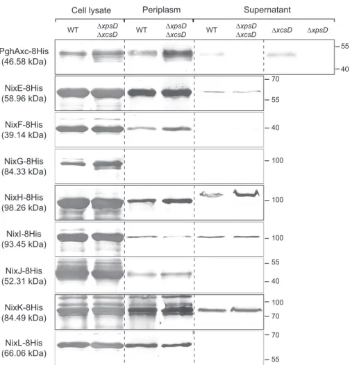
Xccpossesses two type II secretion systems (Xps and Xcs), the first one playing a major role in the secretion of plant cell wall degradative enzymes (38). Therefore, subcellular localization of Nix enzymes was analyzed in WT strain and strains with non-functional Xps and/or Xcs machinery (⌬xpsD, ⌬xcsD and ⌬xpsD ⌬xcsD mutants). We performed Western analyses using different fractions (total cell lysate, periplasmic-enriched frac-tion, and concentrated supernatant) prepared from Xcc strains overexpressing 8His-tagged Nix proteins; for each strain, frac-tions loaded on the gel were normalized to correspond to the same number of bacterial cells. The polygalacturonase PghAxc (XCC3459) was used as a control. Indeed, in Xcc strain 8004, PghAxc is secreted via the type II secretion system in an Xps-dependent and Xcs-inXps-dependent manner (39). In agreement with these observations in strain 8004, we detected PghAxc-8His from strain ATCC33913 in the supernatant fractions of both WT and⌬xcsD strains but not in ⌬xpsD or ⌬xpsD ⌬xcsD mutants (Fig. 6). All NixX-8His enzymes were detected in total cell lysates. Except NixG-8His, all enzymes were also detected in the periplasmic enriched fraction of Xcc cells. NixE-, NixH-, NixI-, and NixK-8His were also detected in the culture super-natant of both WT and⌬xpsD ⌬xcsD strains (Fig. 6), suggesting that these proteins are secreted in a type II secretion system-independent manner, as previously observed for amylase and cellulase activities in X. campestris pv. vesicatoria (40). NixF-8His was weakly detected in the culture supernatant of⌬xpsD ⌬xcsD strain but not of the WT strain, which is probably due to the detection limits of the experiment. Finally, the absence of detection of NixG-, NixJ-, and NixL-8His in the supernatant and NixG-8His in periplasmic-enriched fractions of either WT or⌬xpsD ⌬xcsD strains could be due to the detection limits of the experiment. Alternatively, NixG-8His could be cytoplasmic or associated with one membrane whereas NixJ- and NixL-8His would not be secreted in the supernatant and either remain in the periplasm or may be associated to the outer membrane.
at INRA Institut National de la Recherche Agronomique on May 6, 2019
http://www.jbc.org/
Surprisingly, NixH-8His was detected in the supernatant at an apparent molecular mass higher than that of the protein detected in the cell lysate and in the periplasmic enriched frac-tion (Fig. 6), suggesting that NixH-8His is modified during its translocation through the outer membrane.
DISCUSSION
Xcc, the causal agent of black rot disease on Brassica plants, produces an extensive repertoire of GHs, lyases, and esterases
(41) that target linkages present in plant cell wall polysaccha-rides such as cellulose, mannan, xylan, galacturonan, and pec-tin. In this paper, we characterized Nix GHs belonging to the
Xcc GlcNAc utilization system on pNP synthetic substrates, and we demonstrated their ability to degrade a plant N-glycan. This is the first evidence of in vitro N-glycan degradation by a plant pathogen suggesting that N-glycans may by metabolized by phytopathogenic bacteria during infection. By using Xcc total extracts or His-tagged proteins purified from Xcc, we t0 t24h t4h t28h t52h t24h t48h t72h 2200 2400 2600 m/z 2800 3000 3200 0 50 100 3006.31 0 50 100 2844.25 0 50 100 2712.21 0 50 100 2550.15 0 50 100 + NixE/F/G/H/I or J + NixK In te n s it y ( % ) In te ns it y (%) In te n s it y ( % ) In te ns it y (%) In te n s it y ( % ) + NixI + NixJ t0 m/z 0 50 100 3006.31 0 50 100 2550.15 0 50 100 2404.09 0 0 50 10 100 2200 2204.0 2262.4 2300.8 2339.2 2377.6 2416.0 2400 2600 2800 3000 3200 2388.09 2242.03 In te n s it y ( % ) Int ens it y (%) Int ens it y (%) In te ns it y (%) + NixK/I and J + NixE + NixF/G and H
C
3006.31 2550.15 2550.15 α-1,3 α-1,6 β-1,4 β-1,4 β-1,2 α-1,3 M M M N N F X NixI (2) NixE (4/5) NixH (4/5) NixK (1) NixJ (3) 2242.03 2388.09FIGURE 5. Sequential degradation of the N-glycan from a N-glycopeptide by purified NixX-8His enzymes from Xcc. N-Glycopeptides were analyzed by MALDI-TOF MS at different time points after sequential addition of purified NixX-8His enzymes. A, identification of the three first cleavage steps of the N-glycan. When added individually, no cleavage was observed when either NixE-, NixF-, NixG-, NixH-, NixI-, or NixJ-8His was added (top panel). Deglycosylation of the N2M3FX N-glycopeptide was initiated by NixK-8His (␣-1,3-mannosidase), then NixI-8His eliminated the xylose residue, and finally NixJ-8His removed the
␣-1,6-mannose residue. B, identification of final cleavage steps. NixK-, NixI-, and NixJ-8His were added simultaneously, and after 4 h, the product (N2MF)
obtained was used as substrate for other NixX-8His enzymes. NixE-8His (␣-fucosidase) and NixH-8His (-mannosidase) were able to cleave fucose and mannose residues, respectively. The inset corresponds to an enlargement of the mass spectra showing peaks corresponding to N2and N2F
N-glyco-peptides. C, schematic representation of the N-glycopeptide used in this study. Cleavages observed with purified Xcc NixX-8His enzymes are indicated. The numbers in parentheses indicate the order of action of enzymes. The blue squares represent GlcNAc (N), the green circles represent mannose (M, with different color tones according to different glycosidic linkages), the red triangles represent fucose (F), and the orange star represents xylose (X). The peptide moiety is represented by a gray line.
at INRA Institut National de la Recherche Agronomique on May 6, 2019
http://www.jbc.org/
demonstrated that N-glycan processing is sequential. On the N2M3FX N-glycopeptide substrate, NixK initiates the
deglyco-sylation by removing the ␣-1,3-mannose, NixI acts on the -1,2-xylose in a second step, and then NixJ releases the ␣-1,6-mannose. NixE and NixH are assumed to act at a later stage, by removing the ␣-1,3-fucose and the -1,4-mannose, respec-tively. This sequential mode of action corroborates literature data reporting that, in the context of fruit ripening, the removal of␣-1,3-mannose from N-glycan is a prerequisite for hydrolysis of the-1,2-xylose by a tomato xylosidase (42). In addition, in the human N-glycan degrading Firmicutes, a sequential model is proposed, with the removal of the␣-1,3-mannose by a GH38 as a first step necessary for the activity of a GH125 subsequently involved in the removal of the␣-1,6-mannose (19). Such com-mon features suggest that N-glycan processing is of widespread importance, in particular for pathogens in a variety of host contexts.
In Xcc, although the N-glycan cluster potentially encodes all the enzymes required for complete degradation of the N2M3FX
N-glycan, the N2MF product accumulates, suggesting that in
our conditions, there is a limiting step for the N2MF
glycopep-tide degradation process. The reasons for this limiting step may be multiple. Indeed, it could be due to inappropriate condition
assays or to the fact that the polypeptidic part of the glycopep-tide provides a steric hindrance for NixE, NixF, and/or NixH activities. It is also possible that NixE, NixF, and/or NixH are not in a form active on N-glycans or that NixF is not able to accommodate in its active site the N-glycopeptide used in this study. Furthermore, we cannot exclude that other secreted crit-ical enzyme(s), not encoded by the N-glycan cluster, are required for full N-glycan processing. Finally, this limiting step could be a strategy for the bacterium to partially deglycosylate plant N-glycoproteins. Indeed, in S. pneumoniae strain D39, although the genome codes for ␣- and -mannosidases (belonging to GH2, GH38, GH92, and GH125 families; Carbo-hydrate-Active EnZymes database), deglycosylation of human complex-type N-glycoconjugates is partial with only removal of terminal sialic acid, galactose and GlcNAc residues. This led to the exposure of mannose residues on host glycoproteins, which plays a role in adherence of unencapsulated pneumococci on human cells (43).
The results obtained here suggest that in Xcc, as observed in S. oralis (17) and S. pneumoniae (43), deglycosylation is sequential because of the action of exo-enzymes. However, in human pathogens such as E. faecalis (16), S. pyogenes (44),
B. fragilis(15), and C. canimorsus (10, 21), and in infant
gut-NixK-8His (84.49 kDa) NixL-8His (66.06 kDa) NixJ-8His (52.31 kDa) NixI-8His (93.45 kDa) NixH-8His (98.26 kDa) NixG-8His (84.33 kDa) NixF-8His (39.14 kDa) NixE-8His (58.96 kDa) PghAxc-8His (46.58 kDa) 100 100 100 70 100 55 70 55 40 40 55 70 55 40 WT ΔxpsDΔxcsD WT ΔxpsDΔxcsD WT ΔxpsDΔxcsD ΔxcsD ΔxpsD
Cell lysate Periplasm Supernatant
FIGURE 6. Subcellular localization of Xcc NixX-8His enzymes. Western blot analysis using anti-His antibody was carried out on total cell lysates, periplasmic enriched fractions, and cell-free supernatants of Xcc WT and type II secretion system mutants (⌬xpsD, ⌬xcsD and/or ⌬xpsD-⌬xcsD strains) overexpressing NixX-8His enzymes from replicative plasmids. PghAxc-8His was used as a control for protein secretion dependent on the Xps type II secretion system in Xcc. Molecular masses are indicated in parentheses beneath each protein name correspond to the predicted mass of the His-tagged protein without the signal peptide. Molecular mass markers (in kDa) are indicated on the right.
at INRA Institut National de la Recherche Agronomique on May 6, 2019
http://www.jbc.org/
associated Bifidobacteria (45), the ability to release N-glycans from host proteins has been mainly associated with endo- -N-acetylglucosaminidase activity. In these bacteria, it seems that deglycosylation is initiated by the cleavage of the N-glycan between the two GlcNAc residues common to all N-glycans. Because the two first enzymes acting on the N2M3FX substrate
(i.e. NixK and NixI) in Xcc were detected in the supernatant-enriched fraction of bacterial cultures, we can propose that
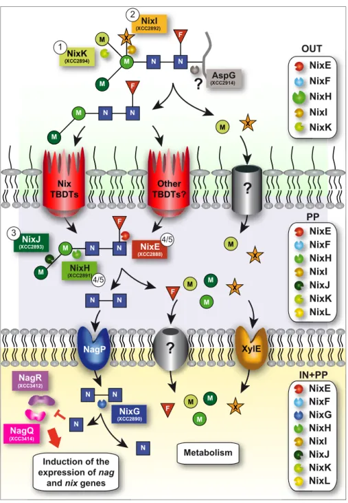
deg-radation of the N2M3FX N-glycan is initiated outside the cell
(Fig. 7). Possibly after cleavage of the peptide by the asparagi-nase AspG (XCC2914, proposed in the KEGG pathway data-base to cleave the N-glycan from the asparagine residue of
N-glycoproteins), uptake of partially degraded N-glycans through the outer membrane could be achieved by Nix TonB-dependent transporters. Indeed, we have recently observed that these active transporters belonging to the GlcNAc utilization
X M M M M N N F M M N N F OUT OUT NixE NixF NixI NixK NixH N N N N N N M M F M X X N N NixJ (XCC2893) 3 NixH (XCC2891) 4/5 NixE (XCC2888) 4/5 PP PP NixE NixL NixF NixI NixJ NixK NixH NixG (XCC2890) NagR (XCC3412) NagQ (XCC3414) gQ Metabolism N N M M M F X N N Induction of the expression of nag
and nix genes
IN+PP XylE NagP Other TBDTs? Nix TBDTs
?
?
IN+PP NixE NixL NixK N N N NixF NixG NixI NixJ NixH N N N?
M M M N N F X X NixI (XCC2892) 2 M M?
?
(XCC2914)AspG M NixK (XCC2894) 1FIGURE 7. Model of the N2M3FX N-glycopeptide degradation and utilization in Xcc ATCC33913 strain. NixK and NixI initiate deglycosylation of the
N-glycopeptide outside the cell. We propose that AspG (XCC2914) would cleave the peptide moiety, and the resulting N2M2F N-glycan would be transported
through Nix and/or other TonB-dependent transporters to the periplasm where NixJ, NixH, and NixE continue its degradation. Uptake of chitobiose (N2)
through the inner membrane occurs via NagP, and NixG would be responsible for its degradation inside the cell. Free GlcNAc would then directly or indirectly inhibit repressors NagR and NagQ, leading to the induction of the expression of all nag and nix genes. Uptake of xylose through the inner membrane is performed by XylE. Transporters responsible for the uptake of xylose and mannose through the outer membrane and fucose and mannose through the inner membrane are not known. Free monosaccharides (fucose, mannose, GlcNAc, and xylose) would then be utilized for the metabolism of the bacterium. Numbers in circles indicate the order of action of Nix enzymes as determined in this paper. The boxes on the right indicate NixX-8His enzymes detected in total cell lysates (IN⫹PP), in periplasmic-enriched fraction (PP) and in the supernatant (OUT) of Xcc. F, fucose; M, mannose; N, GlcNAc; X, xylose; Nix, N-acetylglucosamine induced in Xanthomonas; TBDT, TonB-dependent transporter.
at INRA Institut National de la Recherche Agronomique on May 6, 2019
http://www.jbc.org/