HAL Id: hal-01341580
https://hal-univ-rennes1.archives-ouvertes.fr/hal-01341580
Submitted on 10 Sep 2020HAL is a multi-disciplinary open access archive for the deposit and dissemination of sci-entific research documents, whether they are pub-lished or not. The documents may come from teaching and research institutions in France or abroad, or from public or private research centers.
L’archive ouverte pluridisciplinaire HAL, est destinée au dépôt et à la diffusion de documents scientifiques de niveau recherche, publiés ou non, émanant des établissements d’enseignement et de recherche français ou étrangers, des laboratoires publics ou privés.
Studies on plant cell toxicity of luminescent silica
nanoparticles (Cs2[Mo6Br14]@SiO2) and its constitutive
components
Francisco Cabello-Hurtado, Maria Dolorès Lozano-Baena, Chrystelle Neaime,
Agnès Burel, Sylvie Jeanne, Pascal Pellen-Mussi, Stéphane Cordier, Fabien
Grasset
To cite this version:
Francisco Cabello-Hurtado, Maria Dolorès Lozano-Baena, Chrystelle Neaime, Agnès Burel, Sylvie Jeanne, et al.. Studies on plant cell toxicity of luminescent silica nanoparticles (Cs2[Mo6Br14]@SiO2) and its constitutive components. Journal of Nanoparticle Research, Springer Verlag, 2016, 18 (3), pp.69. �10.1007/s11051-016-3381-6�. �hal-01341580�
1
Studies on plant cell toxicity of luminescent silica nanoparticles
1
(Cs
2[Mo
6Br
14]@SiO
2) and its constitutive components
2 3
Francisco Cabello-Hurtado a,*, María Dolores Lozano-Baena a, Chrystelle Neaime b, Agnès Burel
4
c, Sylvie Jeanne b, Pascal Pellen-Mussi b, Stéphane Cordier b, Fabien Grasset b, d
5 6
a UMR UR1-CNRS 6553 ECOBIO, Mechanisms at the Origin of Biodiversity Team, University of
7
Rennes 1, 263 av. du Général Leclerc, 35042 Rennes, France
8
b UMR UR1-CNRS 6226 Institut des Sciences Chimiques de Rennes, Solid State Chemistry and
9
Materials Group, University of Rennes 1, 263 av. du Général Leclerc, 35042 Rennes, France
10
c Electronic Microscopy Department, University of Rennes 1, 2 av. du Professeur Léon-Bernard,
11
Campus de Villejean, 35043 Rennes, France
12
d CNRS-Saint Gobain, UMI 3629, Laboratory for Innovative Key Materials and Structures-Link,
13
National Institute of Material Science (NIMS), GREEN/MANA Room 512, 1-1 Namiki, 305-0044
14
Tsukuba, Japan
15 16
* Corresponding author: e-mail: francisco.cabello@univ-rennes1.fr; phone: +33223235022; fax:
17 +33223235026 18 19 Acknowledgment 20 21
This work was supported by the French National Research Agency (Project CLUSTOP 2011
22
BS0801301). Authors thank Marie Thérèse Lavault for technical assistance (MRic, UR 1), Juan B.
23
Arellano for kindly providing Arabidopsis cells (IRNASA-CSIC), and Vincent Dorcet for
24
nanoparticle TEM microcraphs (Plateforme THEMIS, UR1).
25 26
Journal of Nanoparticle Research
Preprint--2 Abstract
27 28
As part of the risk evaluation before potential applications of nanomaterials, phytotoxicity of newly
29
designed multifunctional silica nanoparticles (CMB@SiO2, average diameter of 47 nm) and their
30
components, i.e. molybdenum octahedral cluster bromide units (CMB, 1 nm) and SiO2
31
nanoparticles (nSiO2, 29 nm), has been studied using photosynthetic Arabidopsis thaliana cell
32
suspension cultures. CMB clusters presented toxic effects on plant cells, inhibiting cell growth and
33
negatively affecting cell viability and photosynthetic efficiency. Nevertheless, we showed that
34
neither nSiO2 nor CMB@SiO2 have any significant effect on cell growth and viability or
35
photosynthetic efficiency. At least part of the harmful impact of CMB clusters could be ascribed to
36
their capacity to generate an oxidative stress since lipid peroxidation greatly increased after CMB
37
exposure, which was not the case for nSiO2 or CMB@SiO2 treatments. Exposure of cells to CMB
38
clusters also lead to the induction of several enzymatic antioxidant activities (i.e. superoxide
39
dismutase, guaiacol peroxidase, glutathione peroxidase, glutathione reductase, and glutathione
S-40
transferase activities) compared to control and the other treatments. Finally, using electron
41
microscopy, we showed that Arabidopsis cells internalize CMB clusters and both silica
42
nanoparticles, the latter through, most likely, endocytosis-like pathway as nanoparticles were
43
mainly found incorporated into vesicles.
44 45 46 Keywords 47 48
Nanotoxicity; Silica nanoparticles; Molybdenum clusters; Arabidopsis cells; Oxidative stress.
49 50
3 Introduction
51 52
Manufactured nanoparticles (NPs) (one dimension < 100 nm) are being increasingly produced for a
53
wide range of applications and are present in hundreds of nanotechnology products (Buzea et al.
54
2007). However, they are also bringing new toxic effects on human and environmental health
55
(Buzea et al. 2007; Colvin 2003). Among nanomaterials, the use of functional synthetic amorphous
56
silicon dioxide or silica nanoparticles (nSiO2) in information technology, biotechnology and
57
medicine is becoming increasingly accepted for a variety of therapeutic, diagnostic and imaging
58
applications (Selvan et al. 2010). The challenge for nanotechnologies at this point is to elaborate
59
non-toxic and aging resistant phosphorescent silica nanoparticles emitting in the near infrared
60
region (NIR). For this purpose, new functional silica nanoparticles incorporating luminescent
61
molybdenum hexanuclear cluster bromide units (Cs2Mo6Br14, noted CMB, as the cluster precursor)
62
inside monodispersed and size-controlled silica nanoparticles (noted CMB@SiO2) have been
63
recently developed in our group (Aubert et al. 2013). Besides, Mo6-based clusters are already
64
involved in several patents for applications in biotechnology as contrast agents (Long et al. 1998),
65
oxygen sensors (Baker et al. 2010) and in display technologies (Cordier et al. 2015).
66
Silica nanoparticles are used as matrices because of their versatility and their relative
67
biocompatibility (Fruijtier-Pölloth 2012). However, this point of nSiO2 safety is controversial, and
68
different studies reported toxic effects in some cells or organisms like humans (Brown et al. 2015;
69
Guarnieri et al. 2014; Napierska et al. 2010), other animals (Debnath et al. 2011; Lee et al. 2009;
70
Parveen et al. 2014), algae (van Hoecke et al. 2011) or bacteria (Adams et al. 2006). Concerning
71
higher plants, most of the studies reported null or positive effects (Le et al. 2014; Lee et al. 2010;
72
Lin et al. 2004; Nair et al. 2011; Siddiqui and Al-Whaibi 2014; Slomberg and Schoenfisch 2012),
73
nSiO2 toxicity being only observed at very high concentrations (Le et al. 2014; Lee et al. 2010). The
74
toxic mechanisms of nSiO2 exposure remain far from clear, but in some cases nSiO2 toxic effects
75
were related to interactions with cellular surfaces (membrane or cuticle), oxidative stress and/or
4
genotoxicity (Adams et al. 2006; Brown et al. 2015; Debnath et al. 2011; Fruijtier-Pölloth 2012;
77
Napierska et al. 2010; Parveen et al. 2014).
78
SiO2 nanoparticles are a common nanomaterial which is used (either or not in admixture with
79
other elements) for a variety of applications in the medical (biomedicine, biosensor, disease
80
labeling) and technological (food processing, ceramics synthesis, industrial and household
81
applications) fields, but also in the environmental (wastewater treatment, water purification,
82
environmental remediation) and agriculture fields. In agriculture, silica nanoparticles are used in
83
different formulations, mainly as carriers in chemical delivery, or in uptake and translocation of
84
nutrient elements, and as active ingredients against insect pests (Gogos et al. 2012), thereby
85
fostering their dispersion in the environment. In this context, luminescent properties of CMB@SiO2
86
nanoparticles are not without interest for biotechnological uses that could also be applied to plants,
87
or to be combined with silica nanoparticles intended for agronomical uses. From these current and
88
potential uses of silica nanoparticles, it is obvious that their potential to harm the environmental is a
89
relevant issue. Plants, as important environmental components and sinks in terrestrial and aquatic
90
ecosystems, are essential living organisms for testing ecological effects of nanoparticles. Hence, it
91
is of great importance to study the impact of new functional silica nanoparticles on plant cells, and
92
to anticipate new potential risks derived from their accumulation into plants and their subsequent
93
fate within food chains. In earlier studies on the impact of CMB@SiO2 nanoparticles and of CMB
94
clusters in plant growth (Aubert et al. 2012; 2013), we showed that silica nanoparticles containing
95
clusters have no effect on plant growth, whereas CMB clusters penetrated into roots and negatively
96
impacted growth. In these studies, roots were always much more affected than aerial parts, certainly
97
due to the root direct contact with clusters and the very low translocation of clusters into aerial part.
98
The latter makes it difficult to analyze the direct impact of these nanomaterials on photosynthetic
99
cells in these root-treated systems. Indeed, the absence of adverse effects reported in most works
100
evaluating silica nanoparticle phytotoxicity could be partially linked to low or no occurrence of
101
silica nanoparticles in photosynthetic cells, rather than the lack of inherent hazards. At this respect
5
in particular, the use of plant cell cultures provides a way for in vitro exposing photosynthetic cells
103
directly to the action of nanoparticles. In fact this model system can mimic, as regards with plant
104
cell interaction with nanoparticles, the situation that could be found in photosynthetic cells of aerial
105
parts of plants exposed to CMB@SiO2, nSiO2 or CMB in a chronic way, where a higher
106
accumulation of nanoparticles in leaves can be achieved after a long-time exposure.
107
In order to go further in CMB@SiO2, nSiO2 and CMB toxicological research, and with the aim
108
of exploring the impact of weak doses on photosynthetic cells, we have chosen light-grown
109
Arabidopsis thaliana cell suspension cultures (ACSC) as a valuable cellular system in which to 110
investigate oxidative damage and cell response. ACSC join uniformity, homogeneity, repeatability,
111
decoupling of cellular processes from development and slow systemic effects between cells
112
(Menges et al. 2003), to the convenience of application of nanomaterial treatments. Here, we
113
present studies on biochemical and oxidative stress factors on A. thaliana cells under exposure to
114
functional CMB@SiO2 nanoparticles and their components, nSiO2 nanoparticles and CMB clusters.
115
The in vitro cytotoxicity of these nanomaterials was examined by investigating their influence on
116
cell growth and viability, photosynthesis, lipid peroxidation, and antioxidant enzyme activities (i.e.
117
superoxide dismutase (SOD), guaiacol peroxidase (POD), glutathione peroxidase (GPX),
118
glutathione reductase (GR), and glutathione S-transferase (GST) activities). Finally, the fate of
119
nanoparticles in the medium and their penetration into plant cells was detected by transmission
120
electron microscopy (TEM).
121
122 123
Materials and Methods 124
125
Chemicals, cluster units, and silica nanoparticles
126 127
6
Polyoxyethylene (4) lauryl ether (Brij30) and tetraethoxysilane (TEOS, 99.00%) were purchased
128
from Sigma-Aldrich. Ammonia (28 wt % in water) and n-heptane (99.00%) were purchased from
129
VWR. Ethanol (99.80%) was purchased from Fluka. Cs2Mo6Br14 was used as the precursor of
130
[Mo6Br14]2− cluster units.
131
Hexamolybdenum cluster units are nanometric building blocks (1 nm) constituted of a Mo6
132
octahedral cluster bonded to 8 inner Bri (i = inner) ligands capping the faces of the octahedron and 6
133
apical Bra (a = apical) ligands in terminal positions. The negative charge of the [Mo
6Br14]2- cluster
134
unit is counter balanced by two Cs+ cations. In solid state, the cluster units co-crystallize with the
135
cations to form a cluster compound denoted Cs2Mo6Br14. The Cs2Mo6Br14 cluster compound can be
136
dispersed as nanosized entities in ethanolic solution (Grasset et al. 2008).
137
All the silica nanoparticles have been prepared using a water-in-oil (W/O) microemulsion
138
process developed by our group since the earlier 2000 (Aubert et al. 2010; Grasset et al. 2002). In
139
this work, the complex water phase was prepared by dissolving the Cs2[Mo6Br14] cluster compound
140
in a mixture of ethanol and distilled water (1:1 volume ratio). The concentration of the cluster sol
141
was 0.02 M. For pure nSiO2, the complex water phase was free of cluster. Finally, the nanoparticles
142
were collected and washed by several centrifugation cycles to remove surfactant molecules before
143
to be dispersed in purified water at concentration around 15 g L–1. The average hydrodynamic size
144
of the silica nanoparticles in water solution was estimated by dynamic light scattering
145
(Supplementary Fig. 1) using a Malvern Zetasizer Nano ZS apparatus. All the samples were studied
146
by TEM (Supplementary Fig. 2) using a microscope JEOL 2100 LaB6 at 200 kV or JEOL
JEM-147
1400 microscope operating at 120 kV. Samples for TEM analysis were prepared by placing a drop
148
of the diluted solution in mesh copper grids, allowing the solvent in the grid to evaporate at room
149
temperature.
150 151
ACSC growth conditions and nanomaterial treatments
152 153
7
The Arabidopsis thaliana (ecotype Col-0) cell suspension cultures were kindly provided by the
154
Institute of Natural Resources and Agronomy from Salamanca (IRNASA-CSIC, Spain). ACSC
155
were maintained at 24°C under sterile conditions in 200 mL of liquid growth medium (Axelos et al.
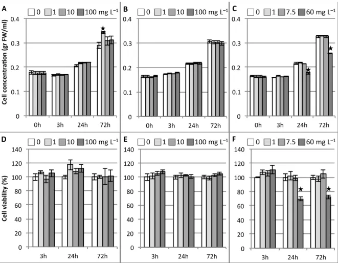
156
1992; Jouanneau et al. 1967) by agitation at 120 rpm and under continuous illumination (50 µE m–2
157
s–1) in an incubator shaker (Innova 42R, NBS). For nanoparticle toxicity tests, we applied
158
luminescent silica nanoparticles (CMB@SiO2) and their constituents (i.e. CMB clusters and nSiO2)
159
into 9 days-old ACSC (cell density of 150-200 mg mL–1). For these studies, three different
160
concentrations of CMB@SiO2 and nSiO2 (1, 10 and 100 mg L–1), and of CMB clusters (1, 7.5 and
161
60 mg L–1) were tested. Treated ACSC were incubated for up to three days under normal growth
162
conditions, and sample aliquots collected at 3, 24 and 72 h. Collected samples were centrifuged at
163
4000 rpm for 5 min, the supernatant was removed, and the cell pellet was weighed, frozen in liquid
164
nitrogen and finally stored at -80°C until further analysis.
165
For stock solutions to be used for nanoparticle toxicity tests, CMB@SiO2 and nSiO2 were
166
suspended in water at 5 g L–1, and CMB in 50% ethanol at 60 g L–1. CMB@SiO2 used here are
167
composed of 7.5% clusters and 92.5% SiO2 (Aubert et al. 2013). Thus, the intermediate CMB
168
concentration (7.5 mg L–1) used for treatments corresponds to the cluster content associated to the
169
intermediate concentration of CMB@SiO2 nanoparticles (100 mg L–1). From this intermediate
170
CMB concentration, we have set the lowest CMB concentration at 1 mg/L, which correspond
171
(rounded to the nearest unit) to the cluster content present in 10 mg/L of CMB@SiO2, and the
172
highest CMB concentration at 60 mg/L (in order not to exceed 0.05% of ethanol in cell culture
173
medium). As control, we used ACSC without added nanomaterial but containing the equivalent
174
volume of the corresponding solvent or medium as used for the nanomaterial tested.
175 176
Cell viability assay
177 178
8
Cell mitochondria and metabolic activities were measured by the MTT
(3-(4,5-dimethylthiazol-2-179
yl)-2,5-diphenyl tetrazolium bromide) test following manufacturer’s indications (Cell growth
180
determination kit CGD-1, Sigma-Aldrich). Cell density was adjusted to 25 mg mL–1 at the
181
beginning of the treatment, and cell dilution for MTT assay was the same for all the samples.
182
Relative cell viability was expressed as the percentage of control untreated cells and calculated by
183
[Absorbance 570 nm – Absorbance 690 nm]test / [A570 – A690]control ×100.
184 185
Pigment analysis
186 187
The chlorophyll and carotenoid contents were determined spectrophotometrically following
188
Lichtenthaler and Wellburn (1983) equations. Collected cells were lyophilised (Christ ALPHA
1-189
2LDplus) and pigments extracted from 20 mg dry weight (DW) by overnight pure acetone
190
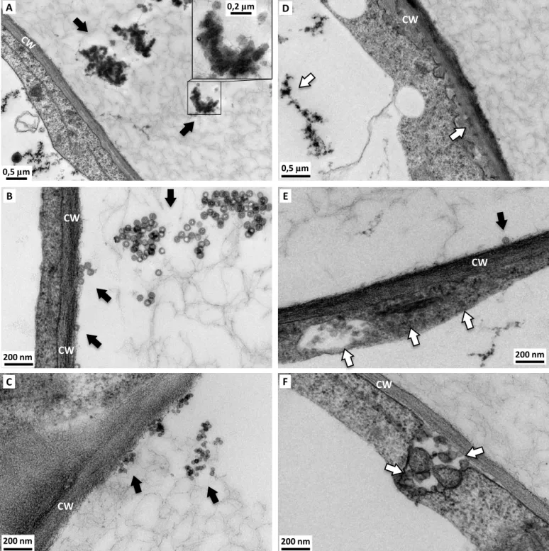
extraction at 4°C. The absorbance was quantified at 470, 645 and 663 nm using a micro plate
191
spectrophotometer (SAFAS, Xenius).
192 193
Chlorophyll fluorescence measurements
194 195
Modulated chlorophyll fluorescence measurements were made in ACSC (previously dark adapted
196
for 30 min) with a PAM-210 chlorophyll fluorometer (Heinz Walz). Maximum quantum yield of
197
photosynthesis was estimated by the Fv/Fm ratio from dark-adapted ACSC, where Fv is calculated
198
subtracting the minimal fluorescence (Fo) to the maximal fluorescence (Fm).
199 200
Lipid peroxidation
201 202
9
The level of lipid peroxidation was determined by measuring the amount of TBARS (thiobarbituric
203
acid reactant species) produced by the thiobarbituric acid (TBA) reaction, according to the corrected
204
TBA method as described by Hodges et al. (1999) adapted to 96-well plates.
205 206
Antioxidant enzyme extraction and activity assays
207 208
Enzyme extracts correspond to supernatants obtained after homogenizing A. thaliana cells in
209
sodium phosphate buffer (50 mM, pH 7.5) with Na-EDTA (1 mM), polyvinyl-pyrrolidone (5 %
210
w/v), sodium ascorbate (5 mM) and Protease Inhibitor Cocktail (0.5 % v/v, Sigma-P9599) at a ratio
211
of 1 mL per 20 mg DW. Protein contents were determined according to Bradford (Bradford 1976),
212
using bovine serum albumin as the standard protein. Enzyme extracts were frozen in liquid nitrogen
213
and kept at -80°C until their use for enzymatic assays. All enzyme assays were adapted to 96-well
214
plates (final reaction volume of 300 μL).
215
SOD activity was determined based on the inhibition of the reduction of nitro-blue tetrazolium
216
(NBT) into formazan in the presence of riboflavin as described by Giannopolitis and Ries (1977).
217
Formazan formation was determined measuring the absorbance at 560 nm after 10 min of
218
incubation under white light at 25 ºC. The reaction mixture consisted of 10 μL of enzyme extract,
219
potassium phosphate buffer (50 mM, pH 7.8), EDTA (0.1 mM), NBT (75 μM), methionine (13
220
mM) and riboflavin (2 μM).
221
POD activity was measured by the method of Srivastava and van Huystee (1977) with a reaction
222
mixture consisting of 5 µL of enzyme extract, potassium phosphate buffer (100 mM, pH 6.5), H2O2
223
(0.05% v/v) and guaiacol (15 mM). The enzymatic activity was determined from the maximum rate
224
of tetragaiacol formation by monitoring the increase in absorbance at 470 nm (εTetragaiacol = 26.6
225
mM−1 cm−1).
226
GPX activity was measured by a coupled assay system in which oxidation of GSH was coupled
227
to NADPH oxidation catalyzed by glutathione reductase according to the method of Floh and
10
Günzler (1984). The reaction mixture consisted of 5 µL of enzyme extract, potassium phosphate
229
buffer (100 mM, pH 7.0), cumene hydroperoxide (0.5 mM), GSH (4 mM), NADPH (0.2 mM) and
230
0.5 units of yeast glutathione reductase. The enzymatic activity was determined at 25ºC from the
231
maximum rate of NADPH oxidation by monitoring the decrease in absorbance at 340 nm (εNADPH =
232
6.22 mM−1 cm−1).
233
GR activity was measured according to the method of Carlberg and Mannervik (1985), following
234
the oxidized glutathione (GSSG)-dependent oxidation of NADPH. The assay mixture consisted of
235
10 µL of enzyme extract, HEPES buffer (50 mM, pH 8.0), EDTA (0.5 mM), GSSG (0.5 mM) and
236
NADPH (0.25 mM). The enzymatic activity was determined at 25ºC from the maximum rate of
237
NADPH oxidation by monitoring the decrease in absorbance at 340 nm (εNADPH = 6.22 mM−1 cm−1).
238
GST activity was measured by the method of Habig and Jacoby (1981) using CDNB (l-chloro-2,
239
4-dinitrobenzene) as the substrate. The assay mixture consisted of 5 µL of enzyme extract,
240
potassium phosphate buffer (100 mM, pH 7.4), GSH (1 mM) and CNDB (1 mM). The enzymatic
241
activity was determined at 25ºC from the maximum rate of GSH/CNDB conjugate formation by
242
monitoring its absorbance at 340 nm (εCNDB = 9.6 mM−1 cm−1).
243
All enzymatic activities but SOD were expressed as nkat mg−1 protein. SOD activity was
244
expressed as U mg−1 protein, U (a unit) being the amount of enzyme causing 50% inhibition of the
245
NBT reduction observed in the absence of enzyme.
246 247
ACSC TEM analysis
248 249
TEM samples were prepared following standard procedures. Roughly, collected cell samples were
250
centrifuged at 1700 g for 5 min, the supernatants were removed, and the cell pellets were washed
251
once with cacodylate buffer, chemically prefixed in 2.5 % (v/v) glutaraldehyde for 1.5 h, washed 3
252
times in sodium cacodylate buffer (0.2 M, pH 7.1), then post fixed in 0.5 % (v/v) osmium tetroxide
253
for 1 h, and washed 3 times in sodium cacodylate buffer (0.2 M, pH 7.1). The samples were then
11
included in low melting agar (4%) and dehydrated in several ethanol baths with increasing
255
concentrations. The specimens were embedded in an Araldite/Epon epoxy resin from which
256
ultrathin sections (thickness: 90 nm) were cut using an ultramicrotome (LEICA UC7) and directly
257
deposited on copper grids. The grids were visualized in a JEOL 1400 microscope operated at 120
258
kv and using a Gatan 2kX2k Orius camera. Image analysis on the silica nanoparticles and clusters
259
was carried out on 35 TEM images. The processing of the image files was performed on more than
260
500 particles using standard ImageJ analysis software (http://rsbweb.nih.gov/ij/). Particle size is
261
presented as mean standard deviation (SD).
262
263
Statistical analysis
264 265
Statistical analyses were performed with R software version 3.2.1 (http://www.r-project.org/).
266
Normality and homoscedasticity were confirmed with Shapiro and Bartlett tests for each assay. The
267
results are presented as mean standard error of the mean (SEM) of three independent experiments.
268
Differences between means were evaluated for significance by Student’s t-test for pairwise
269
comparisons, and by one-way analysis of variance (ANOVA) followed of Tukey’s test for multiple
270
comparisons. Statistical significance was accepted when p < 0.05.
271 272 273 Results 274 275 Particle characterization 276 277
The hydrodynamic diameter of the two types of silica nanoparticles was found to be comprised
278
between 40-60 nm from the dynamic light scattering data in aqueous dispersion at pH = 7.4, which
279
indicates that the nanoparticles are not or slightly aggregated in the solution. The result obtained for
12
CMB@SiO2 is represented in Supplementary Fig. 1 as example. These results are in the same range
281
as the size observed by scanning electron microscopy (not shown) and TEM. The TEM images of
282
the nSiO2 and CMB@SiO2 are as shown in Supplementary Fig. 2. Diameter sizes of ‘as produced’
283
CMB@SiO2 and nSiO2 obtained from the TEM image are of 47 ± 3 and 29 ± 2 nm, respectively.
284 285
Impacts of nanomaterials on cell growth and cell viability
286 287
In our conditions, ACSC grew with a doubling time of about 2.1 days and had a cell density around
288
200 mg mL–1 at the beginning of the stationary phase (between 9 to 11 days after subculture). No
289
changes in cell growth or viability were observed 3 h after exposure, regardless of nanomaterial
290
(Fig. 1a-c). However, depending on the type of nanomaterial, significant changes in cell growth
291
were detected during longer treatment periods. Thus, while ACSC exposed to CMB@SiO2 or nSiO2
292
at concentrations up to 100 mg L–1 are capable to continue normal growth up to 72 h after treatment
293
(Fig. 1a, b), 60 mg L–1 CMB significantly impacted ACSC growth (18.5 and 21.3 % of growth
294
inhibition after 24 and 72 h of treatment, respectively) (Fig. 1c).
295
On the other hand, Arabidopsis cell viability was assessed by the MTT assay (Fig. 1d-f). In
296
agreement with the impact of nanomaterials on cell growth, only CMB clusters at their highest used
297
concentration had significant cytotoxic effects, provoking a 45.6 and 27.7 % decrease of cell
298
viability after 24 and 72 h of exposure respectively (Fig. 1f).
299 300
Changes in chloroplast pigment content and photosynthetic efficiency
301 302
The impact of nanomaterials on chloroplasts was evaluated through the analyses of chlorophyll and
303
carotenoid contents, and PSII photochemical efficiency. Light-grown ACSC used for nanomaterial
304
treatments were pale green and contained about 240 µg chlorophyll and 85 µg carotenoid per gram
305
DW. Chlorophyll content remained unchanged during treatment in light-grown control, and in
13
CMB@SiO2 and nSiO2 treated ACSC, whereas ACSC treated with 60 ppm CMB experienced a 13
307
and 21% decline in chlorophyll content after 24 and 72 h of treatment respectively (Fig. 2a–c).
308
Concerning carotenoid content, we roughly observed a similar behavior as for chlorophyll, CMB
309
clusters being the only nanomaterial to have a significant impact on it (Fig. 2d–f). Finally, it should
310
also be noted that the chlorophyll a/b and chlorophyll/carotenoid mass ratios in control ACSC at the
311
beginning of the treatment were approximately 3.7 and 2.7 respectively, and that they were not
312
affected by any of the nanomaterials (data not shown).
313
The maximum quantum yield of photosynthesis is generally influenced by stress situations, and
314
is usually estimated by the ratio Fv/Fm. The maximum quantum yield of photosynthesis in
light-315
grown ACSC reached a level of around 0.45-0.55 in 9-days-old ACSC used for nanomaterial
316
treatments, and changed little during treatment time course. CMB@SiO2 treatments did not affected
317
Fv/Fm values (Fig. 2g, h) but CMB significantly did (Fig. 2i) through 24-72 hours of treatment at all
318
the tested concentrations.
319 320
Oxidative impact and enzymatic antioxidant response
321 322
We studied the mechanisms of cytotoxicity caused by nanomaterials with respect to oxidative stress
323
through the oxidative impact on lipids (lipid peroxidation) and the antioxidant response (antioxidant
324
enzymatic activities). The oxidative degradation of lipids by reactive oxygen species (ROS), called
325
lipid peroxidation, results in the formation of highly reactive and unstable lipid peroxides which
326
decomposed into TBARS, including malondialdehyde (MDA). Thus, TBARS level give a
327
convenient estimation of the relative lipid peroxide content. The TBARS content of control
9-day-328
old ACSC was ≈14-16 nmol MDAequivalents g-1. After nanomaterial exposure, a significant increase
329
of lipid peroxidation was only observed for 60 mg L-1 CMB-treated cells (Fig. 3). In this case, lipid
330
peroxidation increased with time treatment, being of 119% after 24 h and 143% after 72 h of
331
treatment.
14
In order to understand the adaptability and to determine the nature of the antioxidant responses
333
of A. thaliana cells to the different nanomaterials, we analyzed the activities of five antioxidant
334
enzymes, i.e. superoxide dismutase (SOD; EC 1.15.1.1), guaiacol peroxidase (POD; EC 1.11.1.7),
335
glutathione peroxidase (GPX; EC 1.11.1.9), glutathione reductase (GR; EC 1.8.1.7) and glutathione
336
S-transferase (GST; EC 2.5.1.18), in ACSC treated for 3, 24 and 72 hours with investigated
337
nanoparticles at different concentrations. The only nanomaterial affecting SOD (Fig. 4a–c), POD
338
(Fig. 4d–f) and GR (Fig. 4g–i) activities were CMB clusters. Thus, A. thaliana cells undergoing 60
339
ppm CMB treatment showed, relative to control, a 50% transitory increase in SOD activity after 24
340
h, and a marked increase in POD (2 and 1.9 times) and GR (1.6 and 1.5 times) activities after 24
341
and 72 h of treatment. On the other hand, GPX activity (Fig. 4j–l) was increased by all the
342
nanomaterials tested, but with different induction patterns. Thus, while 60 ppm CMB clusters
343
induced GPX after 24 and 72 h of treatment, CMB@SiO2 and nSiO2 slightly induced GPX after 3
344
h, and nSiO2 was able to provoke a second wave of inductions in a concentration-dependent way
345
after 72 h. Finally, concerning GST activity (Fig. 4m–o), all the nanomaterials at the different
346
concentrations were able to early induce GST (3 h after treatment). The highest increase of GST
347
activity was obtained after nSiO2 treatment, but only 60 ppm CMB maintained GST induction over
348
time.
349 350
Nanoparticle interaction with plant cells as examined by TEM
351 352
In culture medium (‘as exposed’ state), CMB clusters showed tendency to aggregate forming
353
particles of a diameter around 83 ± 14 nm, which agglomerate to form different shape branched
354
structures under the micrometer range (Fig. 5a). On the other hand, ‘as exposed’ CMB@SiO2 and
355
nSiO2 stayed non aggregated and spherical in shape with diameters, as measured from TEM
356
micrographs, of 44 ± 4 and 27 ± 2 nm, respectively (Fig. 5b, c). These nanoparticle sizes were not
15
significantly different from ‘as produced’ sizes measured from TEM micrographs (Supplementary
358
Fig. 2).
359
In addition, Arabidopsis cells were also observed by TEM to determine if the different
360
nanomaterials entered the plant cells. In the case of CMB treated cells (Fig. 5a, d), they display
361
altered cell wall ultrastructure, presenting a loosely structured cell wall (reduced electron density)
362
but more than twice as thick compared with that of control cells (Supplementary Fig. 3). Cluster
363
aggregates seem to be present inside this loose cell wall and central vacuole, but are not observed
364
inside vesicles. Furthermore, we cannot rule out the presence of individual nanometric clusters
365
which, as already mentioned, are not detectable when they are at their synthesis size (1 nm
366
diameter) due to resolution limit of the TEM available for this work. In contrast, CMB@SiO2 (Fig.
367
5b, e) and nSiO2 (Fig. 5c, f) were observed inside plant cells and seemed to conserve their ‘as
368
produced’ and ‘as exposed’ sizes. Furthermore, CMB@SiO2 and nSiO2 were observed inside
369
vesicles (Fig. 5d, e), pointing endocytosis as a mechanism of cell uptake for these nanoparticles.
370 371 372 Discussion 373 374
Luminescent functional silica nanoparticles based on Mo6 clusters possess a huge potential for
375
application in the field of nanobiotechnology or nanophotonics (Cordier et al. 2015). For a
376
reasonable and responsible development of their use, potential toxic effects must be deeply studied.
377
Here, the cytotoxicity of low/medium doses of functional silica nanoparticles and their components
378
was investigated under in vitro conditions using photosynthetic A. thaliana cell cultures. CMB
379
clusters at 60 ppm concentration negatively impacted cells, significantly reducing cell growth and
380
viability (Fig. 1) in agreement with their reported negative impact on plant root growth (Aubert et
381
al. 2012). In the case of silica nanoparticles, none negative effect were observed on ACSC growth
382
or viability after neither CMB@SiO2 nor ‘empty’ nSiO2 treatments at any tested concentration.
16
Slomberg and Schoenfisch (2012) already showed that nSiO2 did not caused toxic effects on A.
384
thaliana plants up to 1 g L–1, but in this case nSiO2 contact with photosynthetic cells was negligible
385
since they reported minimal upward translocation to foliage.
386
The impact of CMB@SiO2, nSiO2 and CMB on chloroplast functioning has never been
387
evaluated. We have first showed that although photosynthesis is not necessary for ACSC survival,
388
the photosynthetic electron transport chain of thylakoid membranes in light-grown ACSC is active
389
(Fig. 2). Thus, we measured quantum yield of photosynthesis (Fv/Fm) values around 0.6 for control
390
ACSC, which is in agreement with those from the literature for A. thaliana cell cultures
(González-391
Pérez et al. 2011) and indicates that A. thaliana cell cultures produce functional chloroplasts, even if
392
this ratio is lower than the 0.8 determined for green leaves (Zhang et al. 2008). In addition, the
393
levels of chlorophylls (240 µg g−1 DW) and carotenoids (85 µg g−1 DW) were in perfect agreement
394
with those described in the literature for A. thaliana cell cultures (González-Pérez et al. 2011; Doyle
395
et al. 2010) and represent, respectively, about 2.5% and 5.5 % of the levels described in leaf tissues
396
(Zhang et al. 2008; Doyle et al. 2010). Furthermore, while the chlorophyll a/b ratio (around 3.7)
397
was close to those for mature chloroplasts of A. thaliana leaves (around 3.3), the
398
chlorophyll/carotenoid mass ratio (around 2.7) was much lower than in A. thaliana leaves (around
399
6.4) (Zhang et al. 2008). Photosynthetic apparatus parameters such as pigment content (chlorophylls
400
and carotenoids), pigment ratios, and photosynthesis yield are good indicators for stress detection
401
and tolerance (Doyle et al. 2010; Zhang et al. 2008). In our work, only CMB clusters significantly
402
impacted these parameters, decreasing chlorophyll and carotenoid contents as well as Fv/Fm values,
403
but without affecting Chl a/b or chlorophyll/carotenoid ratios. It is worth to be noted that a
404
significant decreased in maximum quantum yield of photosynthesis was observed for CMB doses as
405
low as 1 ppm. This photosynthetic unbalance can generate excess energy, which is extremely
406
harmful and dangerous for plant cell metabolism, notably because it provokes the accumulation of
407
ROS which may lead to damages in the thylakoid membranes and protein modulation (Ruban
408
2015). In the light of the foregoing, photosynthetic apparatus seems to be more sensitive to CMB
17
under light conditions than cell growth or viability. This can be attributed to the fact that light
410
enhances ROS production by clusters, precisely 1O2 (Aubert et al. 2013), and that this could
411
synergically interact with ROS production in different organelles, notably those associated to
412
photosynthetic light-driven process: 1O2 in PSII, superoxide radical (O2.−) in PSI, and hydrogen
413
peroxide (H2O2) in the chloroplast stroma (Gill and Tuteja 2010).
414
We have shown in previous work that 1O2 production involving CMB clusters can be prevented,
415
to some extent, by the encapsulation of the cluster units in silica nanoparticles (Aubert et al. 2013).
416
However, the capacity of CMB to provoke an oxidative stress in cells, and the impact of silica
417
encapsulation on this, has never been studied. It is well known that the generation of ROS as natural
418
by-products during cell metabolism is enhanced in the different plant cell compartments after the
419
exposure of plants to environmental stresses, provoking subsequent damage in cell biomolecules
420
and metabolism. We chose to follow MDA production (through TBARS quantification) because
421
MDA is a product of the peroxidation of unsaturated fatty acids and it has been used as an indicator
422
of free radical damage to cell membranes under stress conditions (Gill and Tuteja 2010). Under
423
nanomaterial treatment, the TBARS content was found to be increased only after CMB cluster
424
exposure. This could be explained on the basis of the above mentioned 1O2 production by CMB
425
under light conditions, as it has been shown that 1O
2 mediate lipid peroxidation (Triantaphylidès et
426
al. 2008), and matches with previous studies showing that the CMB@SiO2 nanoparticles are
427
particularly stable and do not liberate clusters (Aubert et al. 2013).Furthermore, even if oxygen has
428
been shown to still have access to some cluster units from CMB@SiO2 and produce 1O2, we
429
showed here that silica encapsulation of CMB clusters prevents 1O2 production at levels able to
430
provoke lipid peroxidation in A. thaliana cells.
431
To protect themselves against ROS production and uncontrolled lipid peroxidation, plant cells
432
possess and induce an array of antioxidant defense systems (Gill and Tuteja 2010). We analyzed the
433
activities of an array of antioxidant enzymes (SOD, POD, GPX, GR and GST) under nanomaterial
434
treatment conditions. Within these activities, SOD, which catalyze disproportionation of O2.− into
18
H2O2 and O2, belong to the first line of defense. The H2O2 produced in the cell, by SOD or other
436
processes, may be scavenged by catalases and peroxidases, the latter including POD and GPX.
437
Additionally, GPX may also reduce lipid hydroperoxides. For their part, GST catalyze the
438
conjugation of electrophilic substrates to reduced glutathione, and can also function as glutathione
439
peroxidases. Finally, the glutathione oxidized in cells is regenerated by GR utilizing NADPH. The
440
increment in the activity of SOD after 24 h of exposure to CMB, and the higher increase of POD
441
and GR activities after CMB treatment at 24 and 72 h, suggested their role in the defense system
442
against CMB induced oxidative stress, either by the removal of ROS and of toxic products of
443
organic peroxidation. Moreover, the induction of POD, which are mainly considered extracellular
444
proteins, in interplay with apoplastic SOD could participate in initial oxidative burst and signal
445
transduction pathways (Francoz et al. 2015) as well as cell wall loosening (Minibayeva et al. 2015).
446
Interestingly, the latter have been observed in CMB treated cells (Fig. 5 and Supplementary Fig. 3).
447
It should also be pointed out that, in agreement with the absence of physiological (cell growth and
448
viability, pigments, and quantum yield of photosynthesis) and oxidative (lipid peroxidation)
449
impacts, CMB@SiO2 and nSiO2 treatments did not have a marked impact on antioxidant activities,
450
exception done of the induction of GR activity by nSiO2 after a long exposure period (72 h), and the
451
early induction of GST activity by both silica nanoparticles.
452
It is well-known that the properties of nanomaterials can change from the form in which they are
453
synthesized to the form to which biological test systems are exposed, and that these potential
454
changes in size, shape or aggregation, among other, could influence toxicity. In previous studies we
455
showed that the CMB clusters and cluster aggregates can be found in a wide range of sizes
456
depending on the dispersing medium, and that their concentration-dependent toxicity depends on
457
their aggregation state (Aubert et al. 2012). Consequently, here we analyzed using TEM the
458
different nanomaterials in the exposure medium and in intimate contact with living cells. Even if
459
silica nanoparticles can have tendency to agglomerate and aggregate in high ionic strength medium
460
like growth medium (Guarnieri et al. 2014), we observed that hydrophilic CMB@SiO2 and nSiO2
19
do not aggregate in plant cell growth medium and presented similar size and shape that ‘as
462
produced’ (Fig. 5b, c). It should be mentioned that due to the resolution limit of the TEM available
463
for this work, it is not possible to see the nanosized metal cluster inside the CMB@SiO2 silica
464
nanoparticles. For this particular point, the reader should see Grasset et al. (2008). In contrast, as
465
expected from our previous work, 1 nm CMB clusters aggregate forming structures with
“spheric-466
like” shapes that further agglomerates into ramified structures of different shapes and sizes (Fig.
467
5a). Actually, even if cluster units are nanosized entities, they are hydrolyzed in presence of water
468
and co-precipitate with water molecules to form the crystalline compound
469
[(Mo6Bri8)(OH)a4(H2O)a2]12H2O. However, it is worth to be noted that the sizes of aggregates (up
470
to one hundred nanometers) in present work conditions were smaller than in previous ones (from
471
several hundred nanometers to few micrometers), and that shapes are also different to the disc-like
472
aggregates previously observed. The TEM grid preparation could be one reason to explain this
473
difference.
474
The uptake and bioaccumulation of nanoparticles by plants is crucial in many respects, such as
475
environmental issues, food-chain transfer, biotechnological applications and interaction with cell
476
organelles or toxicity. There have been only a few studies examining silica nanoparticle uptake by
477
plants. These studies reported different degrees of nanoparticle root uptake and internalization onto
478
plant cells, and more rarely their upward translocation into shoots (Le et al. 2014; Nair et al. 2011;
479
Slomberg and Schoenfisch 2012; Torney et al. 2007; Vivero-Escoto et al. 2012). In our system, we
480
observed that both silica nanoparticles intimately interact with the cell wall, and seem to be
481
internalized into Arabidopsis cells by endocytosis since they were mainly found encapsulated in
482
vesicles (Fig. 5e, f). Indeed, recent studies have shown that plant cells are able to accomplish
483
endocytosis for the internalization of molecules from the extracellular environment in a process
484
resembling mammalian cell endocytosis (Fan et al. 2015). In contrast, CMB clusters were observed
485
forming aggregates inside cell walls and vacuoles, but not inside vesicles, suggesting that clusters
20
mainly penetrate by passive diffusion as nanosized entities. We already described this situation in
487
root cells of A. thaliana seedlings treated with CMB (Aubert et al. 2012).
488
A final consideration concerns the potential participation of ion release in the toxicity of the
489
evaluated nanomaterials. The observed perturbation of cell growth and metabolism in response to
490
Mo-based clusters cannot be ascribed to the eventual release of metal ions as we already showed
491
that only the apical Br ligands (6 atoms) and the Cs counter cations (2 atoms) were liberated from
492
clusters in culture medium, but no Mo was released in the solution as ionic species. At the highest
493
concentration of CMB clusters (60 mg L-1 = 0.0306 mM) used in the present study, levels of Cs+
494
and Br− ions liberated would be 0.0612 and 0.1836 mM respectively, which is far down toxic
495
concentrations for these ions (Aubert et al. 2012).
496 497 498 Conclusion 499 500
ACSC showed to be an appropriate screening system to assess plant biological responses to
501
nanomaterials, allowing proper interactions of the biological system with the evaluated
502
nanomaterials. We showed in this study that Mo6-based clusters, even at low doses, present a
503
significant toxicity for plant cells, negatively affecting growth, viability and photosynthesis, and
504
increasing oxidative impact, which provoked stimulation of antioxidant enzymatic activities. Based
505
on the results presented here, it is also concluded that the encapsulation of the clusters into silica,
506
which showed to be biologically compatible in our conditions, protected the plant cells by avoiding
507
direct contact of harmful clusters with cellular structures and the generation of oxidative stress.
508
Thus, deleterious impacts were not observed after CMB@SiO2 nanoparticle exposure, and nSiO2
509
nanoparticles neither showed cytotoxic effects, despite intimate contact with cells and their
510
internalization.
511 512
21 513
References 514
515
Adams LK, Lyon DY, Alvarez PJJ (2006) Comparative eco-toxicity of nanoscale TiO2, SiO2, and
516
ZnO water suspensions. Water Res 40:3527–3532. doi:10.1016/j.watres.2006.08.004
517
Aubert T, Burel A, Esnault M-A, Cordier S, Grasset F, Cabello-Hurtado F (2012) Root uptake and
518
phytotoxicity of nanosized molybdenum octahedral clusters. J Hazard Mater 219-220:111−118.
519
doi:10.1016/j.jhazmat.2012.03.058
520
Aubert T, Cabello-Hurtado F, Esnault M-A, Neaime C, Lebret-Chauvel D, Jeanne S, Pellen P,
521
Roiland C, Le Polles L, Saito N, Kimoto K, Haneda H, Ohashi N, Grasset F, Cordier S (2013)
522
Extended investigations on luminescent Cs2[Mo6Br14]@SiO2 nanoparticles: physico-structural
523
characterizations and toxicity studies. J Phys Chem C 117:20154–20163.
524
doi:10.1021/jp405836q
525
Aubert T, Grasset F, Mornet S, Duguet E, Cador O, Cordier S, Molard Y, Demange V, Mortier M,
526
Haneda H (2010) Functional silica nanoparticles synthesized by water-in-oil microemulsion
527
processes. J Colloid Interface Sci 341:201–208. doi:10.1016/j.jcis.2009.09.064
528
Axelos M, Curie C, Mazzolini L, Bardet C, Lescure B (1992) A protocol for transient gene
529
expression in Arabidopsis thaliana protoplasts isolated from cell suspension cultures. Plant
530
Physiol Biochem 30:123–128.
531
Baker GL, Ghosh RN, Osborn DJ (2010) Sol–gel encapsulated hexanuclear clusters for oxygen
532
sensing by optical techniques. U.S. Patent 7,858,380
533
Bradford MM (1976) A rapid and sensitive method for the quantitation of microgram quantities of
534
protein utilizing the principle of protein-dye binding. Anal Biochem 72:248–54
535
Buzea C, Pacheco II, Robbie K (2007) Nanomaterials and nanoparticles: sources and toxicity.
536
Biointerphases 2:MR17–MR71. doi:10.1116/1.2815690
537
Brown DM, Varet J, Johnston H, Chrystie A, Stone V (2015) Silica nanoparticles and biological
22
dispersants: genotoxic effects on A549 lung epithelial cells. J Nanopart Res 17:1–16.
539
doi:10.1007/s11051-015-3210-3
540
Carlberg I, Mannervik B (1985) Glutathione reductase. Methods Enzymol 113:484–490.
541
Colvin V-L (2003) The potential environmental impact of engineered nanomaterials. Nat
542
Biotechnol 21:1166–1170. doi:10.1038/nbt875
543
Cordier S, Grasset F, Molard Y, Amela-Cortes M, Boukherroub R, Ravaine S, Mortier M, Ohashi
544
N, Saito N, Haneda H (2015) Inorganic molybdenum octahedral nanoclusters, versatile
545
functionnal building block for nanoarchitectonics. J Inorg Organomet Polym Mater 25:189–
546
204. doi:10.1007/s10904-014-0112-2
547
Debnath N, Das S, Chandra DSR, Bhattacharya SCh, Goswami A (2011) Entomotoxic effect of
548
silica nanoparticles against Sitophilus oryzae (L.). J Pest Sci 84:99–105.
doi:10.1007/s10340-549
010-0332-3
550
Doyle SM, Diamond M, McCabe PF (2010) Chloroplast and reactive oxygen species involvement
551
in apoptotic-like programmed cell death in Arabidopsis suspension cultures. J Exp Bot 61:473–
552
482. doi:10.1093/jxb/erp320
553
Fan L, Li R, Pan J, Ding Z, Lin J (2015) Endocytosis and its regulation in plants. Trends Plant Sci
554
20:388–397. doi:10.1016/j.tplants.2015.03.014
555
Floh L, Günzler WA (1984) Assays of glutathione peroxidase. Methods Enzymol 105:114–121
556
Francoz E, Ranocha P, Nguyen-Kim H, Jamet E, Burlat V, Dunand C (2015) Roles of cell wall
557
peroxidases in plant development. Phytochem 112:15–21.
558
doi:10.1016/j.phytochem.2014.07.020
559
Fruijtier-Pölloth C (2012) The toxicological mode of action and the safety of synthetic amorphous
560
silica-A nanostructured material. Toxicology 294:61–79. doi:10.1016/j.tox.2012.02.001
561
Giannopolitis CN, Ries SK (1977) Superoxide dismutase I. Occurrence in higher plants. Plant
562
Physiol 59:309–314.
563
Gill SS, Tuteja N (2010) Reactive oxygen species and antioxidant machinery in abiotic stress
23
tolerance in crop plants. Plant Physiol Biochem 48:909–930. doi:10.1016/j.plaphy.2010.08.016
565
Gogos A, Knauer K, Bucheli TD (2012) Nanomaterials in plant protection and fertilization: current
566
state, foreseen applications, and research priorities. J Agr Food Chem 60:9781−9792.
567
doi:10.1021/jf302154y
568
González-Pérez S, Gutiérrez J, García-García F, Osuna D, Dopazo J, Lorenzo O, Revuelta JL,
569
Arellano JB (2011) Early transcriptional defense responses in Arabidopsis cell suspension
570
culture under high-light conditions. Plant Physiol 156:1439–1456. doi/10.1104/pp.111.177766
571
Guarnieri D, Malvindi MA, Belli V, Pompa PP, Netti P (2014) Effect of silica nanoparticles with
572
variable size and surface functionalization on human endothelial cell viability and angiogenic
573
activity. J Nanopart Res 16:1–14. doi:10.1007/s11051-013-2229-6
574
Grasset F, Dorson F, Cordier S, Molard Y, Perrin C, Marie AM, Sasaki T, Haneda H, Bando Y,
575
Mortier M (2008) Water-in-oil microemulsion preparation and characterization of
576
Cs2Mo6X14@SiO2 phosphor nanoparticles based on transition metal clusters (X = Cl, Br, and I).
577
Adv Mater 20:143–148. doi:10.1002/adma.200701686
578
Grasset F, Labhsetwar N, Li D, Park DC, Saito N, Haneda H, Cador O, Roisnel T, Mornet S,
579
Duguet E, Portier J, Etourneau J (2002) Synthesis and magnetic characterization of zinc ferrite
580
nanoparticles with different environments: powder, colloidal solution and zinc ferrite-silica
581
core-shell nanoparticles. Langmuir 18:8209–8216. doi:10.1021/la020322b
582
Habig WH, Jakoby WB (1981) Assays for differentiation of glutathione-S-transferases. Methods
583
Enzymol 77:398–405
584
Hodges DM, DeLong JM, Forney CF, Prange RK (1999) Improving the thiobarbituric
acid-585
reactive-substances assay for estimating lipid peroxidation in plant tissues containing
586
anthocyanin and other interfering compounds. Planta 207:604–611. doi:
587
10.1007/s004250050524
588
Jouanneau JP, Péaud-Lenoël C (1967) Growth and synthesis of proteins in cell suspensions of a
589
kinetin dependent tobacco. Physiol Plant 20:834–850
24
Le V, Rui Y, Gui X, Li X, Liu S, Han Y (2014) Uptake, transport, distribution and Bio-effects of
591
SiO2 nanoparticles in Bt-transgenic cotton. J Nanobiotechnology 12:50.
doi:10.1186/s12951-592
014-0050-8
593
Lee CW, Mahendra S, Zodrow K, Li D, Tsai YC, Braam J, Alvarez PJ (2010) Developmental
594
phytotoxicity of metal oxide nanoparticles to Arabidopsis thaliana. Environ Toxicol Chem
595
29:669-675. doi:10.1002/etc.58
596
Lee SW, Kim SM, Choi J (2009) Genotoxicity and ecotoxicity assays using the freshwater
597
crustacean Daphnia magna and the larva of the aquatic midge Chironomus riparius to screen
598
the ecological risks of nanoparticle exposure. Environ Toxicol Phar 28:86–91.
599
doi:10.1016/j.etap.2009.03.001
600
Lichtenthaler HK, Wellburn AR (1983) Determination of total carotenoids and chlorophyll a and b
601
of leaf extract in different solvents. Biochem Soc T 11:591–592
602
Lin BS, Diao SQ, Li CH, Fang LJ, Qiao SC, Yu M (2004) Effect of TMS (nanostructured silicon
603
dioxide) on growth of Changbai larch seedlings. J For Res-CHN 15:138–140. doi:
604
10.1007/BF02856749
605
Long JR, Xheng X, Holm RH, Yu S-B, Droege M, Sanderson WA (1998) Contrast agents. U.S.
606
Patent 5,804,161
607
Menges M, Hennig L, Gruissem W, Murray JAH (2003) Genome-wide gene expression in an
608
Arabidopsis cell suspension. Plant Mol Biol 53:423–442. doi:
609
10.1023/B:PLAN.0000019059.56489.ca
610
Minibayeva F, Beckett RP, Ilse K (2015) Roles of apoplastic peroxidases in plant response to
611
wounding. Phytochem 112:122–129. doi:10.1016/j.phytochem.2014.06.008
612
Nair R, Poulose A, Nagaoka Y, Yoshida Y, Maekawa T, Kumar DS (2011) Uptake of FITC labeled
613
silica nanoparticles and quantum dots by rice seedlings: effects on seed germination and their
614
potential as biolabels for plants. J Fluoresc 21:2057–2068. doi:10.1007/s10895-011-0904-5
615
Napierska D, Thomassen LC, Lison D, Martens JA, Hoet PH (2010) The nanosilica hazard: another
25
variable entity. Part Fibre Toxicol 7:39. doi:10.1186/1743-8977-7-39
617
Parveen A, Rizvi SHM, Mahdi F, Tripathi S, Ahmad I, Shukla RK, Khanna VK, Singh R, Patel
618
DK, Mahdi AA (2014) Silica nanoparticles mediated neuronal cell death in corpus striatum of
619
rat brain: implication of mitochondrial, endoplasmic reticulum and oxidative stress. J Nanopart
620
Res 16:1–15. doi:10.1007/s11051-014-2664-z
621
Ruban AV (2015) Evolution under the sun: optimizing light harvesting in photosynthesis. J Exp Bot
622
66: 7–23. doi:10.1093/jxb/eru400
623
Selvan ST, Tan TT, Yi DK, Jana NR (2010) Functional and multifunctional nanoparticles for
624
bioimaging and biosensing. Langmuir 26:11631–11641. doi:10.1021/la903512m
625
Siddiqui MH, Al-Whaibi MH (2014) Role of nano-SiO2 in germination of tomato (Lycopersicum
626
esculentum seeds Mill.). Saudi J Biol Sci 21:13–17. doi:10.1016/j.sjbs.2013.04.005
627
Slomberg DL, Schoenfisch MH (2012) Silica nanoparticle phytotoxicity to Arabidopsis thaliana.
628
Environ Sci Technol 46:10247−10254. doi:10.1021/es300949f
629
Srivastava OP, van Huystee RB (1977) IAA oxidase and polyphenol oxidase activities of peanut
630
peroxidase isoenzymes. Phytochem 16:1527–1530
631
Torney F, Trewyn BG, Lin VS-Y, Wang K (2007) Mesoporous silica nanoparticles deliver DNA
632
and chemicals into plants. Nat Nanotech 2:295–300. doi:10.1038/nnano.2007.108
633
Triantaphylidès C, Krischke M, Hoeberichts FA, Ksas B, Gresser G, Havaux M, van Breusegem F,
634
Mueller MJ (2008) Singlet oxygen is the major reactive oxygen species involved in
635
photooxidative damage to plants. Plant Physiol 148:960–968. doi:10.1104/pp.108.125690
636
van Hoecke K, de Schamphelaere KAC, Ramirez-Garcia S, van der Meeren P, Smagghe G, Janssen
637
CR (2011) Influence of alumina coating on characteristics and effects of SiO2 nanoparticles in
638
algal growth inhibition assays at various pH and organic matter contents. Environ Int 37:1118–
639
1125. doi:10.1016/j.envint.2011.02.009
26
Vivero-Escoto JL, Huxford-Phillips RC, Lin W (2012) Silica-based nanoprobes for biomedical
641
imaging and theranostic applications. Chem Soc Rev 41:2673–2685.
642
doi:10.1039/C2CS15229K
643
Zhang X, Wollenweber B, Jiang D, Liu F, Zhao J (2008) Water deficits and heat shock effects on
644
photosynthesis of a transgenic Arabidopsis thaliana constitutively expressing ABP9, a bZIP
645
transcription factor. J Exp Bot 59:839–848. doi:10.1093/jxb/erm364
27 FIGURE CAPTIONS
647 648
Fig. 1 A. thaliana cell biomass concentration (g fresh weight mL-1) (a–c) and cell viability (d–f). ACSC 649
were treated with CMB@SiO2 (a, d), nSiO2 (b, e) and CMB clusters (c, f) at different concentrations for 3, 650
24 and 72 hours. Relative cell viability is expressed as percentage related to control at each time point. * 651
Significant differences between nanomaterial treatment and control (p < 0.05) 652
653
Fig. 2 ACSC chlorophyll (a–c) and carotenoid (d–f) contents (µg g−1 dry weight), and PSII maximum 654
quantum yield (Fv/Fm) (g–i). ACSC were treated with CMB@SiO2 (a, d, g), nSiO2 (b, e, h) and CMB 655
clusters (c, f, i) at different concentrations for 3, 24 and 72 hours. * Significant differences between 656
nanomaterial treatment and control (p < 0.05) 657
658
Fig. 3 Level of lipid peroxidation in A. thaliana cells. ACSC were treated with CMB@SiO2 (a), nSiO2 (b) 659
and CMB clusters (c) at different concentrations for 3, 24 and 72 hours. TBARS content is expressed as 660
percentage related to control at each time point. * Significant differences between nanomaterial treatment 661
and control (p < 0.05) 662
663
Fig. 4 Antioxidant enzymatic activities in A. thaliana cells. ACSC were treated with CMB@SiO2 (a, d, g, j, 664
M), nSiO2 (b, e, h, k, n) and CMB clusters (c, f, i, l, o) at different concentrations for 3, 24 and 72 hours. 665
Different letters above bars indicate statistical significance (p < 0.05) at each time point 666
667
Fig. 5 TEM images of A. thaliana cells in culture medium after 72 hours of treatment with 60 mg L-1 CMB 668
clusters (a, d), 100 mg L-1 CMB@SiO
2 (b, e), and 100 mg L-1 nSiO2 (c, f). The cell wall (CW), and the 669
nanomaterials outside (black arrows) and inside cells, cell wall or vesicles (white arrows) are shown 670
Fig. 1 0 1 7.5 60 mg L–1 0 1 10 100 mg L–1 0 1 10 100 mg L–1 0 1 7.5 60 mg L–1 0 1 10 100 mg L–1 0 1 10 100 mg L–1 Figure 1
0 1 7.5 60 mg L–1 0 1 10 100 mg L–1 0 1 10 100 mg L–1 0 1 7.5 60 mg L–1 0 1 10 100 mg L–1 0 1 10 100 mg L–1 0 1 7.5 60 mg L–1 0 1 10 100 mg L–1 0 1 10 100 mg L–1 Figure 2
0 1 7.5 60 mg L–1
0 1 10 100 mg L–1
0 1 10 100 mg L–1
a a a a a a a a a a a aa a aaa aaaa a aaa a a a a a a a b a a a b aa a a a a a b aaa a a aa a a a a a a a a a aa a a a a a a a a a a a aa a a a a b aa a b a a aa a aa a a a a a a a aa a a a a a a a a a a a a a a a b a b b b a b a b a a b aa a aaa a b bb a aa b a aa b a bb a a b a a a a a a a a bb a aa a aaa a aa a a a b b cc a bab b 0 1 7.5 60 mg L–1 0 1 10 100 mg L–1 0 1 10 100 mg L–1 0 1 7.5 60 mg L–1 0 1 10 100 mg L–1 0 1 10 100 mg L–1 0 1 7.5 60 mg L–1 0 1 10 100 mg L–1 0 1 10 100 mg L–1 0 1 7.5 60 mg L–1 0 1 10 100 mg L–1 0 1 10 100 mg L–1 0 1 7.5 60 mg L–1 0 1 10 100 mg L–1 0 1 10 100 mg L–1 Figure 4
Fig. 5 0,5 µm A 0,5 µm D 200 nm B 200 nm E 200 nm C 200 nm F CW CW CW CW CW CW 0,2 µm Figure 5