HAL Id: hal-01609937
https://hal.uca.fr/hal-01609937
Submitted on 4 Oct 2017
HAL is a multi-disciplinary open access
archive for the deposit and dissemination of
sci-entific research documents, whether they are
pub-lished or not. The documents may come from
teaching and research institutions in France or
abroad, or from public or private research centers.
L’archive ouverte pluridisciplinaire HAL, est
destinée au dépôt et à la diffusion de documents
scientifiques de niveau recherche, publiés ou non,
émanant des établissements d’enseignement et de
recherche français ou étrangers, des laboratoires
publics ou privés.
Specific Detection and Localization of Microsporidian
Parasites in Invertebrate Hosts by Using In Situ
Hybridization
Aurore Dubuffet, Judith E. Smith, Leellen Solter, M. Alejandra Perotti, Henk
R. Braig, Alison M. Dunn
To cite this version:
Aurore Dubuffet, Judith E. Smith, Leellen Solter, M. Alejandra Perotti, Henk R. Braig, et al..
Spe-cific Detection and Localization of Microsporidian Parasites in Invertebrate Hosts by Using In Situ
Hybridization. Applied and Environmental Microbiology, American Society for Microbiology, 2013,
79 (1), pp.385-388. �10.1128/AEM.02699-12�. �hal-01609937�
Published Ahead of Print 19 October 2012.
10.1128/AEM.02699-12.
2013, 79(1):385. DOI:
Appl. Environ. Microbiol.
Alejandra Perotti, Henk R. Braig and Alison M. Dunn
Aurore Dubuffet, Judith E. Smith, Leellen Solter, M.
Hybridization
In Situ
Hosts by Using
Microsporidian Parasites in Invertebrate
Specific Detection and Localization of
http://aem.asm.org/content/79/1/385
Updated information and services can be found at:
These include:
SUPPLEMENTAL MATERIAL
Supplemental material
REFERENCES
http://aem.asm.org/content/79/1/385#ref-list-1
at:
This article cites 37 articles, 12 of which can be accessed free
CONTENT ALERTS
more»
articles cite this article),
Receive: RSS Feeds, eTOCs, free email alerts (when new
http://journals.asm.org/site/misc/reprints.xhtml Information about commercial reprint orders:
http://journals.asm.org/site/subscriptions/ To subscribe to to another ASM Journal go to:
on January 21, 2013 by INIST-CNRS BiblioVie
http://aem.asm.org/
Specific Detection and Localization of Microsporidian Parasites in
Invertebrate Hosts by Using In Situ Hybridization
Aurore Dubuffet,a,bJudith E. Smith,cLeellen Solter,dM. Alejandra Perotti,e,fHenk R. Braig,eAlison M. Dunna
School of Biology, University of Leeds, Leeds, United Kingdoma; UMR CNRS 6282 BioGéoSciences, Université de Bourgogne, Dijon, Franceb; School of Environmental and
Life Sciences, University of Salford, Salford, United Kingdomc; Illinois Natural History Survey, Urbana, Illinois, USAd; School of Biological Sciences, Bangor University, Bangor,
United Kingdome; School of Biological Sciences, University of Reading, Reading, United Kingdomf
We designed fluorescence in situ hybridization probes for two distinct microsporidian clades and demonstrated their applica-tion in detecting, respectively, Nosema/Vairimorpha and Dictyoceola species. We used them to study the vertical transmission of two microsporidia infecting the amphipod Gammarus duebeni.
M
icrosporidia are important parasites of invertebrates that cause losses in beneficial insects such as pollinators (1,2) and farmed Crustacea (3,4), and they are also utilized for biolog-ical control of insect pests (5). While many microsporidian species are transmitted horizontally to new hosts, others are transmitted vertically (i.e., female-to-offspring transmission) or by a combi-nation of the two modes (6, 7). Such variation in the mode of transmission affects the evolution of virulence, which is generally reduced in vertically transmitted microsporidia (6,7). For exam-ple, parasites of the genus Nosema that infect crustacean hosts cause little pathogenesis; they form low-burden localized infec-tions in the reproductive tissue, are vertically transmitted, and cause feminization of the host offspring, leading to distorted sex ratios (8, 9). In contrast, Vairimorpha disparis (also from theNosema clade) causes high-density infection of the fat body,
lead-ing to death of the gypsy moth host and subsequent horizontal transmission (10).
Understanding and managing the impact of these parasites on their hosts requires an ability to both discriminate between strains and map the distribution and burden within host tissues. PCR-based detection of microsporidian parasites is well established and can be combined with either restriction fragment length polymor-phism (11) or sequencing analysis (12) to identify parasite species. Although quantitative PCR techniques have been developed to monitor parasite burdens in hosts (13), they are a weak tool for the investigation of tissue distribution. Transmission electron mi-croscopy (TEM) is an excellent tool for the visualization of para-sites within tissues but is very time-consuming when looking at the distribution of microorganisms across a whole organism or whole tissues because only thin (⬍100-nm) sections of tissues can be visualized. Since its development as a method to detect micro-organisms (14), fluorescence in situ hybridization (FISH) has been used to detect and localize many endosymbionts, notably in arthropod hosts (15, 16). It allows the precise localization and distribution of microorganisms within a particular tissue, as well as evaluation of their density. FISH probes target particular re-gions of the rRNA and can be designed to detect a broad range of microorganisms (if the targeted rRNA region is well conserved across these microorganisms) or to be very specific (if the targeted region is unique); they are thus considered to be phylogenetic stains (17). While this method is widely used to detect and localize bacteria, only a few studies have applied this method to micro-sporidia (18–20). This is partly due to the fact that microsporidia
are eukaryotes with unusual rRNAs (21,22) and many of the tools developed for bacteria to design FISH probes are therefore unsuit-able (23–25).
In this article, we present two FISH probes designed to detect microsporidia of the genera Nosema/Vairimorpha and Dictyocoela. We tested the specificity of these probes by applying them to three economically important microsporidia of the genus Nosema and to two unrelated clades. We then used these probes to study the distri-bution of two vertically transmitted microsporidia, Nosema
granulo-sis and Dictyocoela duebenum (26), within the ovaries of the crusta-cean host Gammarus duebeni in order to elucidate the route of transmission to developing oocytes.
To design these probes, we first used MAFFT (27) to align 34 microsporidian small rRNA sequences across the microsporidian phylogeny, including several Dictyocoela and Nosema species (ac-cession numbers are shown inFig. 1) in order to identify the regions that were conserved among the members of the genus of interest but distinct from others. rRNA regions are known to be more or less accessible to FISH probes (23). As an accessibility map does not exist yet for microsporidia, whose small rRNA is distinct in structure from that of other eukaryotes and from that of bacteria, we chose to target rRNA regions that are known to be ac-cessible for both yeasts and bacteria. These regions were identified for our species by using the secondary structure of the microsporidian small subunit (SSU) found at the comparative RNA website (http: //www.rna.ccbb.utexas.edu/) (28). Finally, we checked the in silico specificity of the two probes by using ProbeCheck (29) and BLAST. By using this method, we designed two probes, Ng02 (ATAGGTCA AGTTTCGCCC), with specificity for the Nosema/Vairimorpha clade, and Dd04 (GACCTTGGTCCTGGTAGC), with specificity to the genus Dictyoceola. The probes matching the targeted areas of each species are illustrated inFig. 1.
We used the probes on tissues of G. duebeni infected with either
Received 2 September 2012 Accepted 12 October 2012 Published ahead of print 19 October 2012
Address correspondence to Aurore Dubuffet, aurore_dubuffet@yahoo.fr. Supplemental material for this article may be found athttp://dx.doi.org /10.1128/AEM.02699-12
Copyright © 2013, American Society for Microbiology. All Rights Reserved.
doi:10.1128/AEM.02699-12
January 2013 Volume 79 Number 1 Applied and Environmental Microbiology p. 385–388 aem.asm.org 385
on January 21, 2013 by INIST-CNRS BiblioVie
http://aem.asm.org/
D. duebenum or N. granulosis (infection confirmed by PCR-RLFP
[11]). Tissues were fixed with acetone or Carnoy fixative and held at⫺20°C until use. The tissues were subsequently rehydrated in PBS-T (phosphate-buffered saline [pH 7.4] with 0.05% Triton X-100), incubated in a 1:1 solution of PBS-T and hybridization buffer (HB; 20 mM Tris-HCl [pH 8], 0.9 M NaCl, 0.01% SDS, 1⫻ Denhardt’s solution, 30% [for Dd04] or 35% [for Ng02] deion-ized formamide [P040.1; Roth]) for 10 min at 20°C, in HB for 20 min at 20°C, and then in HB for 30 min at 45°C before the addition of 5=-Cy5-labeled probes (Jena Bioscience) Ng02 and Dd04 at concentrations of 0.5M (Ng02) and 0.25 M (Dd04). Following incubation at 45°C overnight, samples were washed in HB at 45°C 2⫻ 30 min and at 20°C 1 ⫻ 30 min and in HB–PBS-T at 20°C 1 ⫻ 30 min, stained for 30 min with PBS-T containing 300 nM 4=,6-diamidino-2-phenylindole (DAPI), and mounted in 40% glyc-erol. Images were taken with a Zeiss LSM510 confocal inverted microscope (Carl Zeiss Ltd., Herts, United Kingdom) and pro-cessed using Gimp. DAPI staining was visualized with a 405-nm laser and a 420- to 480-nm bandpass filter and Cy5-labeled probes using a 633-nm laser and a 650-nm low-pass filter. Autofluores-cence of spores was transiently visible with a 488-nm laser and a
530- to 600-nm bandpass filter. Using our probes, we were able to detect all stages of these microsporidia (vegetative and spore stages; see Fig. S1 and S2 in the supplemental material). FISH is therefore a more powerful method to detect microsporidia than is immunofluorescence, which detects only spores (12,30).
We assessed the specificity of these probes by applying them to
G. duebeni whole-mount embryos infected with N. granulosis or D. duebenum and to spores of five other microsporidian species
that had been stored in 50% glycerol (31). These included N.
cera-nae and N. apis, both of which cause significant disease in
honey-bees (1); Vairimorpha disparis, which also belongs to the Nosema clade (32) and is a parasite studied for biological control of the gypsy moth (5); and parasites from two additional unrelated clades, Paranosema whitei, which is a parasite of flour beetles (33), and Thelohania contejeani, which causes porcelain disease in cray-fish (34). The protocol described above was used. The pattern of hybridization perfectly matched the phylogenetic associations (Fig. 1; see Fig. S3 and S4 in the supplemental material). Dd04 hybridized only with D. duebenum, and there was no cross-reac-tivity with other genera. Ng02 reacted with all three members of the Nosema clade; N. apis, N. ceranae, and V. disparis, and there
FIG 1 Use of probes Ng02 and Dd04 as phylogenetic markers. (A). Phylogenetic tree showing the relationships of microsporidian species belonging to various
clades defined by Vossbrinck and Debrunner-Vossbrinck (39). The species with which probes Ng02 and Dd04 hybridized are highlighted in light and dark gray, respectively. The alignment of the small-subunit rDNA sequences of these 34 microsporidian species was performed with MAFFT (27). The tree, obtained by using the neighbor-joining method, was based on 578 positions. (B) Alignment of the microsporidian species in the tree in panel A focusing on the rRNA regions targeted by probes Ng02 and Dd04. The target region of each probe is shown. Periods indicate nucleotides in that region that match the corresponding probe, while letters indicate mismatches. A dash indicates a deletion in the SSU sequence. The species tested in this study are in bold. A check mark indicates that a hybridization signal was observed, and a boxed X indicates absence of hybridization.
Dubuffet et al.
386 aem.asm.org Applied and Environmental Microbiology
on January 21, 2013 by INIST-CNRS BiblioVie
http://aem.asm.org/
was no cross-reactivity with other genera. These results not only show that these probes can be used as phylogenetic tools to detect
Dictyocoela or Nosema species but highlight their potential use to
detect microsporidian species of economic relevance. These probes could also be used to detect these species in environmental samples such as honey, soil, or water in order to follow their spread in the environment.
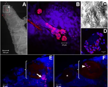
Finally, we applied these probes to whole-mount ovarian tis-sues of G. duebeni to investigate the mechanism of transovarial transmission by N. granulosis and D. duebenum. We observed a high density of N. granulosis spores in follicle cells (see Fig. S5 in the supplemental material), which are adjacent to developing oocytes, in accord with previous TEM studies that suggested that spores invade secondary oocytes during their maturation (35). Furthermore, we observed a similar proliferation of D. duebenum spores in follicle cells, as well as the presence of meronts in matur-ing oocytes (Fig. 2). These data led us to conclude that these two phylogenetically distant microsporidia have evolved convergent vertical transmission strategies.
Our study shows that FISH can be applied successfully to detect and precisely localize microsporidian species within host tissues. So far, most of the few studies that have applied FISH to microspo-ridia have focused on the detection of spores of microspomicrospo-ridia infecting vertebrates, especially humans (18,19,36). Our study shows that not only the spores but all of the stages of the microspo-ridian life cycle can be detected by FISH (see Fig. S1 and S2 in the supplemental material). Moreover, previous studies applied this method to stool samples, intestinal biopsy samples, or environ-mental samples (18,36). Our study shows that this method is also suitable for use with whole-mount tissues, allowing the study of
the dynamics of cell invasion of microsporidia within tissues. Ap-plications of FISH to microsporidia are broad. For example, coin-fections with different microsporidian species have been reported in many hosts (13,37,38). With the use of specific probes, one could easily determine the tissue specificity of multiple microspo-ridian species within a host to understand their respective impact on the host’s biology. Moreover, as vertical transmission is wide-spread among microsporidia (9), FISH could help to decipher the various mechanisms used to achieve such vertical transmission. Owing to the diversity of rRNA sequences in microsporidia (39), FISH probes could also be designed for other important clades or species. We believe that the FISH method applied to microspo-ridia is only at its beginning and that a variety of studies will ben-efit from its application.
Probe sequence accession numbers. The sequences of probes Ng02 and Dd04 have been deposited in ProbeBase (29) under accession numbers pB-03882 and pB-03883.
ACKNOWLEDGMENTS
We thank Gareth Howell for his advice on confocal microscopy and Greg-ory D. Hurst for fruitful discussions.
This work was funded by NERC/BBSRC grant NE/D011000/1. REFERENCES
1. Evans JD, Schwarz RS. 2011. Bees brought to their knees: microbes affecting honey bee health. Trends Microbiol. 19:614 – 620.
2. Evison SEF, Roberts KE, Laurenson L, Pietravalle S, Hui J, Biesmeijer
JC, Smith JE, Budge G, Hughes WOH. 2012. Pervasiveness of parasites
in pollinators. PLoS One 7:e30641. doi:10.1371/journal.pone.0030641. 3. Morado JF. 2011. Protistan diseases of commercially important crabs: a
review. J. Invertebr. Pathol. 106:27–53.
4. Stentiford GD, Neil DM, Peeler EJ, Shields JD, Small HJ, Flegel TW,
Vlak JM, Jones B, Morado F, Moss S, Lotz J, Bartholomay L, Behringer DC, Hauton C, Lightner DV. 2012. Disease will limit future food supply
from the global crustacean fishery and aquaculture sectors. J. Invertebr. Pathol. 110:141–157.
5. Solter LF, Becnel JJ, Oi DH. 2012. Microsporidian entomopathogens, p 221–263. In Vega FE, Kaya HK (ed), Insect pathology, 2nd edition. Elsevier, San Diego, CA.
6. Dunn AM, Smith JE. 2001. Microsporidian life cycles and diversity: the relationship between virulence and transmission. Microbes Infect. 3:381– 388.
7. Dunn AM, Terry RS, Smith JE. 2001. Transovarial transmission in the microsporidia. Adv. Parasitol. 48:57–100.
8. Haine ER, Motreuil S, Rigaud T. 2007. Infection by a vertically-transmitted microsporidian parasite is associated with a female-biased sex ratio and survival advantage in the amphipod Gammarus roeseli. Parasi-tology 134:1363–1367.
9. Terry RS, Smith JE, Sharpe RG, Rigaud T, Littlewood DT, Ironside JE,
Rollinson D, Bouchon D, MacNeil C, Dick JT, Dunn AM. 2004.
Widespread vertical transmission and associated host sex-ratio distortion within the eukaryotic phylum Microspora. Proc. Biol. Sci. 271:1783–1789. 10. Goertz D, Hoch G. 2008. Vertical transmission and overwintering of microsporidia in the gypsy moth, Lymantria dispar. J. Invertebr. Pathol.
99:43– 48.
11. Hogg JC, Ironside JE, Sharpe RG, Hatcher MJ, Smith JE, Dunn AM. 2002. Infection of Gammarus duebeni populations by two vertically trans-mitted microsporidia; parasite detection and discrimination by PCR-RFLP. Parasitology 125:59 – 63.
12. Franzen C, Müller A. 1999. Molecular techniques for detection, species differentiation and phylogenetic analysis of microsporidia. Clin. Micro-biol. Rev. 12:243–285.
13. Chen Y, Evans JD, Zhou L, Boncristiani H, Kimura K, Xiao T,
Lit-kowski AM, Pettis JS. 2009. Asymmetrical coexistence of Nosema ceranae
and Nosema apis in honey bees. J. Invertebr. Pathol. 101:204 –209. 14. Amann R, Fuchs BM, Behrens S. 2001. The identification of
microor-ganisms by fluorescence in situ hybridisation. Curr. Opin. Biotechnol.
12:231–236. FIG 2 Transovarial transmission of D. duebenum in its G. duebeni host. (A) G.
duebeni ovary. The red signal (FISH) indicates the presence of D. duebenum.
(B) High magnification of the G. duebeni ovary shown in panel A. Clusters of microsporidia are visible. (C, D) Group of microsporidia. Spore walls are visible in panel C. (E) A follicle cell containing D. duebenum spores (arrow) is visible in the vicinity of a maturing oocyte (bracket). Oocyte yolk is lightly autofluorescent. (F) Same tissue as in panel E observed in a deeper z plane. An immature oocyte is visible (*), while the maturing oocyte contains D. duebeni meronts (arrowhead). Panel C, differential interference contrast image; B, D, E, and F, fluorescence microscopy images; A, overlay of differential interfer-ence contrast and fluorescinterfer-ence images; red, FISH signal; blue, DAPI staining.
Design and Use of FISH Probes in Microsporidia
January 2013 Volume 79 Number 1 aem.asm.org 387
on January 21, 2013 by INIST-CNRS BiblioVie
http://aem.asm.org/
15. Bourtzis K, Miller TA. 2003. Insect symbiosis. CRC Press, Boca Raton, FL. 16. Perotti MA, Allen JM, Reed DL, Braig HR. 2007. Host-symbiont
inter-actions of the primary endosymbiont of human head and body lice. FASEB J. 21:1058 –1066.
17. Amann RI, Ludwig W, Schleifer K-H. 1995. Phylogenetic identification and in situ detection of individual microbial cells without cultivation. Microbiol. Rev. 59:143–169.
18. Graczyk TK, Johansson MA, Tamang L, Visvesvara GS, Moura LS,
DaSilva AJ, Girouard AS, Matos O. 2007. Retrospective species
identi-fication of microsporidian spores in diarrheic fecal samples from human immunodeficiency virus/AIDS patients by multiplexed fluorescence in situ hybridization. J. Clin. Microbiol. 45:1255–1260.
19. Hester JD, Varma M, Bobst AM, Ware MW, Lindquist HD, Schaefer
FW, III. 2002. Species-specific detection of three human-pathogenic
mi-crosporidial species from the genus Encephalitozoon via fluorogenic 5= nuclease PCR assays. Mol. Cell. Probes 16:435– 444.
20. Troemel ER, Felix MA, Whiteman NK, Barriere A, Ausubel FM. 2008. Microsporidia are natural intracellular parasites of the nematode
Caeno-rhabditis elegans. PLoS Biol. 6:2736 –2752.
21. Caetano-Anollés G. 2002. Tracing the evolution of RNA structure in ribosomes. Nucleic Acids Res. 30:2575–2587.
22. Hartskeerl RA, Schuitema ARJ, Dewachter R. 1993. Secondary structure of the small subunit ribosomal-RNA sequence of the microsporidium
Encephalitozoon cuniculi. Nucleic Acids Res. 21:1489.
23. Behrens S, Ruhland C, Inacio J, Huber H, Fonseca A, Spencer-Martins
I, Fuchs BM, Amann R. 2003. In situ accessibility of small-subunit rRNA
of members of the domains Bacteria, Archaea, and Eucarya to Cy3-labeled oligonucleotide probes. Appl. Environ. Microbiol. 69:1748 –1758. 24. Kumar Y, Westram R, Behrens S, Fuchs B, Glockner FO, Amann R,
Meier H, Ludwig W. 2005. Graphical representation of ribosomal RNA
probe accessibility data using ARB software package. BMC Bioinformatics
6:61. doi:10.1186/1471-2105-6-61.
25. Pruesse E, Quast C, Knittel K, Fuchs BM, Ludwig W, Peplies J,
Glöck-ner FO. 2007. SILVA: a comprehensive online resource for quality
checked and aligned ribosomal RNA sequence data compatible with ARB. Nucleic Acids Res. 35:7188 –7196.
26. Ironside JE, Smith JE, Hatcher MJ, Sharpe RG, Rollinson D, Dunn AM. 2003. Two species of feminizing microsporidian parasite coexist in popu-lations of Gammarus duebeni. J. Evol. Biol. 16:467– 473.
27. Katoh K, Misawa K, Kuma K, Miyata T. 2002. MAFFT: a novel method for rapid multiple sequence alignment based on fast Fourier transform. Nucleic Acids Res. 30:3059 –3066.
28. Cannone JJ, Subramanian S, Schnare MN, Collett JR, D’Souza LM, Du
Y, Feng B, Lin N, Madabusi LV, Müller KM, Pande N, Shang Z, Yu N,
Gutell RR. 2002. The comparative RNA web (CRW) site: an online
data-base of comparative sequence and structure information for ribosomal, intron, and other RNAs. BMC Bioinformatics 3:2. doi:10.1186/1471-2105-3-2.
29. Loy A, Arnold R, Tischler P, Rattei T, Wagner M, Horn M. 2008. ProbeCheck—a central resource for evaluating oligonucleotide probe coverage and specificity. Environ. Microbiol. 10:2894 –2896.
30. Garcia L. 2002. Laboratory identification of the microsporidia. J. Clin. Microbiol. 40:1892–1901.
31. Maddox JV, Solter LF. 1996. Long-term storage of infective microspo-ridian spores in liquid nitrogen. J. Eukaryot. Microbiol. 43:221–225. 32. Vavra J, Hylis M, Vossbrinck CR, Pilarska DK, Linde A, Weiser J,
McManus ML, Hoch G, Solter LF. 2006. Vairimorpha disparis n. comb.
(Microsporidia: Burenellidae): a redescription and taxonomic revision of
Thelohania disparis Timofejeva 1956, a microsporidian parasite of the
gypsy moth Lymantria dispar (L.) (Lepidoptera: Lymantriidae). J. Eu-karyot. Microbiol. 53:292–304.
33. Sokolova YY, Dolgikh VV, Morzhina EV, Nassonova ES, Issi IV, Terry
RS, Ironside JE, Smith JE, Vossbrinck CR. 2003. Establishment of the
new genus Paranosema based on the ultrastructure and molecular phylog-eny of the type species Paranosema grylli gen. nov., comb. nov. (Sokolova, Selezniov, Dolgikh, Issi 1994), from the cricket Gryllus bimaculatus Deg. J. Invertebr. Pathol. 84:159 –172.
34. Imhoff EM, Mortimer RJG, Christmas M, Dunn AM. 2010. Non-lethal tissue sampling allows molecular screening for microsporidian parasites in signal, Pacifasticus leniusculus (Dana), and white-clawed crayfish,
Aus-tropotamobius pallipes (Lereboullet). Freshw. Crayfish 17:145–150.
35. Terry RS, Dunn AM, Smith JE. 1997. Cellular distribution of a feminiz-ing microsporidian parasite: a strategy for transovarial transmission. Par-asitology 115:157–163.
36. Graczyk TK, Sunderland D, Tamang L, Shields TM, Lucy FE, Breysse
PN. 2007. Quantitative evaluation of the impact of bather density on levels
of human-virulent microsporidian spores in recreational water. Appl. En-viron. Microbiol. 73:4095– 4099.
37. Pilarska DK, Solter LF, Kereselidze M, Linde A, Hoch G. 2006. Mi-crosporidian infections in Lymantria dispar larvae: interactions and effects of multiple species infections on pathogen horizontal transmission. J. In-vertebr. Pathol. 93:105–113.
38. Weigl S, Korner H, Petrusek A, Seda J, Wolinska J. 2012. Natural distribution and co-infection patterns of microsporidia parasites in the
Daphnia longispina complex. Parasitology 139:870 – 880.
39. Vossbrinck CR, Debrunner-Vossbrinck BA. 2005. Molecular phylogeny of the microsporidia: ecological, ultrastructural and taxonomic consider-ations. Folia Parasitol. (Praha) 52:131–142.
Dubuffet et al.
388 aem.asm.org Applied and Environmental Microbiology