HAL Id: hal-01087342
https://hal.archives-ouvertes.fr/hal-01087342
Submitted on 25 Nov 2014HAL is a multi-disciplinary open access archive for the deposit and dissemination of sci-entific research documents, whether they are pub-lished or not. The documents may come from teaching and research institutions in France or abroad, or from public or private research centers.
L’archive ouverte pluridisciplinaire HAL, est destinée au dépôt et à la diffusion de documents scientifiques de niveau recherche, publiés ou non, émanant des établissements d’enseignement et de recherche français ou étrangers, des laboratoires publics ou privés.
PRODUCTION OF 3-10 µm MICROBUBBLES
SUITED FOR ULTRASONIC IMAGING BY 2.5D
NANOFLUIDIC DEVICES
Sébastien Méance, Lucas Pages, Anne Marie Gué, Pierre Joseph
To cite this version:
Sébastien Méance, Lucas Pages, Anne Marie Gué, Pierre Joseph. PRODUCTION OF 3-10 µm MI-CROBUBBLES SUITED FOR ULTRASONIC IMAGING BY 2.5D NANOFLUIDIC DEVICES. 18th International Conference on Miniaturized Systems for Chemistry and Life Sciences (MicroTAS 2014), Oct 2014, San Antonio, United States. �hal-01087342�
PRODUCTION OF 3-10 µm MICROBUBBLES SUITED FOR
ULTRASONIC IMAGING BY 2.5D NANOFLUIDIC DEVICES
S. Méance
1,2, L. Pages
1,2, A.-M. Gué
1,2, and P. Joseph
1,2*1
CNRS, LAAS, 7 avenue du colonel Roche, F-31400 Toulouse, France
2
Univ de Toulouse, LAAS, F-31400 Toulouse, France
ABSTRACT
In this paper, we propose a novel route to generate monodisperse microbubbles (MB) in the range size of 3-10 µm. Using deep reactive ion etching, we designed several “terraces” emulsification devices with depths of 200-1000 nm. We performed characterizations and systematical investigations on critical parameters such as gas pressure, liquid flow rate, and chip geometry. We experimentally show here that MB size is mainly dependent on nanochannel height. The robust monodisperse MB production and the biocompatibility of the materials opens an opportunity to integrate such systems in the future diagnostics routines.
KEYWORDS: Ultrasound contrast agents, Nanochannels, Nanofluidics INTRODUCTION
For their use in diagnostic echocardiography, ultrasound contrast agents (UCA) have been widely studied for several decades. Injection of UCA MB through the circulatory system imposes a size limitation of about 8 µm, that does not exceed that of pulmonary capillaries. Conversely the ultrasound scattering efficiency is a function of the diameter MB. Hence, commercially produced UCA have a mean size of 2-6 µm. They also have a relatively narrow distribution but they are still polydisperse [1]. In order to improve the sensitivity of the imaging systems, Hettiarachchi et al. proposed a microfluidic flow-focusing chip for monodisperse UCA generation [2]. The drawback of this approach is that the device depends on a critical dimension: a narrow orifice that can clog with PDMS debris accumulation. Another method based on 2.5D geometries has recently been proposed by Stoffel et al.[3]. It is driven by surface tension and relies on the shape instability at a confined-unconfined junction. This mechanism is particularly robust independently on flow rates, and gas pressure. However the MB diameter of 12-27 µm measured in their work was not in the range required for use as ultrasound contrast agents. In this work we experimentally demonstrate the MB generation with size that fits the requirement of contrast echography.
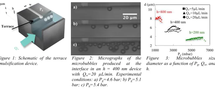
Figure 1: Schematic of the terrace emulsification device.
Figure 2: Micrographs of the microbubbles produced at the interface in an h = 400 nm device with Qw=20 µL/min. Experimental
conditions: a) Pg=4.6 bar; b) Pg=5.1
bar; c) Pg=5.4 bar.
Figure 3: Microbubbles size diameter as a function of Pg, Qw, and
h. Qw=5µL/min ▲Qw=10µL/min Qw=20µL/min Pg (mbar) d (µm) Terrace Qw Pg a) b) c)
EXPERIMENTAL
The devices were realized by standard microfabrication. Nanochannel (height h between 200 nm and 1 µm, lateral dimension 10 µm x 60 µm) and microchannels (20 µm x 100 µm) were etched in silicon by dry etching. Holes were sand blasted; anodic bonding ensured sealing to a glass substrate. Gas (nitrogen) pressure Pg was imposed using a pressure controller. The liquid (deionized water + 2% Tween20) was
injected at a flow rate Qw using a syringe pump. Chip design is shown in Figure 1.
RESULTS AND DISCUSSION
In the nanochannel, the curvature introduces a pressure jump, known as the Laplace pressure, between the inside and the outside of a gas-liquid interface. Assuming full wetting, in this case it is written ΔP = 2γ/h where γ is the surface tension. When Pg > ΔP, the water-gas meniscus advances to the terrace. Once the
interface is pinned to the terrace edge, an increase in pressure creates a meniscus unbalanced, causing an overflow of the gas in the microchannel, followed by pinch-off and formation of a new bubble.
Figure 2 shows MB generation for three different gas pressures. When Pg is slightly higher than ΔP,
only a few MB are formed. Above this pressure, the system is stable and production is monodisperse (Figure 2-b). For Pg>5400 mbar, the frequency is too high and the MB are facing an environment
increasingly confined. They start to be polydisperse (Figure 2-c).
Figure 3 summarizes all the parameters (gas pressure, flow rate, nanochannel height) involved in this experiment. The first observation relates to the influence of the pressure on the MB size. Indeed when the pressure increases, the diameter increases to its maximum and then decreases. This is consistent with the semi empirical physical model of [3] but could also be due to the deformation of the MB in the confined environment. The second point concerns the influence of flowrate Qw. When it increases, the diameter of the
MB decreases. In such situation, the geometry can be compared with a T-junction microfluidic device. Then, the higher the flowrate, the greater the shear forces are important to the terrace level causing detachment of the MB. Nevertheless the variations remain weak and the MB size has little dependency on the gas pressure and the liquid flowrate. Finally, the most important parameter is the nanochannel height. As we can observe the size of MB formed by terrace emulsification is roughly proportional to the confining dimension (around 10 times the nanochannel depth).
CONCLUSION
In agreement with existing literature, we show that MB size have little dependency on production frequency. Moreover, we experimentally show that the nanochannel depth is a critical parameter to reach the desired size. Finally, we have implemented a terrace emulsification chip for manufacturing monodisperse MB in the size range desired for ultrasonic imaging.
ACKNOWLEDGEMENTS
We acknowledge CNRS and French National Research Agency for funding (Smart-US program). REFERENCES
[1] J. R. Lindner, “Microbubbles in medical imaging: current applications and future directions.” Nature
Reviews Drug Discovery, 3, 527-533, 2004.
[2] K. Hettiarachchi, E.Talu, M. L. Longo, P. A. Dayton, and A. P. Lee, “On-chip generation of microbubbles as a practical technology for manufacturing contrast agents for ultrasonic imaging.”
Lab Chip, 7, 463-468, 2007.
[3] M. Stoffel, S. Wahl, E. Lorenceau, R. Höhler, B. Mercier, and D. E. Angelescu, “Bubble production mechanism in a microfluidic foam generator.” Physical review letters, 108, 198302, 2012.
CONTACT
![[PDF] Débuter avec le logiciel SPIP étape par étape | Cours Informatique](data:image/gif;base64,R0lGODlhAQABAIAAAP///wAAACH5BAEAAAAALAAAAAABAAEAAAICRAEAOw==)