Cardnogenesis Vol.5 no.3 pp.403-406, 1984
Methylation of DNA by incubation with methylamine and nitrite
Kurt W. Huber and Werner K. Lutz
1 Institute of Toxicology, ETH and University of Zurich, CH-8603 Schwerzenbach, Switzerland'To whom reprint requests should be sent
DNA was incubated in septum-dosed reaction vials with
[
14C]methylamine and nitrite. The DNA was purified,
hydro-lysed with hydrochloric acid, and the purines were anahydro-lysed
by h.p.l.c. 7-Methylguanine was detectable as a result of
DNA methylation in experiments performed in 100 mM
acetate at pH 4. Using different concentrations of amine and
nitrite a first order reaction for total amine and a second
order for total nitrite could be shown. A study on the pH
dependence using 100 mM malonate buffer, pH 2.0 — 6.0,
revealed a maximum rate at pH 3.5, with steep slopes above
and below this pH value, in agreement with a mathematical
analysis of the reaction equations. The data show that the
alkylating agent formed spontaneously by nitrosation and
deamination of a primary amine has a long enough lifetime to
react with DNA in vitro. Using the reaction orders established
here, an extrapolation to lower concentrations found in the
stomach can now be performed. Future in vivo experiments
on the methylation of gastro-intestinal DNA then would
show to what extent DNA in a cell is protected from
alkyl-ation.
Introduction
The generation of nitrosamines from amines and nitrite under
acidic conditions could represent an important mechanism
for the formation of carcinogens in the stomach (1) from
ubi-quitous dietary amines (2) and unavoidable salivary nitrite
(3,4). With primary amines, chemically unstable products are
formed which react readily with nucleophiles. This instability
must have been the reason for the negligible interest of
toxicologists in primary amines as opposed to the secondary
amines known to produce chemically stable nitrosamines.
Nevertheless it seemed important to investigate a DNA
damage exerted by the nitrosation products of methylamine,
a dietary constituent of fish (5) and vegetables (2). Since the
chemistry predicted the formation of a methylating agent (6),
methylated DNA bases were searched using techniques
established in earlier work with dimethylamine (7).
Establishing dose dependences both for the amine and for
nitrite and investigating the pH dependence seemed to be
in-dicated because preliminary experiments using [
14C]methyl-amine allowed the detection of DNA methylations (8). The
results reported here allowed us to form an equation for the
reaction kinetics at different pH values, information which
will ultimately be required for an extrapolation to the lower
concentrations encountered in a human stomach.
Materials and methods
Chemicals and apparatusL( + ySodium ascorbate and potassium nitrate were obtained from Fluka AG
(Buchs, Switzerland). DNA from calf thymus (sodium salt type I; highly poly-merized) and 7-methylguanine were purchased from Sigma Chemical Com-pany (St. Louis, MO). All other chemicals were purchased in the highest puri-ty available from Merck (Darmstadt, FRG). [14C]Methylamine hydrochloride
(mol. wt. 67.5) with a specific activity of 50 mCi/mmol, dissolved in ethanol (100 /tCi/ml), was obtained from New England Nuclear (Boston, MA). The radiochemical purity was 99%, as determined by t.l.c. on cellulose (Merck, Darmstadt, FRG) using methanol:diethylether:lN hydrochloric acidrwater (10+ 10+ 1 +3) as the solvent system. The ethanol from the original [14
C]-methylamine hydrochloride solution was evaporated with nitrogen and the salt redissolved in incubation buffer to give a stock solution with a specific radioactivity of 245 fiCi/ml. Dialysis tubing (Visking Type 20/32, mol. wt. ex-clusion at 12000—14000 daltons; diameter 17 mm) was obtained from Union Carbide (Chicago, 1L).
Radioactivity measurements were carried out in 10 ml of Insta-Gel (Packard Instruments, Downers Grove, IL) in a liquid scintillation counter, model Packard Tri Carb 460 CD. The h.p.l.c. analysis of the DNA bases was performed on a semipreparative jtBondapak C18 column, 300 x 7.8 mm
(Waters Associates, MiUford, MA) equipped with two h.p.l.c. pumps (model LC Pump 410 from Kontron, Zurich, Switzerland) controlled by a Kontron Programmer 200 to generate a linear gradient of two eluents.
General methods
Incubation system. Stock solutions of unlabelled methylamine, ["C]methyl-amine, nitrite, nitrate and DNA were prepared in 100 mM potassium acetate/ acetic acid pH 4 or 100 mM malonic acid/sodium hydroxide or hydrochloric acid (pH dependence). Appropriate volumes of amine and DNA (where ap-plicable) were mixed to give the concentrations wanted in a final volume of 10 ml in 24.6 ml glass vials tightly closed with a septum. The reaction was started by injection of potassium nitrite dissolved in the same buffer. The solution was kept at 37°C, slowly stirred with a magnetic stirrer, and the reac-tion was stopped after 30 min by addireac-tion of sodium ascorbate in 2-fold molar excess of the nitrite concentration (9). Immediately before stopping the reac-tion, a 1 ml sample of the gas phase above the reaction mixture was taken out with a syringe and injected into a 2 ml serum vial prefilled with 1 g methanol and closed with a septum. After thorough shaking, 1 ml of this methanol solution was pulled up into a syringe and mixed into 10 ml Insta-Gel for scin-tillation counting.
Isolation of DNA. The incubation buffer was dialysed three times at 4°C against 10 1 0.2 M NaCl for 5 - 1 0 h in order to remove most of the non-covalently bound radioactive species. The DNA was precipitated by adding 2 volumes ethanol and storing at - 2 0 ° C overnight. After centrifugation for 20 min at 1000 g the supernatant was decanted and the DNA was dried in vacuo for 2 - 3 h. The DNA was dissolved in 10 mM MgCl* 10 mM Tris/HCl, pH 7.0 and reprecipitated once with ethanol. The amount of DNA was determin-ed by u.v. spectroscopy using as standard an absorbance of 20 at 260 nm for a solution of 1 mg DNA per ml.
Depurination of DNA and h.p.l.c. analysis. DNA was hydrolysed for 1 h at 70°C with 0.1 N HC1 to liberate the purines (10). 7-Methylguanine was added as standard, the mixture was filtered through a 0.2 /*m filter (Millipore, Bed-ford, MA) and loaded on a reverse-phase h.p.l.c. system. Elution medium was a 10 mM ammonium phosphate buffer, pH 4.0, containing methanol (1% for 20 min, followed by a linear gradient to 100% methanol in 40 min). The flow rate was 3.5 ml/min and the optical density of the duate was record-ed at 260 nm. Fractions of 2 min were collectrecord-ed and the radioactivity of each fraction was determined by scintillation counting. To avoid phase separation between the scintillation cocktail and the eluate, fractions 19, 20 and 21 were diluted with 1 g water, 1 g methanol and 0.5 g methanol, respectively. The retention times of the purines, guanine, adenine and 7-methylguanine were 10.2, 14.8 and 20.4 min, respectively. The recovery of the injected radio-activity ranged from 90 to 110%. To compare the results of the different ex-periments, a 7-methylguanine index was defined as micromol 7-methyl-guanine/mol DNA nucleotide.
Time dependence of the deamination. Three experiments were performed with 2.5 mM, 1.5 mM, 3.0 mM ["CJmethylamine hydrochloride (sp. act. 50 /iCi/mmol) in 100 mM acetate buffer pH 4.0, incubated with 78.8 mM, 78.8 mM and 50.4 mM potassium nitrite, respectively. The control
K.W.Huber and W.K.Lutz
tion (3.0 mM [14C]methylamine hydrochloride) was 74.2 mM in potassium nitrate. The radioactivity in the gas phase was determined at different times up to 6 h.
Nitrite concentration dependence. A solution of 1.2 mM ["C]methylamine hydrochloride (sp. a n . 2.56 mCi/mmol) and 1.05 mg/ml calf thymus DNA (equivalent to 3.4 mM in nudeotides) was incubated with 0.0,4.4, 13.0, 22.0, 44.0 and 66.0 mM sodium nitrite. In order to reach a constant ionic strength in the incubations, potassium nitrate was added to give a total concentration of 66 mM for nitrite plus nitrate.
Amine concentration dependence. Solutions of 15 JIM, 45 yM, 0.14 mM, 0.38 mM and 1.2 mM [14C]methylamine hydrochloride (sp. act. 58.6, 58.6, 6.31, 6.31,0.68 mCi/mmol, respectively) were incubated with 66 mM sodium nitrite in the presence of 1.02 mg/ml calf thymus DNA. The control contained' 1.2 mM ["Clmethylamine hydrochloride (sp. act. 0.68 mCi/mmol), 3.3 mM DNA nucleotide and 82 mM potassium nitrate.
pH dependence. The reaction vials contained 1 mM ["C]methylamine hydro-chloride (sp. act. 0.58 mCi/mmol) in 100 mM malonate buffer at the follow-ing pH values: 2.0, 2.6, 3.3, 3.7, 4.6, 5.0, 6.0. Sodium nitrite was added to a final concentration of 70 mM to start the deamination reaction. Two controls with 66 mM potassium nitrate instead of nitrite were run at pH 1.5 and 5.1. No change in pH was noticed during the 30 min incubation period.
Results
Time-dependence of the deamination of methylamine
The reaction of [
14C]methylamine with nitrite is known to
yield [
14C]methanol and [
14C]methyl nitrite (11), two volatile
products expected to appear in the gas phase above the
reac-tion mixture. The simple determinareac-tion of the radioactivity in
the headspace could therefore be used to follow the
ap-pearance of the reaction products in order to assess the rate of
the formation of electrophilic intermediates such as the
methyl diazonium ion. Figure 1 shows a linear
time-dependence up to ~ 1 h using three different sets of
concen-trations of methylamine and nitrite. For the following
studies, incubation times of 30 min were therefore chosen to
make sure that no changes of the reaction kinetics by side
reactions would confuse the analysis of the data. The control
experiment with nitrate instead of nitrite (open circles in
Figure 1) showed that the incubation of [
14C]methylamine
with nitrate did not lead to the generation of volatile
radio-labelled reaction products.
100 200 300
R e a c t i o n time (min.]
Fig. 1. Rate of formation of volatile reaction products determined in the
closed air volume above a solution of ["CJmethylamine hydrochloride and potassium nitrite in a 100 mM acetate buffer, pH 4.0. The concentrations of amine and nitrite, respectively, were: • • , 0.0025 M, 0.0788 M; C - - - C , 0.0015 M, 0.0788 M; • • • • • • , 0.003 M, 0.0504 M; O - - - - O , control incubation with 0.003 M ["CJmethylamine hydrochloride and 0.072 M potassium nitrate.
404
In vitro methylation of DNA
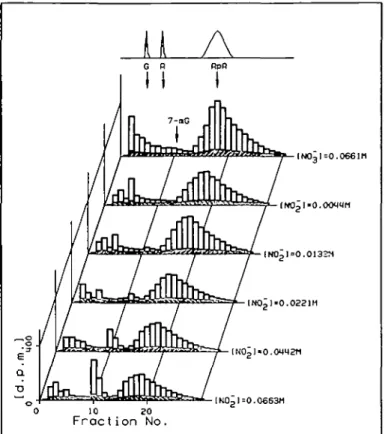
Nitrite concentration dependence. Figure 2 shows the h.p.l.c.
radioactivity elution profile of depurinated DNA after
in-cubation with ["CJmethylamine and different concentrations
of potassium nitrite. The top chart is the control DNA
digest obtained when using nitrate in the place of nitrite. Sixty
to seventy percent of the radioactivity always coeluted with
•INOpl-O.OOMMM
0 10 20 Fraction No.
Fig. 2. H.p.l.c. elution profile of acid-hydrolysed DNA isolated 30 min
after incubation with 1.2 mM ["CJmethylamine and different concentra-tions of nitrite. Separate top chart: optical density profile at 260 nm, showing elution region for guanine (G), adenine (A), 7-methylguanine (7-mG) and apurinic acid (ApA). Top radioactivity profile: control incuba-tion with nitrate instead of the highest concentraincuba-tion of nitrite.
o.ooq 0.02S o.oso N i t r i t e c o n e . (Ml
Fig. 3. Formation of 7-methylguanine in DNA incubated for 30 min in
0.1 M acetate buffer pH 4.0, containing 1.2 mM ["CJmethylamine and different concentrations of potassium nitrite. The ionic strength of all solutions was made equal by appropriate additions of potassium nitrate.
• • , experimental data; — , theoretical dose dependence obtained by using the equation 7-mG-lndex = k' x [total amine] x [total nitrite]2 with k' = 4.0 x 10* M~3 (see Discussion section).
DNA methjlation by meth)1ainiiK and nitrite
the apurinic acid, probably representing DNA
phosphate-alkylammonium salts. At the retention of 7-methylguanine
(fractions 10 and 11), increasing amounts of radioactivity
ap-peared with higher nitrite concentrations. Net counts in the
fractions 10 and 11 were determined by subtraction of a
back-ground calculated as the mean radioactivity of the fractions 9
and 12. The control DNA digest as well as the incubation
with the lowest concentration of nitrite contained no
7-methylguanine. The remaining counts were spread over the
front fractions, the region known to contain guanine
(frac-tion 5) and adenine (frac(frac-tions 7 and 8). Upon conversion of
the net radioactivity in fractions 10 plus 11 to methyl groups
on the basis of the known specific activity of the methylamine
used and the amount of DNA hydrolysed, a 7-methylguanine
index ( = fimo\ 7-methylguanine/mol DNA nucleotide) was
defined and plotted in Figure 3 against the nitrite
concentra-tion. The dashed line which was calculated according to the
square of the nitrite concentration nicely overlaps with the
ex-perimental points and is a clear indicator of a second order
reaction of nitrite with methylamine to produce DNA
methylations.
Amine concentration dependence. Using the highest nitrite
concentration taken above and varying the amine
concentra-tion, an analogous experiment was performed. Figure 4
clear-ly shows the linear dependence of the DNA methylations on
the amine concentration.
0.00000 0.00050 0.00100 Methglamine cone. (Ml
Fig. 4. Formation of 7-methylguanine in DNA incubated for 30 min in 0.1 M acetate buffer pH 4.0, containing 66.0 mM potassium nitrite and different concentrations of ["C]methylamine. Correlation coefficient for a linear regression analysis: R = 0.999.
o 3- . nj Inde x 16. 0 i o Eo
rU"
O / / 0 50 100 150 Heodspoce r o d i o o c t . 1 d . p . m .Fig. 5. Correlation of the methylation of DNA with the formation of volatile radiolabelled products. O O, data from the dose dependence for methylamine, R = 0.998; • — • , data from the dose dependence for nitrite, R = 0.998.
pH dependence. In both experiments described above, the
headspace radioactivity was determined at the end of the
30-min incubation period, before the quantification of DNA
methylations. Figure 5 shows the linear relationship between
these two experimental endpoints. Since headspace activities
can be determined using relatively little radioactivity, these
values were taken for the determination of the pH
dependence of the nitrosation reaction. The proportionality
to the DNA methylations makes it possible to use headspace
data obtained from short incubation times as a substitute for
the 7-methylguanine-index. Figure 6A clearly shows that the
generation of volatile [
14C]methyl derivatives after incubation
of [
14C]methylamine and nitrite has a maximum value at a pH
of - 3 . 5 with very steep slopes above and below.
Discussion
We have shown that the rate of formation of a
DNA-methylating agent by incubation of methylamine with DNA
in the presence of nitrite follows the equation
d[7-methylguanine]/dt = k x [total amine] x [total nitrite]
2Using the present data on the formation of
7-methyl-guanine in our standard incubation system, the equation
reads
7-methylguanine index = k' x [total amine] x [total nitrite]
2with k' = 4.0 x 10
8M~
3determined as an average of all
single experimental values. The reaction order, therefore, is in
accordance with the mechanism of nitrosation of secondary
E • tr Oo 2 O D a in T> O 2 . 0 pH in 14.0 incubate 0.0 pH {or 2 . 0 M . 0 6 . 0 nitrosation reaction
Fig. 6. Formation of volatile radiolabelled reaction products at different pH values. A, Experimental values; B, Curve calculated on the basis of equation (2).
K.W.Huber and W.K.Lutz
amines elucidated by Ridd (6) and Mirvish (12). The reactions
which can take place in our incubation system are compiled in
a review by Douglass et al. (13) where he discusses the
dif-ferent nitrosating agents. The fact that the reaction is second
order in nitrite is an indication favoring N2O3 as the most
im-portant nitrosating agent in our system. Since N ^ can be
regarded as the anhydride of two nitrous acid molecules it is
obvious that its concentration increases with decreasing pH.
The amine reactant must be in the neutral form to be
nitrosatable. The ratio of neutral to protonated form is
dependent on the pH and the basicity of the amine. The lower
the pH, the less nitrosatable amine is available. The situation
is therefore characterized by the following reaction kinetics
dC/dt = k" x [A] x [HNO
2]
2(1)
The concentrations of the reactive species [A] and [HNO2] in
solution can be calculated in terms of the total amine [A]
totand total nitrite [NO
2~]
totconcentration, given that the latter
values are equal to the sum of the protonated and the
depro-tonated forms for each component. Substitution into the
respective acid dissociation equilibria and rearrangement
yields:
K, x[H + P Ov 2j= = k" x [A]1OI x [NO,-]t0,* x — p ^
-Using Kj = 2 . 3 x l O ~
na n d K
2= 4.3 x 10~* for the acidity
of methylammonium ion and of nitrous acid, respectively,
the graphical representation of this equation in terms of the
reaction rate as a function of the pH is shown in Figure 6B.
Shape and position of the maximum are almost identical to
the experimentally observed data (Figure 6A).
7-Methylguanine is the most abundant methylated base
formed upon reaction of DNA with methylating carcinogens,
representing ~809/o of all DNA methylations (10), and was
the only methylation product detectable in the present assay.
The level of DNA methylation in the 7-position of guanine is
not correlated with mutagenic or carcinogenic effects. Other
methylation products, such as O
6-methylguanine, seem to be
more important. Nevertheless, the present data are
inter-pretable in terms of the formation of this promutagenic lesion
because the ratio between 7- and O
6-methylations is known to
be 10 to 1 for methyl diazonium ion, the ultimate methylating
agent, well known from dimethylnitrosamine studies (10) and
expected to be formed here, too.
Methylation of DNA by methylamine and nitrite has
already been reported two decades ago, but this was in the
chemical context of DNA derivatization by diazomethane
(14). The toxicological aspect so far has received little
atten-tion (15) as compared with the formaatten-tion of carcinogenic
nitrosamines from secondary amines (16). These latter
com-pounds are chemically stable and are converted to reactive
derivatives only upon oxidative enzymatic dealkylation. DNA
alkylation is therefore expected primarily in cells which
con-tain the appropriate enzyme systems. The nitrosation of
primary amines is known to the chemist to yield highly
unstable intermediates which spontaneously react with water
to yield the corresponding alkyl alcohols. This rapid
inactiva-tion at the site of formainactiva-tion was probably the reason why
biologists did not investigate the potential DNA alkylating
ac-tivity. Indeed, the extent of DNA methylations was very low
(of the order of 0.01 % with respect to methylamine) in our in
vitro system, even using high concentrations of nitrite and
op-timum pH, and incubating the DNA in situ. Sporadic reports
have shown, otherwise, that a mutagenicity to
micro-organisms can be generated by incubation with primary
amines and nitrite (17,18). In addition, we have reported
preliminary data on a DNA methylation of gastro-intestinal
DNA in rats that had been administered [
14C]methylamine
and nitrite (8). All these experiments have been performed
using a nitrite concentration which was 2 — 3 orders of
magnitude higher than the one expected to be encountered in
a stomach. More refined in vivo work will therefore be
re-quired before the role of primary amines in the etiology of
cancer can be assessed.
Acknowledgement
We thank the Swiss Cancer League for the graduate fellowship awarded to K.W.Huber.
References
1. Lijinsky.W. (1979), Current concepts in the toxicology of nitrates, nitrites, and nitrosamines, in Mehlman.M.A., Shapiro.R.E. and Blumenthal.H. (eds.), Advances in Modern Toxicology, Vol. 1, Part 2, Hemisphere Publishing Corporation, Washington, New York and London, pp. 149-164.
2. Neurath,G.B., Duenger.M., Pein.F.G., Ambrosius.D. and Schreiber.O. (1977), Primary and secondary amines in the human environment, Fd. Cosmet. Toxicol., 15, 275-282.
3. Walters.C.L., Carr.F.P.A., Dyke.C.S., Saxby.M.J., Smith.P.L.R. and Walker.R. (1979), Nitrite sources and nitrosamine formation in vitro and in vivo, Fd. Cosmet. Toxicol., 17, 473-479.
4. Walters.C.L. (1980), The exposure of humans to nitrite, Oncology, 37, 289-296.
5. Lin,J.-K., Lee,Y.-J. and Chang.H.W. (1983), High concentrations of dimethylamine and methylamine in squid and octopus and their implica-tions in tumour aetiology, Fd. Chem. Toxicol., 21, 143-149.
6. Ridd.J.H. (1961), Nitrosation, diazotisation and deamination, Quarterly Reviews, 15, 418-441.
7. Meier-Bratschi,A., Lutz.W.K. and Schlatter.Ch. (1983), Methylation of liver DNA of rat and mouse by N-nitrosodimethylamine formed in vivo from dimethylamine and nitrite, Fd. Chem. Toxicol., 21, 285-289. 8. Huber.K.W., Lutz,W.K. and Schlatter.Ch. (1982), Methylation of DNA
by N-methylnitrosamine formed in vivo from methyiamine and nitrite, Experientia, 38, 755.
9. Dahn.H., Loewe.L. and Bunton.C.A. (1960), Ueber die Oxidation von Ascorbinsaeure durch salpetrige Saeure Teil VI: Uebersicht und Diskus-sion der Ergebnisse, Helv. Chim. Ada, 43, 320-333.
10. Lawley.P.D. (1976), Methylation of DNA by carcinogens: some applica-tions of chemical analytical methods, in Montesano.R., Bartsch.H. and Tomatis,L. (eds.), Screening Tests in Chemical Carcinogenesis (IARC Scientific Publications No. 12), International Agency for Research on Cancer, Lyon, France, pp. 181-208.
11. Austin.A.T. (1960), The action of nitrous acid on aliphatic amines, Nature, 188, 1086-1088.
12. Mirvish,S.S. (1975), Formation of N-nitroso compounds: chemistry, kinetics and in vivo occurrence, Toxicol. Appl. Pharmacol., 31, 325-351. 13. Douglass.M.L., Kabakoff.B.L., Anderson.G.A. and Cheng,M.C. (1978), The chemistry of nitrosamine formation, inhibition and destruction, J. Soc. Cosmet. Chem., 29, 581-606.
14. Kriek.E. and Emmelot.P. (1964), Methylation of deoxyribonuclek acid by diazomethane, Btochim. Biophys. Ada, 91, 59-66.
15. Obiedzinski.M.W., Wishnok.J.S. and Tannenbaum.S.R. (1980), N-Nitroso compounds from reactions of nitrite with methylamine, Fd. Cosmet. Toxicol., 18, 585-589.
16. Preussmann.R. (1975), Chemische Carcinogene in der menschlichen Umwelt, in Grundmann,E. (ed.), Handbuch der allgemeinen Pathologie, Vol. 6, Part 6, Springer Verlag, Berlin, Heidelberg, New York, pp. 421-507.
17. Hussain.S. and Ehrenberg.L. (1974), Mutagenicity of primary amines combined with nitrite, Mutat. Res., 26, 419-422.
18. Boido.V., Benniceili.C., Zanacchi.P. and De Flora.S. (1980), Formation of mutagenic derivatives from nitrite and two primary amines, Toxicol. Lett., 6, 379-383.
(Received on 10 October 1983; accepted on 30 November 1983)