ORIGINAL ARTICLE
Postmortem computed tomography angiography
vs. conventional autopsy: advantages and inconveniences
of each method
Chevallier Christine&Doenz Francesco&Vaucher Paul&
Palmiere Cristian&Dominguez Alejandro&Binaghi Stefano&
Mangin Patrice&Grabherr Silke
Received: 25 September 2012 / Accepted: 20 December 2012 / Published online: 6 January 2013 # Springer-Verlag Berlin Heidelberg 2013
Abstract
Purpose Postmortem computed tomography angiography (PMCTA) was introduced into forensic investigations a few years ago. It provides reliable images that can be con-sulted at any time. Conventional autopsy remains the refer-ence standard for defining the cause of death, but provides only limited possibility of a second examination. This study compares these two procedures and discusses findings that can be detected exclusively using each method.
Materials and methods This retrospective study compared radiological reports from PMCTA to reports from conven-tional autopsy for 50 forensic autopsy cases. Reported find-ings from autopsy and PMCTA were extracted and compared to each other. PMCTA was performed using a modified heart–lung machine and the oily contrast agent Angiofil® (Fumedica AG, Muri, Switzerland).
Results PMCTA and conventional autopsy would have drawn similar conclusions regarding causes of death. Nearly 60 % of all findings were visualized with both techniques. PMCTA demonstrates a higher sensitivity for identifying skeletal and vascular lesions. However, vascular occlusions due to postmortem blood clots could be falsely assumed to be vascular lesions. In contrast, conventional autopsy does not detect all bone fractures or the exact source of bleeding. Conventional autopsy provides important information about organ morphology and remains the only way to diagnose a vital vascular occlusion with certitude.
Conclusion Overall, PMCTA and conventional autopsy pro-vide comparable findings. However, each technique presents advantages and disadvantages for detecting specific findings. To correctly interpret findings and clearly define the indica-tions for PMCTA, these differences must be understood.
Keywords Forensic medicine . X-Ray computed tomography . Autopsy . Perfusion imaging . Angiography
Introduction
The use of multi-detector computed tomography (MDCT) in postmortem investigations has become routine in many centers of legal medicine [1–6]. MDCT is a rapid and easy way to look inside the body and document findings. It offers the opportunity to consult data at any time, even after cremation or burial of the body. Furthermore, the handling of an MDCT unit is relatively easy, and its maintenance costs are affordable for certain institutes of legal medicine. The diagnostic value of MDCT compared to conventional autopsy has been addressed in multiple publications [7,8].
C. Christine (*)
:
P. Cristian:
D. Alejandro:
M. Patrice:
G. SilkeUniversity Centre of Legal Medicine Lausanne–Geneva,
University of Lausanne, Rue du Bugnon 21, 1011 Lausanne, Switzerland
e-mail: [email protected]
C. Christine
:
D. Francesco:
B. StefanoDepartment of Diagnostic and Interventional Radiology, University Hospital of Lausanne, Rue du Bugnon 46, 1011 Lausanne, Switzerland
V. Paul
:
M. PatriceUniversity Centre of Legal Medicine Lausanne–Geneva,
University of Geneva, Michel-Servet 1, 2011 Geneva 4, Switzerland
D. Alejandro
University of Health Sciences (HESAV), Rue du Bugnon 19, 1011 Lausanne, Switzerland
Although native or unenhanced MDCT (without the in-jection of contrast agent) can show major vascular lesions such as aortic rupture [9], its most important limitation is its low ability to visualize the vascular system and soft tissue. By injecting contrast agent into the vessels, different parts of the vascular system are rendered visible [10–13]. Recent advances have made it possible to visualize the entire vas-cular system in a minimally invasive way [14, 15]. The performance of postmortem computed tomography angiog-raphy (PMCTA) appears to have important advantages, especially in detecting sources of hemorrhage [16,17]. In the early twenty-first century, postmortem angiography was performed with little success due to the imaging and perfu-sion techniques [18,19]. In 2008, Grabherr at al. introduced the use of a modified heart–lung machine to establish post-mortem circulation, in turn allowing the injection of a con-trast agent and constant perfusion of a body [20]. This technique has been further developed and standardized. Nowadays, it consists of the execution of at least one native computed tomography scan and three angiographic phases (arterial, venous, and dynamic). This multi-phase PMCTA (MPMCTA) [21] has been introduced as a routine investi-gation method at the University Centre of Legal Medicine in Lausanne. The combination of MPMCTA with conventional autopsy appears to increase the results of postmortem inves-tigations, as has been stated regarding the examination of the coronary arteries [22].
However, MPMCTA is still a new technique. In contrast, conventional autopsy consists of a systematic approach that has been known and used for centuries—one reason why it is recognized as the optimal method with which to detect findings that indicate an individual’s cause of death. To define the limits, advantages, and overall diagnostic value of MPMCTA, it is necessary to compare the performance of this new method to the well-known technique of conven-tional autopsy. To our knowledge, this is the first such study. This study will evaluate the diagnostic value of MPMCTA compared with native MDCT and conventional autopsy, and describe findings that can be detected exclu-sively by each method to define method-specific limitations and advantages.
Materials and methods
Subjects
A total of 50 human corpses with postmortem intervals ranging from a few hours to several days were included retrospectively in the study. The first nine bodies were donated by anatomical institutes, and a summary of the medical history of the diseased was made available. The other 41 cadavers underwent a forensic autopsy in our
institute between September 2008 and February 2010. A consecutive sample method with no specific exclusion cri-teria was used to include autopsy cases. No choice was made concerning the indication for the medico-legal autop-sy. Therefore, causes of death included trauma (e.g., stab wounds or traffic accidents), intoxication (e.g., drug or carbon monoxide intoxication), and natural death (e.g., heart attack or cancer-related complications). In each case, a ra-diological investigation including native CT scan and post-mortem CT angiography was performed before the conventional autopsy. This study was approved by the local justice department and the ethics committee.
Radiological examination
Before starting any invasive incisions, an external examina-tion of the body was performed by the forensic pathologist in charge of the case, and a native CT scan was done by the forensic radiographer of the institute. MDCT scans were performed using an eight-row CT unit (CT LightSpeed 8; GE Healthcare, Milwaukee, WI, USA) with the following scan parameters: field of view (FOV), 50 cm; slice thick-ness, 2.5 mm; interval of reconstruction, 2 mm; 120 kV; 280 mA (modulated); and noise index, 15. The scan was performed following the standard protocol of the institute, from the cerebral vertex to the pubic symphysis. Standard lung and bone filter reconstructions were acquired, and bone-reformatted images of the spine were extracted.
After the collection of postmortem liquid samples under CT guidance for toxicological analysis and the analysis of clinical biomarkers [21, 23], cannulation of the femoral vessels of one side was performed using 16F diameter cannulas for arteries and 18F cannulas for veins (MAQUET GmbH & Co. KG, Rastatt, Germany). MPMCTA was per-formed according to the standardized protocol proposed by Grabherr et al. [21], using a contrast agent mixture com-posed of 6 % of the oily liquid Angiofil® (Fumedica AG, Muri, Switzerland) and paraffin oil (paraffinum liquidum) with a Virtangio® perfusion device (Fumedica). For the arterial phase of MPMCTA, 1,200 ml of contrast-agent mixture has been injected into the femoral artery using a flow rate of 800 ml/min. Once this volume was injected, data acquisition of the arterial phase was completed using the following scan parameters: field of view, 50 cm; slice thickness, 1.25 mm; interval of reconstruction, 0.6 mm; 120 kV; 280 mA (modulated); and noise index, 15.
For the venous phase, 1,800 ml of contrast-agent mixture has been injected at a flow rate of 800 ml/min. Data acqui-sition was then obtained using the following scan parame-ters: field of view, 50 cm; slice thickness, 2.5 mm; interval of reconstruction, 1.2 mm; 120 kV; 280 mA (modulated); and noise index, 15. In order to perform the dynamic phase of MPMCTA, further 500 ml of contrast-agent mixture were
injected at a flow rate of 200 ml/min. The data acquisition of this phase was performed during the ongoing injection in order to mimic in vivo conditions. For this acquisition, the same scan parameters were used as in the venous phase of angiography.
Radiological interpretation
A complete radiological report, including native CT scan and CT angiography, was written jointly by two board certified radiologists (one specialized in vascular radiology and one specialized in neuroradiology) and one board certi-fied forensic pathologist specialized in forensic radiology. These specialists did get major information about the case such as age of the deceased, circumstances of death or discovery of the body, short medical history if available, information obtained by witnesses, and most important find-ings of the external examination (same information as the forensic pathologist had prior to autopsy).
Findings that were identified and reported in the autopsy report but not in the radiological report were then cross-checked by one radiologist to define whether the finding was imperceptible or simply not reported during the first lecture.
Conventional autopsy
Autopsies were performed by the forensic pathologists in charge of the case (one board-certified forensic pathologist and one forensic pathologist-in-training). These experts were informed about the most important radiological find-ings prior to the autopsy, enabling them to adequately adapt their autopsy technique. The autopsy complied with local standards (examination of the cranial, thoracic, and abdom-inal cavities) and was in accordance with European stand-ards [24]. A complete autopsy report was edited by the two forensic pathologists.
Data extraction
To compare the two procedures (radiological examination and autopsy), only macroscopic autopsy findings (excluding those from external examination, histology, and toxicology) were taken into account and extracted from the autopsy reports.
For each case, all reported signs were extracted from both radiological and autopsy reports by an independent observ-er. This process made it possible to recognize findings that were identified and reported by both methods, as well as those that were only mentioned by a single method.
All findings were also categorized by the type of tissue in which they were observed. Therefore, we distinguished between vascular (e.g. leak, rupture, and occlusion), bone
(e.g., fracture, arthrosis, and defect), soft tissue (e.g., hema-toma…), and parenchymal (e.g., lung or liver nodules, and lacerations) findings.
To define the importance of each finding to the solution of the respective case, two board-certified forensic patholo-gists independently classified each finding, without know-ing if it was extracted from autopsy or radiological report, on a three-step Likert scale: “essential”, “useful”, and “not important”. Experts were asked to "subjectively evaluate the importance of each finding in defining the cause of death". For example, in one case "Fracture of the 4th, 5th and 6th rib, and fracture of the sternum" was judged as "useful findings" only as these fractures were due to unsuccessful cardiopulmonary resuscitation that took place after death had occurred whereas in a cases of thoracic trauma, such fractures would be judged as“essential findings”. To give another example, the presence of coronary sclerosis would be esteemed as essential finding in a case of sudden cardiac disease whereas it would only be classified as“useful find-ing” in a case of trauma. Typical “not important findings” were the description of degenerative changes of the spine or the presence of small gallstones. When experts disagreed, an intermediate category was generated. Therefore, in total, there were five ordinal values corresponding to “Essential finding” (important to solve the case), “Very useful finding” (helped to solve the case),“Useful finding” (good to know), “Less important finding” (may be mentioned), and “Not important finding” (not useful to mention).
Causes of death were defined from the overall conclu-sion, which includes all examinations. Cause of death was then categorized as due to polytrauma, other violent death, sudden cardiac death, or other natural death.
Statistical analysis
Kappa statistics were used to assess the reliability of pathol-ogists’ judgments when defining the relevance of each sign used to determine the cause of death. Next, we used the chi-squared test to test the significance level of observed differ-ences between groups that had been classified by relevance of signs, nature of tissue, and cause of death. The signifi-cance level was set at 0.05, and P values did not take into consideration the lack of independence between signs from the same case. Results were confirmed using a generalized estimate equation that controlled for clustering effects. All statistics were performed by a certified statistician, using STATA 12.0 (StataCorp LP, College Station, TX, USA).
Results
Details of the 50 cases assessed are summarized in Table1. From these 50 cases, a total of 582 findings were reported.
On a three-step Likert scale, overall agreement be-tween the two assessors regarding the relevance of findings to the conclusion of the cause of death was 61.5 %. Two hundred eighteen of the discrepancies (97.3 %) concerned neighboring categories, while the six remaining findings were classified with a discrepan-cy of two categories. Therefore, even when Cohen’s kappa was low (k=0.34; 95 % CI = 0.28, 0.41), the assessment was shown to be reliable when the ordinal
value of the scale was taken into consideration (ICC2,3=
0.573; 95 % CI = 0.481, 0.647).
Of the 463 findings reported at autopsy, 340 were initially reported by the radiologists (73.4 %; 95 % CI = 69.2, 77.4). Five radiological findings out of 459 (1.1 %; 95 % CI = 0.4, 2.5) were “missed” during the first lecture and identified during the second reading. Apparently, the ability to detect findings from the autopsy was not related to the cause of death (χ2
=1.73; df=3; P=0.630), but was mostly related to the nature of the lesion itself (χ2
=35.4; df=4; P<0.001). The proportion of findings detected by CT angiography was high-est (91.3 %) for findings high-estimated as essential to defining the cause of death. This trend was independent of the type of tissue studied and the cause of death (Table2).
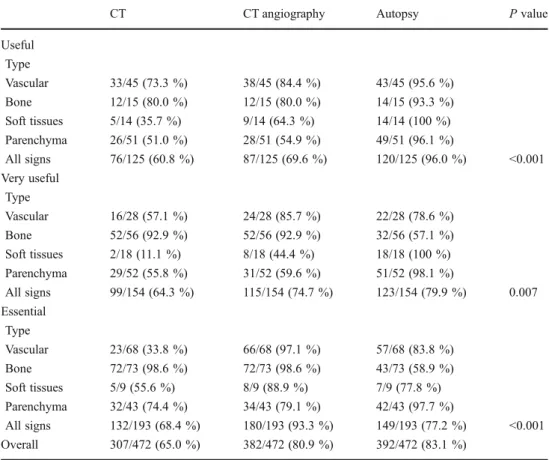
Compared to autopsy or CT, MPMCTA was most effi-cient in detecting essential findings (Table 3). MPMCTA was highly efficient in detecting vascular signs (97.1 %) and bone findings (98.6 %, with bone filter reconstructions). However, it demonstrated some limitations regarding the detection of parenchymal findings (79.1 %), while autopsy demonstrated major limitations in detecting bone findings (58.9 %). These two methods were complementary, as 97.7 % of findings in parenchyma were detected by autopsy. Useless findings for defining the cause of death were reported in both radiological and autopsy reports (Table4), and varied from 14.7 % of reported findings for MPMCTA to 17.9 % for native CT scan.
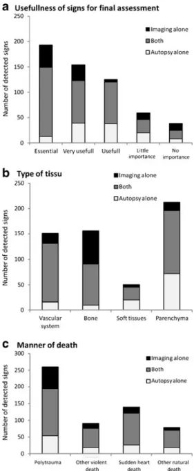
Figure1 illustrates the comparative abilities of imaging and autopsy to detect findings. Thirty of the 73 essential bone signs were detected by imaging alone, while nine of the 43 essential parenchymal findings were detected by
Table 1 Characteristics of observed cases (n=50)
Characteristic Description
Sex, number of males (%) 39 (76.5 %)
Age
Mean (SD) 52 years (19.6 years)
Median (range) 51 (15–89)
Cause of death, n (%)
Polytrauma 11 (22 %)
Other violent death 13 (26 %)
Sudden cardiac death 17 (34 %)
Other natural death 9 (18 %)
Number of observed signs
Mean (SD) 11.2 signs (8.9) Median (range) 8 (1–49) <3 1 (3.9 %) 3–10 28 (54.9 %) 11–20 15 (29.4 %) >20 6 (11.8 %)
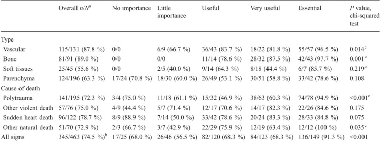
Table 2 Autopsy findings detected by CT angiography
Overall n/Na No importance Little
importance
Useful Very useful Essential P value,
chi-squared test Type Vascular 115/131 (87.8 %) 0/0 6/9 (66.7 %) 36/43 (83.7 %) 18/22 (81.8 %) 55/57 (96.5 %) 0.014c Bone 81/91 (89.0 %) 0/0 0/0 11/14 (78.6 %) 28/32 (87.5 %) 42/43 (97.7 %) 0.001c Soft tissues 25/45 (55.6 %) 0/0 2/5 (40.0 %) 9/14 (64.3 %) 8/18 (44.4 %) 6/7 (85.7 %) 0.219c Parenchyma 124/196 (63.3 %) 17/24 (70.8 %) 18/30 (60.0 %) 26/49 (53.1 %) 30/51 (58.8 %) 33/42 (78.6 %) 0.108 Cause of death Polytrauma 141/195 (72.3 %) 3/4 (75.0 %) 11/18 (61.1 %) 15/32 (46.9 %) 38/63 (60.3 %) 74/78 (94.9 %) <0.001c
Other violent death 57/76 (75.0 %) 4/9 (44.4 %) 5/7 (71.4 %) 12/17 (70.6 %) 14/17 (82.3 %) 22/26 (84.6 %) 0.175
Sudden heart death 96/122 (78.7 %) 8/9 (88.9 %) 7/14 (50.0 %) 33/42 (78.6 %) 20/24 (83.3 %) 28/33 (84.8 %) 0.075
Other natural death 51/70 (72.9 %) 2/3 (66.7 %) 3/7 (42.9 %) 22/29 (75.9 %) 12/19 (63.4 %) 12/12 (100 %) 0.035c
All signs 345/463 (74.5 %)b 17/25 (68.0 %) 26/46 (56.5 %) 82/120 (68.3 %) 84/123 (68.3 %) 136/149 (91.3 %) <0.001
a
n the number of reported signs from CT angiography, N the total number of signs reported from the autopsy. n is a subset of N b
The proportion of detected signs was case dependent (P<0.05; chi-squared test) c
autopsy alone. Thirty of the 38 findings that were judged of no importance were of parenchymal origin.
As the radiological investigation showed clear advan-tages especially concerning bone findings, we investigated these findings in more details (Table 5). One hundred twenty-three fractures were observed on 22 cases. The num-ber of fractures per case ranged from one to 33 fractures (median = 2). Thirty-nine fractures (31.7 %) were not reported during the autopsy. Missed fractures most often concerned the scapula (3/3; 100 %), the pelvis (5/7;
71.4 %), the skull (17/36; 47.2 %), and the spine (13/30; 43.3 %), and least of all the ribs, sternum and collar bone (1/40; 2.5 %), the lower limb (0/3; 0 %), and the hyoid bone (0/3; 0 %).
Discussion
The present study evaluated the diagnostic value of MPMCTA, particularly its additional value compared with
Table 3 Ability of CT alone, CT angiography, and autopsy to detect useful signs
CT CT angiography Autopsy P value
Useful Type Vascular 33/45 (73.3 %) 38/45 (84.4 %) 43/45 (95.6 %) Bone 12/15 (80.0 %) 12/15 (80.0 %) 14/15 (93.3 %) Soft tissues 5/14 (35.7 %) 9/14 (64.3 %) 14/14 (100 %) Parenchyma 26/51 (51.0 %) 28/51 (54.9 %) 49/51 (96.1 %) All signs 76/125 (60.8 %) 87/125 (69.6 %) 120/125 (96.0 %) <0.001 Very useful Type Vascular 16/28 (57.1 %) 24/28 (85.7 %) 22/28 (78.6 %) Bone 52/56 (92.9 %) 52/56 (92.9 %) 32/56 (57.1 %) Soft tissues 2/18 (11.1 %) 8/18 (44.4 %) 18/18 (100 %) Parenchyma 29/52 (55.8 %) 31/52 (59.6 %) 51/52 (98.1 %) All signs 99/154 (64.3 %) 115/154 (74.7 %) 123/154 (79.9 %) 0.007 Essential Type Vascular 23/68 (33.8 %) 66/68 (97.1 %) 57/68 (83.8 %) Bone 72/73 (98.6 %) 72/73 (98.6 %) 43/73 (58.9 %) Soft tissues 5/9 (55.6 %) 8/9 (88.9 %) 7/9 (77.8 %) Parenchyma 32/43 (74.4 %) 34/43 (79.1 %) 42/43 (97.7 %) All signs 132/193 (68.4 %) 180/193 (93.3 %) 149/193 (77.2 %) <0.001 Overall 307/472 (65.0 %) 382/472 (80.9 %) 392/472 (83.1 %)
Table 4 Proportion of signs
reported as not useful Native CT CT angiography Autopsy P value
Type Vascular 7/79 (8.9 %) 7/135 (5.2 %) 9/131 (6.9 %) Bone 10/146 (6.8 %) 10/146 (6.8 %) 2/91 (2.2 %) Soft tissues 4/16 (25.0 %) 5/30 (16.7 %) 6/91 (2.2 %) Parenchyma 46/133 (34.6 %) 44/137 (32.1 %) 54/196 (27.5 %) Cause of death Polytrauma 16/171 (9.4 %) 16/204 (7.8 %) 22/195 (11.3 %)
Other violent death 16/55 (29.1 %) 16/72 (22.2 %) 16/76 (21.0 %)
Sudden heart death 28/97 (28.9 %) 28/114 (24.6 %) 23/122 (18.8 %)
Other natural death 7/51 (13.7 %) 6/58 (10.3 %) 10/70 (14.3 %)
native CT and advantages and limitations compared with conventional autopsy. We compared findings extracted from radiological reports with those extracted from conventional autopsy reports without taking into consideration the results of histology, toxicology, or additional examinations because the study’s aim was to compare the results of autopsy and imaging, and not those of the entire medico-legal investiga-tion to imaging alone.
To determine the overall value of MPMCTA, medico-legal cases were selected according to the availability of technical personnel during the initial implementation of this
new technique, rather than in response to specific indica-tions. Therefore, MPMCTA was applied to some cases for which this type of investigation would not have been indi-cated in routine practice. This consequence can be assimi-lated as a limitation of the study. However, different types of medico-legal cases are examined. We therefore limited bias concerning the application of this technique to only one kind of case. In addition, this procedure permitted to determine in which cases the performance of MPMCTA is of use, and therefore to define indications for its application.
To examine the impact of MPMCTA, we categorized different findings according to their importance for solving respective medico-legal cases. Although the two board-certified forensic pathologists who performed this grading have graduated from different European medico-legal schools, their estimations were similar regarding the impor-tance of each finding.
As suspected at the beginning of our study, these results demonstrate that MPMCTA is highly efficient in the detec-tion of vascular findings, particularly regarding hemorrhage sources (Table 3). However, some vascular findings were only detected by conventional autopsy. Such findings were bleedings into the vascular wall or ruptured atherosclerotic plaques that were too small to be detected by CT because they were essentially located in the coronary vessels. A similar observation has already been described by Michaud et al., who investigated the use of MPMCTA to examine coronary arteries [22].
The most important weakness of MPMCTA was its in-ability to visualize parenchymal findings. Although the per-formance of an MPMCTA can significantly improve the detection of parenchymal findings compared with native MDCT (from 65 % to 80.9 %), it still remains less sensitive than conventional autopsy. This difference can be easily explained by the fact that, even with the injection of contrast agent, contrast of the parenchyma is not high enough to diagnose lesions of inner organs. The implementation of MRI has already been proposed to overcome this disadvan-tage of forensic imaging, and initial studies have had prom-ising results [25–27]. However, gaining access to an MRI unit may be even more difficult for forensic institutions than the performance of an MPMCTA.
Concerning the detection of bone findings, the radiolog-ical examination appears to clearly overcome conventional autopsy (Table2). As shown by our results and in Table5, standard autopsy can miss fractures in different regions of the body, especially in those which are difficult to access (scapula, spine, pelvis, and skull). Concerning the skull, missed fractures were mostly located in the facial bones which are not investigated routinely by a standard conven-tional autopsy. By using an appropriated filter of reconstruc-tion, bone findings can be obtained by performing either native CT scan or MPMCTA.
Fig. 1 The number of signs detected by each method (by postmortem radiology = MDCT + PMCTA, by both postmortem radiology and conventional autopsy, and by conventional autopsy alone) depending on importance (a), type of tissue (b), and manner of death (c)
One surprising result of this study was that MPMCTA appears to have greater sensitivity regarding soft tissue findings, such as small hemorrhages in subcutaneous fatty tissue or muscular tissue. This fact may be explained by the high sensitivity of contrast agents to detect the smallest extravasations (e.g., hematomas) but also by the fact that we have compared radiological data to findings from stan-dard conventional autopsy, including opening three body cavities and examining soft tissue in the thorax and abdo-men, but not including the dissection of soft tissue of the back. While the complete and detailed dissection of
subcutaneous and muscular tissue of the back requires a special indication in conventional autopsy, the soft tissue of the back is always visualized during a standard CT acquisition, which includes the head, thorax, and abdomen. Therefore, MPMCTA may detect small hemorrhages in the muscle and subcutaneous tissue that would not necessarily be searched for during a conventional autopsy. This possi-bility may indicate that MPMCTA could also be used as a screen to decide whether dissection of the back is necessary. Native CT scan and MPMCTA provided nearly the same percentage of non-useful findings (Table 4). The mean
Table 5 Fractures
af the number of fractures missed
at the autopsy, F the total num-ber of fractures reported. f is a subset of F
bFour fractures from the leg from
two cases detected by both au-topsy and CT scan, three frac-tures of the scapula from two cases all undetected at the autop-sy, and three hyoidal bone frac-ture all seen at the autopsy
Case no. Skull f/Fa Spine f/Fa Rib, sternum,
and collar bone
f/Fa Pelvis f/Fa Othersb f/Fa All fractures f/Fa 5 0/1 1/1 1/2 11 0/2 0/2 0/4 13 10/13 3/8 0/5 1/1 2/6 16/33 14 0/1 0/1 0/2 16 3/9 0/4 0/1 3/14 5/11 0/2 17 2/3 0/5 3/3 19 0/2 22 0/1 0/1 23 1/4 1/3 2/7 24 5/8 0/1 5/9 26 1/2 1/2 29 0/1 0/1 0/2 0/2 0/2 0/1 3/10 0/2 0/2 0/1 0/6 31 0/2 35 0/2 36 0/2 38 0/1 39 2/3 0/2 0/2 0/1 1/2 40 0/2 41 0/2 42 0/1 44 0/2 0/2 0/2 45 1/1 2/2 0/2 0/1 0/1 3/7 Total missed at autopsy 17/36 (47.22 %) 13/30 (43.33 %) 1/40 (2.5 %) 5/7 (71.43 %) 3/10 (30 %) 39/123 (31.71 %)
number of findings reported as non-useful for conclusions of sudden cardiac death and other violent death is slightly higher for imaging than for autopsy, possibly because radi-ologists are used to a very detailed description of patholog-ical changes that may play a role in clinpatholog-ical investigations, but not in forensic ones. A typical example is the description of degenerative lesions of the vertebral spine, which are mostly present in elderly subjects. As a matter of fact, elderly subjects are mostly victims of sudden cardiac death, and therefore the description of such“useless findings” may be observed more often in this group. Cases of other violent death were typically cases of intoxication. These also, showed more preexisting pathological changes that were described by radiologists without influencing the cause of death.
According to the results of our study, the indication to perform MPMCTA depends on the findings that are expected. MPMCTA is indicated if lesions are suspected in the vascular system according to the circumstances of death and the medical anamnesis (e.g., cases of polytrauma or sudden cardiac death). Cases in which a hemorrhage is suspected, or in which the vascular system has been modi-fied by a surgical intervention (e.g., after coronary bypass), comprise a very special group of cases in which a detailed examination of the vascular anatomy is of importance. The performance of MPMCTA does not make sense in cases in which no vascular lesions are expected (e.g., cases of intox-ication, drowning, or hanging). A vascular lesion should be suspected as a result of the reported data concerning the case, as well as after viewing the data from a native CT scan. In our institute, the decision to perform MPMCTA is made by the forensic pathologist in charge of the case, who has all information necessary to know the circumstan-ces of death and who will view radiological images of the native MDCT before starting the autopsy. This new tech-nique can be implemented easily, particularly in institutes with access to a CT unit. Performance of a complete MPMCTA takes 1 to 1.5 h (depending on the MDCT unit used), including sample collections performed prior to the injection of the contrast agent. Thus, the entire postmortem investigation would be prolonged by up to 1.5 h, a change that appears to be possible, especially considering the advantages that can be obtained by using this technique. In our institute, the entire radiological examination is per-formed by forensic radiographers [26], which have been added to the forensic team. The multi-phase exploration can increase on one hand the quality of the obtained images as it allows a nearly complete filling of the vessels of the head, the thorax, and the abdomen and, on the other hand, it ameliorates also the radiological interpretation as it allows to verify findings by comparing different angiographic phases as described by Grabherr et al. [21]. However, the perfor-mance of the technique and the radiological interpretation of the images require specialized personnel.
If we consider all findings regardless of tissue type and importance, MPMCTA and conventional autopsy demon-strate nearly the same sensitivity concerning the detection of important findings. MPMCTA can increase the sensitivity of native CT scan from 65 % to 80.9 %, while conventional autopsy detects 83.1 % of all findings (Table3). The result concerning essential findings is more surprising: autopsy reported 77.2 % and MPMCTA reported 93.3 % of all findings. For this reason, we esteem MPMCTA as a very powerful and useful tool that should be implemented as part of the routine medico-legal examination, if possible.
Acknowledgments This study was financially supported by the
Pro-motion Agency for Innovation of the Swiss Confederation (KTI Nr.10221.1 PFIW-IW) and the Leenards Foundation, Lausanne, Switzerland.
Conflicts of interest None
References
1. Dirnhofer R, Jackowski C, Vock P et al (2006) VIRTOPSY: mini-mally invasive, imaging-guided virtual autopsy. Radiographics 26
(5):1305–1333, Review
2. Thali M, Dirnhofer R, Vock P (eds) (2009) The virtopsy approach: 3D optical and radiological scanning and reconstruction in forensic medicine. CRC, New York
3. Weustink AC, Hunink MGM, van Dijke CF et al (2009) Minimally invasive autopsy: an alternative to conventional autopsy? Radiology 250(3):897–904
4. Jeffery AJ (2010) The role of computed tomography in adult postmortem examinations: an overview. Diagn Histopathol 16 (12):546–551
5. O’Donnell C (2010) An image of sudden death: utility of routine
postmortem computed tomography scanning in medico-legal
au-topsy practice. Diagn Histopathol 16(12):552–555
6. Poulsen K, Simonsen J (2007) Computed tomography as a routine
in connection with medico-legal autopsies. Forensic Sci Int 171(2–
3):190–7
7. Jacobsen C, Lynnerup N (2010) Craniocerebral trauma-congruence between postmortem computed tomography diagnoses and autopsy
results: a 2-year retrospective study. Forensic Sci Int 194(1–3):9–14
8. Roberts IS, Benamore RE, Benbow EW et al (2012) Postmortem imaging as an alternative to autopsy in the diagnosis of adult
deaths: a validation study. Lancet 379(9811):136–42
9. Aghayev E, Sonnenschein M, Jackowski C et al (2006) Postmortem radiology of fatal hemorrhage: measurements of cross-sectional areas of major blood vessels and volumes of aorta and spleen on MDCT and volumes of heart chambers on MRI. AJR 187(1):209–215
10. Kennedy D, Laing C, Tseng L et al (2010) Detection of active gastrointestinal hemorrhage with CT angiography: a 4(1/2)-year
retrospective review. J Vasc Interv Radiol 21(6):848–855
11. Karhunen PJ, Servo A (1993) Sudden fatal or non-operable bleed-ing from ruptured intracranial aneurysm: evaluation by post-mortem angiography with vulcanising contrast medium. Int J
Legal Med 106(2):55–59
12. Roberts I, Benamore R, Peebles C et al (2011) Diagnosis of coronary artery disease using minimally invasive autopsy: evalu-ation of a novel method of post-mortem coronary CT angiography.
13. Saunders S, Morgan B, Raj V et al (2011) Targeted post-mortem computed tomography cardiac angiography: proof of concept. Int J
Legal Med 125(4):609–616
14. Jackowski C, Thali M, Sonnenschein M et al (2005) Virtopsy: postmortem minimally invasive angiography using cross section techniques—implementation and preliminary results. J Forensic Sci 50(5):1175–1186
15. Grabherr S, Djonov V, Friess A et al (2006) Postmortem angiog-raphy after vascular perfusion with diesel oil and a lipophilic
contrast agent. AJR 187(5):W515–W523
16. Kominato Y, Fujikura T, Hata Y et al (2006) A case of postoper-ative hemorrhage after a hysterectomy in which a bleeding point of the left uterine artery was identified by postmortem angiography.
Legal Med 6(3):187–189
17. Ruder T, Ketterer T, Preiss U et al (2011) Suicidal knife wound to the heart: challenges in reconstructing wound chan-nels with postmortem CT and CT-angiography. Legal Medicine
13(2):91–94
18. Grabherr S, Djonov V, Yen K et al (2007) Postmortem angi-ography: review of former and current methods. AJR 188
(3):832–838
19. Saunders S, Morgan B, Raj V, Rutty G (2010) Post-mortem com-puted tomography angiography: past, present and future. Forensic Sci Med Pathol 7(3):271–277
20. Grabherr S, Gygax E, Sollberger B et al (2008) Two-step
post-mortem angiography with a modified heart–lung machine:
prelim-inary results. AJR 190(2):345–351
21. Grabherr S, Doenz F, Steger B et al (2011) Multi-phase post-mortem CT angiography: development of a standardized protocol. Int J Legal Med 125(6):791–802
22. Michaud K, Grabherr S, Doenz F, Mangin P (2012) Evaluation of postmortem MDCT and MDCT-angiography for the investigation of sudden cardiac death related to atherosclerotic coronary artery
disease. Int J Cardiovasc Imaging 28(7):1807–1822
23. Schneider B, Chevallier C, Dominguez A (2011) The forensic radiographer: a new member in the medicolegal team. Am J
Forensic Med Pathol 33(1):30–36
24. Brinkmann B (1999) Harmonisation of medico-legal autopsy rules.
Int J Legal Med 113(1):1–14
25. Jackowski C, Warntjes MJ, Berge J et al (2011) Magnetic resonance imaging goes postmortem: noninvasive detection and assessment of
myocardial infarction by postmortem MRI. Eur Radiol 21(1):70–8
26. Yen K, Thali M, Aghayev E et al (2005) Strangulation signs: initial correlation of MRI, MSCT and forensic neck findings. J Magn
Reson Imaging 22(4):501–510
27. Bouchireb K, Teychene AM, Rigal O et al (2010) Post-mortem MRI reveals CPT2 deficiency after sudden infant death. Eur J Pediatr 169(12):1561–1563