M A J O R A R T I C L E
Histone Deacetylase Inhibitors Impair
Antibacterial Defenses of Macrophages
Matteo Mombelli,1Je´roˆme Lugrin,1Ivana Rubino,1Anne-Laure Chanson,1Marlyse Giddey,2Thierry Calandra,1and
Thierry Roger1
1Infectious Diseases Service, Department of Medicine, Centre Hospitalier Universitaire Vaudois and University of Lausanne; and2Department of
Fundamental Microbiology, University of Lausanne, Switzerland
Histone deacetylases (HDACs) control gene expression by deacetylating histones and nonhistone proteins. HDAC inhibitors (HDACi) are powerful anticancer drugs that exert anti-inflammatory and immunomod-ulatory activities. We recently reported a proof-of-concept study demonstrating that HDACi increase susceptibility to bacterial infections in vivo. Yet, still little is known about the effects of HDACi on antimicrobial innate immune defenses. Here we show that HDACi belonging to different chemical classes inhibit at multiple levels the response of macrophages to bacterial infection. HDACi reduce the phagocytosis and the killing of Escherichia coli and Staphylococcus aureus by macrophages. In line with these findings, HDACi decrease the expression of phagocytic receptors and inhibit bacteria-induced production of reactive oxygen and nitrogen species by macrophages. Consistently, HDACi impair the expression of nicotinamide adenine dinucleotide phosphate (NADPH) oxidase subunits and inducible nitric oxide synthase. These data indicate that HDACi have a strong impact on critical antimicrobial defense mechanisms in macrophages.
The innate immune system plays a crucial role in host defenses against invasive microorganisms. Professional phagocytes are key sentinel cells of the innate immune system. Pathogen recognition relies on the capacity of phagocytes to sense microbial molecular motifs (eg, lipopolysaccharide, peptidoglycan, lipopeptides, mannans, glucans, flagellin, and nucleic acids) via pattern-recognition receptors comprising Toll-like receptors (TLRs), nucleotide-binding oligomerization domainlike receptors (NLRs), retinoic acid–inducible gene I (RIG-I)– like receptors (RLRs), C-type lectin receptors (CLRs), and scavenger receptors [1]. The engagement of phagocytic receptors, either through a direct interaction with
microbial motifs or through the recognition of opsonized infectious agents, stimulates the engulfment and the de-livery of the pathogen to the phagosome. Phagosome maturation by fission and fusion with endosomes and lysosomes generates the phagolysosome that provides a powerful microbicidal microenvironment, usually re-sulting in efficient microbial killing [2,3]. The release of proinflammatory cytokines during an infection stimulates the production of powerful phagocyte activating mole-cules like interferon c (IFNc).
Reversible acetylation of the e amino groups of lysine residues from histones and nonhistone proteins (such as a-tubulin, steroid receptors, HSP90, and regulators of nuclear import and transcription) is controlled by histone acetyltransferases and histone deacetylases (HDACs). Generally, acetylated histones are associated with active gene transcription, whereas deacetylated histones are associated with transcription repression [4–6]. The 18 mammal HDACs have been classified into class I (HDAC1–3 and 8), class IIa (HDAC4, 5, 7 and 9), class IIb (HDAC6 and 10), class III (SIRT1–7), and class IV (HDAC11) HDACs [4,7]. Small-molecule inhibitors of class I, II, and IV HDACs were originally identified
Received 10 May 2011; accepted 24 June 2011.
Correspondence: Thierry Roger, PhD, Infectious Diseases Service, BH19-111, Centre Hospitalier Universitaire Vaudois, Rue du Bugnon 46, CH-1011 Lausanne, Switzerland (thierry.roger@chuv.ch).
The Journal of Infectious Diseases 2011;204:1367–74
Ó The Author 2011. Published by Oxford University Press on behalf of the Infectious Diseases Society of America. All rights reserved. For Permissions, please e-mail: journals.permissions@oup.com
0022-1899 (print)/1537-6613 (online)/2011/2049-0010$14.00 DOI: 10.1093/infdis/jir553
for their potential to induce cellular differentiation, growth ar-rest, and apoptosis of transformed cells. HDACi targeting class I and II HDACs have been reported to counteract cancer de-velopment by reducing tumor angiogenesis, metastasis, and invasion and antitumor immunity [4–6].
Besides their anticancer properties, HDACi exert immuno-modulatory activities that have been exploited for the treatment of inflammatory and autoimmune disease [8]. Recently, we re-ported that HDACi interfere with the response of innate im-mune cells stimulated with TLR agonists and increase the mortality of mice to microbial sepsis [9]. Yet, whether HDACi impair the phagocytosis and the killing of bacteria by phagocytes remains unknown. To more deeply characterize the influence of HDACi on innate immune responses, we investigated whether HDACi have an impact on key antibacterial defense mechanisms of macrophages. We report that HDACi reduce the expression of phagocytic and opsonophagocytic receptors and inhibit the phagocytosis of Escherichia coli and Staphylococcus aureus, 2 of the most common infectious agents, by macrophages. Moreover, HDACi impair the generation of reactive oxygen and nitrogen species by macrophages infected with bacteria, resulting in a marked reduction of bacterial killing.
MATERIALS AND METHODS Cells and Reagents
Animal procedures were approved by the Office Ve´te´rinaire du Canton de Vaud (authorizations n° 876.6) and performed ac-cording to institution guidelines for animal experiments. We purchased 8- to 10-week-old female BALB/c mice from Charles River Laboratories . We obtained mouse bone marrow–derived macrophages (BMDMs) and thioglycolate-elicited macrophages as previously described [10,11]. RAW 264.7 macrophages were cultured in Roswell Park Memorial Institute (RPMI) 1640 me-dium containing 2 mmol/L glutamine and 10% fetal calf serum (FCS) [12]. E. coli O18:K1:H7 (E. coli) and S. aureus AW7 (S. aureus) are clinical isolates obtained from septic patients hospitalized at the Centre Hospitalier Universitaire Vaudois. We purchased trichostatin A (TSA) and valproic acid (VPA) from Sigma-Aldrich, Salmonella minnesota Ultra Pure lipopolysac-charide (LPS) from List Biologicals Laboratories , and IFNc from R&D Systems. The concentrations of TSA (dissolved in ethanol) and VPA (dissolved in phosphate-buffered saline [PBS]) used in this study were selected based on previous publications [13–18] and did not affect the viability (Trypan blue staining and 3-[4,5-dimethylthiazol-2-yl]-2,5-diphenyltetrazolium bromide [MTT] Cell Proliferation and Viability Assay) of BMDMs ($ 85% cell recovery after 18 hours of culture with 20–40 nmol/ L TSA and 1–2 mmol/L VPA with or without bacteria; n 5 6–9 determinations; P . .5 for all conditions). Ethanol and PBS ve-hicle controls were performed in each experiment. For simplicity only 1 set of data is presented in each (Figures 2,4,5, and6).
Assay for Bacterial Uptake and Bacterial Killing
E. coli and S. aureus were grown overnight at 37°C in tryptic soy broth (BD Biosciences), washed in PBS, and adjusted to 107 colony-forming units (CFU)/mL in RPMI medium containing 10% FCS. BMDMs (4 3 105cells in 24-well cell-culture plates, Costar) were treated with TSA or VPA for 18 hours. Medium was changed and cells were incubated for 1 hour with bacteria at a multiplicity of infection of 20 bacteria per macrophage. Nonadherent bacteria were removed by washing with PBS. Ex-tracellular bacteria were killed by a 30-minute exposure to either 100 lg/mL of gentamicin (E. coli) or 10 lg/mL of lysostaphin (S. aureus). We washed and lysed BMDMs. We plated serial dilutions of cell lysates on agar plates and enumerated colonies to calculate the number of phagocytosed bacteria. In parallel wells, BMDMs were treated as previously except that, after 30 minutes of incubation with antibiotics, cells were washed and incubated for a further 24 hours in culture medium containing 20 lg/mL gentamicin or 10 lg/mL lysostaphin. Bacteria were enumerated and results expressed as percent changes in bacterial counts using the following formula: (count after 24 hours/count after 1 hour) 3 100. Of note, neither TSA nor VPA at the concentrations used in these assays were toxic for bacteria. RNA Analysis by Quantitative Real-Time Polymerase Chain Reaction
RNA was isolated using the RNeasy kit (Qiagen). Reverse tran-scription was performed using the ImProm II RT System kit (Promega). Quantitative real-time polymerase chain reaction (PCR) was performed with a 7500 Fast Real-Time PCR System using the Power SYBR Green PCR Master Mix (Applied Bio-systems) and primer pairs (Supplementary Table 1) as previously described [19]. We tested samples in triplicate. For each mea-surement, we processed in parallel a standard made of successive dilutions of a reference complementary DNA. The relative ex-pression levels of NADPH oxidase subunits and inducible nitric oxide synthase (iNOS) were reported to the relative expression of glyceraldehyde-3-phosphate dehydrogenase (Gapdh) and ex-pressed in arbitrary units (AU). The expression of phagocytic re-ceptors and TLRs was calculated with the comparative Ct (DDCt) method . The expression of the target gene was first normalized to the endogenous control (Gapdh) and then to that of a calibrator (ie, data obtained from cells cultured with vehicle set at 1). Im-portantly, the Ct values of Gapdh were not affected by TSA or VPA (19.24 6 0.13, 19.10 6 0.03, 19.30 6 0.48, 19.21 6 0.06, and 19.10 60.15 in BMDMs cultured for 18 hours with medium, 20 nmol/L TSA, 40 nmol/L TSA, 1 mmol/L VPA, and 2 mmol/L VPA, re-spectively; n 5 6 determinations; P . .05 for all conditions compared with cells cultured in medium). In selected experiments, results were validated using Hprt as an endogenous control. Flow Cytometric Analysis
BMDMs cultured for 18 hours with TSA (40 nmol/L) and VPA (2 mmol/L) were incubated 30 minutes at 4°C in PBS containing
5% FCS, 5 mM ethylenediaminetetraacetic acid (EDTA), 2.4G2 monoclonal antibody, and monoclonal antibodies specific for macrophage scavenger receptor 1 (Msr1/CD204), CD11c, CD14, and major histocompatibility complex II (MHC-II) [20]. Ac-quisition and analysis were performed using a FACSCalibur (BD Biosciences) and FlowJo 8.5.3 software (FlowJow).
Analysis of Oxidative Burst Using the Dichlorofluorescein Diacetate Fluorescence Assay
BMDMs (4 3 105 cells in 24-well cell-culture plates) were cultured as previously described [21] and incubated for 18 hours with TSA and VPA. Dichlorofluorescein diacetate (DCFDA) (20 lmol/L, Sigma-Aldrich) was added to the cultures followed 15 minutes later by bacteria (5 3 108CFU/mL). After 30 mi-nutes, cell fluorescence was measured by flow cytometry. Western Blot Analysis
Cell lysates were electrophoresed through polyacrylamide gels and transferred onto nitrocellulose membranes as previously described [12]. Membranes were incubated with antibodies di-rected against iNOS (BD Biosciences), p47phox(Santa Cruz) and tubulin (Sigma). After washing, membranes were incubated with horseradish peroxidase (HRP)–conjugated secondary antibody (Pierce). Signals were revealed using the ECL Western blotting analysis system (GE Healthcare).
Nitrite/Nitrate Measurements
BMDMs (105 cells in 96-well cell-culture plates) were pre-incubated for 1 hour with TSA and VPA and stimulated with LPS (100 ng/mL), IFNc (100 U/mL), E. coli (108CFU/mL), and S. aureus (108CFU/mL). Cell-culture supernatants were collected after 24 hours. The concentrations of nitrite/nitrate were measured using the Griess reagent.
Statistics
Statistical analyses were performed using PRISM (GraphPad Software). Comparisons between the different groups were performed by analysis of variance and appropriate post hoc analyses. P values are 2-sided, and values of , .05 were con-sidered to indicate statistical significance.
RESULTS
HDACi Inhibit Bacterial Phagocytosis by Macrophages
We recently reported that HDACi impair host defenses to bacterial infection in vivo [9]. Whether HDACi impact the phagocytosis and the killing of bacteria is currently unknown. To fill this gap, we first analyzed the phagocytosis of gram-negative (E. coli) and gram-positive (S. aureus) bacteria by BMDMs pretreated with 2 chemically unrelated HDACi: TSA, a hydroxamate widely used as a prototypical broad-spectrum HDACi, and VPA, a clinically relevant short fatty acid. The dose and duration of treatment with TSA and VPA were in the range
of those used in cancer preclinical studies or measured in pa-tients enrolled in cancer clinical trials (VPA) (see for example [13–18]). We evaluated phagocytosis after 1 hour of contact between bacteria and macrophages. As shown in Figure 1, HDACi dose dependently reduced the number of E. coli (2- to 4-fold; P , .05) and S. aureus (1.5- to 2-fold; P , .05) phago-cytosed by BMDMs.
HDACi Impair the Expression of Phagocytic Receptors
Macrophages express phagocytic scavenger receptors, including macrophage scavenger receptor 1 (Msr1/SR-AI/CD204), CD14, CD36, and C-type lectins such as Dectin-1 (encoded by Clec7a) that mediate the recognition of microbial ligands expressed at the surface of pathogens and initiate phagocytosis. Macrophages also express opsonic phagocytic receptors of the integrin family (integrinaX/Itgax/CD11c, integrinb2/Itgb2/CD18, and integrina5/
Itga5/CD49e) that facilitate the uptake of microorganisms coated with opsonins like the mannose-binding lectin, comple-ment subcomponents, growth arrest specific 6, ficolins, and pentraxins [2,22]. TSA and VPA reduced 2- to 10-fold Msr1,
Figure 1. Histone deacetylase inhibitors impair the phagocytosis of Escherichia coli (A) and Staphylococcus aureus (B ). Mouse bone marrow– derived macrophages (BMDMs) were incubated for 18 hours with increasing concentrations of trichostatin A (TSA) and valproic acid (VPA) before the addition of 107 colony-forming units (CFU) of E. coli or 1.5 3 107CFU of S. aureus. The number of bacteria ingested by BMDMs
was determined 1 hour later. Data are presented as mean 6 standard deviation (SD) of quadruplicate samples from 1 experiment representative of 2–3 experiments. *, .05 , P , .005; **, P , .005.
CD14, Dectin-1 and Itgax messenger RNA (mRNA) levels in BMDMs (Figure 2A). TSA inhibited Itgb2 expression more ef-ficiently than did VPA (2.4-fold with 40 nM TSA vs 1.4-fold with 2 mM VPA), whereas HDACi did not affect CD36, Itga5, or Itga6 expression. Flow cytometry analyses confirmed that TSA and VPA inhibited the expression of Msr1, CD11c and CD14 by BMDMs (Figure 2B). As a control of nonspecific broad in-hibitory effects of HDACi, MHC-II expression was not affected by HDACi. All together, these data suggest that reduced ex-pression of phagocytic receptors may contribute to impair the phagocytosis of E. coli and S. aureus in macrophages treated with HDACi.
HDACi Inhibit Bacterial Killing
TLRs play crucial roles in the sensing of invasive microorganisms and in transmitting signals involved in the maturation of phag-osomes [23]. Interestingly, we observed that HDACi strongly reduced baseline expression of TLR1–7 and TLR9 in BMDMs (Figure 2C). Pathogen delivery to phagolysosomes usually results in effective microbial killing [2,24]. In agreement, , 5% of E. coli and 25% of S. aureus phagocytosed by BMDMs were recovered 24 hours later in macrophages (P , .001; Figure 3). TSA and VPA reduced 5-fold and 3-fold E. coli and S. aureus killing, re-spectively (ie, increasing bacteria recovery to 25% and 75% of the ingested inoculum; P , .05). Thus, HDACi inhibit both the phagocytosis (Figure 1) and the killing (Figure 3) of bacteria by macrophages, in agreement with the observation that HDACi increased the susceptibility of mice to microbial infection [9]. HDACi Interfere With the Generation of Reactive Oxygen Species
In response to microbial challenge, macrophages produce highly toxic reactive oxygen species (ROS) that contribute to pathogen destruction [25]. The generation of ROS in BMDMs was ana-lyzed by flow cytometry using the cell permeable nonfluorescent dye 2#,7#-dichlorofluorescein diacetate (DCFDA) that is trans-formed on oxidation into the highly fluorescent DCF. E. coli and S. aureus strongly increased DCF fluorescence in BMDMs, which was inhibited 2- to 3-fold by TSA or VPA (Figure 4A and 4B). HDACi also inhibited the production of ROS in BMDMs stimulated with phorbol myristate acetate (.10-fold reduction, data not shown) indicating that HDACi inhibit the oxidative burst induced by microbial and nonmicrobial stimuli.
In macrophages, ROS are generated during the respiratory burst through the action of the phagocytic NADPH oxidase, an enzymatic complex composed of 2 membrane-associated subunits (gp91phox/NOX2 and p22phox), 3 cytosolic subunits (p47phox, p40phox, and p67phox), and the Rac2 regulatory subunit [26,27]. Cytokines, particularly IFNc, and microbial products released during an infection prime and amplify macrophage respiratory burst through the induction of NADPH oxidase subunits [21]. Real-time PCR and Western blot analyses re-vealed that TSA and VPA dose-dependently inhibited baseline
expression of NADPH oxidase subunits and potently inhibited the upregulation of the catalytic gp91phoxand regulatory p47phox Figure 2. Histone deacetylase inhibitors inhibit the expression of phagocytic and Toll-like receptors. A, Real-time polymerase chain reaction (PCR) analysis of Msr1, CD14, CD36, Clec7a, Itgax, Itgb2, Itga5, and Itga6 messenger RNA expression in mouse bone marrow–derived macrophages (BMDMs) incubated for 8 hours with increasing concentrations of trichostatin A (TSA) and valproic acid (VPA). Data are presented as mean 6 standard deviation (SD) of triplicate samples from 1 experiment and are representative of 2 independent experiments. B, Flow cytometry analysis of Msr1, CD14, CD11c, and major histocompatibility complex II (MHC-II) expression by BMDMs incubated for 18 hours with medium (gray area), TSA (dashed line), and VPA (solid line). Results are representative of 2 independent experiments. C, Real-time PCR analysis of Toll-like receptors in BMDMs incubated for 8 hours with increasing concentrations of TSA and VPA. Data are presented as mean 6 standard deviation (SD) of triplicate samples from 1 experiment and are representative of 2 independent experiments. AU, arbitrary units; *, .05 , P , .005; **, P , .005.
subunits in LPS1IFNc–stimulated macrophages (Figure 5). All together, these data provide compelling evidence that HDACi inhibit ROS production in macrophages.
HDACi Inhibit Nitric Oxide Production and iNos Gene Expression
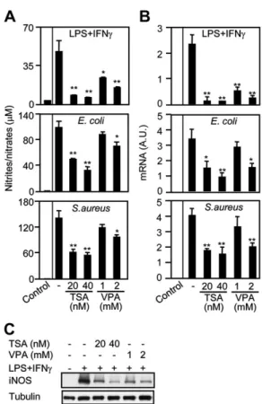
Nitric oxide (NO) is produced during the nitrosative burst by iNOS and represents an important antimicrobial effector mechanism [25,27]. TSA and VPA dose-dependently inhibited the production of NO by BMDMs stimulated with LPS1IFNc (50%–80% reduction; P , .05), in line with previous work showing that HDACi inhibit cytokine-induced NO release [28, 29]. More relevant for microbial infection, TSA and to a lesser extent VPA inhibited NO production induced by E. coli and S. aureus (50%–60% reduction using 20 nmol/L and 40 nmol/L TSA and 30%–35% reduction using 2 mmol/L VPA; P , .05) (Figure 6A). In agreement with these findings, real-time PCR and Western blot analyses demonstrated that TSA and VPA inhibited iNOS mRNA and protein expression in BMDMs
(Figure 6B and 6C). Similar results were obtained using thio-glycolate-elicited peritoneal and RAW 264.7 macrophages (data not shown). Taken together, these results suggest that HDACi impair NO production by BMDMs in response to bacterial challenge by interfering with iNOS expression.
DISCUSSION
In this study, we report for the first time to our knowledge that HDACi inhibit the phagocytosis and the killing of bacteria, the expression of phagocytic receptors, and the generation of oxi-dative and nitrosative bursts induced by bacteria in macro-phages. These data extend our previous work demonstrating that HDACi interfere with cytokine production by macrophages and impair host defenses to bacterial infection [9].
The inhibition of E. coli and S. aureus phagocytosis by HDACi was associated with a reduced expression of phago-cytic receptors, among which is Msr1 (scavenger receptor A1). Msr1 binds a wide range of microbial ligands and mediates Figure 3. Histone deacetylase inhibitors impair the killing of
Escherichia coli and Staphylococcus aureus. Mouse bone marrow– derived macrophages (BMDMs) were cultured for 18 hours with increasing concentrations of trichostatin A (TSA) and valproic acid (VPA) before the addition of 107colony-forming units (CFU) of E. coli (A) or
1.5 3 107CFU of S. aureus (B ). The number of bacteria recovered from macrophages after 24 hours was divided by the number of bacteria recovered after 1 hour and expressed in percentage using the formula (count after 24 hours/count after 1 hour) 3 100. Data are presented as mean 6 standard deviation (SD) of quadruplicate samples from 1 experiment representative of 2–3 experiments. *, .05 , P , .005; **, P , .005.
Figure 4. Histone deacetylase inhibitors interfere with the generation of reactive oxygen species. Mouse bone marrow–derived macrophages (BMDMs) were incubated with trichostatin A (TSA; 40 nmol/L) and valproic acid (VPA; 2 mmol/L) and exposed to Escherichia coli or Staphylococcus aureus as described in Material and Methods. A, The generation of reactive oxygen species (ROS) was quantified by flow cytometry by measuring dichlorofluorescein (DCF) diacetate oxidation into fluorescent DCF. B, Data are means 6 standard deviation (SD) of 2 independent determinations. P 5 .03 and .008 for E. coli and S. aureus vs control. *, P 5 .05; **, .05 , P , .005 vs E. coli– and S. aureus–treated cells.
nonopsonic phagocytosis of E. coli and S. aureus [30]. Moreover, Msr12/2mice are more susceptible than wild-type mice to S. aureus infection [31]. Thus, HDACi-mediated in-hibition of Msr1 expression may well help impair bacterial phagocytosis, although HDACi may target other phagocytic receptors such as the mannose receptor, MARCO, or CD14. Of note, HDACi decreased Dectin-1 expression in BMDMs. Considering that Dectin-1 is a major receptor involved in the recognition of b-glucan, we speculate that HDACi may affect the phagocytosis of yeast. In line with this hypothesis, we observed that VPA increases mortality of mice infected with Candida albicans [9].
The ax and b2 integrin subunits contribute to the structure of complement receptor (CR) 3 and CR4 that mediate the recog-nition of opsonized microorganisms by phagocytes. b2 integrins play an important role in antimicrobial defenses as suggested by the observation that patients with leukocyte adhesion deficiency type I (LADI) syndrome (ie, patients deficient in functional b2 integrin) have defects in phagocytosis and are prone to bacterial infections [32]. All together, inhibition of the expression of ax and b2 integrins and scavenger and lectin receptors by HDACi
support the contention that HDACi interfere with bacterial opsonic and nonopsonic phagocytosis.
HDACi powerfully inhibited the killing of E. coli and S. aureus by macrophages. This observation is congruent with the fact that VPA treatment increased the proportion and the magnitude of bloodstream infections in mice infected with Klebsiella pneu-moniae [9]. Reactive oxygen and nitrogen species are among the most deleterious components produced by phagocytes and implicated in the destruction of microorganisms [2, 24]. De-ficiency in members of the NADPH oxidase complex or in iNOS, which control the generation of superoxide (O22°) and
nitric oxide (NO°) radicals, impair the killing of E. coli and S. aureus by innate immune cells and compromise mouse Figure 5. Histone deacetylase inhibitors inhibit nicotinamide adenine
dinucleotide phosphate (NADPH) oxidase subunits expression. A, Real-time polymerase chain reaction (PCR) analysis of gp91, p22, p47, p67, p40, and Rac2 messenger RNA expression in mouse bone marrow–derived macro-phages (BMDMs) cultured for 8 hours with trichostatin A (TSA) and valproic acid (VPA). Results are expressed as the ratio of the gene of interest to that of glyceraldehyde-3-phosphate dehydrogenase. Real-time PCR (B ) and Western blot (C ) analyses of gp91 and p47 expression in BMDMs cultured for 1 hour with TSA and VPA and then stimulated for 6 hours with lipopolysaccharide (LPS; 100 ng/mL) 1 interferon c (IFNc; 100 U/mL). Data are presented as means 6 standard deviation (SD) of triplicate samples from 1 experiment and are representative of 2 independent experiments (A, B ). Abbreviation: AU, arbitary units.*, P , .005 vs control (A) and LPS1IFNc (B ).
Figure 6. Histone deacetylase inhibitors impair nitric oxide production and iNos gene expression. Mouse bone marrow–derived macrophages (BMDMs) were cultured for 1 hour with trichostatin A (TSA) and valproic acid (VPA) and then stimulated with lipopolysaccharide 1 interferon c (100 ng/mL and 100 U/mL), Escherichia coli (108 colony-forming units [CFU]/mL), and
Staphylococcus aureus (108 CFU/mL) for 24 hours (A) or 8 hours (B–C ). A, Nitrite/nitrate concentration in cell culture supernatants was measured using the Griess reagent. Data are presented as mean 6 standard deviation (SD) of triplicate samples from 1 experiment and are representative of 4 independent experiments. Real-time polymerase chain reaction (PCR) (B ) and Western blot (C ) analyses of inducible nitric oxide synthase (iNOS) expression. Results are expressed as the ratio of iNOS mRNA levels to that of glyceraldehyde-3-phosphate dehydrogenase. Data are means 6 SD of triplicate samples from 1 experiment and are representative of 3 independent experiments. Abbreviation: AU, arbitrary units. *, .05 , P , .005; **, P , .005.
survival [33–36]. Moreover, germ-line mutation in 1 of the components of NADPH oxidase complex results in chronic granulomatous disease characterized by recurrent bacterial and fungal infections and reduced life expectancy [25]. Inhibition of NADPH oxidase subunit and iNOS expression and of reactive oxygen and nitrogen species generation by HDACi likely rep-resents an effective mechanism by which these drugs impair the killing of bacteria.
In agreement with the notion that phagocytosis is coupled with a proinflammatory cytokine response and with the obser-vation that HDACi inhibit TLR expression, HDACi strongly impaired the secretion of cytokines and chemokines (TNF, IL-6, IL-12p40, and MIP-2a, data not shown) by macrophages in-fected with E. coli and S. aureus. These data expand on recent studies showing that HDACi inhibit cytokine production in-duced by cytokines and purified microbial products in innate immune cells [14, 15, 28, 37, 38]. Considering that proin-flammatory mediators released during the course of an infection coordinate the development of innate and adaptive immunity, one may expect that HDACi interfere with the generation of pathogen-specific adaptive immune response.
HDACi have been reported to interfere with signaling path-ways controlling the expression of genes particularly relevant for this study. Indeed, HDACi down-regulate the expression of PU.1 transcription factor [37,39], which regulates constitutive expression of HDACi-target genes encoding for integrins, scavenger receptors, TLR4, CD14, and p40, p47, and p67 NADPH oxidase subunits [20,40]. Moreover, HDACi have been reported to interfere with the activation of mitogen-activated protein kinases, NF-jB, and AP-1, which control inflammatory and antimicrobial host responses [15,41]. HDACi also impair gene expression through chromatin modifications or aceytlation-dependent recruitment of transcriptional repressors, albeit these mechanisms are less well characterized. For example, TSA in-hibits the expression of the proinflammatory cytokine macro-phage migration inhibitory factor through a local deacetylation of migration inhibitory factor-promoter–associated histones impairing the recruitment of the basal transcriptional machinery [42, 43]. Finally, we have recently shown that TSA inhibits macrophage response to LPS stimulation by inducing the ex-pression of Mi-2b and the activity of the Mi-2/NuRD tran-scriptional repressor complex [9].
HDACi have been used to treat inflammatory diseases in mouse models [8]. Abundant preclinical and clinical studies indicate that interfering with critical mediators of innate or adaptive immunity increases the risk of infections. Thus, one may question whether HDACi might affect natural host defenses in patients, as could be anticipated from the powerful immu-nomodulatory and anti-inflammatory activities of HDACi in vivo [8] and the increased susceptibility to bacterial and fungal infections of mice treated with HDACi [9]. Patients treated with HDACi (suberoylanilide hydroxamic acid, MS-275, valproate,
and ITF2357) in phase I and II clinical trials have developed episodes of severe infection even in the absence of treatment-induced neutropenia [44–49], suggesting the need to monitor the immune status and susceptibility to infection of patients treated with HDACi, especially immunosuppressed cancer patients [4–6,50].
In summary, this study demonstrates that HDACi impair the capacity of macrophages to ingest and destroy gram-positive and gram-negative bacteria. The fact that HDACi impede the expression of phagocytic receptors, the generation of oxygen-and nitrogen-reactive species, oxygen-and the release of proin-flammatory cytokines provide a plausible mechanism whereby HDACi negatively impact critical antimicrobial functions of innate immune cells and increase the susceptibility of mice to bacterial and fungal infection [9].
Supplementary Data
Supplementary dataare available at The Journal of infections Diseases online (http://www.oxfordjournals.org/our_journals/jid/).
Supplementary materials consist of data provided by the author that are published to benefit the reader. The posted materials are not copyedited. The contents of all supplementary data are the sole responsibility of the authors. Questions or messages regarding errors should be addressed to the author.
Notes
Financial support. This work was supported by the Swiss National Science Foundation (310000_114073 and 310030_132744 to T. R. and 310000_118266 to T. C.); a Merck Sharp & Dohme-Chibret award from the Swiss Society for Infectious Diseases; and the Leenaards Foundation (to T. R.).
Potential conflicts of interest. All authors: No reported conflicts. All authors have submitted the ICMJE Form for Disclosure of Potential Conflicts of Interest. Conflicts that the editors consider relevant to the content of the manuscript have been disclosed.
References
1. Ishii KJ, Koyama S, Nakagawa A, Coban C, Akira S. Host innate im-mune receptors and beyond: Making sense of microbial infections. Cell Host Microbe 2008; 3:352–63.
2. Stuart LM, Ezekowitz RA. Phagocytosis: Elegant complexity. Immunity 2005; 22:539–50.
3. Flannagan RS, Cosı´o G, Grinstein S. Antimicrobial mechanisms of phagocytes and bacterial evasion strategies. Nat Rev Microbiol 2009; 7:355–66.
4. Bolden JE, Peart MJ, Johnstone RW. Anticancer activities of histone deacetylase inhibitors. Nat Rev Drug Discov 2006; 5:769–84. 5. Glozak MA, Seto E. Histone deacetylases and cancer. Oncogene 2007;
26:5420–32.
6. Minucci S, Pelicci PG. Histone deacetylase inhibitors and the promise of epigenetic (and more) treatments for cancer. Nat Rev Cancer 2006; 6:38–51.
7. Yang XJ, Seto E. The Rpd3/Hda1 family of lysine deacetylases: From bacteria and yeast to mice and men. Nat Rev Mol Cell Biol 2008; 9:206–18.
8. Haberland M, Montgomery RL, Olson EN. The many roles of histone deacetylases in development and physiology: Implications for disease and therapy. Nat Rev Genet 2009; 10:32–42.
9. Roger T, Lugrin J, Le RD, et al. Histone deacetylase inhibitors impair innate immune responses to Toll-like receptor agonists and to in-fection. Blood 2011; 117:1205–17.
10. Roger T, Froidevaux C, Le RD, et al. Protection from lethal gram-negative bacterial sepsis by targeting Toll-like receptor 4. Proc Natl Acad Sci U S A 2009; 106:2348–52.
11. Roger T, David J, Glauser MP, Calandra T. MIF regulates innate im-mune responses through modulation of Toll-like receptor 4. Nature 2001; 414:920–4.
12. Roger T, Chanson AL, Knaup-Reymond M, Calandra T. Macrophage migration inhibitory factor promotes innate immune responses by suppressing glucocorticoid-induced expression of mitogen-activated protein kinase phosphatase-1. Eur J Immunol 2005; 35:3405–13. 13. Atmaca A, Al-Batran SE, Maurer A, et al. Valproic acid (VPA) in
patients with refractory advanced cancer: A dose escalating phase I clinical trial. Br J Cancer 2007; 97:177–82.
14. Bode KA, Schroder K, Hume DA, et al. Histone deacetylase inhibitors decrease Toll-like receptor–mediated activation of proinflammatory gene expression by impairing transcription factor recruitment. Im-munology 2007; 122:596–606.
15. Cao W, Bao C, Padalko E, Lowenstein CJ. Acetylation of mitogen-activated protein kinase phosphatase-1 inhibits Toll-like receptor sig-naling. J Exp Med 2008; 205:1491–503.
16. Daud AI, Dawson J, DeConti RC, et al. Potentiation of a topoisomerase I inhibitor, karenitecin, by the histone deacetylase inhibitor valproic acid in melanoma: Translational and phase I/II clinical trial. Clin Cancer Res 2009; 15:2479–87.
17. Mu¨nster P, Marchion D, Bicaku E, et al. Phase I trial of histone de-acetylase inhibition by valproic acid followed by the topoisomerase II inhibitor epirubicin in advanced solid tumors: A clinical and trans-lational study. J Clin Oncol 2007; 25:1979–85.
18. Mu¨nster P, Marchion D, Bicaku E, et al. Clinical and biological effects of valproic acid as a histone deacetylase inhibitor on tumor and sur-rogate tissues: Phase I/II trial of valproic acid and epirubicin/FEC. Clin Cancer Res 2009; 15:2488–96.
19. Delaloye J, Roger T, Steiner-Tardivel QG, et al. Innate immune sensing of modified vaccinia virus Ankara (MVA) is mediated by TLR2-TLR6, MDA-5 and the NALP3 inflammasome. PLoS Pathog 2009; 5:e1000480. 20. Roger T, Miconnet I, Schiesser AL, Kai H, Miyake K, Calandra T. Critical role for Ets, AP-1 and GATA-like transcription factors in regulating mouse Toll-like receptor 4 (Tlr4) gene expression. Biochem J 2005; 387:355–65.
21. Anrather J, Racchumi G, Iadecola C. NF-kappaB regulates phagocytic NADPH oxidase by inducing the expression of gp91phox. J Biol Chem 2006; 281:5657–67.
22. Areschoug T, Gordon S. Scavenger receptors: Role in innate immunity and microbial pathogenesis. Cell Microbiol 2009; 11:1160–9. 23. Blander JM. Signalling and phagocytosis in the orchestration of host
defence. Cell Microbiol 2007; 9:290–9.
24. Underhill DM, Ozinsky A. Phagocytosis of microbes: Complexity in action. Annu Rev Immunol 2002; 20:825–52.
25. Fang FC. Antimicrobial reactive oxygen and nitrogen species: Concepts and controversies. Nat Rev Microbiol 2004; 2:820–32.
26. Bedard K, Krause KH. The NOX family of ROS-generating NADPH oxidases: Physiology and pathophysiology. Physiol Rev 2007; 87:245–313. 27. Lambeth JD. NOX enzymes and the biology of reactive oxygen. Nat Rev
Immunol 2004; 4:181–9.
28. Leoni F, Zaliani A, Bertolini G, et al. The antitumor histone deacetylase inhibitor suberoylanilide hydroxamic acid exhibits antiinflammatory properties via suppression of cytokines. Proc Natl Acad Sci U S A 2002; 99:2995–3000.
29. Reilly CM, Mishra N, Miller JM, et al. Modulation of renal disease in MRL/lpr mice by suberoylanilide hydroxamic acid. J Immunol 2004; 173:4171–8.
30. Peiser L, Mukhopadhyay S, Gordon S. Scavenger receptors in innate immunity. Curr Opin Immunol 2002; 14:123–8.
31. Thomas CA, Li Y, Kodama T, Suzuki H, Silverstein SC, El Khoury J. Protection from lethal gram-positive infection by macrophage scavenger receptor-dependent phagocytosis. J Exp Med 2000; 191:147–56.
32. Bunting M, Harris ES, McIntyre TM, Prescott SM, Zimmerman GA. Leukocyte adhesion deficiency syndromes: adhesion and tethering defects involving beta 2 integrins and selectin ligands. Curr Opin Hematol 2002; 9:30–5.
33. Shiloh MU, MacMicking JD, Nicholson S, et al. Phenotype of mice and macrophages deficient in both phagocyte oxidase and inducible nitric oxide synthase. Immunity 1999; 10:29–38.
34. Jackson SH, Gallin JI, Holland SM. The p47phox mouse knock-out model of chronic granulomatous disease. J Exp Med 1995; 182:751–8. 35. Ellson CD, Davidson K, Ferguson GJ, O’Connor R, Stephens LR, Hawkins PT. Neutrophils from p40phox-/- mice exhibit severe defects in NADPH oxidase regulation and oxidant-dependent bacterial killing. J Exp Med 2006; 203:1927–37.
36. Sasaki S, Miura T, Nishikawa S, Yamada K, Hirasue M, Nakane A. Protective role of nitric oxide in Staphylococcus aureus infection in mice. Infect Immun 1998; 66:1017–22.
37. Aung HT, Schroder K, Himes SR, et al. LPS regulates proinflammatory gene expression in macrophages by altering histone deacetylase ex-pression. FASEB J 2006; 20:1315–27.
38. Brogdon JL, Xu Y, Szabo SJ, et al. Histone deacetylase activities are required for innate immune cell control of Th1 but not Th2 effector cell function. Blood 2007; 109:1123–30.
39. Laribee RN, Klemsz MJ. Loss of PU.1 expression following inhibition of histone deacetylases. J Immunol 2001; 167:5160–6.
40. Gallant S, Gilkeson G. ETS transcription factors and regulation of immunity. Arch Immunol Ther Exp (Warsz) 2006; 54:149–63. 41. Calao M, Burny A, Quivy V, Dekoninck A, Van Lint C. A pervasive role
of histone acetyltransferases and deacetylases in an NF-kappaB-signaling code. Trends Biochem Sci 2008; 33:339–49.
42. Calandra T, Roger T. Macrophage migration inhibitory factor: a regu-lator of innate immunity. Nat Rev Immunol 2003; 3:791–800. 43. Lugrin J, Ding XC, Le Roy D, et al. Histone deacetylase inhibitors
repress macrophage migration inhibitory factor (MIF) expression by targeting MIF gene transcription through a local chromatin deacety-lation. Biochim Biophys Acta 2009; 1793:1749–58.
44. Ryan QC, Headlee D, Acharya M, et al. Phase I and pharmacokinetic study of MS-275, a histone deacetylase inhibitor, in patients with advanced and refractory solid tumors or lymphoma. J Clin Oncol 2005; 23:3912–22. 45. Kelly WK, O’Connor OA, Krug LM, et al. Phase I study of an oral
histone deacetylase inhibitor, suberoylanilide hydroxamic acid, in pa-tients with advanced cancer. J Clin Oncol 2005; 23:3923–31. 46. Gojo I, Jiemjit A, Trepel JB, et al. Phase 1 and pharmacologic study of
MS-275, a histone deacetylase inhibitor, in adults with refractory and relapsed acute leukemias. Blood 2007; 109:2781–90.
47. Candelaria M, Gallardo-Rinco´n D, Arce C, et al. A phase II study of epigenetic therapy with hydralazine and magnesium valproate to overcome chemotherapy resistance in refractory solid tumors. Ann Oncol 2007; 18:1529–38.
48. Rocca A, Minucci S, Tosti G, et al. A phase I–II study of the histone deacetylase inhibitor valproic acid plus chemoimmunotherapy in pa-tients with advanced melanoma. Br J Cancer 2009; 100:28–36. 49. Galli M, Salmoiraghi S, Golay J, et al. A phase II multiple dose
clinical trial of histone deacetylase inhibitor ITF2357 in patients with relapsed or progressive multiple myeloma. Ann Hematol 2010; 89: 185–90.
50. Song W, Tai YT, Tian Z, et al. HDAC inhibition by LBH589 affects the phenotype and function of human myeloid dendritic cells. Leukemia 2011; 25:161–8.