. . . .
. . . .
Upgrading to resynchronization therapy after
chronic right ventricular pacing improves left
ventricular remodelling
Georg Fro
¨ hlich
1†, Jan Steffel
1†, David Hu¨rlimann
1, Frank Enseleit
1,
Thomas F. Lu¨scher
1, Frank Ruschitzka
1, William T. Abraham
2, and
Johannes Holzmeister
1,2*
1
Cardiovascular Center, Cardiology, University Hospital Zurich, Raemistrasse 100, Zurich CH-8091, Switzerland; and2
Division of Cardiovascular Medicine, Davis Heart and Lung Research Institute, The Ohio State University, Columbus, OH, USA
Received 29 October 2009; revised 21 December 2009; accepted 5 January 2010; online publish-ahead-of-print 16 March 2010
This paper was guest edited by Jeroen J. Bax, Leiden University Medical Center, Leiden, The Netherlands
Aims Chronic right ventricular (RV) pacing may impose ventricular dyssynchrony leading to LV remodelling and is
associ-ated with increased morbidity and mortality. Upgrading patients with chronic RV pacing to cardiac resynchronization therapy (CRT) may be considered to restore synchronicity and prevent these deleterious effects.
Methods and results
A total of 172 patients from two tertiary centres were analysed over a mean follow-up of 21.7 and 23.5 months after primary CRT implantation (n ¼ 102) and CRT upgrade (n ¼ 70), respectively. In the latter group, mean duration of RV pacing before CRT upgrade was 80.3 months, and ventricular stimulation was .95%. A significant improvement in left ventricular (LV) ejection fraction (10 and 11% absolute increase in primary CRT vs. upgrades, respectively),
LV end-diastolic diameter index (20.15 cm/m2 vs. 20.2 cm/m2), and LV end-systolic diameter (26.0 vs.
27.0 mm) was observed in both groups, which did not differ between primary CRT recipients and CRT upgrades. Response to CRT upgrade was independent of the underlying rhythm, QRS duration, duration of prior RV pacing, or LV function and size at baseline. Of note, even seven of nine patients with RV pacing .12 years responded favourably to CRT.
Conclusion The current study demonstrates that CRT reverses LV remodelling in heart failure patients with chronic RV pacing in a similar way as in primary CRT recipients, even after very long periods of RV pacing. Our data, therefore, may have important implications for the treatment of pacemaker-dependent patients with heart failure, and support the use of CRT in this setting.
-Keywords Cardiac resynchronization therapy † Right ventricular pacing † CRT upgrade
Introduction
Biventricular pacing (cardiac resynchronization therapy, CRT) has been shown to reduce morbidity and mortality in heart failure patients with a left ventricular (LV) ejection fraction (LVEF)
,35% and a wide QRS complex (.120 ms).1–3In these patients,
LV function is impaired as a result of asynchronous contractions of the different segments within the left ventricle as well as between the right and left ventricle. Concomitant pacing with a coronary
sinus lead implanted in a lateral or posterolateral vein resynchro-nizes the contraction, resulting in reverse LV remodelling with a
decrease in LV size and an increase in LVEF.4–6 The extent of
LV reverse remodelling is of crucial importance, as it directly
relates to the long-term prognosis after CRT.7,8
Right ventricular (RV) apical pacing by a permanent pacemaker is the standard treatment for patients with severe bradyarrhythmias. However, RV pacing (like left bundle branch block) leads to a delayed activation of lateral LV segments resulting in intra- and
†The first two authors contributed equally to the study.
*Corresponding author. Tel:+41 44 255 24 38, Fax: +41 44 255 44 01, Email: [email protected]
. . . . . . . . . . . . . . . . . . . . . . . . . . . . Table 1 Baseline parameters
Characteristic Primary CRT CRT upgrade P-value
Patients (n, %) 102/172 (59.3) 70/172 (40.7)
Demographics
Age at implantation (years) 61.0 (54 – 67)a 66.5 (57.0– 75.0)a 0.005
Male (n, %) 82 (80.4) 51 (72.9) 0.25
Underlying heart disease (n, %) 0.74
Ischaemic cardiomyopathy 41 (40.2) 36 (51.4) Non-ischaemic cardiomyopathy 61 (59.8) 34 (48.6) Duration of right ventricular pacing (months) 60.0 (43.8– 96.0) Average ventricular stimulation (% of time) 99 (90 – 100)
NYHA class (n, %) 0.2
I 0 0
II 20 (20) 20 (29)
III 76 (75) 48 (69)
IV 5 (5) 2 (3)
Echocardiographic baseline parameters
LV ejection fraction (%) 20.0 (16.8 – 29.0) 24.0 (17.8– 30.0) 0.12 End-diastolic volume (mL)b 244 (175 – 305) 219 (141 – 259) 0.07 End-diastolic volume index (mL/m2)b 120 (92 – 144) 104 (82 – 134) 0.15 End-systolic volume (mL)b 183 (134 – 252) 164 (95 – 217) 0.12 End-systolic volume index (mL/m2)b 92.2 (66.9 – 115) 83.1 (53.8– 111) 0.18 LV end-diastolic diameter (mm) 70.0 (60.0 – 78.8)a 64.0 (57.5– 71.0)a 0.008 LV end-diastolic diameter index (cm/m2) 3.5 (3.0 – 3.9) 3.2 (3.0 – 3.6) 0.03 LV end-systolic diameter (mm) 60.0 (52.0 – 69.0)a 54.5 (46.0– 62.0)a 0.002 Left ventricular muscle mass index (g/m2)b 165 (150 – 194) 149 (109 – 201) 0.48 Pulmonary artery pressure (mmHg) 34.5 (25.0 – 40.8) 31.0 (23.3– 38.8) 0.46 Left atrium index (cm/m2) 2.4 (2.2 – 2.8) 2.6 (2.2 – 3.1) 0.35
Degree of mitral regurgitation (n, %) 0.31
Mild 73 (74) 41 (66)
Moderate 18 (18) 14 (23)
Severe 8 (8) 7 (11)
Rhythm at time of implantation
Sinus rhythm/VAT pacing 70 (68.6) 33 (47.1) 0.005 Atrial fibrillation (n, %) 32 (31.4) 37 (52.9) 0.005 QRS width (ms) 154 (133 – 178)a 184 (163 – 205)a ,0.001
Pharmacologic therapy at time of implantation (n, %)
Beta-blockers 91 (89.2) 63 (90) 0.87 ACE-inhibitors 77 (75.5) 45 (64.3) 0.11 AT receptor blockers 20 (19.6) 22 (31.4) 0.08 Diuretics 84 (82.4) 49 (70.0) 0.09 Aldosterone antagonists 53 (52) 33 (47.1) 0.54 Aspirin 58 (56.9) 36 (51.4) 0.49 Oral anticoagulation 43 (42.2) 41 (58.6) 0.03
Number of patients (%) and median (25 – 75 percentile) are shown for categorical and continuous data, respectively. AF, atrial fibrillation; ACE, angiotensin converting enzyme; AT, angiotensin; CRT, cardiac resynchronization therapy; LV, left ventricular; NYHA, New York Heart Association.
a
Not normally distributed.
b
interventricular dyssynchrony. As such, chronic RV pacing has been shown to induce LV remodelling, impair LV function, and increase
the severity of mitral regurgitation.9–12As a result, long-term RV
pacing may be deleterious and has been associated with increased
morbidity and mortality.13,14 ‘Upgrading’ these patients to CRT
therefore intuitively appears reasonable to correct dyssynchrony. Previous studies examining the effect of biventricular pacing on LV remodelling, however, were limited due to small patient
numbers and relatively short follow-up periods.15–18The current
study was hence designed to assess the long-term effect of CRT on reverse LV remodelling in a large cohort of heart failure patients upgraded to CRT after chronic RV pacing, and compare it with that observed after primary CRT implantation.
Methods
Study population
We retrospectively reviewed 69 consecutive patients at the University Hospital of Zurich and 103 consecutive patients at Ohio State Univer-sity, who underwent either primary CRT implantation or CRT upgrade between 2001 and 2008, and for whom echocardiographic follow-up data at least 6 months after CRT implantation were available. At the time of implantation, patients were managed by a heart failure special-ist and were on optimal medical heart failure therapy including a beta-blocker as well as an angiotensin converting enzyme inhibitor or an angiotensin receptor blocker (unless contraindicated or not tolerated, Table1).
Cardiac resynchronization therapy upgrade recipients had a pace-maker implanted for a standard bradycardia indication or after atrio-ventricular (AV) node ablation for refractory atrial fibrillation. Mean duration of RV pacing before the upgrade procedure was 80.3 months; average percent of ventricular stimulation had to be in excess of 50% for at least 6 months for a patient to be included (Table 1). Patients were selected for CRT implantation based on current standard criteria and guidelines.19
Echocardiography
Only subjects in whom echocardiographic follow-up data were avail-able were included in the study. Patients underwent baseline echocar-diography before the implantation procedure and at least 6 months thereafter. Standard parameters including LVEF (as assessed by biplane Simpson’s rule in apical four- and two-chamber view), LV end-diastolic diameter and LV end-end-diastolic diameter index, LV end-systolic diameter, left atrial index, the degree of mitral regurgitation (as recorded on a semi-quantitative scale), and pulmonary artery pressure (as estimated from tricuspid regurgitation using CW-Doppler) were recorded whenever available. Patients demonstrating an absolute increase in LVEF of at least 10% at follow-up were considered respon-ders to CRT.
Biventricular pacemaker implantation
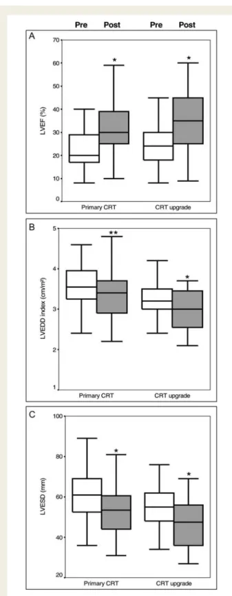
Biventricular pacemaker implantation was performed following stan-dard protocols, using either a left or right cephalic or subclavian venous access. Retrograde venous angiography was performed to facilitate optimal positioning of the LV lead into the coronary sinus. The LV lead was preferably implanted in the lateral or postero-lateral mid-ventricular region of the left ventricle whenever possible. Figure 1 Response to CRT in primary and CRT upgrade
recipi-ents. A significant improvement in left ventricular ejection fraction (LVEF, A), left ventricular end-diastolic diameter (LVEDD, B), and left ventricular end-systolic diameter (LVESD, C) was observed both after primary CRT implantation and after CRT upgrades. *P , 0.0001 vs. baseline in primary CRT group or in CRT upgrade group, **P ¼ 0.001 vs. baseline in primary CRT group. ‘Pre’ and ‘Post’ denote values before and after CRT implantation, respectively.
Statistics
Comparison of categorical variables was performed byx2and Fisher’s
exact test (in case of low sample sizes). The influence of the aetiology of heart failure, underlying heart rhythm, QRS width, and duration of preceding RV pacing on CRT response rate was assessed using Pearson’s x2 test. Continuous variables within the same group (i.e.
comparison of variables before and after CRT implantation) were analysed by two-sided paired Student’s t-test (for normally distributed variables) or Wilcoxon signed-rank test (for non-normally distributed variables). Continuous variables between groups (i.e. comparison between primary CRT implantation and CRT upgrade) were analysed by unpaired two-sided Student’s t-test (for normally distributed
variables) or Mann – Whitney U test (for non-normally distributed vari-ables). Confidence intervals were calculated using the t-test. A P-value , 0.05 was considered significant. Statistical analysis was per-formed using SPSS Ver 17.0 (SPSS, Inc., Chicago, IL, USA).
Results
Baseline parameters
Baseline characteristics are summarized in Table 1. Patients with
primary CRT implantation were younger than those receiving CRT upgrades. The distribution of the underlying cardiomyopathy
. . . .
. . . .
. . . .
. . . . Table 2 Follow-up of primary cardiac resynchronization therapy recipients and upgrades
Characteristic Primary CRT CRT upgrade P-value
Time of last FUP (months after CRT implantation; mean + SD) 21.7 + 14.2 23.5 + 15.2 0.43
D At last FUP D At last FUP
Echocardiographic parameters
LV ejection fraction (%) 10.0 (3.8 – 15.0) 10.0 (5.0 – 20.0) 0.61 End-diastolic volume (mL)a 234.0 (272.5 to 13.0) 220.0 (273 to 21.0) 0.67 End-diastolic volume index (mL/m2)a 216.0 (233.8 to 7.0) 211.0 (235.5 to 0.0) 0.55 End-systolic volume (mL)a 235.3 (275.1 to 26.9) 226.0 (278.6 to 26.1) 0.68 End-systolic volume index (mL/m2)a 217.4 (235.5 to 23.3) 214.9 (238.3 to 22.5) 0.61 LV end-diastolic diameter index (cm/m2) 20.15 (20.4 to 0.08) 20.2 (20.6 to 0) 0.66 LV end-diastolic diameter (mm) 22.0 (212.5 to 1.0) 25.0 (213.0 to 0) 0.56 LV end-systolic diameter (mm) 26.0 (213.8 to 20.25) 27.0 (216.0 to 20.75) 0.89 Left ventricular muscle mass index (g/m2)a 211 (240 to 26) 214.0 (256.5 to 9.5) 0.67 Pulmonary artery pressure (mmHg) 0 (215.3 to 8.0) 25.0 (211.0 to 1.0) 0.42 Left atrium index (cm/m2) 20.1 (20.3 to 0) 20.1 (20.2 to 0) 0.52
Change in mitral regurgitation (n, %) 0.22
Improved by two levels 3 (3) 1 (2)
Improved by one level 15 (16) 16 (28)
no change 71 (74) 38 (66)
worsened 7 (7) 3 (5)
Change in NYHA functional class (n, %) 0.51
Improved by two or more classes 4 (4) 2 (3)
Improved by one class 48 (47) 35 (50)
No change 43 (42) 30 (43)
Worsened 3 (3) 2 (3)
QRS duration (ms) 211.0 (226.5 to 14.5)b 228.0 (244.0 to 26.0)b 0.001
Pharmacological therapy at follow-up (n, %, change to baseline, n)
Beta-blockers 92 (90) 63 (90) 0.65 ACE-inhibitors 69 (68) 40 (57) 0.11 AT receptor blockers 26 (26) 25 (36) 0.18 Diuretics 81 (79) 48 (69) 0.44 Aldosterone antagonists 59 (58) 29 (41) 0.03 Aspirin 54 (53) 38 (54) 0.97 Oral anticoagulation 48 (47) 47 (67) 0.01
Number of patients (%) and median (25 – 75 percentile) are shown for categorical and continuous data, respectively, unless indicated otherwise. CRT, cardiac resynchronization therapy; FUP, follow-up; NYHA, New York Heart Association; LV, left ventricular.
a
n ¼ 69.
b
was similar among the two groups. Patients receiving primary CRT implantation had higher LV end-diastolic and end-systolic diameters, whereas end-diastolic and end-systolic volumes were similar. Atrial fibrillation (as well as concomitant oral anticoagulation) was more common in patients with CRT upgrade procedures. Pharmacological heart failure therapy was optimized in both groups.
Echocardiographic and clinical follow-up
Left ventricular ejection fraction, LV end-diastolic diameter index, and LV end-systolic diameter at baseline and at the time of lastfollow-up are shown in Figure1. Left ventricular ejection fraction
increased from 20.0 to 30.0% and from 24.0 to 35.0% in primary CRT and in CRT upgrade recipients, respectively. Likewise, LV end-diastolic diameter index and LV end-systolic diameter improved in
primary CRT recipients (3.5 – 3.2 cm/m2and 6.0 – 5.2 cm,
respect-ively) and in CRT upgrade patients (3.2 – 3.0 cm/m2 and 5.5 –
4.5 cm, respectively).
Comparison of follow-up data after primary CRT implantation
and CRT upgrade are presented in Table2. Mean follow-up was
21.7 and 23.5 months after primary CRT implantation and CRT upgrade, respectively. Both groups displayed a similar improvement in LVEF, LV end-systolic and end-diastolic diameters, pulmonary artery pressure, left atrial size, and degree of mitral regurgitation.
Improvement in NYHA functional class was equally comparable. As expected, pre-CRT QRS duration was significantly longer in
patients with chronic RV pacing (Table1) and was reduced more
dramatically after CRT implantation (Tables2and3).
Comparison of primary CRT responders with CRT upgrade
responders are presented in Table3. No difference in the rate of
responders, the aetiology of the underlying heart failure, or the improvement in echocardiographic parameters was observed between the two groups, except for a slightly more pronounced improvement in the degree of mitral regurgitation in the CRT upgrade group.
Responder characteristics
Baseline characteristics of responder patients receiving CRT upgrades
are summarized in Table4, separated according to responder status.
Cardiac resynchronization therapy upgrade patients with underlying ischaemic heart failure were less likely to respond to CRT when com-pared with those with a non-ischaemic aetiology of reduced LV func-tion. No other baseline parameters, including underlying rhythm, QRS duration, duration of prior RV pacing, or echocardiographic par-ameters were distinctly predictive of response to CRT.
Baseline characteristics of responder patients receiving primary
CRT are summarized in Table5. No baseline parameter, including
. . . . . . . .
. . . .
. . . . Table 3 Follow-up parameters in cardiac resynchronization therapy responders
Characteristic Primary CRT CRT upgrade P-value
Responders, n (%) 57/102 (55.9) 39/70 (55.7) 0.98
Aetiology of heart failure 0.32
Ischaemic heart failure 20/57 (35.1) 15/39 (38.5) Non-ischaemic heart failure 37/57 (64.9) 24/39 (61.5)
Echocardiographic parameters
D LV ejection fraction (%) 15.0 (11.5 – 20.5) 17.0 (13.0 – 22.0) 0.58 D LV end-diastolic diameter index (cm/m2) 20.3 (20.5 to 0) 20.3 (20.7 to 20.1) 0.90 D LV end-diastolic diameter (mm) 27.0 (214.0 to 21.0) 211.0 (214.5 to 22.5) 0.84 D LV systolic diameter (mm) 210 (219.8 to 23.5) 212.0 (216.0 to 23.0) 0.82 D Left atrium index (cm/m2) 20.1 (20.38 to 0) 0 (20.15 to 0.25) 0.49
Change in mitral regurgitation 0.02
Improved by two levels 0 1 (3)
Improved by one level 7 (13) 9 (28)
No change 45 (79) 22 (69)
Worsened 2 (4) 0
D Pulmonary artery pressure (mmHg) 21.0 (216.0 to 6.75) 25.5 (212.8 to 1.0) 0.31
Change in NYHA functional class (n, %) 0.18
Improved by two or more classes 3 (6) 2 (5) Improved by one class 34 (62) 24 (63)
No change 18 (33) 28 (29)
Worsened 0 1 (3)
D QRS (ms) 26.0 (222 to 12)a 224 (243 to 26.0)a 0.02
Number of patients (%) and median (25 – 75 percentile) are shown for categorical and continuous data, respectively. CRT, cardiac resynchronization therapy; NYHA, New York Heart Association; LV, left ventricular.
a
the aetiology of heart failure, underlying rhythm, QRS duration, or echocardiographic parameters were predictive of CRT response in this group of patients.
Discussion
Chronic RV pacing has been shown to induce pathologic LV remo-delling, including LV dilation and a reduction in LVEF, due to
‘extrinsically’ imposed dyssynchrony.9,11‘Upgrading’ these patients
to CRT to restore synchronicity may therefore be a reasonable therapeutic option. Our data demonstrate that CRT upgrade after chronic RV pacing results in a similar improvement in LVEF and reduction in LV dimensions as observed in heart failure patients after primary CRT implantation. Interestingly, our data further indicate that CRT significantly reverses LV remodelling even after long-term (up to 10 years) RV pacing. These results are of particular interest as long-term survival after CRT has
been shown to be predicted by the degree of LV reverse
remodel-ling in patients undergoing primary CRT implantation.7,8
A few small-scale single centre studies have previously investi-gated the short-term changes in LV remodelling after upgrading chronically RV-paced heart failure patients to CRT. One study demonstrated an acute improvement in LVEF and LV size in 15
patients upgraded to CRT.16 Similar results were observed in a
group of 20 heart failure patients with atrial fibrillation and
AV-node ablation 6 months after being upgraded to CRT.15
Although no direct comparison with primary CRT recipients was performed in these early investigations, two more recent short-term studies compared the effect of CRT on LV remodelling in patients with CRT upgrade and primary CRT implantation. In the first study, LVEF improved at least 5% in both groups after
3 months of follow-up.18 In the second very recent study, an
increase in LVEF of 13.7 and 8.7% was observed after 1-year follow-up with primary vs. upgrade CRT implantation,
respect-ively.20 All of the above-mentioned studies, however, are limited
. . . .
. . . .
. . . .
. . . .
. . . . Table 4 Baseline parameters of cardiac resynchronization therapy upgrade patients
Characteristic Responder Non-responder P-value
Underlying cardiomyopathy Ischaemic 15/36 (41.7) 21/36 (58.3) 0.01 Non-ischaemic 24/34 (70.6) 10/34 (29.4) Underlying rhythm 0.30 VAT pacing 20/33 (60.6) 13/33 (39.4) Atrial fibrillation 19/37 (51.4) 18/37 (48.6) QRS duration at implantation 0.57 ,120 ms 1/1 (100) 0/1 (0) 121 – 150 5/5 (100) 0/5 (0) 151 – 180 ms 8/21 (38.1) 13/21 (61.9) .180 ms 23/38 (60.5) 15/38 (39.5) QRS (ms) 186 (163 – 209) 181 (174 – 201) 0.90
Duration of right ventricular pacing 0.74
,24 months 4/6 (66.7) 2/6 (33.3) 25 – 60 months 17/31 (54.8) 14/31 (45.2) 61 – 96 months 9/18 (50) 9/18 (50) 96 – 144 months 2/6 (33.3) 4/6 (66.7) . 144 months 7/9 (77.8) 2/9 (22.2) Duration of RV pacing 60.0 (36.0 – 96.0) 60.0 (48.0 – 85.0) 0.53 Echocardiographic parameters LVEF at implant 25.0 (20.0 – 30.0) 22.0 (16.0 – 30.0) 0.15 ,20% 8/18 (44.4) 10/18 (55.6) 0.39 20 – 30% 24/40 (60) 16/40 (40) .30% 7/12 (58.3) 5/12 (41.7) LVEDD index (cm/m2) 3.0 (2.8 – 3.7) 3.3 (3.0 – 3.5) 0.82 LVESD (mm) 50.0 (45.0 – 60.0) 56.0 (52.5 – 64.0) 0.07 Left atrium index (cm/m2) 2.6 (2.1 – 3.1)a 2.7 (2.3 – 3.1)a 0.90 Pulmonary artery pressure (mmHg) 31.0 (23.3 – 37.5) 30.5 (23.5 – 40.0) 0.67
Number of patients (%) and median (25 – 75 percentile) are shown for categorical and continuous data, respectively. AF, atrial fibrillation; LVEF, left ventricular ejection fraction; LVEDD, left ventricular end-diastolic diameter; LVESD, left ventricular end-systolic diameter; RV, right ventricular.
a
by a rather low patient number, short follow-up period, and lack of comprehensive analysis of LV remodelling. In contrast, our data demonstrate for the first time the beneficial effect of CRT on several parameters of LV remodelling in a large cohort of patients receiving CRT upgrade after chronic RV pacing and show that a similar response may be expected as after primary CRT implan-tation. Importantly, all patients were managed by heart failure specialists, and were maintained on optimal medical therapy both
at the time of implantation (Table1) as well as during follow-up
(Table2).
There is currently no gold standard for the definition of
response to CRT.21 Previous studies have frequently used
com-bined clinical and/or echocardiographic criteria to assess response
to CRT upgrade after chronic RV pacing.20,22 The use of clinical
endpoints, however, may in itself be problematic, especially in a non-randomized, non-blinded study design. Furthermore, LV remodelling, but not clinical improvement, is related to and may
predict long-term survival after CRT.7,8 Therefore, an absolute
increase in LVEF of ≥10% was used to define response to CRT
in our study. Using this cut-off, which in our opinion clearly resembles a significant therapeutic effect, the rate of responders to CRT was similar between the two groups. Furthermore, we also observed no difference in the proportion of responders between the two groups when using a different cut-off value (e.g. increase in LVEF . 5%) for response (data not shown).
Of note, some differences in baseline characteristics were observed between patients receiving primary CRT implantation when compared with those with CRT upgrades (e.g. age, LV end-diastolic and end-systolic diameter, rhythm at time of implan-tation), which may have introduced a certain degree of bias in the intergroup analyses in this non-randomized study. However, a comparable absolute response to CRT in the intra-group ana-lyses was observed with a similar improvement in LV reverse remodelling. Indeed, the overall response to CRT within each group was largely independent of the underlying rhythm, QRS dur-ation, LVEF, LV size, and duration of prior RV pacing, indicating a similar response potential in all subgroups of patients. In this retro-spective analysis, LV volumes were only available in 69 patients. There was no difference in baseline LV volumes or the improve-ment in LV volumes between primary CRT recipients and CRT upgrades. Together with the similar improvement in LVEF and LV diameters between the two groups, these data strongly suggest that CRT indeed results in a comparable degree of LV reverse remodelling in both primary CRT and CRT upgrade recipients.
Patients with CRT upgrades were older than those with primary CRT implantation, and more frequently presented with atrial
fibril-lation, which is in line with previous data,20and may be explained
by a subgroup of patients in the CRT upgrade group, who initially underwent pacemaker implantation and AV nodal ablation due to
. . . .
. . . .
. . . .
. . . . Table 5 Baseline parameters of primary cardiac resynchronization therapy recipients
Characteristic Responder Non-responder P-value
Underlying cardiomyopathy 0.24 Ischaemic 20/41 (48.8) 21/41 (51.2) Non-ischaemic 37/61 (60.7) 24/61 (39.3) Underlying rhythm 0.38 Sinus rhythm 41/70 (58.6) 29/70 (41.4) Atrial fibrillation 16/32 (50) 16/32 (50) QRS duration at implantation 0.34 ,120 ms 9/13 (69.2) 4/13 (30.8) 121 – 150 19/31 (61.3) 12/31 (38.7) 151 – 180 ms 14/31 (45.2) 17/31 (54.8) .180 ms 10/17 (58.8) 7/17 (41.2) QRS (ms) 148 (129 – 178) 166 (135 – 178) 0.24 Echocardiographic parameters LVEF at implantation 20.0 (15.0– 27.0) 23.0 (17.0 – 30.0) 0.63 ,20% 20/36 (55.6) 16/36 (44.4) 0.99 20 – 30% 31/55 (56.4) 24/55 (43.6) .30% 6/11 (54.5) 5/11 (45.5) LVEDD index (cm/m2) 3.4 (3.0 – 3.9) 3.6 (3.2 – 3.9) 0.80 LVESD (mm) 60.0 (50.5– 67.0) 61.0 (52.0 – 75.3) 0.15 Left atrium index (cm/m2) 2.25 (2.0 – 2.4)a 2.6 (2.2 – 2.8)a 0.01 Pulmonary artery pressure (mmHg) 31.0 (25.0– 41.0) 36.0 (28.0 – 40.5) 0.36
Number of patients (%) and median (25 – 75 percentile) are shown for categorical and continuous data, respectively. AF, atrial fibrillation; LVEF, left ventricular ejection fraction; LVEDD, left ventricular end-diastolic diameter; LVESD, left ventricular end-systolic diameter.
a
refractory atrial fibrillation. The response to CRT, however, was independent of the underlying rhythm both in patients receiving CRT upgrades and after primary CRT implantation. As expected, baseline QRS duration was longer in patients with chronic RV pacing compared with primary CRT patients. As a consequence, the reduction of QRS duration was more dramatic in the first when compared with the latter group. However, response to CRT in both groups was largely independent of baseline QRS dur-ation, and was observed in patients both with little and with pro-nounced conduction delay.
Patients with ischaemic cardiomyopathy receiving CRT upgrades were less likely to respond to CRT than those with a non-ischaemic aetiology as the cause of ventricular dysfunction. On the other hand, patients receiving primary CRT implantation in our cohort had a similar response rate independent of the under-lying type of cardiomyopathy, which is somewhat in contrast to large-scale primary CRT studies such as CARE-HF, in which the presence of ischaemic heart disease was predictive of a worse
outcome.23This difference may, however, be related to the
echo-cardiographic criteria used to define response to CRT in our study (when compared with the combined clinical endpoint in CARE-HF). Furthermore, several criteria known to affect response
such as myocardial viability or presence of myocardial scars24,25
were routinely taken into consideration before primary CRT implantation in patients with ischaemic cardiomyopathy, and may have resulted in a selection of patients who are more likely to respond to CRT. In this context, a priori identification of patients most likely to respond to CRT upgrade would be of interest. It is conceivable that the response to CRT upgrade, similar to primary CRT implantation, is largely influenced by the extent of scar tissue and viable myocardium, especially in the LV pacing lead region. However, the current study was primarily designed to investigate whether CRT upgrade patients show evidence of LV remodelling, and not to comprehensively assess predictors of response; hence, further studies are required in this regard. For the time being and in the absence of conclusive data, it is current practice of the authors to apply the same eligibility and selection criteria for patients receiving CRT upgrades as for primary CRT recipients.
Chronic RV pacing may induce LV remodelling and consequently
lead to worsening LV function.9–12 On an individual basis,
however, it is difficult if not impossible to discern whether a decrease in LV function occurs as a result of RV pacing or as a con-sequence of the natural course of the underlying cardiomyopathy. The current study was hence not designed to address the differen-tial contribution of these two effects to the precedent deterio-ration of LV function but to examine the effect of CRT upgrade on LV remodelling and compare it with primary CRT implantation. We therefore limited our study population to patients in whom echocardiographic follow-up was available at our centres, which may have introduced a selection bias. For the same reason, our study was not suited to comprehensively assess morbidity and mortality endpoints. Very recently, a comparison of heart failure patients undergoing CRT upgrade with primary CRT implantation revealed a similar risk of cardiac events after a mean follow-up of
2.33 and 2.43 years, respectively.20 The mean duration of RV
pacing prior to CRT upgrade, however, was only 2.8 years in this cohort, which is of major importance, because the detrimental
effects of RV pacing increase with increasing duration of RV
pacing.9In contrast, the mean duration of RV pacing in our study
was 6.7 years extending up to over 12 years in nine individuals. Interestingly, the benefit of CRT upgrade appeared independent of the duration of previous RV pacing, and a majority of patients even in the latter group responded to CRT. These data indicate that reverse remodelling may occur even after very long periods of RV pacing and hence support the use of CRT even in this setting. As reverse LV remodelling is associated with a favourable
prognosis after primary CRT implantation,7,8 an improvement in
survival would equally be expected in this patient population. In the current study, we only analysed the change in echocardio-graphic parameters at the maximum follow-up time point available for each patient, and did not perform formal longitudinal analysis of LV remodelling after CRT implantation. We can therefore not unequivocally prove at what time point after implantation the ben-eficial effect of CRT upgrade begins to set in. However, when patients with different maximum follow-up periods were com-pared, the degree of echocardiographic improvement (as well as in NYHA functional class) was similar at all time points investigated (data not shown). Hence despite the lack of a formal longitudinal analysis, our data strongly suggest that the beneficial effects of CRT upgrade on echocardiographic reverse remodelling occur early and are sustained, which compares well with the pattern observed after primary CRT implantation.
In addition to the parameters investigated in the present study, further aspects regarding the response to CRT, including the assessment of RV size and function, diastolic dysfunction, and echocardiographic signs of dyssynchrony may be important and of potential interest in the comparison of primary CRT recipients and CRT upgrades. The present retrospective study, however, was primarily designed to assess the effect of CRT on LV function and LV remodelling, and therefore, further studies are needed to address these particular aspects.
Conclusions
The current study demonstrates that CRT reverses LV remodelling in heart failure patients with chronic RV pacing in a similar way as in primary CRT recipients, even after very long periods of RV pacing. Our data therefore may have important implications for the treat-ment of pacemaker-dependent patients with heart failure, and support the use of CRT in this setting. Whether this translates into an improved prognosis, in particular for patients with very long-term RV pacing, remains to be determined.
Acknowledgements
The authors would like to thank Prof. B. Seifert, Department of Biostatistics, University of Zurich for help with the statistics.
Conflict of interest: D.H.: Educational grants from Boston Scien-tific and Medtronic, speaker honoraria, and consulting fees from St Jude Medical. T.F.L.: Educational grants from Biotronik, Medtronic and St Jude Medical. F.R.: Research grants, speaker honoraria, and consulting fees from Biotronik. W.T.A.: Research grants, speaker honoraria, and consulting fees from Medtronic, St Jude
Medical, and Biotronik. J.H.: Consulting fees from St Jude Medical and Biotronik; research grants and speaker honoraria from Biotro-nik, St Jude and Medtronic.
References
1. Cleland JG, Daubert JC, Erdmann E, Freemantle N, Gras D, Kappenberger L, Tavazzi L. The effect of cardiac resynchronization on morbidity and mortality in heart failure. N Engl J Med 2005;352:1539 – 1549.
2. Bristow MR, Saxon LA, Boehmer J, Krueger S, Kass DA, De Marco T, Carson P, DiCarlo L, DeMets D, White BG, DeVries DW, Feldman AM. Cardiac-resynchronization therapy with or without an implantable defibrillator in advanced chronic heart failure. N Engl J Med 2004;350:2140 – 2150. 3. Holzmeister J, Hurlimann D, Steffel J, Ruschitzka F. Cardiac resynchronization
therapy in patients with a narrow QRS. Curr Heart Fail Rep 2009;6:49 – 56. 4. Sutton MG, Plappert T, Hilpisch KE, Abraham WT, Hayes DL, Chinchoy E.
Sustained reverse left ventricular structural remodeling with cardiac resynchroni-zation at one year is a function of etiology: quantitative Doppler echocardio-graphic evidence from the Multicenter InSync Randomized Clinical Evaluation (MIRACLE). Circulation 2006;113:266 – 272.
5. Steendijk P, Tulner SA, Bax JJ, Oemrawsingh PV, Bleeker GB, van Erven L, Putter H, Verwey HF, van der Wall EE, Schalij MJ. Hemodynamic effects of long-term cardiac resynchronization therapy: analysis by pressure – volume loops. Cir-culation 2006;113:1295 – 1304.
6. Abraham WT, Fisher WG, Smith AL, Delurgio DB, Leon AR, Loh E, Kocovic DZ, Packer M, Clavell AL, Hayes DL, Ellestad M, Trupp RJ, Underwood J, Pickering F, Truex C, McAtee P, Messenger J. Cardiac resynchronization in chronic heart failure. N Engl J Med 2002;346:1845 – 1853.
7. Ypenburg C, van Bommel RJ, Borleffs CJ, Bleeker GB, Boersma E, Schalij MJ, Bax JJ. Long-term prognosis after cardiac resynchronization therapy is related to the extent of left ventricular reverse remodeling at midterm follow-up. J Am Coll Cardiol 2009;53:483 – 490.
8. Yu CM, Bleeker GB, Fung JW, Schalij MJ, Zhang Q, van der Wall EE, Chan YS, Kong SL, Bax JJ. Left ventricular reverse remodeling but not clinical improvement predicts long-term survival after cardiac resynchronization therapy. Circulation 2005;112:1580 – 1586.
9. Nielsen JC, Andersen HR, Thomsen PE, Thuesen L, Mortensen PT, Vesterlund T, Pedersen AK. Heart failure and echocardiographic changes during long-term follow-up of patients with sick sinus syndrome randomized to single-chamber atrial or ventricular pacing. Circulation 1998;97:987 – 995.
10. Tops LF, Schalij MJ, Holman ER, van Erven L, van der Wall EE, Bax JJ. Right ven-tricular pacing can induce venven-tricular dyssynchrony in patients with atrial fibrilla-tion after atrioventricular node ablafibrilla-tion. J Am Coll Cardiol 2006;48:1642 – 1648. 11. Thambo JB, Bordachar P, Garrigue S, Lafitte S, Sanders P, Reuter S, Girardot R,
Crepin D, Reant P, Roudaut R, Jais P, Haissaguerre M, Clementy J, Jimenez M. Det-rimental ventricular remodeling in patients with congenital complete heart block and chronic right ventricular apical pacing. Circulation 2004;110:3766 – 3772. 12. Maurer G, Torres MA, Corday E, Haendchen RV, Meerbaum S. Two-dimensional
echocardiographic contrast assessment of pacing-induced mitral regurgitation: relation to altered regional left ventricular function. J Am Coll Cardiol 1984;3: 986 – 991.
13. Wilkoff BL, Cook JR, Epstein AE, Greene HL, Hallstrom AP, Hsia H, Kutalek SP, Sharma A. Dual-chamber pacing or ventricular backup pacing in patients with an implantable defibrillator: the Dual Chamber and VVI Implantable Defibrillator (DAVID) Trial. J Am Med Assoc 2002;288:3115 – 3123.
14. Saad EB, Marrouche NF, Martin DO, Cole CR, Dresing TJ, Perez-Lugones A, Saliba W, Schweikert RA, Wilkoff BL, Tchou P, Natale A. Frequency and associ-ations of symptomatic deterioration after dual-chamber defibrillator implantation in patients with ischemic or idiopathic dilated cardiomyopathy. Am J Cardiol 2002; 90:79 – 82.
15. Leon AR, Greenberg JM, Kanuru N, Baker CM, Mera FV, Smith AL, Langberg JJ, DeLurgio DB. Cardiac resynchronization in patients with congestive heart failure and chronic atrial fibrillation: effect of upgrading to biventricular pacing after chronic right ventricular pacing. J Am Coll Cardiol 2002;39:1258 – 1263. 16. Horwich T, Foster E, De Marco T, Tseng Z, Saxon L. Effects of resynchronization
therapy on cardiac function in pacemaker patients ‘upgraded’ to biventricular devices. J Cardiovasc Electrophysiol 2004;15:1284 – 1289.
17. Eldadah ZA, Rosen B, Hay I, Edvardsen T, Jayam V, Dickfeld T, Meininger GR, Judge DP, Hare J, Lima JB, Calkins H, Berger RD. The benefit of upgrading chroni-cally right ventricle-paced heart failure patients to resynchronization therapy demonstrated by strain rate imaging. Heart Rhythm 2006;3:435 – 442.
18. Witte KK, Pipes RR, Nanthakumar K, Parker JD. Biventricular pacemaker upgrade in previously paced heart failure patients—improvements in ventricular dyssyn-chrony. J Card Fail 2006;12:199 – 204.
19. Hunt SA, Abraham WT, Chin MH, Feldman AM, Francis GS, Ganiats TG, Jessup M, Konstam MA, Mancini DM, Michl K, Oates JA, Rahko PS, Silver MA, Stevenson LW, Yancy CW, Antman EM, Smith SC Jr, Adams CD, Anderson JL, Faxon DP, Fuster V, Halperin JL, Hiratzka LF, Jacobs AK, Nishimura R, Ornato JP, Page RL, Riegel B. ACC/AHA 2005 Guideline Update for the Diagnosis and Management of Chronic Heart Failure in the Adult: a report of the American College of Cardiol-ogy/American Heart Association Task Force on Practice Guidelines (Writing Com-mittee to Update the 2001 Guidelines for the Evaluation and Management of Heart Failure): developed in collaboration with the American College of Chest Physicians and the International Society for Heart and Lung Transplantation: endorsed by the Heart Rhythm Society. Circulation 2005;112:e154– e235.
20. Nagele H, Dodeck J, Behrens S, Azizi M, Hashagen S, Eisermann C, Castel MA. Hemodynamics and prognosis after primary cardiac resynchronization system implantation compared to ‘upgrade’ procedures. Pacing Clin Electrophysiol 2008; 31:1265 – 1271.
21. Bax JJ, Gorcsan J 3rd. Echocardiography and noninvasive imaging in cardiac resyn-chronization therapy: results of the PROSPECT (Predictors of Response to Cardiac Resynchronization Therapy) study in perspective. J Am Coll Cardiol 2009;53:1933 – 1943.
22. Duray GZ, Israel CW, Pajitnev D, Hohnloser SH. Upgrading to biventricular pacing/defibrillation systems in right ventricular paced congestive heart failure patients: prospective assessment of procedural parameters and response rate. Europace 2008;10:48 – 52.
23. Cleland J, Freemantle N, Ghio S, Fruhwald F, Shankar A, Marijanowski M, Verboven Y, Tavazzi L. Predicting the long-term effects of cardiac resynchroniza-tion therapy on mortality from baseline variables and the early response a report from the CARE-HF (Cardiac Resynchronization in Heart Failure) Trial. J Am Coll Cardiol 2008;52:438 – 445.
24. Ypenburg C, Schalij MJ, Bleeker GB, Steendijk P, Boersma E, Dibbets-Schneider P, Stokkel MP, van der Wall EE, Bax JJ. Impact of viability and scar tissue on response to cardiac resynchronization therapy in ischaemic heart failure patients. Eur Heart J 2007;28:33 – 41.
25. Bleeker GB, Kaandorp TA, Lamb HJ, Boersma E, Steendijk P, de Roos A, van der Wall EE, Schalij MJ, Bax JJ. Effect of posterolateral scar tissue on clinical and echo-cardiographic improvement after cardiac resynchronization therapy. Circulation 2006;113:969 – 976.