Familiality of neural preparation and response
control in childhood attention deficit-hyperactivity
disorder
B. Albrecht1,2*#, D. Brandeis2,3,4,5#, H. Uebel1
, L. Valko3
, H. Heinrich6,7, R. Drechsler3
, A. Heise1 , U. C. Mu¨ller3,8, H.-C. Steinhausen3,9,10, A. Rothenberger1and T. Banaschewski2
1Child and Adolescent Psychiatry, University of Go¨ttingen, Go¨ttingen, Germany
2Department of Child and Adolescent Psychiatry and Psychotherapy, Central Institute of Mental Health, Medical Faculty Mannheim/ Heidelberg University, Mannheim, Germany
3Department of Child and Adolescent Psychiatry, University of Zurich, Zurich, Switzerland 4Center for Integrative Human Physiology, University of Zurich, Zurich, Switzerland 5Neuroscience Center Zurich, University of Zurich and ETH Zurich, Zurich, Switzerland 6Department of Child and Adolescent Mental Health, University of Erlangen, Erlangen, Germany 7Heckscher-Klinik, Munich, Germany
8Interkantonale Hochschule fu¨r Heilpa¨dagogik Zu¨rich, Switzerland
9Aalborg Psychiatric Hospital, Aarhus University Hospital, Aalborg, Denmark
10Department of Clinical Psychology and Epidemiology, University of Basel, Basel, Switzerland
Background.Patients with attention deficit-hyperactivity disorder (ADHD) exhibit difficulties in multiple attentional functions. Although high heritability rates suggest a strong genetic impact, aetiological pathways from genes and environmental factors to the ADHD phenotype are not well understood. Tracking the time course of deviant task processing using event-related electrophysiological brain activity should characterize the impact of familiality on the sequence of cognitive functions from preparation to response control in ADHD.
Method.Preparation and response control were assessed using behavioural and electrophysiological parameters of two versions of a cued continuous performance test with varying attentional load in boys with ADHD combined type (n=97), their non-affected siblings (n=27) and control children without a family history of ADHD (n=43).
Results. Children with ADHD and non-affected siblings showed more variable performance and made more omission errors than controls. The preparatory Cue-P3 and contingent negative variation (CNV) following cues were reduced in both ADHD children and their non-affected siblings compared with controls. The NoGo-P3 was diminished in ADHD compared with controls whilst non-affected siblings were located intermediate but did not differ from both other groups. No clear familiality effects were found for the Go-P3. Better task performance was further associated with higher CNV and P3 amplitudes.
Conclusions.Impairments in performance and electrophysiological parameters reflecting preparatory processes and to some extend also for inhibitory response control, especially under high attentional load, appeared to be familially driven in ADHD and may thus constitute functionally relevant endophenotypes for the disorder.
Received 13 December 2011 ; Revised 12 October 2012 ; Accepted 12 October 2012 ; First published online 3 December 2012 Key words: Attention deficit-hyperactivity disorder, contingent negative variation, Continuous Performance Test, endophenotypes, event-related potentials, NoGo-P3.
Introduction
Attention deficit-hyperactivity disorder (ADHD) goes hand in hand with multiple dysfunctions in neuronal networks responsible for attention and response control (Pennington & Ozonoff, 1996 ; Castellanos &
Tannock, 2002). Heritability estimates exceeding 70 % indicate a strong genetic impact on the expression of the disorder, despite weak associations between ADHD and each individual risk allele, and a limited understanding of developmental pathways from genetic and environmental factors (Castellanos & Tannock, 2002 ; Banaschewski et al. 2005 ; Faraone et al. 2005). Endophenotypes, which are quantitative and heritable vulnerability traits characterized by more fundamental biological properties in between, on the one hand, genetic and environmental risk factors and * Address for correspondence : Dr B. Albrecht, Child and
Adolescent Psychiatry, University of Go¨ttingen, von Siebold-Straße 5, 37075 Go¨ttingen, Germany.
(Email : balbrec@gwdg.de)
on the other hand, the phenotype, may help to clarify these associations. It is expected that endophenotypes show larger and more specific genetic effects than diagnostic phenotypes (Doyle et al. 2005) and may serve as useful intermediate constructs that explain the heterogeneity of the ADHD phenotype (Buitelaar, 2005 ; Banaschewski et al. 2007 ; Rommelse et al. 2007a). This particularly holds for endophenotypes based on neural activity (Meyer-Lindenberg & Weinberger, 2006).
In a recent study, we showed that action monitoring as reflected by several performance and electro-physiological parameters was impaired in boys with ADHD and intermediate in their non-affected siblings compared with controls without a family history of ADHD (Albrecht et al. 2008, 2009). Thus, deficits in these parameters may be a consequence of specific genes or environmental factors that are shared in the families. Similar familial deficits were also reported for performance in a Stroop task and an oral arithmetic test (Doyle et al. 2005), with several parameters of motor inhibition (Slaats-Willemse et al. 2003) and also for quantitative electroencephalography (EEG) par-ameters (Loo et al. 2010).
While action-monitoring tasks require frequent re-sponses to tap cognitive control mechanisms continu-ously, the assessment of preparation and response control requires a different approach that separates these processes in time to capture their temporal progression. In this study, the cued Continuous Performance Test (CPT) combining vigilance and cued Go-NoGo tasks (Rosvold et al. 1956) was used to characterize these aspects of executive functions in ADHD (van Leeuwen et al. 1998 ; Banaschewski et al. 2003). The paradigm requires a simple response only if an infrequent cue stimulus is followed by a pre-specified target. Thus, the rare cues require prep-aration for a Go-NoGo task : if followed by a target a response needs to be executed, but if followed by a non-target, the prepared response must be withheld through inhibitory response control. CPT performance in children with ADHD is typically impaired, sistent with diminished sustained attention and con-trol (Losier et al. 1996 ; Huang-Pollock et al. 2006). Additionally, event-related potentials (ERPs) can di-rectly clarify deviance and timing of the covert brain processes leading to overt performance (Banaschewski & Brandeis, 2007).
Cues elicit a broad positive deflection maximal over parietal areas associated with attentional orienting and resource allocation, which is reduced in children and adults with ADHD (van Leeuwen et al. 1998 ; Banaschewski et al. 2003 ; McLoughlin et al. 2010). This Cue-P3 is followed by a centrally negative slow cortical potential field that terminates with the
presentation of the next stimulus (contingent negative variation ; CNV) and reflects preparation, anticipation and time estimation, which is not present after irrel-evant distractors or non-targets (Walter et al. 1964). Neurophysiological studies have implicated dopami-nergic control of the CNV (Linssen et al. 2011 ; Kratz et al. 2012) and an ensemble of thalamo-cortical struc-tures including the dorsal anterior cingulate cortex, frontal cortex, thalamus and midbrain dopaminergic nuclei involved in CNV generation (Gomez et al. 2003 ; Fan et al. 2007 ; Lutcke et al. 2008). Time processing and preparation problems in patients with ADHD are associated with reduced activation in brain regions implicated in CNV generation (Rubia et al. 1999; Smith et al. 2008) and are considered as a candidate endo-phenotype (Castellanos & Tannock, 2002 ; Rommelse et al. 2007b ; Plummer & Humphrey, 2008 ; Himpel et al. 2009). In line with these considerations, CNV is consistently reduced in ADHD (Hennighausen et al. 2000 ; Perchet et al. 2001 ; Banaschewski et al. 2003, 2008) and may represent a persistent deficit in patients with ADHD (Valko et al. 2009 ; Doehnert et al. 2010).
In trials following such cues, response control is required, and the difference in task demand between Go and NoGo trials is paralleled in the ERP. Following enhanced N2 amplitude in NoGo trials indexing con-flict monitoring (Banaschewski et al. 2004), also the subsequent P3 is modulated (Fallgatter & Strik, 1999). Cued non-targets that require suppressing a prepared response evoke a NoGo-P3 maximal at fronto-central sites. Several authors suggest that it may reflect re-sponse inhibition (Pfefferbaum et al. 1985 ; Fallgatter et al. 1999 ; Fallgatter & Strik, 1999) or termination of motor activation through cortical deactivation in motor areas (Kopp et al. 1996 ; Falkenstein et al. 1999 ; Verleger et al. 2006). Clearly, these aspects are inter-related, and thus the NoGo-P3 may be generated by several processes of terminal inhibitory response con-trol in the medial or anterior cingulate cortex, pre-motor areas and frontal areas (Kiefer et al. 1998 ; Strik et al. 1998 ; Weisbrod et al. 2000 ; Fallgatter et al. 2002 ; Verleger et al. 2006 ; Beste et al. 2008). Clinical studies have shown that the NoGo-P3 is altered in ampli-tude or anteriorization in patients with ADHD, and represents a persistent neurophysiological deficit (Brandeis et al. 2002 ; Fallgatter et al. 2004, 2005 ; Valko et al. 2009 ; Dhar et al. 2010 ; Doehnert et al. 2010).
The more parietally distributed Go-P3 elicited after cued target stimuli has been associated with several attentional functions such as evaluation of stimuli, closure or resource allocation (Polich & Kok, 1995 ; Kok, 2000), and may thus reflect aspects of executive response control. Explicit analyses of the Go-P3 in ADHD are rare, but some instances of diminished
amplitude for patients have been seen (Strandburg et al. 1996 ; Overtoom et al. 1998 ; Banaschewski et al. 2004 ; Lawrence et al. 2005).
Taken together, the CPT elicits several separable phases of attentional and executive processing. Attentional orienting and resource allocation after cue onset are associated with the Cue-P3, and followed by preparation for and anticipation of the next trial as reflected by the CNV. The strength of the CNV has been associated with response speed. If the sub-sequent trial is a Go trial, execution of the prepared response is required, and this target stimulus-evoked Go-P3 reflects perceptual resource allocation to the Go stimulus as well as context updating. NoGo trials instead require the termination of the prepared motor response which is associated with a P3 that has a more anterior topography than the Go-P3 (Banaschewski et al. 2004). The aim of this study was to test whether performance and electrophysiological parameters in the time course from preparation to response ex-ecution or response control show familiality and may thus reflect potential endophenotypes for ADHD. It is further tested whether preparation and response con-trol are particularly impaired in ADHD if additional need for cognitive control is induced by the presen-tation of additional incompatible flanker stimuli in the CPT. Moreover, correlational analyses were per-formed to explore the relationship between perform-ance parameters and brain activity measures, and electrical neuroimaging techniques were used to locate the electrical neuronal sources of the ERPs for clarifying the neuronal networks responsible.
Method Subjects
Recruitment of subjects was carried out as part of the International Multi-Center ADHD Genetics Project (IMAGE). Ethical approval for this study was obtained from the local ethical review boards. Details concern-ing recruitment and the diagnostic procedure can be found elsewhere (Andreou et al. 2007 ; Albrecht et al. 2008). Briefly, recruitment of ADHD sibling pairs was conducted as part of the IMAGE study (Asherson et al. 2007) from specialist clinics for ADHD or child and adolescent psychiatrists in private practice in Zurich, Switzerland and Go¨ttingen, Germany. Controls were recruited from local schools or sports clubs. Following screening by behavioural rating questionnaires ob-tained from both parents and teacher [long versions of Conners’ rating scales (Conners et al. 1998a, b) and the Strengths and Difficulties Questionnaire (SDQ ; Goodman, 1997, Woerner et al. 2004)], a semi-structured clinical interview (Parental Account of
Children’s Symptoms ; Taylor et al. 1986, 1987) was applied by trained investigators in order to verify ADHD diagnosis.
A total of 289 European Caucasian children and adolescents aged 8 to 16 years with an estimated Hamburg–Wechsler Intellegence Test for Children Kinder (HAWIK-IQ) above 80 (Sattler, 1992) and no known child psychiatric disorder that may mimic ADHD were initially included. They belong to one of three subgroups : (1) children and adolescents with a DSM-IV diagnosis of ADHD combined type ; (2) non-affected siblings of those with a DSM-IV diagnosis of ADHD, without a clinical diagnosis of ADHD ; and (3) unrelated healthy controls without a clinical diagnosis or a known family history of ADHD.
Since gender ratio differed across groups (x2 (2)=40.8, p<0.01) and girls with ADHD were outnumbered and furthermore considerably younger, only the sub-sample of boys (n=193) was analysed here. Due to artefacts in the EEG, datasets from three controls, three non-affected siblings and 20 participants with ADHD needed to be excluded, but exclusion ratio did not differ significantly across groups (x2
(2)=3.5, p=0.17). Thus, groups of 97 boys with ADHD, 27 male non-affected siblings of ADHD patients and 43 controls were finally included. The groups were matched for age (F2,164=0.3, p=0.77, g2p<0.01), but showed a tendency towards differences in intelligence quotient (IQ) (F2,164=2.8, p=0.07, g2p <0.03; children with ADHD had lower IQ than controls). Non-affected sibling exhibited only subtle, subclinical increases in hyperactivity scores and parent ratings (for further description of the sample, see Table S1 of the Supplementary material).
Stimuli and task
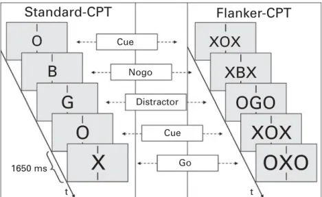
Assessments were carried out on 2 days in video-controlled, noise-shielded and slightly dimmed rooms at both departments, with the order of neurophysio-logical and neuropsychoneurophysio-logical testing following a randomization scheme. To assess preparation and response control and the impact of attentional load thereon, the Standard-CPT as well as a CPT Flanker version (Flanker-CPT) incorporating additional in-compatible stimuli were used (Doehnert et al. 2008, 2010 ; Valko et al. 2009). On each trial of both CPT versions, a bold letter was presented for 150 ms subtending 0.6x viewing angle horizontally and 0.8x vertically at 120 cm viewing distance against a light grey background in the centre of a 17-inch (43 cm) cathode ray tube monitor. Since the Flanker-CPT in-corporates two additional flanker letters that had to be ignored, horizontal viewing angle was approximately 2.0x (see Fig. 1).
In both CPTs, the children had to press the response button with the index finger of their dominant hand only if a central ‘ O ’ (cues, presented 80 times, see Fig. 1) was followed by a central ‘ X ’ (Go-trials, n=40), but to withhold responding if the cue was followed by a non-target (NoGo trials, n=40), or if the ‘X’ was uncued (40 trials). Most trials were neutral distractors (letters B, C, D, E, F, G, J or L, n=20 each, or the letter H, n=80) which also did not require a response, making up a total of 400 trials presented at a rate of 1/1650 ms. The sequences and neutral distractors were pseudo-randomly distributed.
The flankers consisted either of O’s or X’s to induce conflict. Targets and distractors H were flanked by O’s (‘ OXO ’ and ‘ OHO ’), while cues as well as the remaining distractor stimuli were flanked by X’s. As a consequence, cues and cued distractors now required additional response control in terms of inhibition (as they are flanked by target stimuli which can require a response in the context of the CPT paradigm). Cued targets require additional response control in terms of execution (since the flanking cue stimuli would require no response).
Both CPTs lasted 11 min following 25 practice trials. Their order was counter-balanced after 5 min of resting EEG with a 13 min action-monitoring task in between, and short breaks between tasks as desired.
Electrophysiological recording and processing For the subjects assessed in Go¨ttingen, the EEG was recorded with Ag/AgCl electrodes from 24 sites according to an extended 10–20 system using a BrainAmp amplifier (BrainProducts, Germany)
including the FCz recording reference, a 500 Hz sampling-rate, 0.016–100 Hz filters, a 50 Hz notch filter, and a ground electrode placed on the forehead. The electrooculogram (EOG) was recorded from additional electrodes placed above and below the right eye and at the outer canthi. In Zu¨rich, the EEG was recorded in the same way except for including additional channels, and using a SynAmps amplifier (Neuroscan, USA) and Fpz as reference at 500 Hz with 0.1–70 Hz filters. The EOG was recorded from elec-trodes below the eyes. Impedances were kept below 10 kV. Post-processing (see below) eliminated all differences between sites to ensure full compatibility.
Altogether 24 common electrodes were analysed here. After down-sampling to 256 Hz, the EEG was re-referenced to the average reference and filtered offline with 0.1–30 Hz, 24 db/oct Butterworth filters. Occular artefacts were corrected using a linear regression method without raw average subtraction (Gratton et al. 1983). If the amplitude at any EEG electrode ex-ceeded ¡100 mV, a sectionx100 to +800 ms around was excluded from further analyses. Segmentsx125 to 1875 ms around stimulus onset of Cue, Go and NoGo trials with correct responses [i.e. no responses on cues and NoGo trials, responses (‘ hits ’) on Go trials] were subsequently visually checked and aver-aged. All averages contained at least 15 sweeps. To avoid distortion of ERP topography, no baseline subtraction was applied.
Analyses
The performance parameters mean reaction time (RT) of correct target responses and inter-individual RT
OXO
XOX
OGO
XBX
XOX
X
O
G
B
Cue Nogo Distractor Cue 1650 ms t t GoO
Standard-CPT
Flanker-CPT
Fig. 1.Task description. Standard Continuous Performance Test (Standard-CPT) and Flanker-CPT in comparison. In both tasks, a sequence of relevant letters was presented in the centre of a screen between two vertical fixation marks. Participants were instructed to respond if a cue A was followed by X, but to withhold responding in all other cases. In the Flanker-CPT, additional distracting letters X or O were presented alongside.
variability (RT-SD) were analysed in an analysis of variance (ANOVA) with the between-subject factor Group (controls v. non-affected siblings v. children with ADHD) and the within-subject factor Task (Standard-CPT v. Flanker-(Standard-CPT). Since commission errors in Go trials and false alarms in NoGo trials were rare and distribution was not normal, accuracy was analysed non-parametrically with Wilcoxon tests on the factor Task for the total sample, and overall Kruskal–Wallis tests on the factor Group followed by post hoc Mann– Whitney U tests separately for each task.
The CNVs following Cues over centro-parietal electrodes (Fig. 2) were analysed as mean amplitudes from 1200 to 1650 ms at electrodes Cz and Pz. The Cue- and Go-P3 had a parietal maximum and were scored as the most positive peak in the time-frame 250 to 700 ms at electrode Pz (see Figs 2 and 3), while the NoGo-P3 was maximal at fronto-central sites and thus scored 300 to 550 ms at FCz (Fig. 4). The electrophysiological parameters were analysed using ANOVAs with the between-subject factor ‘ Group ’ and within-subject factors ‘ Task ’ and ‘ Site ’ (centro-parietal electrodes Cz and Pz for CNV, fronto-central electrodes Fz, FCz and Cz for NoGo-P3 and electrodes Cz, P3, Pz, P4 and Oz for Cue- and Go-P3 which are maximal over the mid-parietal cortex).
Familiality is indicated by a significant Group main effect or interaction with the factor Group, and by significant differences of the means between controls and ADHD as well as between controls and non-affected siblings, which was tested in post hoc analyses of confidence intervals with p<0.05. The functional significance of the ERP components and their relation to general cognitive ability was explored in the con-trol group through partial correlations between ERP amplitudes, and CPT performance parameters (con-trolled for age as an indicator of developmental trends in the assessed parameters that may lead to artificial correlations).
The scalp topography of ERP group differences was explored using t-maps. ERP sources were calculated using standardized low-resolution electromagnetic tomography (sLORETA) (Fuchs et al. 2002; Pascual-Marqui, 2002) for each group’s average ERP component.
Results
Performance data RTs of correct responses
Correct reactions to cued targets were slower in the Flanker-CPT than in the Standard-CPT (Task : F1,164=50.9, p<0.01, g2p=0.24). There was also a trend towards group differences (Group : F2,164=2.8, p=0.06, g2
p=0.03; non-affected siblings, and as a tendency also
ADHD patients responded slower than controls), but no GrouprTask interaction (F2,164=2.2, p=0.11, g2
p <0.03). For means and standard deviations and further details of the performance data statistics, see Table S2 of the Supplementary material.
RT-SDs of correct responses
Intra-individual standard deviations of RTs (RT-SDs) were higher in the Flanker-CPT than in the Standard-CPT (Task : F1,164=5.5, p=0.02, g2p=0.03). ADHD patients had higher RT-SDs than both non-affected siblings and controls (Group : F2,164=4.4, p=0.01, g2
p=0.05). Hit rate
Since performance accuracy was generally high and close to ceiling, it was analysed non-parametrically. In an overall comparison, the tasks did not differ regarding hit rate (Wilcoxon test, Z=1.0, p=0.31). Patients with ADHD performed less accurately than controls in both tasks, and less accurately than non-affected siblings for the Standard-CPT only (overall Kruskal–Wallis x2
(2)>11.3, p<0.01 and post-hoc Mann–Whitney U tests p<0.03; for all other post-hoc comparisons p>0.14).
False alarms
The Flanker-CPT provoked more false alarms than the Standard-CPT (Wilcoxon test, Z=6.9, p<0.01), but neither task revealed group differences (x2
(2)<2.2, p>0.35).
ERP data Cue-P3
The Cue-P3 with its parietal maximum had sources localized by sLORETA in the superior parietal cortex [Brodmann Area (BA) 5, 7, 31 and 40 ; see Fig. 2]. The latency detected at Pz was prolonged in the Flanker-CPT [marginal means 534 (S.D.=122) ms v. 558
(S.D.=118) ms, Task: F1,164=8.3, p<0.01, g2p=0.05), but similar in the three groups (Group : F2,164=0.2, p>0.50, g2
p <0.01, and TaskrGroup: F2,164=1.5, p=0.22, g2
p=0.02).
Cue-P3 amplitude was largest at the middle-parietal site Pz (compared with the surrounding electrodes ; Site : F4,656=101.4, p<0.01, g2p=0.38), and showed a more anterior topography in the Flanker-CPT (posi-tivity larger at Cz and diminished at Oz, TaskrSite: F4,656=8.1, p<0.01, g2p=0.05). Moreover, the Cue-P3 showed familiality effects (Group : F2,164=4.0, p=0.02, g2
p=0.05), with significantly larger amplitudes in controls than in ADHD, and as a tendency (p=0.08) the intermediate values of the non-affected siblings
Standard-CPT
Cz 5 [µV] 0 0 500 1000 1500 CNV CNV Cue-P3 Cue-P3 [ms] –5 Pz 5 [µV] 0 0 500 1000 1500 [ms] –5 Pz 5 [µV] 0 0 500 1000 1500 [ms] –5 Cz 5 [µV] 0 0 500 1000 1500 [ms] –5Cue-P3
CNV
–8.0 µVNon-affected siblings
8.0 µV 0 –10.0 µV 0 µV 10.0 µVFlanker-CPT
ADHD
Controls
Fig. 2.Topography and sources of the Cue-P3 and the contingent negative variation (CNV). Stimulus-locked grand average waves (above) of controls, non-affected siblings and attention deficit-hyperactivity disorder (ADHD) patients with spline-interpolated maps and standardized low-resolution electromagnetic tomography (sLORETA) source localizations of the Cue-P3 (middle) and mean CNV (below, mean area 1200 to 1650 ms post-cue onset) evoked by correct responded cues. CPT, Standard Continuous Performance Test.
also differed from both groups. T-maps revealed significantly diminished Cue-P3 amplitude in case– control and non-affected siblings–control comparisons at parietal and occipital sites. For further details, see Table S3 and Fig. S1a of the Supplementary material.
Cue-CNV
The CNV following cues shows the characteristic transient negativity with centro-parietal maximum that terminates with the onset of the following stimu-lus. Source localizations using sLORETA detected
activity in the medial frontal cortex and rostral and dorsolateral cingulate cortex (BA 6, 11 and 24 ; see Fig. 2). Particularly for the Flanker-CPT, the CNV mean amplitude following cues was largest at Cz (Site : F1,164=31.4, p<0.01, g2p=0.16, and TaskrSite: F1,164=4.5, p=0.04, g2p=0.03). It was generally dimin-ished for boys with ADHD as compared with both their affected siblings and controls, and non-affected siblings showed smaller CNVs than controls (Group : F2,164=9.7, p<0.01, g2p=0.11). Moreover, this pattern was more prominent for the Flanker-CPT (TaskrGroup: F2,164=3.3, p=0.04, g2p=0.04).
Standard-CPT
Go-P3
Flanker-CPT
Fz 4 0 0 500 1000 1500 [ms] –4 [µV] FCz 4 0 0 500 1000 1500 [ms] –4 [µV] Cz 4 0 0 500 1000 1500 [ms] –4 [µV] Pz 4 0 0 500 –14 µV 0 14 µV 1000 1500 [ms] –4 [µV] Fz 4 0 0 500 1000 1500 [ms] –4 [µV] FCz 4 0 0 500 1000 1500 [ms] –4 [µV] Cz 4 0 0 500 1000 1500 [ms] –4 [µV] Pz 4 0 0 500 1000 Go-P3 Go-P3 1500 [ms] –4 [µV]Non-affected siblings
ADHD
Controls
Fig. 3.Topography and sources of the Go-P3. Stimulus-locked grand average waves of controls, non-affected siblings and attention deficit-hyperactivity disorder (ADHD) patients with spline-interpolated Go-P3 maps evoked by cued targets with correct responses (hits). CPT, Standard Continuous Performance Test.
The t-map comparisons between controls and patients with ADHD or non-affected siblings revealed that the detected effects coincide with the CNV topography. For further details, see Table S3 and Fig. S1b of the Supplementary material.
Go-P3
The Go-P3 had a parietal maximum with sources de-tected in the cingulate gyrus, the superior paracentral and parietal lobulae and the praecuneus (BA 5, 7 and
31 ; see Fig. 4). Its latency was higher for the Flanker-CPT [453 (S.D.=107) ms v. 399 (S.D.=120) ms, Task:
F1,164=29.0, p<0.01, g2p=0.15], and showed no group differences or TaskrGroup interactions (both F2,164 <1, p>0.48, g2
p<0.01).
Go-P3 amplitude was highest at Pz (Site : F4,656= 154.0, p<0.01, g2
p=0.48) and showed a tendency towards higher amplitudes in the Standard-CPT at the left-parietal P3 electrode (TaskrSite: F4,656=2.2, p=0.09, g2
p=0.02), without any overall differences between groups or any further significant interactions
Standard-CPT
Flanker-CPT
Fz 0 0 500 1000 1500 [ms] –4 [µV] FCz 0 0 500 Nogo-P3 Nogo-P3 1000 1500 [ms] –4 [µV] Cz 0 0 500 1000 1500 [ms] –4 [µV] Pz 0 0 500 1000 1500 [ms] –4 [µV] Fz 0 0 500 1000 1500 [ms] –4 [µV] FCz 0 0 500 1000 1500 [ms] –4 [µV] Cz 0 0 500 1000 1500 [ms] –4 [µV] Pz 0 0 500 1000 1500 [ms] –4 [µV] –14 µV 0 14 µVNon-affected siblings
ADHD
Controls
Nogo-P3
Fig. 4.Topography and sources of the NoGo-P3. Stimulus-locked grand average waves of controls, non-affected siblings and attention deficit-hyperactivity disorder (ADHD) patients with spline-interpolated NoGo-P3 maps evoked by cued non-targets with correctly inhibited responses. CPT, Standard Continuous Performance Test.
(all p>0.21). The t-maps showed reduced activity for non-affected siblings at centro-parietal sites (see Table S4 and Fig. S1c of the Supplementary material).
An additional analysis subdividing the Go-P3 into P3a (250–400 ms) and P3b (400–700 ms) components also revealed no familiality effects or interactions.
NoGo-P3
The NoGo-P3 with its fronto-central maximum showed sLORETA sources in the superior and medial frontal cortex and cingulate gyrus (BA 6, 8, 9, 24 and 32 ; see Fig. 3). Its latency was longer for the Flanker-CPT than the Standard-Flanker-CPT [414 (S.D.=60) ms v. 388
(S.D.=48) ms, respectively, Task: F1,164=33.7, p<0.01, g2
p=0.17] without a main effect of Group or an inter-action (both F2,164<1, p>0.85, g2p<0.01).
The amplitude of the NoGo-P3 was higher in the Standard-CPT (Task : F1,164 =7.8, p<0.01, g2p=0.05), and highest at FCz (Site : F2,328=88.5, p<0.01, g2
p=0.35). It was generally diminished for boys with
ADHD as compared with controls, while non-affected siblings did not differ significantly from either of the other groups (Group : F2,164=3.8, p=0.03, g2p=0.04, post hoc controls >ADHD, p<0.05, differences in-volving non-affected siblings both p>0.20). The t-map comparisons showed lower amplitudes for ADHD as compared with controls over central sites for both tasks, and non-affected siblings showed higher amplitudes than ADHD in the Standard-CPT only, but a tendency towards reduction compared with controls in the Flanker-CPT (see Fig. S1d of the Supplementary material).
Fig. 5 summarizes the CNV, Cue-, Go- and NoGo-P3 amplitude findings of the Standard-CPT and Flanker-CPT for all groups.
Correlations between performance and ERP parameters
In the control group, mean RT and RT-SD were posi-tively correlated with mean CNV, while all P3 com-ponent amplitudes showed negative correlations (all 8 Mean Difference 6 4 2 0 –2 (µ V) 12 10 8 6 4 2 0 –2 –4 (µ V) 10 8 6 4 2 0 –2 –4 (µ V) 2 Mean Difference
Mean Difference Mean Difference
0 2 –4 –6 (µ V) Cue-P3 (a) (b) (c) (d) Cue-CNV Go-P3 Nogo-P3
Controls Non-affected siblings ADHD
Fig. 5.Synopsis of electrophysiological parameters. Means and 95 % confidence intervals with p<0.05 for the mean [of Flanker Standard Continuous Performance Test (Flanker-CPT) and Standard-CPT] and difference (Flanker-CPT minus Standard-CPT) event-related potential amplitudes. Impaired cue processing was familially driven in attention deficit-hyperactivity disorder (ADHD), with impairments in (a) Cue-P3 and (b) contingent negative variation (CNV) mean amplitude present in children with ADHD and their non-affected siblings when compared with controls. Patients with ADHD showed (d) further impaired NoGo-P3 while non-affected siblings did not differ significantly from either of the other two groups. (c) No clear group differences were evident for the Go-P3.
p<0.05). The RT and RT-SD differences between the two CPTs correlated negatively with the respective change in the NoGo-P3 amplitude. General cognitive ability was not related to performance (all r<0.12, p>0.43), and only showed by trend a correlation with mean Go-P3 amplitude (see Fig. S2 of the Supplementary material).
Discussion
In this study, the time course of familial ADHD effects upon sequential neural markers from preparation to response control was assessed. Behavioural ratings obtained from parents and teachers with the SDQ confirmed the problems in children with ADHD, while strengths and difficulties were within the normal range for healthy siblings and controls. The mean hyperactivity scores of non-affected siblings also fell within the normal range but were slightly higher than for controls. The aim of this study was to assess brain processes during the time course from prep-aration to response execution or control along with the impact of stimulus incongruency.
Performance
Means and variability of correct response RT as well as false alarm rate confirm that the Flanker-CPT is generally more demanding than the Standard version. The main RT analyses indicate that robust CPT performance impairments in ADHD are limited to the intra-individual RT variability, which is regarded as a particularly sensitive (van Leeuwen et al. 1998 ; Banaschewski et al. 2003 ; Klein et al. 2006), albeit probably not specific, marker of ADHD. Responses to cued targets were slower in the Flanker-CPT than in the Standard-CPT, which probably reflects the ad-ditional attentional load imposed by the flanking tar-gets by cue stimuli that (if presented centrally) would not require a response. Post hoc tests provided some evidence that the Flanker-CPT is also capable of un-covering slower responses in patients with ADHD, as also reported for more demanding tasks tapping cog-nitive control (Albrecht et al. 2008) and for the Flanker-CPT in adults with ADHD (McLoughlin et al. 2010, 2011). Accuracy was lower in the ADHD patients than in the other two groups irrespective of task version. Thus, these results indicate that children with ADHD display several problems in CPT performance, con-sistent with most previous studies (van Leeuwen et al. 1998 ; Banaschewski et al. 2003). However, as non-affected siblings did not consistently display inter-mediate results, overt behavioural CPT performance in ADHD may be less subject to shared familial factors. This is in contrast to our previous findings
regarding action monitoring and may reflect specific qualitative differences in task demands (Albrecht et al. 2008, 2009).
ERPs
Cue processing appears to be the most consistent familially driven electrophysiological deficit in child-hood ADHD in this test. Following the presentation of a cue which indicates that the next stimulus may be relevant for task performance, children with ADHD and also to a smaller degree their non-affected sib-lings showed diminished brain activity related to attentional orienting to the cue as reflected in the Cue-P3 with sources in the parietal lobe. The sub-sequent preparation for or anticipation of the next trial following cues was also familially driven as reflected by a markedly reduced CNV in children with ADHD as compared with controls, while non-affected siblings had intermediate activity. CNV was diminished by increasing conflict through additional flankers in non-affected siblings and in ADHD boys as compared with controls whose CNV remained stable. This indicates that incompatible stimuli implicating response conflict and accordingly increasing attentional load efficiently hampered preparatory processes in ADHD (Valko et al. 2009 ; Doehnert et al. 2010 ; McLoughlin et al. 2010), which is in agreement with previously reported difficulties in action monitoring (Albrecht et al. 2008). Consequently, group differences were particularly pronounced in the more challenging Flanker-CPT, which suggests that impaired processing of conflicting signals shows familiality in ADHD already for pre-paratory activity, and may thus indicate that shared environmental or genetic factors have an impact on cognitive control even during response preparation. Sources of CNV were located in the dorsal and rostral cingulate cortex, consistent with the hypothesis that CNV reductions may be a consequence of impaired dopaminergic modulations. Since several studies have shown that the CNV can be increased by successful neurofeedback training (Heinrich et al. 2004 ; Doehnert et al. 2008 ; Wangler et al. 2010), these findings dem-onstrate that even trait-like endophenotype markers can change with effective behavioural treatment, and may reflect the plasticity of dopaminergic functions which can also be altered by working memory training (McNab et al. 2009). Conversely, one may speculate which shared familial factors influence CNV : it may be that potential genetic factors related to the dopa-minergic system along with environmental factors within the family like psychosocial interactions, par-ticipation in intellectual and physical activities (Nigg et al. 2010 ; Halperin & Healey, 2011) or stress may play important roles.
Both Go- and NoGo-P3s had longer latencies in the Flanker-CPT. Since Go-P3 latency was positively cor-related with Go-RT, this probably reflects additional time needed for conflict resolution due to processing of incongruent stimuli (Kok, 2001). The amplitude of the NoGo-P3 was diminished after incongruent stimuli but reflected less clearly familiality of ADHD. It was significantly diminished in boys with ADHD as compared with controls and intermediate in non-affected siblings, but the siblings’ scores did not differ significantly from either of the other two groups. Impairments detected for NoGo-P3 amplitude did not extend towards the Go-P3. Executive response control in Go trials was associated with a large positive de-flection maximal at posterior electrodes that peaked (like the NoGo-P3) earlier in the Standard-CPT than in the Flanker-CPT. We found no Go-P3 differences in latency or amplitude between groups, consistent with previous findings using this type of CPT (van Leeuwen et al. 1998 ; Banaschewski et al. 2003), although such reductions have also been reported in highly co-morbid ADHD samples (Overtoom et al. 1998 ; Banaschewski et al. 2003).
The functional meaning of these ERP parameters was assessed in the control sample, in order to avoid artificial relationships due to group differences or potential confounds in ADHD symptom severity. In general, CPT performance and the assessed ERP components were not related to differences in cogni-tive ability in the control group. This supports our approach to conduct analysis which does not control for IQ differences between the groups and suggests that IQ cannot explain the reported findings. Moover, a diminished IQ as often reported in ADHD re-search may be a consequence of the symptomatology, which would question the need for a statistical ad-justment. Larger (negative) CNV as well as (positive) Go- and NoGo-P3 amplitudes were associated with faster and more homogeneous RTs in both task ver-sions ; the additional cognitive control needed in the Flanker-CPT compared with the Standard-CPT led to slower and more variable performance, which was significantly correlated with the respective reduction in NoGo-P3 amplitude. This highlights that brain activity reflected in these ERP parameters may be crucial for proper performance.
These ERP parameters and the underlying con-structs also relate to several components of the cogni-tive energetic model of ADHD (Sergeant, 2005) : particularly the diminished Cue-P3 and CNV ampli-tudes correlate with performance and may index attentional orienting and subsequent activation prob-lems in ADHD and also non-affected siblings. Higher cognitive load due to additional incompatible flankers may affect effort available for processing the target
stimuli, and consequently increase familially driven problems in ADHD.
Limitations
The current study has a number of limitations that need to be considered in the evaluation of the results. Although the sample size of the current study (n 167) is relatively large for a clinical ERP investi-gation, the study may still be limited by statistical power. With a set to 5 %, and power to 80 %, it is possible to detect a medium effect size (g2
p >5.5% explained variance) between the three groups. This statistical power is sufficient to detect the medium to large differences between ADHD cases and controls commonly reported for performance and several electrophysiological parameters in post hoc pairwise comparisons, but differences between the ADHD cases and the (intermediate) non-affected siblings may be of considerably lower effect size. The sample size required to detect small-to-medium effect sizes would be 260, and accepting also a trend with a set to 10 % as a control–sibling difference would require at least 204.
As a consequence, this study may have failed to detect possible small sibling–control effects, but our differences between non-affected siblings and controls were even below Cohen’s suggestion for small effect sizes (d=0.12) for the mean NoGo-P3 amplitude, and for the Go-P3 where no familiality at all was detected, the differences between the three groups were even smaller. Taken together, our conclusion that particu-larly cue processing is familially driven in ADHD re-mains supported despite the limited statistical power of the study. ADHD case–control differences in the CPTs NoGo-P3 related to terminal response control did not show clear familiality : although the non-affected sibling group mean was intermediate between controls and ADHD children, they did not statistically differ from either one, and the small effect size of the sibling–control differences suggests a very limited practical importance. Other tasks with more demands on response inhibition like Stop-NoGo or Go-NoGo tasks with more Go-Go than NoGo trials may be better capable of showing familiality in these ‘ inhibitory ’ parameters. This is further supported by the current data, since the non-affected siblings also showed a strong reduction in NoGo-P3 amplitude with additional incongruent flankers.
A second limitation of the current study is the source localization precision of the assessed ERPs. For a clinical study with children suffering from attention deficits, hyperactivity and impulsivity, it is necessary to limit the preparation and recording time. As a consequence, the spatial sampling of the electrical
field on the scalp was limited to a relatively low number of electrodes that could be attached with good electrical impedance (ensuring quality and stability) within a reasonable time in a multicentre study. The 24 scalp electrodes available for the current study are sufficient to capture the basic scalp topography of the evoked potentials, but the precision of the brain electrical neuroimaging is limited. There is consider-able evidence that sLORETA gives qualitatively valid source localizations in children and adults with lower numbers of electrodes as long as the potential field is adequately covered, although optimal spatial resol-ution would require more (at least 64 to 100) electrodes (Ryynanen et al. 2006 ; Michel & Brandeis, 2009). Therefore, the sLORETA neuroimaging provided here is clearly limited in resolution.
Conclusion
Neural ADHD markers of covert preparation follow-ing cues, and partly markers of response inhibition in NoGo trials were familially driven in ADHD. These familial ADHD markers reflect specific attentional and inhibitory processing stages correlated with perform-ance, and thus appear crucial for the functional understanding of genetic and environmental path-ways leading to ADHD. Particularly preparatory pro-cesses as reflected by the Cue-P3 and CNV associated with attentional orienting, resource allocation and preparation or anticipation were significantly im-paired in non-affected siblings, which indicates that genetic or environmental factors shared in families with ADHD have an impact on these functions. Moreover, as both preparation and response inhibition depend on attentional load, ADHD familiality effects seemed to become larger if the going gets tough.
Supplementary material
For supplementary material accompanying this paper visit http://dx.doi.org/10.1017/S003329171200270X.
Acknowledgements
Recruitment of ADHD sibling pairs was supported by National Institute of Mental Health grant no. R01MH062873 to Stephen V. Faraone. D.B. received support from the Swiss National Science Foundation (grant no. 32–109591).
The authors thank all children and their families for participation. Christa Dahlmann, Renate Kolle and Antonia Seitz conducted ERP recordings ; Anke Fillmer-Otte, Anne Reiners, Gudrun Schneider and Nicola Wo¨stmann performed IQ and further neuro-psychological testing.
Declaration of Interest None.
References
Albrecht B, Brandeis D, Uebel H, Heinrich H, Heise A, Hasselhorn M, Rothenberger A, Banaschewski T(2009). Action monitoring in children with or without a family history of ADHD – effects of gender on an endophenotype parameter. Neuropsychologia 48, 1171–1178.
Albrecht B, Brandeis D, Uebel H, Heinrich H, Mueller UC, Hasselhorn M, Steinhausen HC, Rothenberger A, Banaschewski T(2008). Action monitoring in boys with attention-deficit/hyperactivity disorder, their nonaffected siblings, and normal control subjects : evidence for an endophenotype. Biological Psychiatry 64, 615–625.
Andreou P, Neale BM, Chen W, Christiansen H, Gabriels I, Heise A, Meidad S, Muller UC, Uebel H,
Banaschewski T, Manor I, Oades R, Roeyers H, Rothenberger A, Sham P, Steinhausen HC, Asherson P, Kuntsi J(2007). Reaction time performance in ADHD : improvement under fast-incentive condition and familial effects. Psychological Medicine 37, 1703–1715.
Asherson P, Brookes K, Franke B, Chen W, Gill M, Ebstein RP, Buitelaar J, Banaschewski T, Sonuga-Barke E, Eisenberg J, Manor I, Miranda A, Oades RD,
Roeyers H, Rothenberger A, Sergeant J, Steinhausen HC, Faraone SV(2007). Confirmation that a specific haplotype of the dopamine transporter gene is associated with combined-type ADHD. American Journal of Psychiatry 164, 674–677.
Banaschewski T, Brandeis D(2007). Annotation : what electrical brain activity tells us about brain function that other techniques cannot tell us – a child psychiatric perspective. Journal of Child Psychology and Psychiatry and Allied Disciplines 48, 415–435.
Banaschewski T, Brandeis D, Heinrich H, Albrecht B, Brunner E, Rothenberger A(2003). Association of ADHD and conduct disorder – brain electrical evidence for the existence of a distinct subtype. Journal of Child Psychology and Psychiatry and Allied Disciplines 44, 356–376.
Banaschewski T, Brandeis D, Heinrich H, Albrecht B, Brunner E, Rothenberger A(2004). Questioning inhibitory control as the specific deficit of ADHD – evidence from brain electrical activity. Journal of Neural Transmission 111, 841–864.
Banaschewski T, Hollis C, Oosterlaan J, Roeyers H, Rubia K, Willcutt E, Taylor E(2005). Towards an understanding of unique and shared pathways in the psychopathophysiology of ADHD. Developmental Sciences 8, 132–140.
Banaschewski T, Neale BM, Rothenberger A, Roessner V (2007). Comorbidity of tic disorders & ADHD : conceptual and methodological considerations. European Child and Adolescent Psychiatry 16 (Suppl. 1), 5–14.
Banaschewski T, Yordanova J, Kolev V, Heinrich H, Albrecht B, Rothenberger A(2008). Stimulus context and motor preparation in attention-deficit/hyperactivity disorder. Biological Psychology 77, 53–62.
Beste C, Saft C, Andrich J, Gold R, Falkenstein M(2008). Response inhibition in Huntington’s disease – a study using ERPs and sLORETA. Neuropsychologia 46, 1290–1297. Brandeis D, van Leeuwen TH, Steger J, Imhof K,
Steinhausen H-C(2002). Mapping brain functions of ADHD children. In Recent Advances in Human Brain Mapping (ed. K. Hirata, Y. Koga, K. Nagata and K. Yamazaki), pp. 649–654. Elsevier : Amsterdam. Buitelaar JK(2005). ADHD : strategies to unravel its genetic
architecture. Journal of Neural Transmission. Supplementum 69, 1–17.
Castellanos FX, Tannock R(2002). Neuroscience of attention-deficit/hyperactivity disorder : the search for endophenotypes. Nature Reviews. Neuroscience 3, 617–628. Conners CK, Sitarenios G, Parker JD, Epstein JN(1998a).
The revised Conners’ Parent Rating Scale (CPRS-R) : factor structure, reliability, and criterion validity. Journal of Abnormal Child Psychology 26, 257–268.
Conners CK, Sitarenios G, Parker JD, Epstein JN(1998b). Revision and restandardization of the Conners Teacher Rating Scale (CTRS-R) : factor structure, reliability, and criterion validity. Journal of Abnormal Child Psychology 26, 279–291.
Dhar M, Been PH, Minderaa RB, Althaus M(2010). Information processing differences and similarities in adults with dyslexia and adults with attention deficit hyperactivity disorder during a continuous performance test : a study of cortical potentials. Neuropsychologia 48, 3045–3056.
Doehnert M, Brandeis D, Imhof K, Drechsler R, Steinhausen HC(2010). Mapping attention-deficit/ hyperactivity disorder from childhood to adolescence – no neurophysiologic evidence for a developmental lag of attention but some for inhibition. Biological Psychiatry 67, 608–616.
Doehnert M, Brandeis D, Straub M, Steinhausen HC, Drechsler R(2008). Slow cortical potential neurofeedback in attention deficit hyperactivity disorder : is there neurophysiological evidence for specific effects ? Journal of Neural Transmission 115, 1445–1456.
Doyle AE, Willcutt EG, Seidman LJ, Biederman J, Chouinard VA, Silva J, Faraone SV(2005).
Attention-deficit/hyperactivity disorder endophenotypes. Biological Psychiatry 57, 1324–1335.
Falkenstein M, Hoormann J, Hohnsbein J(1999). ERP components in Go/NoGo tasks and their relation to inhibition. Acta Psychologica 101, 267–291.
Fallgatter AJ, Bartsch AJ, Herrmann MJ(2002).
Electrophysiological measurements of anterior cingulate function. Journal of Neural Transmission 109, 977–988. Fallgatter AJ, Ehlis AC, Rosler M, Strik WK, Blocher D,
Herrmann MJ(2005). Diminished prefrontal brain function in adults with psychopathology in childhood related to attention deficit hyperactivity disorder. Psychiatry Research 138, 157–169.
Fallgatter AJ, Ehlis AC, Seifert J, Strik WK, Scheuerpflug P, Zillessen KE, Herrmann MJ, Warnke A(2004). Altered response control and anterior cingulate function in attention-deficit/hyperactivity disorder boys. Clinical Neurophysiology 115, 973–981.
Fallgatter AJ, Mueller TJ, Strik WK(1999). Age-related changes in the brain electrical correlates of response control. Clinical Neurophysiology 110, 833–838.
Fallgatter AJ, Strik WK(1999). The NoGo-anteriorization as a neurophysiological standard-index for cognitive response control. International Journal of Psychophysiology 32, 233–238.
Fan J, Kolster R, Ghajar J, Suh M, Knight RT, Sarkar R, McCandliss BD(2007). Response anticipation and response conflict : an event-related potential and functional magnetic resonance imaging study. Journal of Neuroscience 27, 2272–2282.
Faraone SV, Perlis RH, Doyle AE, Smoller JW, Goralnick JJ, Holmgren MA, Sklar P(2005). Molecular genetics of attention-deficit/hyperactivity disorder. Biological Psychiatry 57, 1313–1323.
Fuchs M, Kastner J, Wagner M, Hawes S, Ebersole JS (2002). A standardized boundary element method volume conductor model. Clinical Neurophysiology 113, 702–712.
Gomez CM, Marco J, Grau C(2003). Preparatory visuo-motor cortical network of the contingent negative variation estimated by current density. Neuroimage 20, 216–224.
Goodman R(1997). The Strengths and Difficulties
Questionnaire : a research note. Journal of Child Psychology and Psychiatry and Allied Disciplines 38, 581–586.
Gratton G, Coles MG, Donchin E(1983). A new method for off-line removal of ocular artifact. Electroencephalography and Clinical Neurophysiology 55, 468–484.
Halperin JM, Healey DM(2011). The influences of environmental enrichment, cognitive enhancement, and physical exercise on brain development : can we alter the developmental trajectory of ADHD ? Neuroscience and Biobehavioral Reviews 35, 621–634.
Heinrich H, Gevensleben H, Freisleder FJ, Moll GH, Rothenberger A(2004). Training of slow cortical potentials in attention-deficit/hyperactivity disorder : evidence for positive behavioral and neurophysiological effects. Biological Psychiatry 55, 772–775.
Hennighausen K, Schulte-Korne G, Warnke A, Remschmidt H(2000). Contingent negative variation (CNV) in children with hyperkinetic syndrome – an experimental study using the Continuous Performance Test (CPT) [article in German]. Zeitschrift fur Kinder- und Jugendpsychiatrie und Psychotherapie 28, 239–246.
Himpel S, Banaschewski T, Gru¨ttner A, Becker A, Heise A, Uebel H, Albrecht B, Rothenberger A, Rammsayer T (2009). Duration discrimination in the range of milliseconds and seconds in children with ADHD and their unaffected siblings. Psychological Medicine 39, 1745–1751.
Huang-Pollock CL, Nigg JT, Halperin JM(2006). Single dissociation findings of ADHD deficits in vigilance but not anterior or posterior attention systems. Neuropsychology 20, 420–429.
Kiefer M, Marzinzik F, Weisbrod M, Scherg M, Spitzer M (1998). The time course of brain activations during response inhibition : evidence from event-related potentials in a go/no go task. Neuroreport 9, 765–770.
Klein C, Wendling K, Huettner P, Ruder H, Peper M(2006). Intra-subject variability in attention-deficit hyperactivity disorder. Biological Psychiatry 60, 1088–1097.
Kok A(2000). Age-related changes in involuntary and voluntary attention as reflected in components of the event-related potential (ERP). Biological Psychology 54, 107–143.
Kok A(2001). On the utility of P3 amplitude as a measure of processing capacity. Psychophysiology 38, 557–577. Kopp B, Mattler U, Goertz R, Rist F(1996). N2, P3 and the
lateralized readiness potential in a NoGo task involving selective response priming. Electroencephalography and Clinical Neurophysiology 99, 19–27.
Kratz O, Studer P, Baack J, Malcherek S, Erbe K, Moll GH, Heinrich H(2012). Differential effects of methylphenidate and atomoxetine on attentional processes in children with ADHD : an event-related potential study using the Attention Network Test. Progress in
Neuro-Psychopharmacology and Biological Psychiatry 37, 81–89. Lawrence CA, Barry RJ, Clarke AR, Johnstone SJ,
McCarthy R, Selikowitz M, Broyd SJ(2005).
Methylphenidate effects in attention deficit/hyperactivity disorder : electrodermal and ERP measures during a continuous performance task. Psychopharmacology 183, 81–91.
Linssen AM, Vuurman EF, Sambeth A, Nave S, Spooren W, Vargas G, Santarelli L, Riedel WJ(2011). Contingent negative variation as a dopaminergic biomarker : evidence from dose-related effects of methylphenidate.
Psychopharmacology 218, 533–542.
Loo SK, Hale ST, Hanada G, Macion J, Shrestha A, McGough JJ, McCracken JT, Nelson S, Smalley SL (2010). Familial clustering and DRD4 effects on electroencephalogram measures in multiplex families with attention deficit/hyperactivity disorder. Journal of the American Academy of Child and Adolescent Psychiatry 49, 368–377.
Losier BJ, McGrath PJ, Klein RM(1996). Error patterns on the continuous performance test in non-medicated and medicated samples of children with and without ADHD : a meta-analytic review. Journal of Child Psychology and Psychiatry and Allied Disciplines 37, 971–987.
Lutcke H, Gevensleben H, Albrecht B, Frahm J (2008). Brain networks involved in early versus late response anticipation and their relation to conflict processing. Journal of Cognitive Neuroscience 21, 2172–2184.
McLoughlin G, Albrecht B, Banaschewski T,
Rothenberger A, Brandeis D, Asherson P, Kuntsi J(2010). Electrophysiological evidence for abnormal preparatory states and inhibitory processing in adult ADHD. Behavioral and Brain Functions 6, 66.
McLoughlin G, Asherson P, Albrecht B, Banaschewski T, Rothenberger A, Brandeis D, Kuntsi J(2011). Cognitive-electrophysiological indices of attentional and inhibitory processing in adults with ADHD : familial effects. Behavioral and Brain Functions 7, 26.
McNab F, Varrone A, Farde L, Jucaite A, Bystritsky P, Forssberg H, Klingberg T(2009). Changes in cortical
dopamine D1 receptor binding associated with cognitive training. Science 323, 800–802.
Meyer-Lindenberg A, Weinberger DR(2006). Intermediate phenotypes and genetic mechanisms of psychiatric disorders. Nature Reviews. Neuroscience 7, 818–827. Michel CM, Brandeis D(2009). Data acquisition standards
for electrical neuroimaging. In Electrical Neuroimaging (ed. C. M. Michel, T. Koenig, D. Brandeis, L. R. R. Gianotti and J. Wackermann), pp. 1–24. Cambridge University Press : Cambridge.
Nigg J, Nikolas M, Burt SA(2010). Measured gene-by-environment interaction in relation to attention-deficit/ hyperactivity disorder. Journal of the American Academy of Child and Adolescent Psychiatry 49, 863–873.
Overtoom CC, Verbaten MN, Kemner C, Kenemans JL, van Engeland H, Buitelaar JK, Camfferman G, Koelega HS(1998). Associations between event-related potentials and measures of attention and inhibition in the Continuous Performance Task in children with ADHD and normal controls. Journal of the American Academy of Child and Adolescent Psychiatry 37, 977–985.
Pascual-Marqui RD(2002). Standardized low-resolution brain electromagnetic tomography (sLORETA) : technical details. Methods and Findings in Experimental and Clinical Pharmacology 24 (Suppl. D), 5–12.
Pennington BF, Ozonoff S(1996). Executive functions and developmental psychopathology. Journal of Child Psychology and Psychiatry and Allied Disciplines 37, 51–87. Perchet C, Revol O, Fourneret P, Mauguiere F,
Garcia-Larrea L(2001). Attention shifts and anticipatory mechanisms in hyperactive children : an ERP study using the Posner paradigm. Biological Psychiatry 50, 44–57. Pfefferbaum A, Ford JM, Weller BJ, Kopell BS(1985).
ERPs to response production and inhibition. Electroencephalography and Clinical Neurophysiology 60, 423–434.
Plummer C, Humphrey N(2008). Time perception in children with ADHD : the effects of task modality and duration. Child Neuropsychology 15, 147–162.
Polich J, Kok A(1995). Cognitive and biological determinants of P300 : an integrative review. Biological Psychology 41, 103–146.
Rommelse NN, Altink ME, Oosterlaan J, Buschgens CJ, Buitelaar J, De Sonneville LM, Sergeant JA(2007a). Motor control in children with ADHD and non-affected siblings : deficits most pronounced using the left hand. Journal of Child Psychology and Psychiatry and Allied Disciplines 48, 1071–1079.
Rommelse NN, Oosterlaan J, Buitelaar J, Faraone SV, Sergeant JA(2007b). Time reproduction in children with ADHD and their nonaffected siblings. Journal of the American Academy of Child and Adolescent Psychiatry 46, 582–590. Rosvold HE, Bransome Jr ED, Mirsky AF, Rosvold HE,
Sarason I(1956). A continuous performance test of brain damage. Journal of Consulting Psychology 20, 343–350. Rubia K, Overmeyer S, Taylor E, Brammer M, Williams SC,
Simmons A, Bullmore ET(1999). Hypofrontality in attention deficit hyperactivity disorder during higher-order motor control : a study with functional MRI. American Journal of Psychiatry 156, 891–896.
Ryynanen OR, Hyttinen JA, Malmivuo JA(2006). Effect of measurement noise and electrode density on the spatial resolution of cortical potential distribution with different resistivity values for the skull. IEEE Transactions on Biomedical Engineering 53, 1851–1858.
Sattler JM(1992). Assessment of Children : Wisc-III and Wppsi-R Supplement. Jerome M. Sattler : San Diego.
Sergeant JA(2005). Modeling attention-deficit/hyperactivity disorder : a critical appraisal of the cognitive-energetic model. Biological Psychiatry 57, 1248–1255.
Slaats-Willemse D, Swaab-Barneveld H, de Sonneville L, van der Meulen E, Buitelaar J(2003). Deficient response inhibition as a cognitive endophenotype of ADHD. Journal of the American Academy of Child and Adolescent Psychiatry 42, 1242–1248.
Smith AB, Taylor E, Brammer M, Halari R, Rubia K(2008). Reduced activation in right lateral prefrontal cortex and anterior cingulate gyrus in medication-naive adolescents with attention deficit hyperactivity disorder during time discrimination. Journal of Child Psychology and Psychiatry and Allied Disciplines 49, 977–985.
Strandburg RJ, Marsh JT, Brown WS, Asarnow RF, Higa J, Harper R, Guthrie D(1996). Continuous-processing – related event-related potentials in children with attention deficit hyperactivity disorder. Biological Psychiatry 40, 964–980.
Strik WK, Fallgatter AJ, Brandeis D, Pascual-Marqui RD (1998). Three-dimensional tomography of event-related potentials during response inhibition : evidence for phasic frontal lobe activation. Electroencephalography and Clinical Neurophysiology 108, 406–413.
Taylor E, Everitt B, Thorley G, Schachar R, Rutter M, Wieselberg M(1986). Conduct disorder and hyperactivity : II. A cluster analytic approach to the identification of a behavioural syndrome. British Journal of Psychiatry 149, 768–777.
Taylor E, Schachar R, Thorley G, Wieselberg HM, Everitt B, Rutter M(1987). Which boys respond to stimulant
medication ? A controlled trial of methylphenidate in boys with disruptive behaviour. Psychological Medicine 17, 121–143.
Valko L, Doehnert M, Mu¨ller UC, Schneider G, Albrecht B, Drechsler R, Maechler M, Steinhausen HC, Brandeis D (2009). Differences in neurophysiological markers of inhibitory and temporal processing deficits in children and adults with ADHD. Journal of Psychophysiology 23, 235–246.
van Leeuwen TH, Steinhausen HC, Overtoom CC, Pascual-Marqui RD, van’t Klooster B, Rothenberger A, Sergeant JA, Brandeis D(1998). The continuous performance test revisited with neuroelectric mapping : impaired orienting in children with attention deficits. Behavioural Brain Research 94, 97–110.
Verleger R, Paehge T, Kolev V, Yordanova J, Jas´kowski P (2006). On the relation of movement-related potentials to the go/no-go effect on P3. Biological Psychology 73, 298–313.
Walter WG, Cooper R, Aldridge VJ, McCallum WC, Winter AL(1964). Contingent negative variation : an electric sign of sensorimotor association and expectancy in the human brain. Nature 203, 380–384.
Wangler S, Gevensleben H, Albrecht B, Studer P, Rothenberger A, Moll GH, Heinrich H(2010). Neurofeedback in children with ADHD : specific event-related potential findings of a
randomized controlled trial. Clinical Neurophysiology 122, 942–950.
Weisbrod M, Kiefer M, Marzinzik F, Spitzer M(2000). Executive control is disturbed in schizophrenia : evidence from event-related potentials in a Go/NoGo task. Biological Psychiatry 47, 51–60.
Woerner W, Becker A, Rothenberger A(2004). Normative data and scale properties of the German parent SDQ. European Child and Adolescent Psychiatry 13 (Suppl. 2), II3–II10.