. . . .
. . . .
NOX2-derived reactive oxygen species are crucial
for CD29-induced pro-survival signalling in
cardiomyocytes
Berit I. Rosc-Schlu¨ter
1, Ste´phanie P. Ha¨uselmann
1, Vera Lorenz
1, Michika Mochizuki
1,
Federica Facciotti
2, Otmar Pfister
1,3, and Gabriela M. Kuster
1,3*
1
Myocardial Research, Department of Biomedicine, University and University Hospital Basel, Hebelstrasse 20, 4031 Basel, Switzerland;2
Experimental Immunology, Department of Biomedicine, University and University Hospital Basel, Basel, Switzerland; and3
Division of Cardiology, University Hospital Basel, Basel, Switzerland Received 22 December 2010; revised 2 December 2011; accepted 22 December 2011; online publish-ahead-of-print 23 December 2011
Time for primary review: 25 days
Aims The highly expressed cell adhesion receptor CD29 (b1-integrin) is essential for cardiomyocyte growth and survival,
and its loss of function causes severe heart disease. However, CD29-induced signalling in cardiomyocytes is ill defined and may involve reactive oxygen species (ROS). A decisive source of cardiac ROS is the abundant NADPH oxidase (NOX) isoform NOX2. Because understanding of NOX-derived ROS in the heart is still poor, we sought to test the role of ROS and NOX in CD29-induced survival signalling in cardiomyocytes.
Methods and results
In neonatal rat ventricular myocytes, CD29 activation induced intracellular ROS formation (oxidative burst) as assessed by flow cytometry using the redox-sensitive fluorescent dye dichlorodihydrofluorescein diacetate. This burst was inhibited by apocynin and diphenylene iodonium. Further, activation of CD29 enhanced NOX activity (lucigenin-enhanced chemiluminescence) and activated the MEK/ERK and PI3K/Akt survival pathways. CD29 also induced phosphorylation of the inhibitory Ser9 on the pro-apoptotic kinase glycogen synthase kinase-3b in a PI3K/Akt- and MEK-dependent manner, and improved cardiomyocyte viability under conditions of oxidative stress. The ROS scavenger MnTMPyP or adenoviral co-overexpression of the antioxidant enzymes superoxide dismutase and catalase inhibited CD29-induced pro-survival signalling. Further, CD29-induced protective pathways were lost in mouse cardiomyocytes deficient for NOX2 or functional p47phox, a regulatory subunit of NOX.
Conclusion p47phox-dependent, NOX2-derived ROS are mandatory for CD29-induced pro-survival signalling in cardiomyocytes.
These findings go in line with a growing body of evidence suggesting that ROS can be beneficial to the cell and support a crucial role for NOX2-derived ROS in cell survival in the heart.
-Keywords NOX † Reactive oxygen species † b1-Integrin † Cardiomyocytes † Glycogen synthase kinase-3b
1. Introduction
Connections to extracellular matrix (ECM) components are essential for the survival and the structural and functional integrity of cardio-myocytes.1,2In particular, transmembrane cell surface adhesion
recep-tors (e.g. integrins) bind ECM proteins such as laminin, collagen, and fibronectin.3,4Integrins are heterodimeric transmembrane receptors
composed of an a- and b-subunit. In total, 18 a- and 8 b-chain iso-forms are known, which form 24 distinct receptors that are in a bent conformation in quiescent, and in an extended conformation in activated cells.5,6The b
1-integrin subunit (CD29) represents the
most abundant b-isoform expressed in cardiomyocytes, where it can dimerize with different a-subunits.7–9 In vitro, CD29 is directly activated by the binding of ECM components or activating anti-bodies,10,11which causes the rearrangement of the actin cytoskeleton and recruitment of signalling kinases (e.g. mitogen-activated protein kinases) to the cytosolic part of the integrin receptor4,12. Through ac-tivation of such signalling kinases, CD29 participates in the regulation of cardiomyocyte growth and survival. Specifically, CD29 facilitates a-adrenergic receptor (AR)-stimulated hypertrophy13 and inhibits b-AR-induced apoptosis in vitro.14,15 In vivo, deletion of CD29 is embryonically lethal,2,16and cardiac-specific disruption or knock-out
*Corresponding author. Tel:+41 61 328 77 36; fax: +41 61 265 23 50, Email: gkuster@uhbs.ch
leads to disturbed cardiac function, impaired cardiac differentiation, fi-brosis, and heart failure.16–18 Similarly, mouse hearts deficient in CD29 exhibit increased cardiac myocyte apoptosis in response to b-AR stimulation19and after myocardial infarction20when compared with wild-type (wt) hearts. Furthermore, integrin shedding, which is essential in cardiomyocyte growth, as well as the induction of apopto-tic processes referred to as anoikis are typical features associated with the cellular detachment from ECM components.21Similarly, cell adhe-sion of a5b1-integrin to fibronectin protects against apoptosis in
modified Chinese hamster ovary cells expressing a5b1-integrin.22
These studies clearly point out the importance of CD29 in cardio-myocyte survival, but the understanding of the likely signalling path-ways involved is poor.
Several studies affirmed the production of reactive oxygen species (ROS; oxidative burst) by various stimuli, including growth factors or G protein-coupled receptor agonists as well as by cell adhesion as seen in anchorage-dependent cell growth.23–26These ROS act as es-sential mediators in redox-signalling activating signalling kinases impli-cated in cell growth, adhesion, and survival. They derive from various oxidase enzymes, including lipoxygenase, xanthine oxidase, or NADPH oxidase (NOX), the latter of which is one of the major sources of cardiac ROS.23,27–29 NOX is a multimeric enzyme that consists of the cytosolic subunits p40phox, p47phox, p67phox, and Rac1, all of which translocate to the membrane upon activation, as well as the transmembrane subunits gp91phoxand p22phox. To date, seven gp91phox homologues, NOX1 – 5 and DUOX1 – 2, have been identified in mammals.30NOX2 predominates in cardiomyocytes, al-though NOX1 and NOX4 are also expressed,31NOX4 being consti-tutively active even in the absence of the cytosolic subunits. NOX catalyses a one-electron transfer to molecular oxygen to form the radical superoxide anion (O†−
2 ). O
†−
2 is further reduced to hydrogen peroxide (H2O2), a reaction that occurs either spontaneously or is
catalysed by superoxide dismutase (SOD), and finally degraded into water by catalase (cat).
Due to the emerging role of ROS as regulators of cell survival, the crucial role of NOX as a potent source of ROS in the heart and the limited understanding of NOX-derived ROS in cardiomyocytes, we addressed the question if CD29-induced pro-survival signalling depends on NOX-derived ROS in neonatal cardiomyocytes.
2. Methods
Detailed information on materials and methods is given in the Supplemen-tary material online.
2.1 Animal material
Wistar rats (2007 – 2010) were obtained from Harlan Laboratories and mice from the Jackson Laboratory. From January 2011, Sprague – Dawley rats originating from in-house breeding were used. All animal protocols were approved by the Veterinary Department of Basel (Switzerland) and conformed to the rules of the Swiss Federal Act on Animal Protection and the National Institutes of Health Guide for the Care and Use of La-boratory Animals. Neonatal rat ventricular myocytes (NRVMs) were iso-lated from the hearts of 1- to 3-day-old pups sacrificed by decapitation and isolation was performed as previously described.32,33 Neonatal mouse ventricular myocytes (NMVMs) were isolated from 1- to 2-day-old pup hearts of wt (C57BL/6), p47phox loss-of-function (LOF; homozygous Ncf1m1J) and NOX2 (homozygous Cybbtm1Din) knock-out strains as previously described.34,35Genotyping of mouse tails was per-formed as recommended by the supplier (http://jaxmice.jax.org/pub-cgi/
protocols/protocols.sh). For some experiments, H9c2 cardiomyoblasts (ATCC) were used.
2.2 Viral transfection
Viral constructs were purchased from the Gene Transfer Vector Core, University of Iowa, IA, USA. Serum-starved cells were transfected with the viral constructs Ad5CMVCatalase,36 Ad5CMVSOD137, or
Ad5CMVcytoLacZ38at concentrations of 50 or 100 multiplicity of infec-tions (MOIs) for 36 h prior to a-CD29 (10 mg/mL) treatment for 15 min. Expression levels were 1.8 + 0.3-fold for SOD and 9 + 1.6-fold for cat at 50 MOI.
2.3 Western blotting
Sample protein concentrations were estimated using a Pierce protein BCA kit (Thermo Fisher Scientific). 10 – 35 mg protein was loaded on a 12% sodium dodecyl sulfate– polyacrylamide gel electrophoresis (SDS – PAGE) and transferred onto a polyvinylidene fluoride membrane (GE Healthcare). Immunoreactive bands were quantified with Image J version 1.37 (http://rsb.info.nih.gov/ij/).39
2.4 Flow cytometry
Cardiomyocytes were loaded with 12.5 mmol/L (5-(and-6)-chloromethyl-2′,7′-dichlorodihydrofluorescein diacetate acetyl ester, CM-H2DCF-DA;
Invitrogen) in phenol-red-free (prf) DMEM with Hepes (25 mmol/L) and L-glutamine (4 mmol/L) for 20 min at 378C with 5% CO2 in the
dark. After washing, cells were detached with accutase (PAA Laborator-ies) and kept in 500 mL prf-DMEM on ice. Green fluorescence was mea-sured with FL-1 488 nm excitation and 530 nm emission on a DAKO flow cytometer and DAKO Summit version 4.3 software.
2.5 Lucigenin assay
NOX activity was measured using lucigenin-enhanced chemiluminescence. H9c2 cells were treated with a-CD29 antibody or IgG (10 mg/mL, 15 min), then washed three times with cold PBS and scraped into Jude Krebs buffer (see Supplementary material online) containing protease in-hibitor cocktail (Roche). Lysates were sonicated (3 s, four times on ice) and centrifuged at 3000 rpm at 48C for 4 min. Measurements of NOX ac-tivity were performed by a luminescence assay in a microplate lumi-nometer using 40 mg of lysate with 5 ml DMSO, 10 mmol/L Lucigenin (Sigma), 100 mmol/L NAD(P)H (Sigma), and 50 mmol/L Tiron (Fluka) per well (96-well plate). Data were recorded as relative light units over time, and integrated and calculated as area under the curve using Image J software for statistical analysis.
2.6 RNA interference
NRVMs were transfected with 2 mg/106cells ON-TARGETplus using the Amaxa Neonatal Rat Cardiomyocytes Nucleofector kit (VPE-1002), according to the manufacturer’s instructions, and efficiency of knockdown was assessed using quantitative RT – PCR.
2.7 Cell viability
Cell viability was measured by a tetrazolium (MTT, 3-(4,5-dimethylthiazol-2-yl)-2,5-diphenyltetrazolium bromide)-based colorimetric assay using Cell Proliferation Kit I (Roche) according to the manufacturer’s instructions.
2.8 Statistical analyses
All statistical analyses were performed in Graphpad Prism version 4.03 and P-values ,0.05 were considered to indicate significant differences. Significance was tested in repeated measures one-way ANOVA with Bon-ferroni’s post-test (for sample sizes less than or equal to four) or Newman – Keuls (for more than four samples to compare) correction.
For analysing flow cytometry data, the geometric mean was used and overlays were performed in FloJo software version 7.5.5. (Tri Star Inc.).
3. Results
3.1 CD29 activation induces a
time-dependent oxidative burst, which is
impaired by apocynin and diphenylene
iodonium in cardiomyocytes
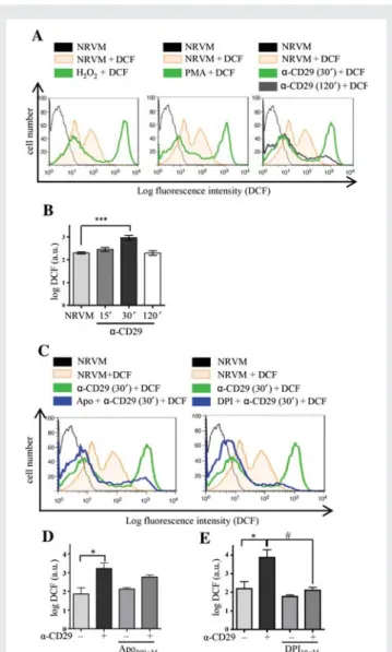
To assess whether CD29 activation generates ROS in NRVM, the redox-sensitive fluorescent dye 5-(and-6)-chloromethyl-2′,7′ -dichlorodihydrofluorescein diacetate acetyl ester (DCF) was used. Flow cytometry analyses demonstrated a significant increase in DCF fluorescence indicative of an oxidative burst after 30 min of in-cubation of NRVM with an activating a-CD29 antibody, which sub-sided after 2 h (Figure1A and B). A qualitatively similar increase in DCF fluorescence was observed in response to exogenous ROS (H2O2, 100 mmol/L) and to 12-phorbol 13-myristate acetate (PMA,
1 mmol/L), which induces endogenous ROS in other cell types40,41 (Figure1A).
We further investigated the NOX-dependency of this burst using the inhibitors apocynin (apo, 500 mmol/L) and diphenylene iodonium (DPI, 10 mmol/L). Apocynin is, besides known to inhibit the regula-tory NOX subunit p47phox in leukocytes, implicated as an antioxi-dant in endothelial and smooth muscle cells,42 and DPI inhibits flavoenzymes, including NOX.43 Both compounds markedly decreased DCF fluorescence intensity when administered prior to a-CD29 (Figure1C – E).
3.2 CD29 activation induces NOX activity
in cardiomyocytes
As both apocynin and DPI are non-specific, H9c2 cardiomyoblasts were treated with a-CD29 or non-immune IgG (10 mg/mL, 15 min) and NOX activity was measured by lucigenin-enhanced chemiluminescence. Cross-linkage of CD29 via activating antibody significantly enhanced NADPH-dependent lucigenin chemilumines-cence in H9c2 cells (Figure 2A and B), thus confirming activation of NOX in response to CD29 stimulation. A similar observation was made in NRVM plated on dishes precoated with laminin, the physiological ligand for CD29. These cells exhibited higher NOX ac-tivity when compared with NRVM plated on non-coated dishes (data not shown).
3.3 CD29 activation induces
phosphorylation of ERK1/2 (Thr202/
Tyr204), Akt (Ser473), and GSK-3b (Ser9)
CD29 activates the MEK/ERK and the PI3K/Akt pathways in various cell types.12 In NRVM, increased phosphorylation of ERK and Akt was observed upon CD29 stimulation, which peaked at 15 min and was no longer detectable at 45 min (Supplementary material online, Figure S1A and B). Phosphorylation of serine 9 of glycogen synthase kinase (GSK)-3b, which is inhibitory to its kinase activity, was used to measure CD29-induced pro-survival signalling, because (i) it is involved in the CD29-induced anti-apoptotic effect in adult cardio-myocytes44 and (ii) it is downstream of the PI3K/Akt pathway.45 Using a phosphorylation site-specific GSK-3b (Ser9) antibody, immu-noreactive bands were observed at 15 min (Supplementary materialonline, Figure S1C). No changes in phosphorylation were detectable when cardiomyocytes were incubated with either an inhibitory a-CD29 antibody or with a non-specific IgG (Supplementary material online, Figure S1A – C).
Figure 1 CD29 activation induces an oxidative burst in NRVM, which is inhibitable by apocynin and DPI. (A and B) (A) Representa-tive flow cytometric histograms depict NRVM loaded with the ROS indicator DCF (12.5 mmol/L). Unstimulated cells (NRVM) without DCF in black. Fluorescence intensity is shown for unstimulated cells (NRVM+ DCF, orange line) and cardiomyocytes stimulated with H2O2(100 mmol/L), PMA (1 mmol/L), or a-CD29 (5 mg/mL)
for 30 min (green line). The shift to the right indicates an increase in ROS. CD29 activation induced a time-dependent burst that peaked at 30 min (A and B, 30′) and declined after 2 h (120′, grey line). (B) Depicts a-CD29-induced fluorescence intensity at different time points. Data are mean + SEM. Unless indicated otherwise, comparisons were not significant. ***P , 0.001 (n ¼ 8). (C – E)
Cells were pretreated (blue) with either apocynin (Apo;
500 mmol/L, 14 – 16 h), or DPI (10 mmol/L, 45 min) prior to a-CD29 administration (5 mg/mL, 30 min, C ). (D and E) Pretreat-ment suppressed the a-CD29-induced oxidative burst partially (apocynin, n ¼ 4) and completely (DPI, n ¼ 5). Data are presented as mean + SEM. Unless indicated otherwise, comparisons were not significant. * and#P , 0.05.
To further elucidate the pathways of kinase activation upon CD29 stimulation, the MEK inhibitor U0126 (U), the PI3K inhibitors Wort-mannin (W) and LY294002 (LY), and the Akt inhibitor Akti1/2 (Akti) were used to inhibit selected parts of the signalling pathway. Of the four inhibitors, U suppressed the a-CD29-induced phosphorylation of ERK (Supplementary material online, Figure S2A – D) and W, LY, and Akti (but not U) suppressed the a-CD29-induced phosphory-lation of Akt (Supplementary material online, Figure S2E – H). No cross talk could be observed between the PI3K/Akt and the MEK/ ERK signalling pathways. However, all inhibitors significantly reduced a-CD29-induced GSK-3b (Ser9) phosphorylation (Supplementary material online, Figure S2I – L). This observation places GSK-3b (Ser9) downstream of both the MEK/ERK and the PI3K/Akt pathways.
3.4 CD29-induced pro-survival signalling
is ROS-dependent
Because activation of CD29 induced an oxidative burst in cardiomyo-cytes (Figure1), the possibility exists that CD29-stimulated signalling is ROS-regulated. To test this hypothesis, MnTMPyP (MnT), a mimetic of the antioxidant enzymes SOD and cat, was used to reduce ROS levels. Pretreatment with MnT significantly inhibited the a-CD29-induced phosphorylation of ERK1/2 (Supplementary material online, Figure S3A), Akt (Supplementary material online, Figure S3B), and GSK-3b (Supplementary material online, Figure S3C). Similarly, co-overexpression of SOD and cat using adenoviral constructs signifi-cantly reduced MEK-, Akt-, and GSK-3b-phosphorylation (Figure3A – C ). To assess the respective contribution of O2†2 and H2O2 to
CD29-induced signalling, similar experiments were performed in NRVM expressing either SOD or cat. Although either enzyme inhi-bited phospho-MEK1/2 and phospho-GSK-3b, persistence of phospho-Akt was observed with overexpression of SOD, but not
of cat, up to 100 MOI (Figure 3D and Supplementary material online, Figure S4).
3.5 CD29-induced pro-survival signalling
is inhibited by apocynin and DPI
Primarily, a likely role of NOX in CD29-induced survival signalling was investigated using apo and DPI. Both inhibitors suppressed CD29-induced ERK1/2- (Supplementary material online, Figure S5A and D) and Akt-phosphorylation (Supplementary material online, Figure S5B and E). Although both apo and DPI also inhibited the CD29-induced GSK-3b phosphorylation, the reliable assessment of this inhibition is hindered, because both substances affected basal kinase phosphorylation (Supplementary material online, Figure S5C and F). However, these findings suggest that NOX might serve as the source of CD29-induced ROS that trigger ROS-dependent pro-survival signalling in cardiomyocytes.
3.6 gp91
phox- and p47
phox-deficient
cardiomyocytes show loss of
a-CD29-induced pro-survival signals
In another set of experiments, NOX was corroborated as source of ROS by examining a-CD29-induced signalling in NMVMs isolated from gp91phox(NOX2) knock-out mice and from mice lacking a func-tional catalytic subunit p47phox. Upon CD29 stimulation, significantly lower levels of phospho-ERK1/2, phospho-Akt, and phospho-GSK-3b were observed in NOX2-knock-out mouse cardiomyocytes when compared with wt (Figure4A – C ). In p47phoxLOF NMVM, CD29 ac-tivation failed to induce phosphorylation of Akt (Ser473; Figure4E) and GSK-3b (Ser9; Figure4F). In contrast, CD29-induced phosphory-lation of ERK1/2 (Figure4D) was unaffected by the loss of functional Figure 2 CD29 induces activation of NOX. H9c2 cells were treated with a-CD29 or non-immune IgG (10 mg/mL) for 15 min and NOX activity was measured by lucigenin-enhanced chemiluminescence. Kinetics of lucigenin-enhanced chemiluminescence is shown in (B). Basal fluorescence was measured five times before adding first NAD(P)H and later tiron, respectively (arrows). Data were recorded as relative light units over time and calculated as area under the curve for statistical analysis (A, n ¼ 3). Data are given as mean + SEM. **P , 0.01.
p47phox, suggesting the activation of a p47phox-independent and/or compensatory pathway.
3.7 The CD29-induced oxidative burst
is impaired in p47
phox-LOF, but not in
NOX2-knock-out cardiomyocytes
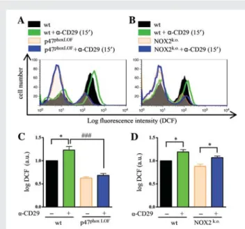
Because NOX2-derived ROS play an important role in CD29-induced pro-survival signalling in NMVM, we tested whether the CD29-induced oxidative burst was impaired in NMVM deficient in NOX2 or p47phox. Upon activation of CD29, a shift in DCF fluores-cence was observed in wt NMVM that was missing in p47phox-LOF NMVM (Figure 5A and C ). In contrast, in NOX2-knock-out cells, CD29 activation enhanced DCF fluorescence (Figure 5B and D).
Therefore, another p47phox-dependent NOX (e.g. NOX1)46 might be involved in CD29-induced ROS formation in NMVM.
4. Discussion
This work demonstrates that NOX-derived ROS regulate cardiomyo-cyte survival pathways. Similar to what has been described for growth factors and cytokines,29transient endogenous ROS were formed in cardiomyocytes in response to CD29 activation (Figure 6) and this ROS formation was impaired by NOX inhibitors. These findings are in agreement with DPI-based data implicating NOX in CD29-induced ROS production in the colic adenocarcinoma cell line (caco-2),47but not with findings from mouse embryonic fibroblasts (NIH-3T3), where synergistically growth factor- and integrin-driven ROS were mostly lipoxygenase-dependent.48 In cardiac myocytes, however, Figure 3 CD29-induced pro-survival signalling depends on ROS. (A – C ) NRVM were infected with adenoviral constructs to express b-galactosidase (LacZ100) at 100 MOI or co-overexpress SOD (S50) and cat (C50) at 50 MOI 36 h prior to a-CD29 administration (10 mg/mL, 15 min). Equal protein
amounts were subjected to western blotting using phosphorylation-specific antibodies for MEK1/2 (Ser217/221), Akt (Ser473), and GSK-3b (Ser9). Data are presented as mean + SEM. *** and###P , 0.001, ** and##P , 0.01, and#P , 0.05 (n ¼ 5 for A and B, n ¼ 3 for C ). Unless indicated other-wise, comparisons were not significant. (D) Western blot of CD29-induced signalling with single-enzyme overexpression at two different MOI, includ-ing the protein expressions of SOD and cat.
CD29 activation induced NOX activity and an additional line of evi-dence for NOX-involvement was obtained using gene knock-outs (see below).
ROS at moderate levels may serve as signalling molecules partici-pating in the regulation of cell survival,24,49,50because of their ability to activate pro-survival related cascades, including the PI3K/Akt and the MEK/ERK pathway.51 A prominent target of Akt is GSK3,52 of which two isoforms, GSK-3a and GSK-3b, are expressed in cardio-myocytes. Inhibition of GSK-3 via the PI3K pathway promotes cell sur-vival in Drosophila or neurones.53 Additionally, CD29-dependent, PI3K-mediated phosphorylation of the inhibitory GSK-3b Ser9 pro-tects adult rat cardiomyocytes from b-AR-induced apoptosis.44 Although GSK-3b is reportedly directly phosphorylated by Akt, several other kinases, including the ERK1/2 target p90 ribosomal S6 kinase have also been implicated in GSK-3b inhibition in the heart.45 Our data show activation of both, the PI3K/Akt and the MEK/ERK pathway upon CD29 engagement in NRVM and uncover GSK-3b as a target of both these pathways.
Clearance of ROS by antioxidants elicited apoptotic pathways in leukaemia cells, inhibiting the phosphorylation of Akt or ERK.54 In accordance with these findings, co-overexpression of both SOD and catalase (Figure 3) significantly diminished CD29-induced
phosphorylation of MEK1/2, Akt, and GSK-3b. It is of note that at higher expression levels single-enzyme overexpression was equally effective with regard to induced phospho-MEK/ERK and -GSK-3b inhibition, implicating both O†−
2 and H2O2as second messengers in
CD29-induced pro-survival signal transduction in NRVM. However, SOD appeared less efficient to suppress Akt-phosphorylation than catalase. Although this difference could be attributed to the lower ex-pression levels of SOD, differential regulation of the PI3K/Akt pathway through O†−
2 and H2O2cannot be ruled out.
DPI and apocynin inhibited the CD29-induced phosphorylation of ERK1/2, Akt, and GSK-3b, implying NOX-derived ROS as signalling mediators. Mice carrying either a knock-out of the transmembrane NADPH oxidase subunit NOX2, or a LOF from the regulatory subunit p47phoxwere used to assess whether NOX2 is required for CD29-induced survival signalling in NMVM. Indeed, NOX2 knock-out NMVM showed reduced levels of CD29-induced ERK1/2 phosphory-lation and the CD29-inducible phosphoryphosphory-lation of Akt and GSK-3b were nearly abolished in both NOX2 knock-out and p47phox-LOF cardiomyocytes (Figure 4A – F ), highlighting the central role of NOX2- and p47phox-derived ROS in CD29-induced survival signalling. In contrast to NOX2 knock-out cardiomyocytes, however, where CD29-induced ERK-phosphorylation was mostly suppressed, Figure 4 CD29-induced signal transduction requires NOX2-derived ROS. a-CD29 administration (10 mg/mL, 15 min) induced phosphorylation of ERK1/2 (p44/42), Akt and GSK-3b in wild-type (wt) NMVM (A – F ). a-CD29-induced phosphorylation was absent in NOX2 knock-out (NOX2k.o.)
cardiomyocytes (A – C ); and partially diminished in p47phoxloss of function (p47LOF) NMVM (D – F ). Data are presented as mean + SEM. Unless indi-cated otherwise, comparisons were not significant. * and#P , 0.05, ** and##P , 0.01, *** and###P , 0.001 (n ¼ 5 for A, C, and D – F; n ¼ 6 for B).
induced phospho-ERK was still solid in p47phox-LOF cardiomyocytes. Although no oxidative burst was detectable in p47phox-LOF cardio-myocytes at 15 min, i.e. at the time of peak signalling, a compensatory pathway involving weaker, NOXO1-supported ROS release may account for ERK activation in the absence of functional p47phox. Alter-natively, lack of ROS may allow for a ROS-independent pathway to activate ERK. In fact, co-existence of ROS-dependent and -independent pathways namely regarding ERK activation has previous-ly been observed in other cell types.55,56
Interestingly, whereas the CD29-induced oxidative burst was absent in p47phox-LOF, it was maintained in NOX2 knock-out cardio-myocytes (Figure 5). Because both NOX1 and NOX2 may use p47phoxto generate ROS in a stimulus-dependent manner,46 preser-vation of the oxidative burst in the absence of NOX2 implies another functional NOX homologue (for example NOX1) as an ad-ditional and/or compensatory ROS source. However, these adad-ditional ROS are unlikely to be involved in CD29-induced survival signalling, because this signalling is lacking in NOX2 knock-out mice. It is note-worthy that reciprocal compensation of NOX2 and NOX4 has been observed in human lung endothelial cells.57Furthermore, there is evi-dence for spatiotemporal-defined localization of NOX isoforms at the plasma membrane (NOX2) or in subcellular compartments (e.g. endosomes, NOX1 and NOX2)58,59 and different NOX isoforms may elicit distinct signalling.60Importantly, NOX2-derived ROS have been implicated as mediators of cardiomyocyte protection against is-chaemia/reperfusion injury conferred by ischaemic preconditioning.61 These findings point out the importance of NOX-derived ROS and are in accordance with our findings that NOX2-derived ROS act as mediators of pro-survival signalling in cardiomyocytes. In fact, CD29 activation improved survival of NOX2-competent cardiomyocytes exposed to oxidative stress (H2O2), but appeared not to do so in
NOX2-deficient cells (Supplementary material online, Figure S6). A Figure 5 Lack of CD29-induced oxidative burst in p47phox-LOF
cardiomyocytes. Serum-starved NMVM were incubated with DCF (12.5 mmol/L, 20 min) before detaching with accutase. Wt cardio-myocytes and cells deficient in functional NOX subunits were treated with a-CD29 antibody (15 mg/mL, 15 min) and DCF inten-sity was measured by flow cytometry. Representative histograms of p47phoxLOF(A and C ) and NOX2 knock-out (B and D). NMVM depict untreated wt (black), a-CD29 treated wt (green), untreated (orange), and treated (blue) (A) p47phoxLOFor (B) NOX2k.o. cardio-myocytes. Quantified data (C and D) are presented as mean + SEM. Unless indicated otherwise, comparisons were not significant. *P , 0.05;###P , 0.001 (n ¼ 4 each).
Figure 6 Proposed model for the NOX2- and ROS-dependency of CD29-induced pro-survival signalling in neonatal cardiomyocytes. For details, see text.
proposed model for the role of NOX2-derived ROS in CD29-induced pro-survival signalling in cardiomyocytes is depicted in Figure6.
In conclusion, CD29-induced pro-survival signalling in cardiomyo-cytes is ROS-dependent and NOX2 acts as source of these ROS. Moreover, CD29-induced inhibition of the pro-apoptotic kinase GSK-3b depends upon NOX2-derived ROS in rat and mouse cardio-myocytes. These findings support a beneficial role for NOX2-derived ROS in cell survival in the heart.
Supplementary material
Supplementary material is available at Cardiovascular Research online.
Acknowledgements
We thank Dr Maria Filippova for providing non-specific IgG; and Prof Lucia Mori De Libero for generously providing Cybb animals. We are grateful to Drs Philipp Schlu¨ter and Stephen Schauer for critical com-ments on the manuscript and to Dr Tracy Glass for English language editing.
Funding
This study was supported by a SCORE career development grant from
the Swiss National Science Foundation (32323B-111352 and
32003B-111353 to G.M.K.), and project grants from the Swiss Heart Foundation and the SwissLife Jubila¨umsstiftung (all to G.M.K.), as well as in part by the Mach-Gaensslen Foundation Switzerland (to O.P.) and the Stiftung fu¨r kardiovaskula¨re Forschung Basel, Switzerland. The authors would like to acknowledge the University of Iowa Gene Transfer Vector Core, supported in part by the National Institute of Health and the Roy J. Carver Foundation, for viral vector preparations.
Conflict of interest: none declared.
References
1. Meredith J, Fazeli B, Schwartz M. The extracellular matrix as a cell survival factor. Mol Biol Cell 1993;4:953 – 961.
2. Hynes R. Targeted mutations in cell adhesion genes: what have we learned from them? Dev Biol 1996;180:402 – 412.
3. Ross R, Borg T. Integrins and the myocardium. Circ Res 2001;88:1112 – 1119. 4. Hynes R. Integrins: versatility, modulation, and signaling in cell adhesion. Cell 1992;69:
11 – 25.
5. Hynes R. Integrins: bidirectional, allosteric signaling machines. Cell 2002;110:673 – 687. 6. Askari JA, Tynan CJ, Webb SED, Martin-Fernandez ML, Ballestrem C, Humphries MJ.
Focal adhesions are sites of integrin extension. J Cell Biol 2010;188:891 – 903. 7. Carver W, Price R, Raso D, Terracio L, Borg T. Disruption of b-1 integrin in the
devel-oping heart. J Histochem Cytochem 1994;42:167 – 175.
8. Hemler M. VLA proteins in the integrin family: structures, functions, and their role in leukocytes. Annu Rev Immunol 1990;8:365 – 400.
9. Ross R. The extracellular connections: the role of integrins in myocardial remodeling. J Card Fail 2002;8:S326 – S331.
10. Diamond M, Springer T. The dynamic regulation of integrin adhesiveness. Curr Biol 1994;4:506 – 517.
11. Byron A, Humphries JD, Askari JA, Craig SE, Mould AP, Humphries MJ. Anti-integrin monoclonal antibodies. J Cell Sci 2009;122:4009 – 4011.
12. Clark E, Brugge J. Integrins and signal transduction pathways: the road taken. Science 1995;268:233 – 239.
13. Ross R, Pham C, Shai S-Y, Goldhaber J, Fenzik C, Glembotski C et al. b1integrins
par-ticipate in the hypertrophic response of rat ventricular myocytes. Circ Res 1998;82: 1160 – 1172.
14. Communal C, Singh M, Menon B, Xie Z, Colucci W, Singh K. b1 integrins expression in adult rat ventricular myocytes and its role in the regulation of b-adrenergic recep-tor stimulated apoptosis. J Cell Biochem 2003;89:381 – 388.
15. Menon B, Singh M, Ross R, Johnson J, Singh K. b-adrenergic receptor-stimulated apop-tosis in adult cardiac myocytes involves MMP2-mediated disruption of b1 integrin sig-naling and mitochondrial pathway. Am J Physiol Cell Physiol 2005;290:254 – 261.
16. Fa¨ssler R, Rohwedel J, Maltsev V, Bloch W, Lentini S, Guan K et al. Differentiation and integrity of cardiac muscle cells are impaired in the absence of b1 integrin. J Cell Sci 1996;109:2989 – 2999.
17. Keller R, Shai S-Y, Babbitt C, Pham C, Solaro R, Valencik M et al. Disruption of integrin function in the murine myocardium leads to perinatal lethality, fibrosis, and abnormal cardiac performance. Am J Pathol 2001;158:1079 – 1090.
18. Shai S-Y, Harpf A, Babbitt C, Jordan M, Fishbein M, Chen J et al. Cardiac myocyte-specific excission of the b1 integrin gene results in myocardial fibrosis and cardiac failure. Circ Res 2002;90:458 – 464.
19. Krishnamurthy P, Subramanian V, Singh M, Singh K. b1 integrin modulate b-adrenergic receptor stimulated cardiac myocyte apoptosis and myocardial remodeling. Hyperten-sion 2007;49:865 – 872.
20. Krishnamurthy P, Subramanian V, Singh M, Singh K. Deficiency of b1integrins results in increased myocardial dysfunction after myocardial infarction. Heart 2006;92: 1309 – 1315.
21. Goldsmith EC, Carver W, McFadden A, Goldsmith JG, Price RL, Sussman M et al. In-tegrin shedding as a mechanism of cellular adaption during cardiac growth. Am J Physiol Heart Circ Physiol 2003;284:2227 – 2234.
22. Matter ML, Ruoslahti E. A signaling pathway from the a5b1and avb3integrins that
elevate bcl-2 transcription. J Biol Chem 2001;276:27757 – 27763.
23. Chiarugi P, Fiaschi T. Redox signalling in anchorage-dependent cell growth. Cell Signal 2007;19:672 – 682.
24. Finkel T. Oxygen radicals and signaling. Curr Opin Cell Biol 1998;10:248 – 253. 25. Xiao L, Pimentel D, Wang J, Singh K, Colucci W, Sawyer D. Role of reactive oxygen
species and NAD(P)H oxidase in a1-adrenoreceptor signaling in adult rat cardiac
myocytes. Am J Physiol Cell Physiol 2002;282:C926 – C934.
26. Finkel T. Signal transduction by reactive oxygen species in non-phagocytic cells. J Leukoc Biol 1999;65:337 – 340.
27. Sawyer D, Siwik D, Xiao L, Pimentel D, Singh K, Colucci W. Role of oxidative stress in myocardial hypertrophy and failure. J Mol Cell Cardiol 2002;34:379 – 388.
28. Bendall J, Cave A, Heymes C, Gall N, Shah A. Pivotal role of gp91phox
-containing NADPH oxidase in angiotensin II-induced cardiac hypertrophy in mice. Circulation 2002;105:293 – 296.
29. Rhee S. H2O2, a necessary evil for cell signaling. Science 2006;312:1882 – 1883.
30. Lambeth JD. NOX enzymes and the biology of reactive oxygen. Nat Rev Immunol 2004;4:181 – 189.
31. Hingtgen S, Tian X, Yang J, Dunlay S, Peek A, Wu Y et al. Nox2-containing NADPH oxidase and Akt activation play a key role in angiotensin II-induced cardiomyocyte hypertrophy. Physiol Genomics 2006;26:180 – 191.
32. Thaik C, Calderone A, Takahashi N, Colucci W. Interleukin-1b modulates the growth and phenotype of neonatal rat cardiac myocytes. J Clin Invest 1995;96:1093 – 1099. 33. Lim C, Zuppinger C, Guo X, Kuster G, Helmes M, Eppenberger M et al.
Anthracy-clines induce calpain-dependent titin proteolysis and necrosis in cardiomyocytes. J Biol Chem 2004;279:8290 – 8299.
34. Springhorn J, Claycomb W. Preproenkephalin mRNA expression in developing rat heart and in cultured ventricular cardiac muscle cells. Biochem J 1989;258:73 – 78. 35. Xiang F, Sakata Y, Cui L, Youngblood J, Nakagami H, Liao J et al. Transcription factor
CHF1/Hey2 suppresses cardiac hypertrophy through an inhibitory interaction with GATA4. Am J Physiol Heart Circ Physiol 2006;290:1997 – 2006.
36. Lam E, Zwacka R, Seftor E, Nieva D, Davidson B, Engelhardt J et al. Effects of antioxi-dant enzyme overexpression on the invasive phenotype of hamster cheek pouch car-cinoma cells. Free Radiac Biol Med 1999;27:572 – 579.
37. Zwacka RM, Dudus L, Epperly MW, Greenberger JS, Engelhardt JF. Redox gene therapy protects human IB-3 lung epithelial cells against ionizing radiation-induced apoptosis. Hum Gene Ther 1998;9:1381 – 1386.
38. Davidson B, Allen E, Kozarsky K, Wilson J, Roessler B. A model system for in vivo gene transfer into the central nervous system using an adenoviral vector. Nat Genet 1993;3:219 – 223.
39. Burger W, Burge M. Digital imaging processing: an algorithmic introduction using Java. New York: Springer-Verlag; 2007.
40. Babior B. NADPH oxidase: an update. Blood 1999;93:1464 – 1476.
41. Li J-M, Mullen A, Yun S, Wientjes F, Brouns G, Thrasher A et al. Essential role of the NADPH oxidase subunit p47phoxin endothelial cell superoxide production in re-sponse to phorbol esters and tumor necrosis factor-a. Circ Res 2002;90:143 – 150. 42. Heumu¨ller S, Wind S, Barbosa-Sicard E, Schmidt HHHW, Busse R, Schro¨der K et al.
Apocynin is not an inhibitor of vascular NADPH oxidases but an antioxidant. Hyper-tension 2008;51:211 – 217.
43. Lambeth JD, Krause K-H, Clark RA. NOX enzymes as novel targets for drug devel-opment. Semin Immunopathol 2008;30:339 – 363.
44. Menon B, Johnson J, Ross RS, Singh M, Singh K. Glycogen synthase kinase-3b plays a pro-apoptotic role in b-adrenergic receptor-stimulated apoptosis in adult rat ven-tricular myocytes: role of b1-integrin. J Mol Cell Cardiol 2007;42:653 – 661. 45. Sugden S, Fuller S, Weiss S, Clerk A. Glycogen synthase kinase 3 (GSK3) in the heart:
a point of integration in hypertrophic signalling and a therapeutic target? A critical analysis. Br J Pharmacol 2008;153:S137 – S153.
46. Ba´nfi B, Clark RA, Steger K, Krause K-H. Two novel proteins activate superoxide gen-eration by the NADPH oxidase NOX1. J Biol Chem 2003;278:3510 – 3513.
47. Honore´ S, Kovacic H, Pichard V, Briand C, Rognoni J-B. a2b1-integrin signaling by itself control G1/S transition in a human adenocarcinoma cell line (Caco-2) implication of NADPH oxidase-dependent production of ROS. Exp Cell Res 2003;285:59 – 71. 48. Chiarugi P, Pani G, Giannoni E, Taddei L, Colavitti R, Raugei G et al. Reactive oxygen
species as essential mediators of cell adhesion: the oxidative inhibition of a FAK tyro-sine phosphatase is required for cell adhesion. J Cell Biol 2003;161:933 – 944. 49. Chiarugi P. Survival or death: the redox parodox. Antioxid Redox Signal 2009;11:
2651 – 2654.
50. Groeger G, Quiney C, Cotter T. Hydrogen peroxide as a cell survival signaling mol-ecule. Antioxid Redox Signal 2009;11:2655 – 2671.
51. Sugden P, Clerk A. Oxidative stress and growth-regulating intracellular signaling path-ways in cardiac myocytes. Antioxid Redox Signal 2006;8:2111 – 2124.
52. Cross D, Alessi D, Cohen P, Andjelkovich M, Hemmings B. Inhibition of glycogen synthase-3 by insulin mediated by protein kinase B. Nature 1995;378:785 – 789. 53. Frame S, Cohen P. GSK3 takes centre stage more than 20 years after its discovery.
Biochem J 2001;359:1 – 16.
54. Maraldi T, Prata C, Fiorentini D, Zambonin L, Landi L, Hakim G. Induction of apop-tosis in human leukemic cell line via reactive oxygen species modulation by antioxi-dants. Free Radiac Biol Med 2009;46:244 – 252.
55. Lee RL, Westendorf J, Gold MR. Differential role of reactive oxygen species in the activation of mitogen-activated protein kinases and Akt by key receptors on B-lymphocytes: CD40, the B cell antigen receptor, and CXCR4. J Cell Commun Signal 2007;1:33 – 43.
56. Frank GD, Eguchi S, Yamakawa T, Tanaka S, Inagami T, Motley ED. Involvement of reactive oxygen species in the activation of tyrosine kinase and extracellular signal-regulated kinase by angiotensin II. Endocrinology 2000;141:3120 – 3126.
57. Pendayala S, Gorshkova I, Usatyuk P, He D, Pannathur A, Lambeth JD et al. Role of Nox4 and Nox2 in hyperoxia-induced reactive oxygen species generation and migra-tion of human lung endothelial cells. Antioxid Redox Signal 2009;11:747 – 764. 58. Ushio-Fukai M. Localizing NADPH oxidase-derived ROS. Sci STKE 2006;re8. 59. Oakley F, Abbott D, Li Q, Engelhardt J. Signaling components of redox active
endo-somes: the redoxosomes. Antioxid Redox Signal 2009;11:1313 – 1333.
60. Anilkumar N, Weber R, Zhang M, Brewer A, Shah A. Nox4 and Nox2 NADPH oxidases mediate distinct cellular redox signaling responses to agonist stimulation. Arterioscler Thromb Vasc Biol 2008;28:1347 – 1354.
61. Bell RM, Cave AC, Johar S, Hearse DJ, Shah AM, Shattock MJ. Pivotal role of NOX2-containing NADPH oxidase in early ischemic preconditioning. FASEB J 2005; 19:2037 – 2039.