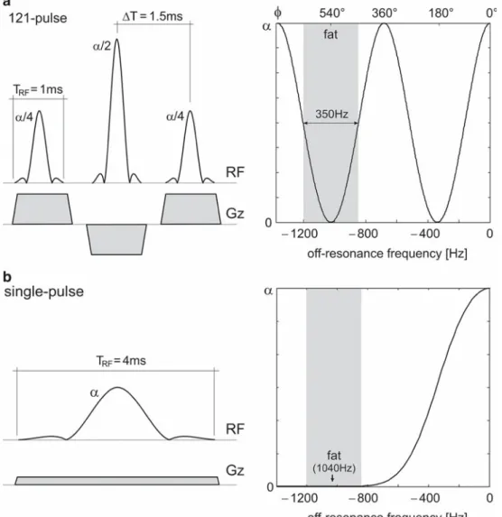
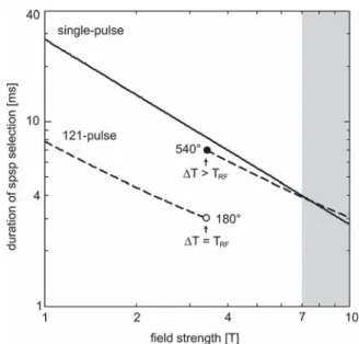
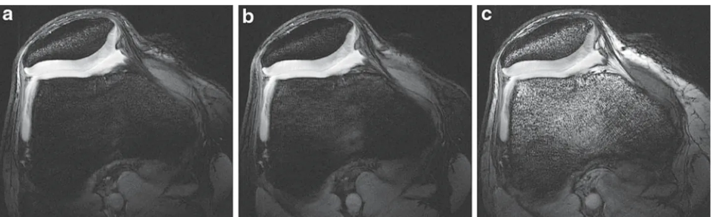
Optimized spectrally selective steady-state free precession sequences for cartilage imaging at ultra-high fields
8
0
0
Texte intégral
Figure

Documents relatifs