SCIENTIFIC ARTICLE
Comparison of radiation dose, workflow, patient comfort
and financial break-even of standard digital radiography
and a novel biplanar low-dose X-ray system for upright
full-length lower limb and whole spine radiography
Tobias J. Dietrich&Christian W. A. Pfirrmann&
Alexander Schwab&Katja Pankalla&Florian M. Buck
Received: 7 November 2012 / Revised: 1 February 2013 / Accepted: 4 March 2013 / Published online: 28 March 2013 # ISS 2013
Abstract
Objective To compare the radiation dose, workflow, patient comfort, and financial break-even of a standard digital radi-ography and a biplanar low-dose X-ray system.
Materials and methods A standard digital radiography sys-tem (Ysio, Siemens Healthcare, Erlangen, Germany) was compared with a biplanar X-ray unit (EOS, EOS imaging, Paris, France) consisting of two X-ray tubes and slot-scanning detectors, arranged at an angle of 90° allowing simultaneous vertical biplanar linear scanning in the upright patient position. We compared data of standing full-length lower limb radiographs and whole spine radiographs of both X-ray systems.
Results Dose–area product was significantly lower for radio-graphs of the biplanar X-ray system than for the standard digital radiography system (e.g. whole spine radiographs; standard digital radiography system: 392.2±231.7 cGy*cm2
versus biplanar X-ray system: 158.4±103.8 cGy*cm2). The mean examination time was significantly shorter for biplanar radiographs compared with standard digital radiographs (e.g. whole spine radiographs: 449 s vs 248 s). Patients’ comfort regarding noise was significantly higher for the standard dig-ital radiography system. The financial break-even point was 2,602 radiographs/year for the standard digital radiography system compared with 4,077 radiographs/year for the biplanar X-ray unit.
Conclusion The biplanar X-ray unit reduces radiation expo-sure and increases subjective noise expoexpo-sure to patients. The biplanar X-ray unit demands a higher number of examinations per year for the financial break-even point, despite the lower labour cost per examination due to the shorter examination time.
Keywords Digital radiography . Radiation dosage . Workflow . Financial management
Introduction
Standard digital radiography systems with X-ray area detec-tors are widely installed and used. Another imaging solution for evaluation of patients with particular musculoskeletal de-formities is a novel biplanar X-ray unit with a vertical biplanar slot-scanning X-ray technique (EOS scanner; EOS Imaging, Paris, France). The first reports about this technique were published in 2005 [1,2]. Although the financial investment needed to purchase, install, run and depreciate such a scanner is substantially higher compared with standard digital
Electronic supplementary material The online version of this article (doi:10.1007/s00256-013-1600-0) contains supplementary material, which is available to authorized users
T. J. Dietrich (*)
:
C. W. A. Pfirrmann:
K. Pankalla:
F. M. Buck Department of Radiology, Orthopedic University Hospital Balgrist, Forchstrasse 340, 8008 Zurich, Switzerland e-mail: [email protected]T. J. Dietrich
:
C. W. A. Pfirrmann:
A. Schwab:
K. Pankalla:
F. M. BuckUniversity of Zurich, Zurich, Switzerland A. Schwab
Department of Finances, Orthopedic University Hospital Balgrist, 8008 Zurich, Switzerland
radiography systems, these biplanar X-ray systems offer ra-diographs without distortions and enable secondary 3D recon-struction [2–4]. Because of these advantages an increasing number of biplanar X-ray systems are being installed with a total of 50 biplanar X-ray systems installed worldwide as of November 2012. Thirty-two units were installed in Europe, 15 in North America and 3 in the remaining continents. So far, no technological assessment addressing workflow and patient comfort parameters of standard digital radiography systems compared with biplanar X-ray systems has been published in the peer-reviewed literature.
Thus, the purpose of our study was to compare the radiation dose, workflow, patient comfort and financial pa-rameters of a standard digital radiography system and a biplanar X-ray system.
Materials and methods
The institutional review board issued a waiver for this study. All the patients included gave written permission for anonymised use of their data before the imaging examination.
Radiographs
Full-length lower limb radiographs and whole spine radio-graphs of a standard digital radiography system were com-pared with radiographs of a biplanar X-ray system (Figs.1,2). In total, 68 consecutive anteroposterior full-length lower limb radiographs and 47 consecutive anteroposterior and lateral whole spine radiographs were obtained using the standard digital radiography system during a 2-month period from May until June 2011. These examinations were compared with 198 anteroposterior full-length lower limb radiographs and 134 anteroposterior and lateral whole spine radiographs of a biplanar X-ray system acquired during a 2-month period from March to April 2011.
Patients
All patients referred for the respective examinations in the time periods mentioned above were included in the study. Mentally disabled patients were not included. The average patient’s height and weight were within the same range for the digital radiography groups compared with the biplanar radiograph study groups. The average patient’s height and weight for full-length lower limb radiographs was slightly lower for the digital radiography group (mean patients’ height and weight: 168.5±9.1 cm, 77.3±17.7 kg) than in patients imaged in the biplanar X-ray system (mean patient’s height and weight: 169.2±10.5 cm, 81.1±21.6 kg) and vice versa for total spine radiographs (digital radiography group mean patient’s height and weight 166.6±10.7 cm, 62.1±
13.0 kg; biplanar X-ray group mean patient’s height and weight 163.6 ± 10.9 cm, 57.7 ± 16.7 kg).
X-ray systems
Both the standard digital radiography system and biplanar X-ray system are commercially available and were evaluated under daily clinical conditions at an University orthopaedic hospital in Switzerland. All radiographs were obtained in an upright standing position.
Standard digital radiography system
The standard digital radiography system (Ysio; Siemens Healthcare, Erlangen, Germany) is equipped with an indi-rect digital radiography image detector consisting of a X-ray scintillator layer of caesium iodide. Based on auto-tracking movements of the X-ray tube and detector, it enables auto-mated acquisition of a craniocaudal image series consisting of up to four separate digital radiographs (digital detector area: 43 × 43 cm) in a single acquisition process in a
Fig. 1 A 65-year-old female patient. a Anteroposterior full-length lower limb radiograph of the standard digital radiography system for planning of a total knee arthroplasty on the right side and b postoper-ative radiograph of the biplanar X-ray system. The patient also underwent posterior interbody fusion of L4 to S1
monoplanar imaging technique. The images of these series are then semi-automatically stitched together by radiographic technicians at a post-processing workplace (Syngo®, Siemens Healthcare, Erlangen, Germany). The isotropic image resolution at the detector is 139 μm. Biplanar X-ray system
The biplanar X-ray system (EOS; EOS imaging, Paris, France) consists of two coupled X-ray tubes and slot-scanning detectors, arranged at an angle of 90° allowing simultaneous vertical biplanar linear scanning by two slit-like, fan-shaped X-ray beams. The linear detectors rely on gaseous micromesh structure technology promoting primary signal amplification through electronic avalanche in the xenon gas [5]. This system allows imaging at low radiation levels [6]. The detector technology has undergone several evolutions since the publication by Després et al. [5] including hardware modification of the detector and output signal processing. These improvements in particular reduce the ripple artefacts. Isotropic image resolution at the detector is 254μm [6]. Imaging parameters
The following imaging parameters were used for a medium-sized patient habitus and were adapted depending on the patient’s weight and size:
– Standard digital radiography system: tube voltage, 75–90 kVp for the anteroposterior and 77–90 kVp for the lateral view; tube current by automatic exposure control; detector-to-tube distance, 300 cm; maximum craniocaudal field of view, 180 cm.
– Biplanar X-ray system: tube voltage, 90 kVp for the anteroposterior and 110 kVp for the lateral view; the tube current was selected manually and the exposure stayed constant from the top to the bottom of the acquisition without automatic exposure control, 250 mAs for the anteroposterior and 320 mAs for the lateral view; detector-to-tube distance, 130 cm; object to tube distance approximately 100 cm for both sources in the standing position; maximum craniocaudal field of view, 175 cm [6]. The scanning time was approximately 13 s for adult full-length lower limb and 10 s for whole spine radiographs.
Data acquisition
Radiation exposure to patients
Both X-ray units are equipped with an integrated dosimeter and the dose–area product (DAP) is shown automatically on the control panel [7].
Fig. 2 An 18-year-old male patient with levoscoliosis at the thoracolumbar level after posterior spinal fusion of the vertebral bodies T4–T12. a, b Standard digital radiographs and c, d biplanar
radiographs of the whole spine in the anteroposterior and lateral views at the 6-month follow-up. The technical image quality of both whole spine radiographs is very good
Radiography technicians’ workflow
Technicians were trained for at least for 6 weeks to use the systems. The examination time was measured and defined as the time period between the moment when the patient stepped inside the examination room and the moment the patient stepped outside the examination room plus the time needed for processing the examination data in the radiology information system and transferring the images to the pic-ture archiving and communications system (PACS).
Using a four-point Likert-type item (answers: very easy, rather easy, rather difficult, very difficult), one question evaluated the workflow as experienced by the radiography technician: was it easy to position the patient? [8]. In addition, the radiography technicians’ workflow
parameters were assessed by a two-item questionnaire, with the following questions:
& Was it necessary to repeat the examination? & Were there any delays or problems? Patients’ comfort
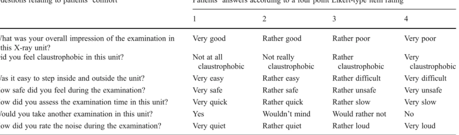
A questionnaire was used for evaluation of patients’ comfort consisting of the following questions using a four-point Likert-type item (Table1) [8].
Financial analysis
The cost effectiveness was evaluated by the calculation of the break-even point based on a maximum number of examinations per annum of each X-ray system. The maximum number of examinations per annum was calculated by recording the time period of standard digital radiographs and biplanar radiographs for 3 days by three different techni-cians in our department. These time period measurements included the time from picking up the patient in the waiting room, changing patients’ clothes, acquisition of the radio-graph, time needed for electronic documentation and bringing the patient back to the waiting room. The cost effectiveness analysis in the present study assumed a theoretical maximum utilization per annum (250 workdays per year) as well as a theoretical maximum reimbursement of both X-ray systems with an equal ratio of whole spine radiographs in anteroposterior/lateral views and anteroposterior full-length lower limb radiographs. Calculations of financial reimburse-ment, fixed costs and variable costs revealed the financial break-even point and the corresponding number of examina-tions per annum. Our financial calculaexamina-tions were based on the preconditions with the following parameters: financial reim-bursement with an equal ratio by health insurances and accident/disability insurances was CHF 182 per examination (TARMED version 1.07.01). The annual interest and
Table 1 Likert-type item rating of patients’ comfort parameters
Questions relating to patients’ comfort Patients’ answers according to a four point Likert-type item rating
1 2 3 4
What was your overall impression of the examination in this X-ray unit?
Very good Rather good Rather poor Very poor
Did you feel claustrophobic in this unit? Not at all claustrophobic Not really claustrophobic Rather claustrophobic Very claustrophobic Was it easy to step inside and outside the unit? Very easy Rather easy Rather difficult Very difficult How safe did you feel during the examination? Very safe Rather safe Rather unsafe Very unsafe How did you assess the examination time in this unit? Very quick Rather quick Rather slow Very slow Would you take another examination in this unit? Yes Wouldn’t mind Would rather not No How did you rate the noise during the examination? Very quiet Rather quiet Rather loud Very loud A questionnaire was used for evaluation of patients’ comfort consisting of the questions listed above by applying a four point Likert type item
Table 2 Dose–area product of full-length lower limb radiographs and whole spine radiographs
Dose–area product Standard digital radiographs Biplanar X-rays P value (Student’s t test) Full-length lower limb radiographs anteroposterior
(cGy*cm2)
(n=66) (n=198)
170.9±104.2 92.1±45.5 <0.001 Whole spine radiographs anteroposterior/lateral
(cGy*cm2)
(n=47) (n=134)
depreciation period was 8 years for both the standard digital radiography system and biplanar X-ray system. Indirect cost was 7.63 % and included mainly back-office tasks and infra-structure provided by the hospital for the radiology depart-ment such as real estate service, information technology, energy and air conditioning, human resources management, telephone switchboard, laundry, cleaning service, technical service, pharmacy, restaurant for employees and centralised purchasing department. Fixed costs included these indirect costs as well as annual interest and depreciation of fixed assets. Variable costs were 46.2 % and included labour costs (76.3 % of the variable costs), picture archiving and commu-nication system including labour costs (4.5 % of variable costs), maintenance expense (9.6 % of variable costs) and consumption of materials (9.6 % of variable costs). Overall costs was the sum of the fixed costs and variable costs.
Statistical analysis
The Student’s t test and Mann–Whitney U test served for statistics. A P value less than 0.05 was considered sufficient to indicate statistical significance between the standard
digital radiography system and the biplanar X-ray system. A computer software package (SPSS, version 17.0, SPSS) was used for statistical calculations.
Results
Radiation exposure to patients
The dose–area product (Table2) of anteroposterior standing full-length lower limb radiographs as well as the dose–area product of the whole spine radiographs including anteroposterior and lateral views was significantly lower for radiographs of the biplanar X-ray system compared with the standard digital radiography system (e.g. spine; standard digital radiography system: 392.2 ± 231.7 cGy*cm2 vs biplanar X-ray system: 158.4±103.8 cGy*cm2, P=<0.001 [Student’s t test]).
Radiography technicians’ workflow
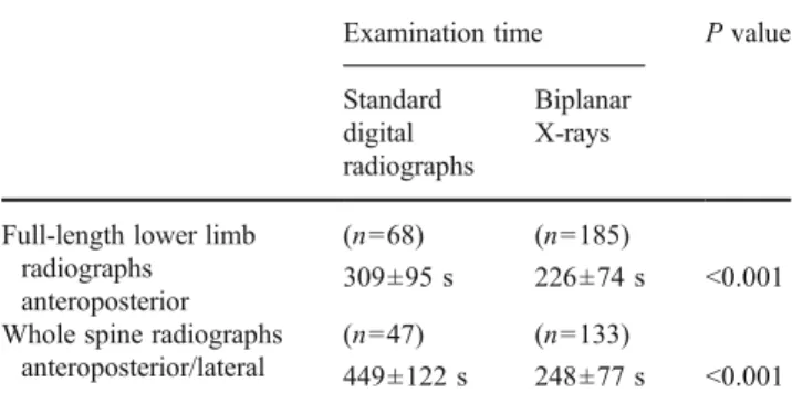
The mean examination time (Table 3) was significantly shorter for biplanar radiographs compared with standard digital radiographs (e.g. whole spine radiographs; standard digital radiography system: 449 s vs biplanar X-ray system: 248 s). The remaining workflow parameters (Table 4) did not reveal any significant difference.
Patients’ comfort
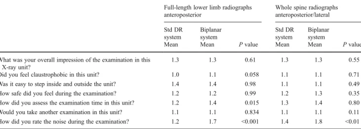
In contrast to the examination time measured, patients under-going full-length lower limb radiographs subjectively assessed the examination time significantly longer in the biplanar X-ray system, whereas there was no significant dif-ference for whole spine radiographs (Table5). The biplanar
Table 3 Examination time of full-length lower limb radiographs and whole spine radiographs
Examination time P value
Standard digital radiographs
Biplanar X-rays
Full-length lower limb radiographs anteroposterior
(n=68) (n=185)
309±95 s 226±74 s <0.001 Whole spine radiographs
anteroposterior/lateral
(n=47) (n=133)
449±122 s 248±77 s <0.001
Table 4 Radiography technicians’ workflow parameters
Full-length lower limb radiographs anteroposterior
Whole spine radiographs anteroposterior/ lateral
Std DR system
Biplanar system Std DR system Biplanar system
Mean Mean P value Mean Mean P value
Was it easy to position the patient? 1.2 1.2 0. 22 1.3 1.2 0.201
Was it necessary to repeat the examination? (Yes)
0 % (0/68) 2.6 % (5/190) 0.18 4.3 % (2/47) 6.7 % (9/134) 0.55 Were there any delays or problems? (Yes) 4.4 % (3/68) 10.5 % (20/190) 0.13 27.7 % (13/47) 22.4 % (30/134) 0.47 Values for the question“Was it easy to position the patient?” are expressed as mean. We used a four-point Likert- type Item (answers: 1=very easy, 2=rather easy, 3=rather difficult, 4=very difficult)
Two radiography technicians’ workflow parameters were binary as yes or no The Mann–Whitney U test served for statistics
X-ray system was significantly noisier compared with the standard digital radiography system (Table 5, P < 0.01 [Mann–Whitney U test]). No other parameters relating to the patients’ comfort showed significant differences between the standard digital radiography system and the biplanar X-ray system. However, patients undergoing full-length lower limb radiography felt considerably more (P=0.058) claustrophobic in the biplanar X-ray system than in the standard digital radiography system. Patients undergoing whole spine radio-graphs, on the other hand, did not feel significantly more claustrophobic in the biplanar X-ray system (P=0.714).
Financial analysis
The theoretical maximum number of examinations per annum was 12,000 radiographs (48 radiographs per day) for the standard digital radiography system compared with 17,250 radiographs (69 radiographs per day) for the biplanar X-ray system. Financial investment was higher for the biplanar X-ray system compared with the standard digital radiography system; therefore, the annual interest and depreciation of fixed assets was lower for the standard digital radiography system (CHF 88,700) than for the
Table 5 Patients’ comfort parameters
Full-length lower limb radiographs anteroposterior
Whole spine radiographs anteroposterior/lateral Std DR system Biplanar system Std DR system Biplanar system
Mean Mean P value Mean Mean P value
What was your overall impression of the examination in this X-ray unit?
1.3 1.3 0.61 1.3 1.3 0.55
Did you feel claustrophobic in this unit? 1.0 1.1 0.058 1.1 1.1 0.71
Was it easy to step inside and outside the unit? 1.4 1.4 0.98 1.1 1.1 0.49
How safe did you feel during the examination? 1.2 1.2 0.99 1.2 1.3 0.35
How did you assess the examination time in this unit? 1.2 1.4 0.015 1.3 1.4 0.80
Would you take another examination in this unit? 1.1 1.1 0.834 1.1 1.1 0.11
How did you rate the noise during the examination? 1.2 1.7 <0.001 1.4 1.8 <0.01
Values are expressed as mean. A questionnaire was used for evaluation of patients’ comfort consisting of the questions listed above by applying a four-point Likert-type Item, e.g.“What was your overall impression of the examination in this X-ray unit?” (Answers: 1=very good, 2=rather good, 3=rather poor, 4=very poor)
The Mann–Whitney U test served for statistics Std DR system standard digital radiography system
Fig. 3 The calculation of the financial break-even point (BE) based on cost and financial reimbursement by health insurance is shown as a graphical illustration. The total fixed costs are lower owing to the smaller financial investment for a the standard
digital radiography systems compared with b the biplanar X-ray system. Therefore, the biplanar X-ray system demands a higher number of examinations per annum to reach the financial break-even point
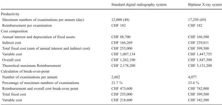
biplanar X-ray system (CHF 160,500). The higher theoret-ical maximum utilization per annum was associated with higher indirect costs for the biplanar X-ray system (CHF 239,011) compared with the standard digital radiography system (CHF 166,269). The break-even point was 2,602 examinations/year for the standard digital radiography sys-tem and 4,077 examinations/year for the biplanar X-ray system (Fig. 3, Table 6). In particular, the significantly shorter examination time in the biplanar X-ray system with a higher patient throughput partly outweighed the difference in the break-even analysis.
Discussion
The biplanar X-ray system reduced radiation exposure to patients in comparison to the standard digital radiography system. Deschênes et al. compared radiation doses of a biplanar X-ray system and a computed radiography system in 50 pa-tients with spinal deformities. They stated a 6 to 9 times reduction of the average skin dose in the thoracoabdominal region when using a biplanar X-ray system instead of computed radiography with phosphor imaging plates [6]. In the present study, we have observed a dose–area product reduction of approximately 50 % in patients examined with the biplanar X-ray system compared with a digital radiography system with
an indirect digital image detector consisting of a X-ray scintil-lator layer of caesium iodide. Detective quantum efficiency (DQE) refers to the efficiency of a detector in converting X-ray energy into an image signal [9,10]. It is known that a computed radiography system with phosphor imaging plates has a DQE comparable to that of conventional analogue X-ray imaging systems (screen-film systems), whereas indirect CsI-based flat-panel detector technology as used in the present study has a better DQE [9,10]. The improved DQE of the indirect CsI-based flat-panel detector in our study compared with the DQE of a computed radiography detector may explain in part the lower dose reduction with the biplanar X-ray system in the present study compared with the study by Deschênes et al. Dose–area product served for measurement of skin entrance exposure in the present study. Parry et al. [7] stated that their measurements obtained with a dose–area product meter strong-ly correlated with those obtained with the thermoluminescent dosimeters. DAP measurement is not the most accurate tech-nique for comparing radiation dose. Luminescence dosimeters also measure radiation due to backscatter, which may increase the entrance surface dose by about 30 % [7, 11]. However, Martin stated that the DAP can audit and compare radiation doses from a wide variety of radiological examinations [11].
So far, there have been no reports about biplanar X-ray systems dealing with examination time, workflow and pa-tients’ comfort analysis available in the literature. Overall,
Table 6 Cost-effective analysis of a theoretical maximum utilization of the standard digital radiography system and biplanar X-ray system per annum (equal ratio of whole spine radiographs in anteroposterior/ lateral views and anteroposterior full-length lower limb radiographs,
assumption of 250 workdays per year). CHF Swiss Francs. Costs underwent rounding according to the system of our department of finances
Standard digital radiography system Biplanar X-ray system Productivity
Maximum numbers of examinations per annum (day) 12,000 (48) 17,250 (69)
Reimbursement per examination CHF 182 CHF 182
Cost composition
Annual interest and depreciation of fixed assets CHF 88,700 CHF 160,500
Indirect cost CHF 166,269 CHF 239,011
Total fixed cost (sum of annual interest and indirect cost) CHF 255,000 CHF 399,500
Variable cost CHF 1,007,134 CHF 1,447,755
Overall cost CHF 1,262,100 CHF 1,847,300
Theoretical maximum Reimbursement CHF 2,178,200 CHF 3,131,200
Calculation of break-even-point
Number of examinations per annum 2,602 4,077
Percentage of maximum numbers of examinations 21.7 % 23.6 %
Reimbursement and overall cost break-even point CHF 473,600 CHF 742,000
Total fixed cost CHF 255,000 CHF 399,500
Variable cost CHF 218,600 CHF 342,500
Financial reimbursement by health insurance; fixed and variable costs were included. Fixed costs included annual interest and depreciation of fixed assets as well as back-office tasks provided by the hospital for the radiology department. Variable costs included labour costs for radiologists, radiography technicians and cost of materials per examination
patients’ comfort with the biplanar X-ray system in compari-son to the standard digital radiography system was equal. Exclusively one comfort parameter revealed significant differ-ences: noise exposure from the standard digital radiography system was significantly lower compared with the biplanar X-ray system. Studies addressing patients’ comfort in the field of diagnostic radiology are rare in the peer-reviewed literature, e.g. breast imaging, CT colonography preparation, dental radi-ography or invasive procedures such as MR hysterosalpingog-raphy and retrograde urethroghysterosalpingog-raphy [12–16]. One may speculate that patients’ comfort in imaging could influence patients’ compliance and thus indirectly image quality.
The examination time of the biplanar X-ray system in our study was significantly shorter than that of the standard digital radiography system implying lower labour costs for radiography technicians per examination and a higher pa-tient throughput per time period. On the other hand the biplanar X-ray system demands a much higher financial investment than standard digital radiography systems.
Given the fact that financial reimbursement for graphs in Switzerland is the same for standard digital radio-graphs and biplanar radioradio-graphs, the shorter examination time and higher financial investment of the biplanar X-ray system requires a higher number of examinations to reach financial break-even. With the biplanar X-ray system it is not possible to obtain radiographs of patients in the prone or supine position. Therefore, the biplanar X-ray system can-not replace a standard digital radiography system. This drawback limits applicability in a general hospital setting.
McKenna et al. [17] carried out a systematic review and economic evaluation of the biplanar X-ray system. The authors suggested that the biplanar X-ray system is not cost-effective [17]. They stated that a patient throughput of 7,530 examinations per year for computed radiography compared with a range of 15,100 to 26,500 examinations per year for the biplanar X-ray system is required to achieve an incremental cost-effectiveness ratio of £30,000 per quality-adjusted life year [17]. The authors also found that the number of examinations for the financial break-even has to be doubled for the biplanar X-ray system compared with computed radiography [17], which is similar to our analysis for a digital radiography system. Our financial analysis is different to that of McKenna et al., who assessed the cost-effectiveness of the biplanar X-ray system without practical assessment in daily practice as in the present study. The theoretical maximum number of examinations per annum is a key parameter for our cost analysis. The higher patient throughput due to the significantly shorter examination time of the biplanar X-ray system partly outweighed the differ-ence in the present break-even analysis. The latter issue was not considered in the study by McKenna et al. Finally, the financial data of McKenna et al. [17] were based on the National Health Service (NHS) of the United Kingdom
whereas our data were based on the healthcare system of Switzerland.
A biplanar X-ray system may be operated cost-effectively in addition to a standard digital radiography system in institutions with a high number of examinations. Institutions with a standard digital radiography system and an additional biplanar X-ray system have the advantage of choosing be-tween both units for each patient individually. In our depart-ment, the predominant types of examinations performed in the biplanar X-ray system were whole spine radiographs and full-length lower limb radiographs. Whole body radiographs and whole femur radiographs were rarely performed. Children and teenagers in our department were preferably examined in the biplanar X-ray unit taking into consideration the special radiation protection issues of young patients.
Advantages of the biplanar X-ray system are radiographs without distortions and the possibility of additional secondary 3D reconstructions with measurement of both internal and external surfaces without acquisition of multiple tomographic images [1, 3, 18–23]. Femur antetorsion can be measured based on the images of the biplanar X-ray system as an alternative to computed tomography [19]. Nevertheless, addi-tional secondary 3D reconstruction is a time-intensive proce-dure consuming between 15 and 30 min for radiologists or radiography technicians to image the entire spine [2].
Our study has limitations. In this study we did not inves-tigate image quality. The quality of the biplanar X-ray system has already been validated for the measurement of skeletal deformities [4, 24–26]. The significantly better image quality of biplanar X-ray systems compared with computed radiography has been reported for spine radio-graphs in the frontal view and lateral view in a previous study [6]. The cost analysis reflects the financial situation of one specific orthopaedic hospital in a single country and may apply for other hospitals in various national health systems.
In summary, the biplanar X-ray system reduces radiation exposure and increases subjective noise exposure to pa-tients. The biplanar X-ray unit demands a higher number of examinations per year for the financial break-even point despite the lower labour costs per examination owing to the shorter examination time. Thus, the biplanar X-ray system may be suitable for institutions with a high number of radiographs in the standing or sitting position.
Conflict of interests The authors declare that they have no conflict of interest.
References
1. Dumas R, Aissaoui R, Mitton D, Skalli W, de Guise JA. Personalized body segment parameters from biplanar low-dose radiography. IEEE Trans Biomed Eng. 2005;52:1756–63.
2. Dubousset J, Charpak G, Dorion I, et al. A new 2D and 3D imaging approach to musculoskeletal physiology and pathology with low-dose radiation and the standing position: the EOS sys-tem. Bull Acad Natl Med. 2005;189:287–300.
3. Illés T, Tunyogi-Csapó M, Somoskeöy S. Breakthrough in three-dimensional scoliosis diagnosis: significance of horizontal plane view and vertebra vectors. Eur Spine J. 2011;20:135–43. 4. Labelle H, Aubin CE, Jackson R, Lenke L, Newton P, Parent S.
Seeing the spine in 3D: how will it change what we do? J Pediatr Orthop. 2011;31:S37–45.
5. Després P, Beaudoin G, Gravel P, de Guise JA. Physical charac-teristics of a low-dose gas microstrip detector for orthopedic x-ray imaging. Med Phys. 2005;32:1193–204.
6. Deschênes S, Charron G, Beaudoin G, et al. Diagnostic imaging of spinal deformities: reducing patients radiation dose with a new slot-scanning X-ray imager. Spine. 2010;35:989–94.
7. Parry CK, Chu RY, Eaton BG, Chen CY. Measurement of skin entrance exposure with a dose-area-product meter at chest radiog-raphy. Radiology. 1996;201:574–5.
8. Uebersax JS. Likert scales: dispelling the confusion. Statistical Methods for Rater Agreement website. 2006. Available via
http://john-uebersax.com/stat/likert.htm. Accessed 6 November 2012.
9. Illers H, Buhr E, Hoeschen C. Measurement of the detective quantum efficiency (DQE) of digital X-ray detectors according to the novel standard IEC 62220–1. Radiat Prot Dosim. 2005;114:39–44.
10. Korner M, Weber CH, Wirth S, Pfeifer KJ, Reiser MF, Treitl M. Advances in digital radiography: physical principles and system overview. Radiographics. 2007;27:675–86.
11. Martin CJ. Radiation dosimetry for diagnostic medical exposures. Radiat Prot Dosim. 2008;128:389–412.
12. Svane G, Azavedo E, Lindman K, et al. Clinical experience of photon counting breast tomosynthesis: comparison with traditional mammography. Acta Radiol. 2011;52:134–42.
13. Liedenbaum MH, Denters MJ, de Vries AH, et al. Low-fiber diet in limited bowel preparation for CT colonography: influence on image quality and patient acceptance. Am J Roentgenol. 2010;195:W31–7. 14. Gonçalves A, Wiezel VG, Gonçalves M, Hebling J, Sannomiya EK. Patient comfort in periapical examination using digital recep-tors. Dentomaxillofac Radiol. 2009;38:484–8.
15. Winter L, Glücker T, Steimann S, et al. Feasibility of dynamic MR-hysterosalpingography for the diagnostic work-up of infertile women. Acta Radiol. 2010;51:693–701.
16. Berná-Mestre JD, Berná-Serna JD, Aparicio-Mesón M, Canteras-Jordana M. Urethrography in men: conventional technique versus clamp method. Radiology. 2009;252:240–6.
17. McKenna C, Wade R, Faria R, et al. EOS 2D/3D X-ray imaging system: a systematic review and economic evaluation. Health Technol Assess. 2012;16:1–188.
18. Morvan G, Mathieu P, Vuillemin V, et al. Standardized way for imaging of the sagittal spinal balance. Eur Spine J. 2011;20:602–8. 19. Than P, Szuper K, Somoskeöy S, Warta V, Illés T. Geometrical values of the normal and arthritic hip and knee detected with the EOS imaging system. Int Orthop. 2012;36:1291–7.
20. Sabourin M, Jolivet E, Miladi L, Wicart P, Rampal V, Skalli W. Three-dimensional stereoradiographic modeling of rib cage before and after spinal growing rod procedures in early-onset scoliosis. Clin Biomech. 2010;25:284–91.
21. Ohl X, Stanchina C, Billuart F, Skalli W. Shoulder bony landmarks location using the EOS® low-dose stereoradiography system: a reproducibility study. Surg Radiol Anat. 2009;32:153–8. 22. Schlatterer B, Suedhoff I, Bonnet X, Catonne Y, Maestro M, Skalli
W. Skeletal landmarks for TKR implantations: evaluation of their accuracy using EOS imaging acquisition system. Orthop Traumatol Surg Res. 2009;95:2–11.
23. Lazennec JY, Rousseau MA, Rangel A, et al. Pelvis and total hip arthroplasty acetabular component orientations in sitting and standing positions: measurements reproducibility with EOS imag-ing system versus conventional radiographies. Orthop Traumatol Surg Res. 2011;97:373–80.
24. Buck FM, Guggenberger R, Koch PP, Pfirrmann CW. Femoral and tibial torsion measurements with 3D models based on low-dose biplanar radiographs in comparison with standard CT measure-ments. Am J Roentgenol. 2012;199:W607–12.
25. Thelen P, Delin C, Folinais D, Radier C. Evaluation of a new low-dose biplanar system to assess lower-limb alignment in 3D: a phantom study. Skeletal Radiol. 2012;41:1287–93.
26. Sutter R, Pfirrmann CW, Espinosa N, Buck FM. Three-dimensional hindfoot alignment measurements based on biplanar radiographs: comparison with standard radiographic measure-ments. Skeletal Radiol. 2013;42:493–98. .