REVIEW
Current management of dentin hypersensitivity
Patrick R. Schmidlin&Phlipp SahrmannReceived: 8 December 2011 / Accepted: 21 November 2012 / Published online: 30 December 2012 # The Author(s) 2012. This article is published with open access at Springerlink.com
Abstract
Objectives The aim of the article was to present an overview of the management strategies of dentin hypersensitivity (DHS) and summarize and discuss the therapeutic options. Materials and methods A PubMed literature search was conducted to identify articles dealing with dentin hypersensitivity prophylaxis and treatment. We focussed on meta-analyses of available or controlled clinical trials. Results DHS therapy should start with noninvasive individual prophylactic home-care approaches. In-office therapy follows with nerve desensitizing, precipitating, or plugging agents. If the hypersensitivity persists, depending on the hard and soft tissue components at reevaluation, i.e., presence or absence of cervical lesions and the gingival contour, adhesive restorations including sealing or mucogingival surgery may be an option. They allow for the establishment of a physicomechanical barrier. As the placebo effect may play an important role, adequate patient management strategies and positive reinforcement may improve the management of DHS in the future.
Conclusions Lifelong maintenance under the premise of strict control of the causative factors is crucial in the management of DHS.
Clinical relevance Clinicians are faced with a broad spectrum of therapeutic options. Therapy should not only focus on pain reduction or better elimination but also on the modification of the exposed cervical dentin area based on the defect type. Keywords Dentin hypersensitivity . Therapy . Review
Background and aim
Dentin hypersensitivity (DHS) describes a painful symptom of the exposed and innervated cervical pulp–dentin complex. Its prevalence greatly varies and ranges from 3 up to 98 % and can be explained—in part—by different evaluation methods and different patient populations, e.g., patients suffering from periodontitis [1].
It is important to acknowledge that localized attachment loss is probably the most widespread and relevant factor leading to any root surface denudation and dental hard tissue sequelae, including DHS. Several predisposing factors of gingival recessions have been identified, e.g., dehiscency or fenestration of the alveolar bone and soft tissue morphotype, but triggering pathological, therapeutic, or iatrogenic factors are also crucial for its development [2]. Loss of dental hard tissue in the coronal aspect, especially enamel, is considered an alternative pathway of cervical dentin exposure and is mainly caused by erosion, abrasion, and abfraction or a com-bination thereof [2].
DHS is mainly a diagnosis of exclusion. Thus, differential diagnostic aspects play a pivotal role and a thorough anam-nestic evaluation is indispensable to identify etiopathogenic factors [3]. This decision process is important to effectively control and treat underlying causative and modifying factors [4].
Different materials and methods have been described to treat DHS. Cause-related therapy primarily aims to mechanically or chemically protect the pulp–dentin complex or to suppress or modify the nerve stimulation while controlling the predisposing factors. However, no treatment has been found so far, which could serve as a defined therapeutic gold standard, which predictably and completely eliminates pain perception, especially in the long term. Root canal therapy may be considered as the last resort, but there is a consensus that this invasive treatment should not be considered in the first line. In
P. R. Schmidlin (*)
:
P. SahrmannClinic of Preventive Dentistry, Periodontology and Cariology, Centre of Dental and Oral Medicine, University of Zurich, Plattenstrasse 11,
8032 Zurich, Switzerland
contrast, treatment should conceptually start with minimally invasive and reversible approaches. If these are not successful at reevaluation, subsequently more invasive and maybe more expensive or even irreversibe treatment options should be envisioned. A crucial question is whether or when does treatment become necessary. Table1gives an overview of a suggested decision tree for intervention and additional differential diagnosis.
If a patient self-reports pain, the necessity of DHS treatment is evident. A pain provocation test should then be performed. Most commonly, air application and/or gentle scratching with a probe are used for this purpose. If this pain provocation test is positive and other pathologies can be excluded (differential diagnosis), DHS therapy should be initiated. In the case of a negative provocation test, special emphasis should be put on the differential diagnosis and the cause of the pain should be carefully evaluated (endodontic or cariologic origin, orofacial pain, etc.). If a patient reports no hypersensitive episodes, a provocation test is optional but recommended during routine dental screening. In the case of exposed cervical areas which are sensitive upon provocation, a treatment can be considered in prophylactic terms. In case of a negative provocation test,
any DHS therapy becomes needless. It is important to acknowledge that not all patients report hypersensitivity experiences, and dentists and prophylaxis personnel should therefore screen for DHS on a routine basis.
This article focuses on the current management of DHS. Different materials and techniques are critically discussed and future perspectives are shown. Whenever possible, systematic reviews were used to document the success/ failure of a method.
Current management of DHS
Prevention is better than cure. Thus, primary prevention of dentin exposure as a result of recession formation and/or dental hard tissue damage by any prophylactic measures or erosive/abrasive diet is the best way to actually treat this phenomenon and its associated painful symptoms [5]. Special focus must thus be set on the prevention of DHS, such as to avoid erosive drinks or foods and choose nonabrasive toothbrushes, brushing techniques, etc.
The management of DHS can be generally divided in two different approaches: self-performed therapy at home (professionally dispensed self-applied or over the counter) or in-office treatment. The latter normally applies more sophisticated noninvasive or invasive methods using professional materials and techniques. In general, all interventions should start with noninvasive, reversible, nonhazardous, easy to perform, and inexpensive options. Only if they prove to be ineffective at reevaluation should more invasive interventions be considered.
Conceptually, treatment of DHS aims either to suppress the nerve impulse by direct neurological interaction or by mechanical blockage of the tubules. Potassium ions can decrease the excitability of A fibers, which surround the odontoblasts, thus resulting in a significant reduction in tooth sensitivity. Toothpastes for instance contain mostly 5 % potassium nitrate [6], but there is still a debate concerning its effectiveness [7].
Table 1 Proposed evaluation and decision steps
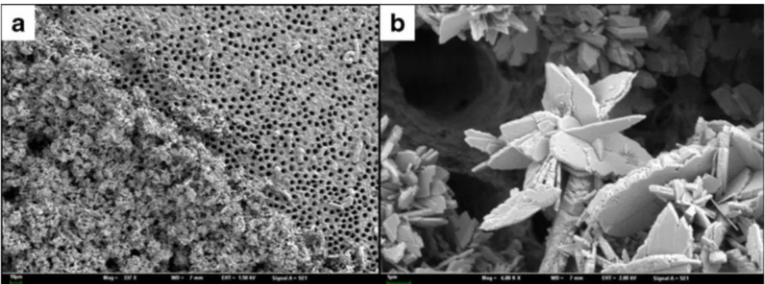
Fig. 1 SEM image of dentin treated with a precipitating agent: Panel a shows treated (bottom left) and untreated (top right) areas. However, despite clear evidence of crystallite deposition, uncovered dentin areas and tubule entrances can be seen (b)
Aside from nerve desensitization, occlusion of the dentinal tubules is the main therapeutic approach (Fig. 1). There is a plethora of agents, materials, and products available on the market for this purpose. Different mechanisms can be identified to modify the dentin surface or tubules by chemical, mechanical, and/or physical means, e.g., protein precipitation, plugging of dentin tubules, sealing, or laser applications.
Aqueous glutaraldehyde-containing solutions have shown to be effective in reducing dentin hypersensitivity
[8]. They have been shown to close dentinal tubules by precipitative intratubular occlusion and thereby to a significant decrease of dentin permeability, even under clinical conditions [9, 10]. As a positive side effect, glutaraldehyde disinfects dentin in vitro and seems compatible with adhesive systems [11,12]. However, potential biocompatibility hazards should not be neglected [13,14].
Different plugging agents have been described and most products belong to this group of action. Among these, oxalates have been used to precipitate and occlude the tubules.
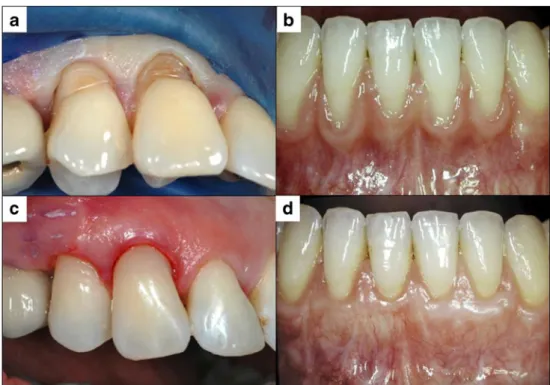
Fig. 2 Restitution of the anatomy of the cervically exposed dentin using a restorative approach with an adhesive class V filling (a/c), thereby covering and protecting the sensitive dentin. As an alternative, a recession coverage using a connective tissue graft before can be indicated, especially if the tooth substance loss is limited and the soft tissue morphology is adequate for a mucogingival approach (panel b before treatment and d 1 year after mucogingival surgery using a coronally advanced flap and connective tissue graft)
Fig. 3 Flow chart of the decision-making process based on the underlying defect. Depending on the dental hard tissue damage and the morphology of the surrounding soft tissues (according to Miller 1985), an adequate therapy can be initiated
However, a systematic review recently revealed that with the possible exception of 3 % monohydrogen monopotassium oxalate, available evidence does not currently support the recommendation of dentin hypersensitivity treatment with oxalates [15].
Products containing arginine/calcium carbonate, bioactive glass, or strontium acetate have been introduced in the market with similar modes of action (occlusion of the tubule openings) and seem to provide good clinical results and pain relief [16–18]. The available home-care products should be considered as the first approach.
xFluoride varnishes have also shown to protect the dentin surface by forming a protective layer of calcium fluoride [19–21]. Whereas lasers have been frequently assessed in the management of DHS, their clinical success is questionable [22]. However, fluoride gels in combination with laser showed some cumulative efficacy [23]. Another more exotic approach is the combination of acidulated fluoride gels with iontophe-resis [24].
A reliable technique to close dentin tubules is the application of a chemomechanical barrier between the dentin and the oral environment based on adhesive bonding techniques [25]. However, most of the bonding agents have no fillers, which potentially leads to wear in exposed areas [26]. The question is therefore how effectively can these lesions be resealed.
All methods described above are indicated especially in cases with limited amounts of dental hard tissue loss, i.e., no classical abrasive or erosive defect characteristics. In cases where a class V filling is indicated, an adhesive filling is a valid option. Regenerative mucogingival therapy also remains an alternative, where hard and soft tissue conditions allow [27] (Fig.2). A suggested strategy for DHS manage-ment, taking these morphological aspects into consideration, is depicted in Fig. 3. It should be noted that adequate diagnosis and prophylactic measures are a prerequisite for any successful management strategy, which is comple-mented with a strict long-term maintenance program in harmony with the special needs of DHS.
Outlook
DHS still is an underestimated problem in daily clinical practice. Most patients develop coping strategies. However, especially in cases where we are confronted with moderately symptomatic and more severe episodes, finding the right solution is difficult, and trial and error strategies are still a daily occurrence. Thus, the development of new materials and methods is greatly needed, including the development of improved precipitating materials, which effectively bind to the exposed dentin wound and reduce the outflow of dentinal fluid. This outflow continues to hamper precipitation or bonding processes. The use of lasers in combination with such materials
could be beneficial, leading to better precipitation or controlled melting mechanisms and impregnation.
In addition, as we are dealing with exposed cervical dental hard tissue areas, a focus also has to be put on improving the caries resistance of these involved surfaces. The development of prophylactic measures with less risk of recession formation or abrasion is also warranted.
Another aspect is the placebo effect, which is an important and potentially beneficial side effect when dealing with pain, and its treatment and management. Using arthritis of the knee as an example in the medical field, it has been impressively shown that sham endoscopic interventions lead to the same reduction of pain and symptoms as conventional treatment modalities [28]. In addition, prescription of differently colored pills resulted in significant differences in pain reduction [29]. Whereas a red placebo tablet, for instance, showed comparable pain relief as the best antirheumatic test pill used, the blue equivalent showed the least effect. Thus, improved psychological co-therapeutic strategies may one day become an important auxiliary aspect in DHS management, especially when it comes to changing patients’ expectations of treatment outcomes and confidence. The psychological training of dental professionals still has some room for development.
Conclusion
DHS is a problem. While there is no necessarily acute pathological risk for the affected tooth site at the moment, patients may suffer greatly and future damage due to the persisting pain challenge and its causative factors, i.e., the co-etiopathological factors, cannot be excluded. Therefore, a therapy should not only focus on pain reduction or better elimination but also on the modification of the exposed cervical dentin area based on the defect type. Preventive, restorative, and periodontal options may be individually indicated to resolve the pain problem, while concomitantly reducing the risk for future damage to the pulp–dentin complex. Adequate home-care products are advisable.
Conflict of interest The authors declare that they have no conflict of interest.
Open Access This article is distributed under the terms of the Creative Commons Attribution License which permits any use, distribution, and reproduction in any medium, provided the original author(s) and the source are credited.
References
1. Splieth CH, Tachou A (2012) Epidemiology of dentin hypersensi-tivity. doi:10.1007/s00784-012-0889-8
2. West NX, Lussi A, Seong J, Hellwig E (2012) Dentin hypersensi-tivity: pain mechanisms and aetiology of exposed cervical dentin. Clin Oral Invest. doi:10.1007/s00784-012-0887-x
3. Gillam DG (2012) Current diagnosis of dentin hypersensitivity in the dental office: an overview. Clin Oral Invest. doi:10.1007/ s00784-012-0911-1
4. Gernhardt CR (2012) How valid and applicable are current diag-nostic criteria and assessment methods for dentin hypersensitivity? An overview. Clin Oral Invest. doi:10.1007/s00784-012-0891-1
5. Jeffcoat MK (1994) Prevention of periodontal diseases in adults: strategies for the future. Prev Med 23(5):704–708
6. Orchardson R, Gillam DG (2000) The efficacy of potassium salts as agents for treating dentin hypersensitivity. J Orofac Pain 14 (1):9–19
7. Poulsen S, Errboe M, Hovgaard O, Worthington HW (2001) Potassium nitrate toothpaste for dentine hypersensitivity. Cochrane Database Syst Rev (2):D001476
8. Kakaboura A, Rahiotis C, Thomaidis S, Doukoudakis S (2005) Clinical effectiveness of two agents on the treatment of tooth cervical hypersensitivity. Am J Dent 18(4):291–295
9. Schupbach P, Lutz F, Finger WJ (1997) Closing of dentinal tubules by Gluma desensitizer. Eur J Oral Sci 105(5):414–421
10. Duran I, Sengun A, Yildirim T, Ozturk B (2005) In vitro dentine permeability evaluation of HEMA-based (desensitizing) products using split-chamber model following in vivo application in the dog. J Oral Rehabil 32(1):34–38
11. Schmidlin PR, Zehnder M, Gohring TN, Waltimo TM (2004) Glutaraldehyde in bonding systems disinfects dentin in vitro. J Adhes Dent 6(1):61–64
12. Stawarczyk B, Hartmann R, Hartmann L, Roos M, Ozcan M, Sailer I, Hammerle CH (2011) The effect of dentin desensitizer on shear bond strength of conventional and self-adhesive resin luting cements after aging. Oper Dent 36(5):492–501
13. Arenholt-Bindslev D, Horsted-Bindslev P, Philipsen HP (1987) Tox-ic effects of two dental materials on human buccal epithelium in vitro and monkey buccal mucosa in vivo. Scand J Dent Res 95(6):467– 474
14. Schweikl H, Schmalz G (1997) Glutaraldehyde-containing dentin bonding agents are mutagens in mammalian cells in vitro. J Biomed Mater Res 36(3):284–288
15. Cunha-Cruz J, Stout JR, Heaton LJ, Wataha JC (2011) Dentin hypersen-sitivity and oxalates: a systematic review. J Dent Res 90(3):304–310 16. Cummins D (2010) Recent advances in dentin hypersensitivity:
clinically proven treatments for instant and lasting sensitivity relief. Am J Dent 23(Spec No A):3A–13A
17. Gendreau L, Barlow AP, Mason SC (2011) Overview of the clinical evidence for the use of NovaMin in providing relief
from the pain of dentin hypersensitivity. J Clin Dent 22(3):90– 95
18. Hughes N, Mason S, Jeffery P, Welton H, Tobin M, O'Shea C, Browne M (2010) A comparative clinical study investigating the efficacy of a test dentifrice containing 8 % strontium acetate and 1040 ppm sodium fluoride versus a marketed control dentifrice containing 8 % arginine, calcium carbonate, and 1450 ppm sodium monofluorophosphate in reducing dentinal hypersensitivity. J Clin Dent 21(2):49–55
19. Ritter AV, de L Dias W, Miguez P, Caplan DJ, Swift EJJ (2006) Treating cervical dentin hypersensitivity with fluoride varnish: a randomized clinical study. J Am Dent Assoc 137 (7):1013–1020
20. Gaffar A (1999) Treating hypersensitivity with fluoride varnish. Compend Contin Educ Dent 20(1 Suppl):27–33
21. Ozen T, Orhan K, Avsever H, Tunca YM, Ulker AE, Akyol M (2009) Dentin hypersensitivity: a randomized clinical comparison of three different agents in a short-term treatment period. Oper Dent 34(1):392–398
22. Sgolastra F, Petrucci A, Gatto R, Monaco A (2011) Effectiveness of laser in dentinal hypersensitivity treatment: a systematic review. J Endod 37(3):297–303
23. Ipci SD, Cakar G, Kuru B, Yilmaz S (2009) Clinical evaluation of lasers and sodium fluoride gel in the treatment of dentine hypersensitivity. Photomed Laser Surg 27(1):85–91
24. Aparna S, Setty S, Thakur S (2010) Comparative efficacy of two treatment modalities for dentinal hypersensitivity: a clinical trial. Indian J Dent Res 21(4):544–548
25. Swift EJJ, May KNJ, Mitchell S (2001) Clinical evaluation of Prime & Bond 2.1 for treating cervical dentin hypersensitivity. Am J Dent 14(1):13–16
26. Schneider F, Hellwig E, Attin T (2008) Influences of acid action and brushing abrasion on dentin protection by adhesive systems. Dtsch Zahnärztl Zeitschr 57:302–306
27. Chambrone L, Pannuti CM, Tu YK, Chambrone LA (2011) Evidence-based periodontal plastic surgery. II. An individual data meta-analysis for evaluating factors in achieving complete root coverage. J Periodontol 83(4):477–490
28. Moseley JB, O'Malley K, Petersen NJ, Menke TJ, Brody BA, Kuykendall DH, Hollingsworth JC, Ashton CM, Wray NP (2002) A controlled trial of arthroscopic surgery for osteoarthritis of the knee. N Engl J Med 347(2):81–88
29. Huskisson EC (1974) Simple analgesics for arthritis. Br Med J 4 (5938):196–200

