_____________________________________________________________________________________________________ *Corresponding author: E-mail: houalitizi@yahoo.fr;
www.sciencedomain.org
Characterization of anti-Listeria innocua. F
Bacteriocins Produced by Lactococcus lactis ssp
raffinolactis Isolated from Algerian Camel Milk
Achour Chergui
1, Ahcene Hakem
2, Nacima Meguenni
1,
Ghenima Aiche Iratni
1, Samira Bouzida
1, Yacine Titouche
1and Karim Houali
1*1
Laboratory of Analytical Biochemistry and Biotechnology (LABAB), Mouloud Mammeri University of Tizi-Ouzou, Algeria. 2
Laboratory of Exploration and Valorization of Steppic Ecosystems, University of Djelfa, Algeria.
Authors’ contributions
This work was carried out in collaboration between all authors. Author AC designed the study, performed and wrote the protocol. Authors AH and NM managed the analyses of the study. Authors GAI, SB and YT managed the literature searches and author KH wrote the first draft of the manuscript and led the work. All authors read and approved the final manuscript.
Article Information DOI: 10.9734/ARRB/2016/31767 Editor(s): (1)J. David Puett, Department of Biochemistry and Molecular Biology, University of Georgia, Athens, USA. (2)George Perry, Dean and Professor of Biology, University of Texas at San Antonio, USA. Reviewers: (1) Layam Anitha, College of Health and Rehabilitation Sciences, Princess Nora University, Riyadh, Saudi Arabia. (2)Mezaini Abdelkader, Université Hassiba Ben Bouali, Chlef 02000, Algérie. Complete Peer review History:http://www.sciencedomain.org/review-history/18022
Received 24th January 2017 Accepted 21st February 2017 Published 2nd March 2017
ABSTRACT
Aims: Our work is focused on the characterization of anti-Listeria innocua. F bacteriocins produced by lactic acid bacteria belonging to the genus Lactococcus isolated from camel milk.
Methodology: We tested the bacteriocins activities by diffusion wells method, followed by protease inactivation. The antibacterial peptides are extracted by adsorption/desorption method and then separated on a PAGE-SDS and their activity is detected by the zymogram technique. On the other hand, genetic characterization of these molecules was realized by the plasmid curing using two antibiotics, Rifampicin and Novobiocin. The cure is checked by extraction of the plasmids followed by a migration on an agarose gel. The Bac-mutants obtained underwent testing activity by well diffusion method and by zymogram technique, using as a positive control, wild strains Bac+.
Results: Lactococcus lactis ssp raffinolactis gave inhibition zones against Listeria innocua F strain with a diameter of 16 mm as well as by the zymogram has inhibition zones between 5 and 10 Kda. Bacteriocins produced are sensitive to the proteases used. The disappearance of the zones of inhibition after the plasmid treatment confirms the plasmid location of the genetic clusters bacteriocins. After cure of the plasmids, it is indicated that the genes for immunity to the parental bacteriocins are also carried by the same plasmid and therefore co-transcribed with genes encoding the bacteriocin. Finally, our work is completed by the determination of CMI of bacteriocins extracts; the value found is 7.14 IU/ml.
Conclusion: Bacteriocins produced are sensitive to trypsin and pepsin, two proteolytic enzymes most commonly used to confirm the protein nature of bacteriocins and gave a protein pattern and a zone of inhibition of 9.26 KDa. This MW is situated between 5 to 10 KDa and it is corresponding to sub-class IIa of bacteriocins. The Minimal inhibitory concentration of bacteriocin was 7.14 IU/ml.
Keywords: Milk camel; lactococcus; bacteriocins; zymogram; Listeria.
1. INTRODUCTION
Camel milk is a microbiological biotope which is very little exploited both on the industrial level and in scientific researches. Serious problems of bacterial resistance to conventional antibacterial agents directed laboratories to new investigations, which tend to use natural sources to find new and more effective molecules [1,2]. One of the most studied sources of these molecules is lactic acid bacteria. Considering its rich and balanced microbiological composition, milk is the ideal medium for seeking for new molecules. By their nature, their variety, and their different mechanisms of action antimicrobial, peptides produced by lactic acid bacteria such as bacteriocins represent an interesting alternative among the new strategies to control bacterial multidrug resistance [3,4,5].
Bacteriocins are peptides or proteins produced by bacterial genera having specific antimicrobial activities directed against the related bacterial species but not lethal to the producing strain [6]. Using bacteriocins in food is advantageous not only for their broad spectrum of action, but also because they are non-toxic, easily degradable by digestive enzymes and do not compromise the medication [7]. Because they are natural substances, their use would be more healthy products without any chemical agents added [8]. A number of bacteriocins have shown synergistic actions in combination with other antimicrobial agents including chemical preservatives, natural phenolic compounds, and even other antimicrobial proteins [9]. The combination of bacteriocins with physical treatments such as high pressure processing or in an electric field provides a more effective preservation of foodstuffs and providing an additional barrier to the most resistant forms such as bacterial endospores [8].
2. MATERIALS AND METHODS 2.1 Bacterial Strains
Lactic acid bacteria isolated from camel milk in the region of Biskra in Algerian Sahara belonging to the genus Lactococcus: Lactococcus lactis ssp
raffinolactis. The identification was carried out by
the study of cultural and biochemical characters based on tests of identification contained in the API 50. The identification of the strain was confirmed by the amplification and sequencing of the 16S rRNA gene. Target bacteria were purchased from University of Nantes in France:
Listeria innocua F.
2.2 Antagonist Activity
We adopted the activity test on solid medium. We followed the well diffusion assay according to the protocol of Nissen-Meyer [10]. To revive the bacterial strains previously stored in the M17 medium, they are first cultured in 1ml of enriched nutrient broth and incubated at 37°C for 18 h, then, taken up in a volume of 10ml of the same medium and incubated under the same culture conditions.
To demonstrate the activity of our bacteriocinogenic strains, the cultures are carried out under anaerobic conditions to eliminate the effect of hydrogen peroxide, the culture supernatants are neutralized to pH7 with a 1N HCl solution to remove the inhibitory effect of lactic acid.
Inoculate 15 ml of enriched nutrient agar with 15
µl of Suspension of Listeria innocua F initially prepared (OD = 0.08 corresponding to 106UFC/ ml at 625 nm). Wells of 6 mm of diameter are made in the agar. Fill the wells with 70µl of the neutralized supernatant. Two wells are made by Petri dish, one containing 70 µl of sterile distilled water (negative control) and the other water,
70 µl of the active supernatant. The dishes were then left for 1 h at room temperature to achieve a pre-diffusion of molecules from the supernatant. Finally, the plates are incubated in the culture conditions of the target strain (at 37°C for 18 h). The results are given by measuring the diameters of the inhibition zones around the wells.
2.3 Sensitivity to Proteases
The enzymes used are trypsin and pepsin. Enzyme solutions at concentrations of 2 mg/ml are prepared and mixed with active supernatant with proportions 1/1 (v/v) to get final concentrations of enzymes, of 1 mg/ml. The mixtures were incubated for 2 hours at 37°C in a water bath to allow the reaction between enzymes and their substrates.
Three wells are made by box, the first containing 70 µl of supernatant of culture, the second 70 µl of enzyme solution and the third 70 µl of mixture enzyme with supernatant of culture.
2.4 Extraction of Bacteriocins
To extract biomolecules, we followed the adsorption desorption protocol [11]. After revitalizing our strains, passing through two successive subcultures, the first in 1ml and the second in 10ml of nutrient broth enriched. Large culture was initiated according to the process of Isleroglu H [12], in 100 ml of medium containing 10% of previous culture and incubated at 37°C for 18h. At 16h of incubation, the culture was removed from the oven and adjusted to pH 6.5 under sterile conditions, this pH value will prevent the adsorption of bacteriocins on bacterial membrane [10].
Culture is left for 2 h at room temperature; these conditions are unfavorable to bacterial growth causing massive production of antibacterial molecules to a competitor interest. The culture is subsequently centrifuged at 6000 g for 30 min at 4°C, the bacterial pellet was dispersed in 4 ml of desorption solution (sterile distilled water adjusted to pH 1.4 with a solution of HCl 1N) and added with 500µl of Tween 80, and then subjected to a second centrifugation at 6000 g for 30 min at 4°C. The supernatant containing the desorbed molecules which is recovered.
2.5 Electrophoretic Separation of Bacteriocins
The bacteriocins extracted are separated on SDS-PAGE according to the protocol of [13]. The
supernatants recovered by the protocol of (11) mixed with running buffer (non-reducing Loading Buffer) at 10 µl of supernatant + 10 µl buffer migration. The samples are distributed in a duplicate plate migration containing three gels: Separating gel with 15% Polyacrylamide, Spacer gel with 10% of Polyacrylamide and the Staking gel with 6% of Polyacrylamide, by using a size marker for migration: PageRulerTM Plus Prestained Protein Ladder (Fermentas LIFE SCIENCE). The migration is performed at 250V, 20 mA for 2h30.
2.6 Activity Test by the Zymogram Method
Half of unstained gel containing molecules migrated is subjected to the test of antibacterial activity. To do this, 20 ml Soft-agar medium (0.8% agarose) inoculated with 20 µl of the bacterial suspension of the target strain at OD = 0.08, are cast on this part of the gel. After incubation under the growth conditions of the target bacterium, interpretation of results will be divided into two steps, the first, the lighter areas of inhibitions on opaque background, and the second, by superimposing these colored bands corresponding to them on the other part of the gel in order to read the value of their molecular weight [13].
2.7 Genetic Location of Genes Encoding Bacteriocins
The genetic variants which plasmids are cleaned are obtained using two antibiotics: Rifampicin and Novobiocin. The elimination of these plasmids is obtained by growing the bacteria in the presence of a sub-inhibitory concentration of these antibiotics; it is the value lying just below the minimum inhibitory concentration (MIC). The strain is inoculated through two sets of tubes in the Elliker broth supplemented with increasing concentrations of each antibiotic chosen. The first series shows the dilution range: 1.875; 2.5; 3.75; 5; 7.5; 10; 15; 20; 30; 40 and 60 (g/ml rifampicin). The second array of dilution is: 5; 2.5; 1.25; 0.625; 0.3125; 0.25 (g/ml of novobiocin). After obtaining the variants with plasmids curing, the strain is streaked onto nutrient agar enriched then taken to perform the anti-Listeria tests. To do this, we followed the same steps realised during the two protocols: diffusion method wells and zymogram while using plasmids curing variants not as positive controls. Thus, for the well diffusion method, we conducted three wells in each box, the first containing 70 µl of sterile
distilled water (negative control), the second contains 70 µl of the active crude culture supernatant variant with plasmids and the third contains 70 µl of the active crude supernatant cure plasmids of the same strain. The disappearance of the zone of inhibition in the plasmid curing variant confirms the plasmid origin of the genes encoding bacteriocins. For the zymogram technique, each two extracts bacteriocins with and without plasmids of the variant strain samples are deposited in two adjacent wells on the electrophoresis gel. In this case also, the disappearance of the zone of inhibition ranging from the plasmids curing variant confirms location of plasmid genes encoding the antibacterial peptide.
2.8 Genetic Localization of Immunity to Bacteriocins
To find the location of the genes encoding the immunity protein, variants curing plasmids are subjected to the action of parental bacteriocin using as a positive control, the variant with plasmids of the same strain. For this purpose, the method of diffusion wells was undertaken. The appearance of the sensitivity to parental bacteriocin in the variants cure plasmids, whereas variant with plasmids resists, shows that
the genes immunity to the bacteriocin are supported by plasmids [14].
2.9 Determination of Minimum Inhibitory Concentrations
We used the method of critical dilution for the determination of the activity on agar medium by the method of diffusion wells. From a dilution series made in half and half of the mother solution of bacteriocins, obtained by the method of adsorption / desorption of YANG [11], 70 µl of each dilution are deposited in a well. The Petri dishes are then incubated at 37°C for 24 h. The concentration of bacteriocins in arbitrary units are reduced to ml, corresponds to the inverse of the final dilution, for which a zone of inhibition is still visible. The dilutions series performed for the extracted bacteriocin performed as follows: 1; 0.5; 0.25; 0.125; 0.0625; 0.0312; 0.016.
3. RESULTS AND DISCUSSION
3.1 Antagonist Activity and Sensitivity to Proteases
Lactococcus lactis ssp raffinolactis expressed
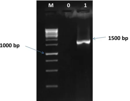
inhibitory activities against Listeria innocua F by the production of bacteriocins that gave zones of inhibition with a diameter of 13 mm. The 16S RNA sequence from producer strain is illustrated in Fig. 1.
Fig. 1. 16S RNA sequence from identification Lactococcus lactis ssp raffinolactis M: Standard (GeneRuler 1KB DNA reader), 0 : Negative control, 1 : Amplified DNA fragment (gene 16S rRNA) of
Lactococcus lactis. Beginning sequence used: s-d-bact-0008-a-S-20; s-dbact-1495-a-A20, Agarose gel concentration: 1.5% (w/v)
Using the same technique, bacteriocins inhibiting
Listeria innocua F and produced by Lactococcus
were characterized in a workshop salting dairy products [15,16,17] who identified bacteriocins produced by the genus Lactococcus which are capable of inhibiting gram-positive strains including Listeria innocua F.
The anti-Listeria activity of a class IIa bacteriocin, the mesenterica 52A was studied by the diffusion test wells by JASNIEWSKI [18]. However, several authors characterized in the same environment using the same method, bacteriocins produced by Lactococcus which is active against bacteria of the genus Listeria. Lactococcin BZ produced by Lactococcus lactis
ssp lactis BA [19], the bozacine 14 produced by Lactococcus lactis ssp lactis B14 [20], the
ST69BZ produced by Lactobacillus plantarum ST69BZ [21].
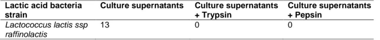
To inactivate bacteriocins by the protease, the inhibition zones are affected by trypsin and pepsin. Indeed, the zone of inhibition has completely disappeared after the enzyme treatment. These results confirm the proteinaceous nature of the biologically active fraction of the bacteriocins produced by this strain [22].
3.2 Extraction of Bacteriocins and Activity Test by the Method of Zymogram
We passed the acid extraction method whose principle is the adsorption and desorption of molecules on cells producing bacteriocins advocated by Yang [11]. The same technique was followed by Allouche [23] in 2010, INA El Harrach, for the purification of bacteriocins produced by strains of thermophilic lactobacilli used in the dairy industry. In 2001, MAO [24] have purified a bacteriocin produced by isolated from pig Lactococcus lactis FS92 and determined its MW with a migration by SDS-PAGE followed by a biological revelation by the zymogram technique.
The bacteriocins separated by SDS-PAGE electrophoresis expressed inhibitory activities against the target strain. By overlaying the two halves (colored and uncolored) of each gel, the inhibition for all active molecules areas are located at MW below 10 kDa. Estimating MW gave a value around 9.26 kDa.
With the same method, PHUONG [25] have detected the presence, in a water-soluble extract of Asiago (Italian cheese), of compounds of molecular weight between 3 kDa and 10 kDa and inhibits the growth of Listeria innocua LRGIA 01. Initially, these compounds are either similar to be bacteriocins or are small fragments of casein with antimicrobial activity, while HAYES [26] demonstrated that the MW of these molecules do not exceed 1000Da, which concludes bacteriocins.
3.3 Characterization of Genes Encoding Bacteriocins
Understanding the molecular mechanisms that regulate the production of bacteriocins against pathogenic strains without attacking beneficial bacteria is very important.
In some cases, it is investigated using non-producing mutants or bacteriocins that exhibit lesser or no activity variants [27,28].
In our work, we compared the activity of wild strains with their variants which are undergoes a cure of plasmid by using of Rifampin and Novobiocin as curing agents. Table 2 shows the results of the MIC for plasmid curing Lactococcus
lactis ssp raffinolactis.
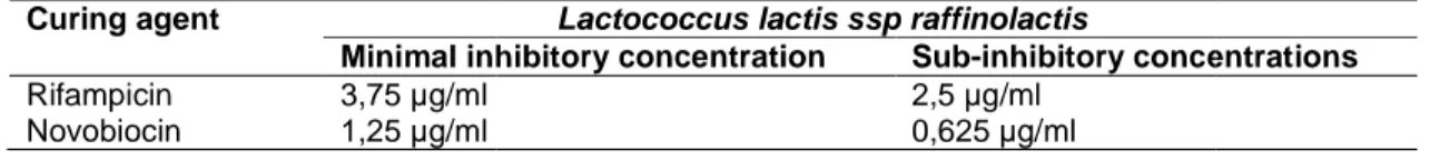
The MIC is the lowest concentration of antibiotic that inhibits all visible growth of a microorganism after incubation for 24 h in a specific growth medium. The sub-inhibitory concentration is the nearest concentration MIC [29]. According to the dilutions used, and from the MIC results, the values of sub-inhibitory concentrations that allowed the cure plasmids of our strain are respectively: 2.5 µg/ml and 0.625 µg/ml for Rifampin and Novobiocin.
Table 1. Results of antagonist activity and protease sensitivity (diameter of inhibitory zone by mm)
Lactic acid bacteria strain
Culture supernatants Culture supernatants + Trypsin Culture supernatants + Pepsin Lactococcus lactis ssp raffinolactis 13 0 0
Table 2. Results of the plasmid curing in MIC and sub Curing agent
Minimal inhibitory concentration Rifampicin 3,75 µg/ml
Novobiocin 1,25 µg/ml
The realization of the activity test by the wells’ diffusion method for the supernatants extracts with neutralized pH and their variants curing plasmids, showed the disappearance of the inhibition zones after plasmid treatment.
Fig. 2. Disappearance of activity after plasmid treatment
T: Culture supernatant of strain before curing plasmids; VC: Culture supernatant of Variant curing
plasmids
BENOIT [30] have demonstrated the localization of the genes encoding the brevicine SB27 as well as those encoding immunity to the bacteriocin. After plasmid curing the strain becomes sensitive to brevicine SB27 and in the same time unable to produce it. The plasmid profile analysis showed the disappearance of a band 3KDa for this variant.
The variants Bac- were obtained from plasmid curing using a Novobiocin at a concentration ranging from 0.25 ug/ml to 5
disappearance of the anti-Listeria
confirmed the plasmid location of the gene encoding this bacteriocin.
To complete the results obtained, the bacteriocins extracts from Lactococcus lactis
raffinolactis and its plasmids curing variants are
subject to migration on SDS-PAGE and test by the zymogram technique was realized in order to compare protein profiles and the zones of inhibition. The disappearance of bacteriocin phenotype after cure plasmids shows that genes
T
Vc
Results of the plasmid curing in MIC and sub-inhibitory concentrations
Lactococcus lactis ssp raffinolactis
Minimal inhibitory concentration Sub-inhibitory concentrations 2,5 µg/ml
0,625 µg/ml The realization of the activity test by the wells’
diffusion method for the supernatants extracts with neutralized pH and their variants curing plasmids, showed the disappearance of the inhibition zones after plasmid treatment.
Fig. 2. Disappearance of activity after plasmid T: Culture supernatant of strain before curing plasmids; VC: Culture supernatant of Variant curing
BENOIT [30] have demonstrated the localization of the genes encoding the brevicine SB27 as well as those encoding immunity to the bacteriocin. After plasmid curing the strain becomes sensitive to brevicine SB27 and in the same time unable to e plasmid profile analysis showed the disappearance of a band 3KDa for this
were obtained from plasmid curing using a Novobiocin at a concentration
µg/ml. The Listeria activity confirmed the plasmid location of the gene
To complete the results obtained, the
Lactococcus lactis ssp
and its plasmids curing variants are PAGE and activity test by the zymogram technique was realized in order to compare protein profiles and the zones of inhibition. The disappearance of bacteriocin phenotype after cure plasmids shows that genes
encoding these bacteriocins are carried by plasmids.
On the part of the gel intended for the activity test, the zone of inhibition disappeared in the variant curing plasmid, as well as the protein band corresponding to it.
The result is illustrated in Fig. 3.
Fig. 3. Electrophoresis separation and zymogramme activity
A: SDS-PAGE profile of protein separation; B: Zymogramme activity.
L. raf : Profile of migration of protein from the Lactococcus lactis ssp raffinolactis strain. L. rafc: Profile of migration of protein from the mutant
Bac- from the same strain
3.4 Genetic Characterization of Immunity to Bacteriocins
We reacted bacteriocin of Lactococcus lactis ssp
raffinolactis to the same strain after plasmid the
results showed the appearance of a large zone of inhibition. These results indicate that, in this strain, the loss of the plasmids carrying the genes for production of bacteriocins is accompanied by the loss of immunit
bacteriocins. We can therefore, conclude that the genes and production of bacteriocin immunity are co-transcribed and carried by the same plasmid.
inhibitory concentrations
inhibitory concentrations
encoding these bacteriocins are carried by
the part of the gel intended for the activity test, the zone of inhibition disappeared in the variant curing plasmid, as well as the protein
Fig. 3. Electrophoresis separation and mme activity
PAGE profile of protein separation; B: Zymogramme activity.
raf : Profile of migration of protein from the Lactococcus lactis ssp raffinolactis strain. rafc: Profile of migration of protein from the mutant
strain
Genetic Characterization of Immunity
bacteriocin of Lactococcus lactis ssp
to the same strain after plasmid the results showed the appearance of a large zone of inhibition. These results indicate that, in this strain, the loss of the plasmids carrying the genes for production of bacteriocins is accompanied by the loss of immunity to their own bacteriocins. We can therefore, conclude that the genes and production of bacteriocin immunity are transcribed and carried by the same plasmid.
Because according to ENNAHAR [31], the common pattern of organization of the genetic systems involved in biosynthesis, transport and regulation of the biosynthesis class IIa consists of a set of genes, involved a group biosynthesis and in immunity to the bacteriocin containing genes encoding the precursor of the bacteriocin (B) and (I) and a group immunity protein involved in the export of the bacteriocin and comprises genes encoding an ABC transporter (D), an accessory protein (E) and sometimes a protein of unknown function (C).
3.5 Determination of Minimum Inhibitory Concentrations of Bacteriocins
The minimum inhibitory concentrations (MICs) are variable depending on the bacterial species and strains [31]. The inhibition spectrum of bacteriocins established from supernatants culture or semi-purified preparations (bacteriocin extracts). The result will be, thus, representative of the overall antibacterial activity of a strain and not of the activity of one bacteriocin. For this reason, when a strain produces several bacteriocins, the interpretation of inhibition spectrum is only possible if it is made from purified bacteriocins [30].
In our case, the anti-Listeria activity disappeared after diluting the extract to a quarter of it. According to the principle of critical dilution, the value of the MIC bacteriocins produced by
Lactococcus lactis ssp raffinolactis is 7.14 IU/ml.
In addition, the inhibitory potency of antimicrobial molecule is inversely proportional to its MIC [30,31].
4. CONCLUSION
Our work has focused on the characterization of anti-Listeria innocua F bacteriocins produced by
Lactococcus lactis ssp raffinolactis isolated from
camel milk in the region of Biskra. We first started by identifying the antibacterial activity of bacteriocin by wells diffusion method, where the strain inhibited the growth of Listeria innocua F, with an inhibition zone of 13 mm.
Bacteriocins produced are sensitive to trypsin and pepsin, two proteolytic enzymes most commonly used to confirm the protein nature of bacteriocins. Later, we performed an extraction of these molecules by adsorption desorption YANG protocol [9] followed by their migration by SDS-PAGE and revealed by a method of zymogram. Lactococcus lactis ssp raffinolactis
gave a protein pattern and a zone of inhibition of 9.26 KDa. This MW is situated between 5 and 10 KDa it is corresponding to sub-class IIa of bacteriocins.
Our results are complemented by a genetic study in order to locate the clusters genes of production and immunity to bacteriocins. To this end, we produced mutants Bac- (not producers of bacteriocins) and Bac S (sensitive to the action of parental bacteriocin), the plasmids are curing by using two antibiotics, Rifampicin and Novobiocin inhibiting nucleic acid synthesis at sub-inhibitory concentration. The disappearance of the anti-Listeria activity and the appearance of the sensitivity of the strain to its own lactic bacteriocin after cure show that the bacteriocin genes and their immunity to those of these latter, are carried by the same plasmids. Finally, we used the method of critical dilutions and testing activity by wells diffusion to determine MIC of bacteriocin extracts the value of the MIC was 7.14 IU/ml.
Finally, it would be interesting to complete this work by the purification of bacteriocins and sequencing the bioactive fraction in order to understand the mechanism of antibacterial activities and study the stability of these molecules to be use in food industries.
COMPETING INTERESTS
Authors have declared that no competing interests exist.
REFERENCES
1. Dimarcq JL et JA. Hoffman. Peptides antimicrobiens: les antibiotiques du futur. Biofutur. 2001;212-21.
2. Papagiani M. Ribosomally synthetized peptides with antimicrobial properties: Biosynthesis, structure, function, and applications. Biotechnol. Advan. 2003;21: 465-99.
3. Twomey DRP, Ross M, Ryan B, Meaney CH. Lantibiotics produced by lactic acid bacteria: Structure, function and applications. Antonie Van Leeuwenhoek. 2002;82:165-85.
4. Trautner BW, Hull RA, Darouiche RO. Colicins prevent colonization of urinary cathéters. J. Antimicrob Chemother. 2005; 56:413-5.
5. Ryan MP, Meaney WJ, Ross RP, Hill C. Evaluation of laciticin 3147 and a teat seal
containing this bacteriocin for inhibition of mastitis pathogens. Applied and Environnement Microbiology. 1998;64: 2287-90.
6. Galvarez A, Abriouel H, Lopez RL, Omar NB. Bacteriocin- based strategies for food bioservation. Int. J. Food Microbiol. 2007; 120:51-70.
7. Grande MJ, Lopez RL, Abriouel H, Valdivia E, Benomar N, Maqueda M, Martinez-Canamero M, Galvez A. Treatment of vegetable sauces with enterocin AS-48 alone and a combinaison with phenolic compounds to inhibit proliferation of
Staphylococcus aureus. J. Food Prot.
2007;70:405-11.
8. Benkerroum N, Oubel H, Zahar M, Dila S, Filalimaltou F. Bacteriocin producing
Lactococcus lactis subsp lactis and
application to control Listeria monocytogenes in Moroccan lben. Journal
of Applied Microbiology. 2000;89:960-6. 9. Yang R, Johnson MC, Ray B. Novel
method to extract large amounts of bacteriocin from lactic acid bacteria. Applied Environ. Micrbiol. 1992;58:3355-9. 10. Nissen-Meyer J, Rogne P, Oppegård C,
Haugen HS, Kristiansen PE. Structure-function relationships of the non-lanthionine-containing peptide (class II) bacteriocins produced by gram-positive bacteria. Curr Pharm Biotechnol. 1992;10: 19-37.
11. Schagger H, Von Jacow G. Tricine-sodium dodecyl sulfate polyacrylamide gel electrophoresis for the separation of proteins in the range from 1 to 100 kDa. Anal. Biochem. 1987;166:368-79.
12. Isleroglu H, Yildirim Z, Tokatli M, Oncul N, Yildirim M. Partial characterisation of enterocin KP produced by Enterococcus
faecalis KP, a cheese isolate. International
Journal of Dairy Technology. 2012;65: 90-7.
13. Yakoubi K, Ammor S, Haydersah J, Chevallier I. Activités antibactériennes des bactéries lactiques isolées d’ateliers fermiers de salaison. Typicité des Produits Alimentaires, ENITA. 2006;11:168-75. 14. Ogunbanwo ST, Sanni AI, Onilude AA.
Characterization of bacteriocin produced
by Lactobacillus plantarum F1 and
Lactobacillus brevis OG1. African Journal
of Biotechnology. 2003;2(8):219-27. 15. Simova ED, Beshkova ZP, Simov ZI. In
vitro and in situ bacteriocin activity of lactic
acid bacteria from Bulgarian dairy products
and methods for making of Lactoba-cillus protective fermented milks with bacteriocin inhibitory substances. Bulg. J. Agric. Sci. 2008;14:28-42.
16. Jasniewski J, Cailliez-Grimal C, Younsi M, Milliere JB, Revol-Junelles AM. Functional differences in Leuconostoc sensitive and resistant strains to mesenterocin 52A, a class IIabacteriocin. FEMS Microbiology Letters. 2008;289:193-201.
17. Şahingil D, İşleroğlu H, Yıldırım Z, Akçelik M, Yıldırım M. Characterization of lactococcin BZ produced by Lactococcus lactis subsp. lactis BZ isolated from boza. Turkish Journal of Biology. 2011;35:21-33. 18. Kabadjova P, Gotcheva I, Ivanova I,
Dousset X. Investigation of bacteriocin activity of lactic acid bacteria isolated from Boza. Biotechnology Equipement. 2000; 14:56-9.
19. Todorov SD. Diversity of bacteriocinogenic lactic acid bacteria isolated from boza, a cereal-based fermented beverage from Bulgaria. Food Control. 2010;21:1011-21. 20. Chen H, Hoover DG. Bacteriocins and their
food applications. Comprehensive Reviews In Food Science And Food Safety. 2003;2: 82-100.
21. Allouche FN, Hellal A, Laraba A. Etude de l’activité antimicrobienne de souches de lactobacilles thermophiles utilisées dans l’industrie laitière. Nature et Technologie. 2010;3:13-20.
22. Mao Y, Muriana PM, Cousin MA. Purification and transpositional inactivation of lacticin FS92, a broad-spectrum bacteriocin produced by Lactococcus lactis FS92. Food Microbiology. 2000;18:165-75. 23. Phuong Nguyen T, Coralie D, Isabelle A, Pascal D, Melanie R. Partial characterisation of peptides inhibiting Listeria growth in two Alpine cheeses. Dairy Science & Technology. 2013;94(1): 61-72.
24. Hayes M, Ross RP, Fitzgerald GF, Hill C, Stanton C. Casein-derived antimicrobial peptides generated by Lactobacillus
acidophilus DPC6026. Applied and
Environmental Microbiology. 2006;72: 2260-4.
25. Richard C, Brillet A, Pilet MF, Prevost H, Drider D. Evidence on inhibition of Listeria
monocytogenes by divercin V41 action.
Letters in Applied Microbiology. 2003;36: 288-92.
26. Nilsson L, Christiansen JN, Jorgensen BL, Grotinum D, Gram L. The contribution of
bacteriocin to inhibition of Listeria
monocytogenes by Carnobacterium
piscicola strains in cold-smoked salmon
systems. Journal of Applied Microbiology. 2004;96:133-43.
27. Fonseca AP, Extremina C, Fonseca AF, Sousa JC. Effect of subinhibitory concentration of piperacillin / tazobactam on Pseudomonas aeruginosa. Journal of Medical Microbiology. 2004;53(9):903-10.
28. Benoit V, Lebrihi A, Milliere JB, Lefebvre G. Purification and partial amino-acid-sequence of brevicin-27, a bacteriocin produced by lactobacillus-brevis sb27. Current Microbiology. 1997;34(3):173- 9.
29. Ennahar S, Deschamps N, Richard J. Natural variation of susceptibility of Listeria strain to class IIa bacteriocin. Current Microbiology. 2000;41(1):1-4.
30. Scannell AGM, Hill C, Ross RP, Marx S, Hartmeier W, Arendt EK. Development of bioactive food packaging materials using immobilised bacteriocins Lacticin 3147 and Nisaplin. Int. Food Microbiol. 2000;60(2-3): 241-249.
31. Izquierdo E, Marchioni E, Aoude-Werner D, Hasselman C, Ennahar S. Smearing of soft cheese with Entercoccus feacium WHE 81, a multi-bacteriocin producer, against Listeria monocytogenes. Food Microbiology. 2008;26:16-20.
_________________________________________________________________________________ © 2016 Chergui et al.; This is an Open Access article distributed under the terms of the Creative Commons Attribution License (http://creativecommons.org/licenses/by/4.0), which permits unrestricted use, distribution, and reproduction in any medium, provided the original work is properly cited.
Peer-review history:
The peer review history for this paper can be accessed here: http://sciencedomain.org/review-history/18022