HAL Id: hal-03245857
https://hal-amu.archives-ouvertes.fr/hal-03245857
Submitted on 2 Jun 2021
HAL is a multi-disciplinary open access
archive for the deposit and dissemination of
sci-entific research documents, whether they are
pub-lished or not. The documents may come from
teaching and research institutions in France or
abroad, or from public or private research centers.
L’archive ouverte pluridisciplinaire HAL, est
destinée au dépôt et à la diffusion de documents
scientifiques de niveau recherche, publiés ou non,
émanant des établissements d’enseignement et de
recherche français ou étrangers, des laboratoires
publics ou privés.
Distributed under a Creative Commons Attribution - NonCommercial - NoDerivatives| 4.0
International License
Draft genome and description of Microvirga
mediterraneensis strain Marseille-Q2068T sp. nov., a
new bacterium isolated from human healthy skin
M Boxberger, M Ben Khedher, S Magnien, N Cassir, B La Scola
To cite this version:
M Boxberger, M Ben Khedher, S Magnien, N Cassir, B La Scola. Draft genome and description
of Microvirga mediterraneensis strain Marseille-Q2068T sp. nov., a new bacterium isolated from
human healthy skin. New Microbes and New Infections, Wiley Online Library 2021, 40, pp.100839.
�10.1016/j.nmni.2021.100839�. �hal-03245857�
Draft genome and description of
Microvirga mediterraneensis strain
Marseille-Q2068T sp. nov., a new
bacterium isolated from human
healthy skin
M. Boxberger1,2, M. Ben Khedher1,2, S. Magnien1,2,
N. Cassir1,2and B. La Scola1,2
1) Aix-Marseille Université, IRD, AP-HM, MEPHI and 2) IHU-Méditerranée Infection, Marseille, France
Abstract
In 2019, by culturing a skin swab from the forehead of a 70-year-old healthy woman via the culturomics method, we isolated the new bacterial strain Marseille-Q2068T (= CSUR-Q2068). Matrix-assisted desorption ionization–time of flight mass spectrometry (MALDI-TOF MS) failed to identify this isolate. Analysis of the 16S ribosomal RNA gene and genome-to-genome comparison suggested that this taxon belongs to a novel bacterial species within the family Methylobacteriaceae in the phylum Proteobacteria. We describe here its main phenotypic characteristics, genome sequence and annotation of Microvirga mediterraneensis strain Marseille-Q2068T, a new member of the Microvirga genus, which we propose as the type strain.
© 2021 The Author(s). Published by Elsevier Ltd.
Keywords: Bacteria, culturomics, genome, Microvirga mediterraneensis, sp. nov., species, taxonogenomics
Original Submission: 14 August 2020; Revised Submission: 9 November 2020; Accepted: 23 November 2020
Article published online: 15 January 2021
Corresponding author: B. La Scola, Aix-Marseille Université, Méditerranée-Infection, 19-21 Boulevard Jean Moulin, 13385, Mar-seille cedex 05, France.
E-mail:bernard.la-scola@univ-amu.fr
Introduction
The genus Microvirga includes 22 species [1], most of which were isolated from diverse environmental samples, such as air [2], soils and springs [2–9], or in samples associated with plants [10–15]. Only one species was isolated from human, Microvirga massiliensis [16], which is the human commensal bacterium with the largest genome. Microvirga mediterraneensis strain Marseille-Q2068T was isolated using the culturomics approach, based on the use of a large panel of culture conditions, in order to describe the culturable microbial composition of a sample [17–19]. A taxonogenomics approach including matrix-assisted laser desorption–ionization time-of-flight mass spectrometry (MALDI-TOF MS), phylogenetic analysis, main phenotypic description and genome sequencing was used to describe this species [20].
The genome of Microvirga mediterraneensis strain Marseille-Q2068T is 5,347,712 bp long with 63.83% G + C content. This new bacterium is most closely related to Microvirga loto-nonidis strain WSM3557 with a 16S ribosomal RNA (rRNA) sequence similarity value of 99.25%. Furthermore, digital DNA-DNA hybridization analysis between the novel organism and the Microvirga lotononidis strain WSM3557 type strain genome revealed an identity of only 39.7%, and genomic comparison using the OrthoANI parameter provided a value of 89.86%. On the basis of these data, we propose Microvirga mediterraneensis strain Marseille-Q2068T, a new member of the Microvirga genus, as the type strain.
Materials and methods
Strain isolation
The subject was registered at the L’Occitane Natural Cosmetic Assessment Center in Marseille (https://cosnat-loccitane.com; CosNat, Marseille France). The skin areas used for sampling were 10 cm2. The samples were collected in a Z-stroked manner [21] using sterile swabs soaked in Culture Top trans-port medium (C-Top Ae-Ana, Eurobio, France). Microvirga mediterraneensis strain Marseille-Q2068T was initially isolated by direct seeding of 50 μL of sample on a homemade R2A incubated in aerobiosis at 31°C. MALDI-TOF MS protein analysis was carried out with a Microflex spectrometer (Bruker Daltonics, Bremen, Germany) [8]. Spectra from strain Marseille-Q2068T were imported into MALDI BioTyper soft-ware (Bruker) and analysed by standard pattern matching using
New Microbe and New Infect 2021; 40: 100839 © 2021 The Author(s). Published by Elsevier Ltd
default parameter settings (Fig. 1). The study was validated by the local ethics committee (ID-RCB 2019-A01508-49).
Phenotypic characterization
Different growth temperatures (20, 30, 37, 45 and 56°C), at-mospheric conditions (anaerobic, aerobic and microaerophilic using CampyGEN, Oxoid, Basingstoke, UK) and pH (5, 6.5, 7.2 and 8.5) were tested. API ZYM, API NE and API 50CH strips (bioMérieux, Marcy l’Etoile, France) were used to evaluate the biochemical properties of the strain according to the manu-facturer’s instructions. For scanning electronic microscopy, a colony was collected from agar and immersed into a 2.5% glutaraldehydefixative solution. The slide was gently washed in water, air dried and examined with approximately 60 cm in height and 33 cm in width to evaluate bacterial structure on a TM4000 microscope (Hitachi High-Technologies, Tokyo, Japan). Motility test was performed using the semisolid TCC media as described by Tittsler and Sandholzer [22].
Genome sequencing
Genomic DNA (gDNA) of Microvirga mediterraneensis strain Marseille-Q2068T was quantified by a Qubit assay with the high sensitivity kit (Life Technologies, Carlsbad, CA, USA) to 0.2 ng/μL.
Genomic DNA was next sequenced on the MiSeq Technology (Illumina, San Diego, CA, USA) with the paired end strategy and was barcoded in order to be mixed respectively with 23 other genomic projects prepared with the Nextera XT DNA sample prep kit (Illumina). To prepare the paired end library, dilution was performed to require 1ng of each genome as input to prepare the paired end library. The tagmentation step fragmented and tagged the DNA. Then limited-cycle PCR amplification (12 cycles) completed the tag adapters and introduced dual-index barcodes. After purification on AMPure XP beads (Beckman Coulter, Full-erton, CA, USA), the libraries were then normalized on specific beads according to the Nextera XT protocol (Illumina). Normal-ized libraries were pooled into a single library for sequencing on the MiSeq. The pooled single strand library was loaded onto the reagent cartridge and then onto the instrument along with theflow cell. Automated cluster generation and paired end sequencing with dual index reads were performed in a single 39-hour run at a 2 × 250 bp read length. Total information of 9.5 Gb was obtained from a 1063K/mm2cluster density, with a cluster passing quality controlfilters of 89.2%. Within this run, the index representation for Microvirga mediterraneensis was determined to index 3.4%. The 20 050 916 paired end reads werefiltered according to the read qualities. 16S RNA gene sequence was extracted, and a
FIG. 1.Matrix-assisted desorption ionization–time of flight mass spectrometry (MALDI-TOF MS) reference mass spectrum. Spectra from 12 indi-vidual colonies of Microvirga mediterraneensis strain Marseille-Q2068T were compared and reference spectrum generated.
2 New Microbes and New Infections, Volume 40 Number C, March 2021
NMNI
© 2021 The Author(s). Published by Elsevier Ltd, NMNI, 40, 100839
phylogenetic tree was obtained using the maximum likelihood method and Kimura two-parameter within MEGA 7 software [11].
Genome annotation and genome comparison
Genome annotation was obtained through the NCBI Prokary-otic Genome Annotation Pipeline [23]. BlastP was used to predict the bacterial proteome (E value of 1e03, coverage of 0.7 and percent identity of 30) according to the Clusters of Orthologous Groups (COGs) database. The Genome-to-Genome Distance Calculator (GGDC) web server (http:// ggdc.dsmz.de) was used to estimate the overall similarity among compared genomes and to replace the wet-lab DNA-DNA hybridization by a digital version. The degree of genomic similarity of Microvirga mediterraneensis strain Marseille-Q2068 with closely related species was estimated using the OrthoANI software [24]. Antibiotic resistance genes (ARG) were searched using the Comprehensive Antibiotic Resistance Database (CARD) [25]. Assembled sequences were searched against the CARD database under moderately stringent con-ditions (e-value of 10−5) for the in silico ARG prediction. The presence of pathogenesis-related proteins was investigated us-ing the virulence factor database (VFDB) [26].
Results
Strain identification and classification
Microvirga mediterraneensis strain Marseille-Q2068T was iso-lated from the forehead skin swab of a 70-year-old healthy
woman. Microvirga mediterraneensis strain Marseille-Q2068T failed to be identified by our systematic MALDI-TOF MS screening, suggesting that the corresponding species was not in our database ( https://www.mediterranee-infection.com/acces-ressources/base-de-donnees/urms-data-base/) (Fig. 1). More-over, Microvirga mediterraneensis strain Marseille-Q2068T exhibited a 99.25% 16S rRNA sequence similarity to the Microvirga lotononidis strain WSM3557 type strain (GenBank accession no. NR_117846.1), the phylogenetically closest bac-terium with standing in nomenclature (Fig. 2).
FIG. 2.16S rRNA–based phylogenetic tree highlighting position of Microvirga mediterraneensis sp. nov., strain Marseille-Q2068T (red), relative to other closely related bacterial taxa. Sequences were aligned using Muscle 3.8.31 with default parameters, and phylogenetic relationship was inferred using maximum likelihood method with 1000 bootstrap replicates within MEGA 7.0 software.
FIG. 3.Scanning electron microscopy of Microvirga mediterraneensis sp. nov., strain Marseille-Q2068T, using TM 4000plus Tabletop microscope (Hitachi, Tokyo, Japan). Scale bar represents 5μm.
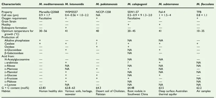
TABLE 1.Differential characteristics of Microvirga mediterraneensis strain Marseille-Q2068T and its most closely related species with standing in nomenclature
Characteristic M. mediterranensis M. lotononidis M. pakistanensis M. calopogonii M. subterranea M.flocculans Property Marseille-Q2068 WSM3557 NCCP-1258 SSW1-57 Fail-4 TFB Cell size (μm) 0.9 × 1.7 0.4–0.56 × 1.0–2.2 NA 0.5–0.9 × 9 1.3–2.0 1 × 1.5–4 0.8 × 1.1 Oxygen requirement Facultative + + Facultative + +
Gram Strain — — — — — —
Motility — — — + + —
Endospore formation — + — — — —
Optimum temperature for growth (°C) 30–56 41 40 20–45 41 10–35 Production of: Alkaline phosphatase + — + NA NA — Catalase + + ? + + + Oxidase — — + + — — α-Glucosidase — + — NA + — β-Galactosidase — — — NA — — Acid from: N-Acetylglucosamine — — — NA NA — L-arabinose — + — ? + NA D-Ribose — NA — NA NA NA D-Mannose — NA + + NA — D-Mannitol — + — NA NA NA D-Glucose — + + NA + — D-Fructose — + + + — NA D-Maltose — + — NA — NA D-Lactose — NA — NA NA NA G + C content (mol%) 63.83 62.8–63 64.3 64.48 63.5 62.2 Habitat Human healthy skin Various: soils, herbage,
seawater
Desert soil of Cholistan, Pakistan
Root nodule in Southwest China
Deep surface Australian thermal aquifer
Air samples
+, positive result;−, negative result; NA, data not available.
FIG. 4. Graphical circular map of genome from strain Marseille-Q2068T obtained by CGView tool [27].
4 New Microbes and New Infections, Volume 40 Number C, March 2021
NMNI
© 2021 The Author(s). Published by Elsevier Ltd, NMNI, 40, 100839
Phenotypic characteristics
Growth of Microvirga mediterraneensis strain Marseille-Q2068T was initially isolated by direct seeding of 50μL of sample on a homemade R2A (Reasoner 2A agar) incubated in aerobiosis at
31°C. Colonies from strain Marseille-Q2068T showed a pink pigmentation and no haemolysis. They were circular, with a diameter of 0.5 to 1.5 mm. Bacterial cells were Gram-negative, nonmotile rods with a length of about 1.70μm and a width of
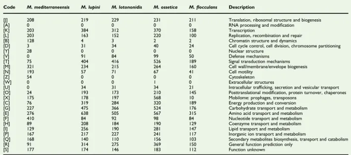
TABLE 2.Number of genes associated with 25 general COGs functional categories of Microvirga mediterraneensis strain Marseille-Q2068 and closely related species Microvirga lupini, Microvirga lotononidis, Microvirga ossetica and Microvirgaflocculans
Code M. mediterraneensis M. lupini M. lotononidis M. ossetica M.flocculans Description
[J] 208 219 229 231 211 Translation, ribosomal structure and biogenesis [A] 0 0 0 0 0 RNA processing and modification
[K] 203 384 312 370 158 Transcription
[L] 203 163 152 220 100 Replication, recombination and repair [B] 128 4 3 2 2 Chromatin structure and dynamics
[D] 3 31 34 40 24 Cell cycle control, cell division, chromosome partitioning
[Y] 28 0 0 0 0 Nuclear structure
[V] 0 91 84 99 50 Defense mechanisms
[T] 75 404 416 526 189 Signal transduction mechanisms [M] 321 234 215 264 160 Cell wall/membrane/envelope biogenesis
[N] 193 57 71 67 41 Cell motility
[Z] 54 0 0 0 0 Cytoskeleton
[W] 0 0 0 1 0 Extracellular structures
[U] 0 34 31 34 21 Intracellular trafficking, secretion and vesicular transport [O] 24 193 173 210 145 Posttranslational modification, protein turnover, chaperones [X] 175 178 197 568 10 Mobilome: prophages, transposons
[C] 76 319 284 320 189 Energy production and conversion [G] 227 475 366 524 176 Carbohydrate transport and metabolism [E] 276 638 505 567 315 Amino acid transport and metabolism [F] 410 84 92 98 84 Nucleotide transport and metabolism [H] 89 208 184 190 129 Coenzyme transport and metabolism [I] 129 256 190 281 147 Lipid transport and metabolism [P] 167 217 227 241 117 Inorganic ion transport and metabolism
[Q] 168 140 110 156 103 Secondary metabolites biosynthesis, transport and catabolism [R] 91 314 275 369 150 General function prediction only
[S] 177 174 146 183 112 Function unknown COGs, Clusters of Orthologous Groups database.
FIG. 5.Distribution of functional classes of predicted genes in Microvirga mediterraneensis, Microvirga lotononidis, Microvirga lupini, Microvirga ossetica and Microvirgaflocculans according to Clusters of Orthologous Groups (COGs) database groups of proteins.
about 0.9μm, as determined by scanning electron microscopy (Fig. 3). Strain Marseille-Q2068T is a facultative aerobe. The sporulation test (20 minutes at 80°C) was negative. Using an API strip, positive reactions were shown for esculin, gelatin, alkaline phosphatase, esterase (C4), esterase lipase (C8), leucine arylamidase, valine arylamidase, cystine arylamidase, trypsin, acid phosphatase and naphtol-AS-BI-phosphohydrolase. All other reactions tested were negative. In addition, this bac-terium shows catalase positivity and oxidase negativity. The results are summarized inTable 1.
Genome properties
The genome size of strain Marseille-Q2068 was 5 347 712 bp long with a 63.83% G + C content. The genome assembly of this strain was achieved on eight contigs. Of the 5099 predicted genes, 4933 were protein-coding genes and 86 were RNAs (five 16S rRNA,five additional 5S rRNAs, five additional 23S rRNAs and 67 transfer RNAs and four noncoding RNAs). A total of 4106 genes (83.2%) were assigned a putative function, and 824
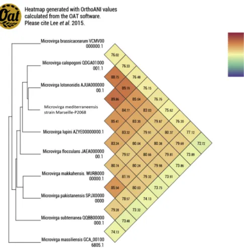
genes (16.7%) were annotated as hypothetical proteins (Fig. 4). The genome properties and distribution of genes into COGs functional categories are detailed in Table 2. The in silico resistome of the strain Marseille-Q2068 obtained by searching the CARD database and the search for virulence factors via the VFDataBase of this strain showed no genes with high identity percentage. Genes with putative function (by COGs analysis) were 3425 (67%). Analysis of the COGs categories showed that the nucleotide transport and metabolism category, the repli-cation, recombination and repair category, and the chromatin structure and dynamics category of the Microvirga medi-terraneensis genome appear to be more numerous than those of the genomes of the Microvirga genus (categories F, L and B respectively) (Fig. 5). Finally, a digital DNA-DNA hybridization analysis between the novel organism and the Microvirga lotono-nidis strain WSM3557 type strain revealed an identity of only 39.7%. Furthermore, genomic comparison using the OrthoANI parameter provided a value of 89.86% with the Microvirga lotononidis strain WSM3557 type strain (Fig. 6).
FIG. 6. Heat map generated with OrthoANI values calculated using OAT software between Microvirga mediterraneensis sp. nov., strain Marseille-Q2068T, and other closely related species with standing in nomenclature.
6 New Microbes and New Infections, Volume 40 Number C, March 2021
NMNI
© 2021 The Author(s). Published by Elsevier Ltd, NMNI, 40, 100839
Discussion and conclusion
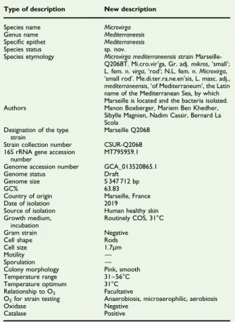
In the past 8 years, the use of the culturomics approach has resulted in the discovery of more than 500 bacterial species [17]. Using the taxonogenomics concept – the combination of the genomic and phenotypic properties of a putative new taxon [27]– we have characterized a new bacterial species within the family Methylobacteriaceae found on forehead human skin. The main characteristics of this strain are summarize inTable 3. It was named Microvirga mediterraneensis strain Marseille-Q2068T, as follows: Mi.cro.vir’ga Gr. adj. mikros, ‘small’; L. fem. n. virga, ‘rod’; N.L. fem. n. Microvirga, ‘a small rod’. Me.di.ter.ra.ne.en’sis, L. masc. adj., mediterraneensis, ‘of Mediterraneum’, the Latin name of the Mediterranean Sea, by which Marseille is located and the bacteria isolated (Table 3).
Deposit in culture collections and sequences database Microvirga mediterraneensis strain Marseille-Q2068T was deposited in CSUR collection under accession number CSUR-Q2068. The 16S rRNA and genome sequences and
annotation are available in GenBank under accession numbers MT795959.1 and GCA_013520865.1 respectively.
Con
flict of interest
None declared.
Acknowledgements
The MB PhD grant is supported by the collaboration between M&L Laboratories and Aix-Marseille Université (reference PVM:2018-200). Supported by the French state, managed by the National Research Agency under the‘Investissements d’avenir’ (Investments for the Future) programme under reference ANR-10-IAHU-03 (Méditerranée Infection) and by the Région Provence-Alpes-Côte-d’Azur and European funding from FEDER PRIMI. The authors are indebted to L. Brechard for sequencing the genome and helping with electron microscopy at IHU-Méditerranée Infection.
References
[1] Parte AC. Lpsn– list of prokaryotic names with standing in nomen-clature (bacterio.net), 20 years on. Int J Syst Evol Microbiol 2018;68: 1825–9.https://doi.org/10.1099/ijsem.0.002786.
[2] Weon HY, Kwon SW, Son JA, Jo EH, Kim SJ, Kim YS, et al. Description of Microvirga aerophila sp. nov. and Microvirga aerilata sp. nov., isolated from air, reclassification of Balneimonas flocculans Takeda et al. 2004 as Microvirgaflocculans comb. nov. and emended description of the genus Microvirga. Int J Syst Evol Microbiol 2010;60:2596–600.https://doi.org/ 10.1099/ijs.0.018770-0.
[3] Liu ZT, Xian WD, Li MM, Liu L, Ming YZ, Jiao JY, et al. Microvirga arsenatis sp. nov., an arsenate reduction bacterium isolated from Tibet hot spring sediments. Antonie Van Leeuwenhoek 2020;113:1147–53. https://doi.org/10.1007/s10482-020-01421-6.
[4] Zhang X, Zhang J, Yao Q, Feng G, Zhu HH. Microvirgaflavescens sp. nov., a novel bacterium isolated from forest soil and emended description of the genus Microvirga. Int J Syst Evol Microbiol 2019;69: 667–71.https://doi.org/10.1099/ijsem.0.003189.
[5] Veyisoglu A, Tatar D, Saygin H, Inan K, Cetin D, Guven K, et al. Microvirga makkahensis sp. nov., and Microvirga arabica sp. nov., isolated from sandy arid soil. Antonie Van Leeuwenhoek 2016;109:287–96. https://doi.org/10.1007/s10482-015-0631-z.
[6] Amin A, Ahmed I, Habib N, Abbas S, Hasan F, Xiao M, et al. Microvirga pakistanensis sp. nov., a novel bacterium isolated from desert soil of Cholistan, Pakistan. Arch Microbiol 2016;198:933–9.https://doi.org/ 10.1007/s00203-016-1251-3.
[7] Huq MdA. Microvirga rosea sp. nov.: a nanoparticle producing bacte-rium isolated from soil of rose garden. Arch Microbiol 2018;200: 1439–45.https://doi.org/10.1007/s00203-018-1558-3.
[8] Dahal RH, Kim J. Microvirga soli sp. nov., an alphaproteobacterium isolated from soil. Int J Syst Evol Microbiol 2017;67:127–32.https://doi. org/10.1099/ijsem.0.001582.
TABLE 3. Description of Microvirga mediterraneensis sp. nov. strain Marseille-Q2068T
Type of description New description Species name Microvirga Genus name Mediterraneesis Specific epithet Mediterraneesis Species status sp. nov.
Species etymology Microvirga mediterraneensis strain Marseille-Q2068T. Mi.cro.vir’ga, Gr. adj. mikros, ‘small’; L. fem. n. virga,‘rod’; N.L. fem. n. Microvirga, ‘small rod’. Me.di.ter.ra.ne.en’sis, L. masc. adj., mediterraneensis,‘of Mediterraneum’, the Latin name of the Mediterranean Sea, by which Marseille is located and the bacteria isolated. Authors Manon Boxberger, Mariem Ben Khedher,
Sibylle Magnien, Nadim Cassir, Bernard La Scola
Designation of the type strain
Marseille Q2068 Strain collection number CSUR-Q2068 16S rRNA gene accession
number
MT795959.1 Genome accession number GCA_013520865.1 Genome status Draft
Genome size 5 347 712 bp
GC% 63.83
Country of origin Marseille, France Date of isolation 2019
Source of isolation Human healthy skin Growth medium,
incubation
Routinely COS, 31°C Gram strain Negative
Cell shape Rods Cell size 1.7μm
Motility —
Sporulation — Colony morphology Pink, smooth Temperature range 31–56°C Temperature optimum 31°C Relationship to O2 Facultative
O2for strain testing Anaerobiosis, microaerophilic, aerobiosis
Oxidase Negative Catalase Positive
[9] Kanso S, Patel BKC. Microvirga subterranea gen. nov., sp. nov., a moderate thermophile from a deep subsurface Australian thermal aquifer. Int J Syst Evol Microbiol 2003;53:401–6.https://doi.org/10. 1099/ijs.0.02348-0.
[10] Jiménez-Gómez A, Saati-Santamaría Z, Igual JM, Rivas R, Mateos PF, García-Fraile P. Genome insights into the novel species Microvirga brassicacearum, a rapeseed endophyte with biotechnological potential. Microorganisms 2019;7:354. https://doi.org/10.3390/ microorganisms7090354.
[11] Wang F, Yang L, Deng J, Liu X, Lu Y, Chen W, et al. Microvirga calo-pogonii sp. nov., a novel alphaproteobacterium isolated from a root nodule of Calopogonium mucunoides in Southwest China. Antonie Van Leeuwenhoek 2019;112:1593–602. https://doi.org/10.1007/s10482-019-01285-5.
[12] Ardley JK, Parker MA, De Meyer SE, Trengove RD, O’Hara GW, Reeve WG, et al. Microvirga lupini sp. nov., Microvirga lotononidis sp. nov. and Microvirga zambiensis sp. nov. are alphaproteobacterial root-nodule bacteria that specifically nodulate and fix nitrogen with geographically and taxonomically separate legume hosts. Int J Syst Evol Microbiol 2012;62:2579–88.https://doi.org/10.1099/ijs.0.035097-0. [13] Safronova VI, Kuznetsova IG, Sazanova AL, Belimov AA, Andronov EE,
Chirak ER, et al. Microvirga ossetica sp. nov., a species of rhizobia iso-lated from root nodules of the legume species Vicia alpestris Steven. Int J Syst Evol Microbiol 2017;67:94–100.https://doi.org/10.1099/ijsem.0. 001577.
[14] Msaddak A, Rejili M, Durán D, Mars M, Palacios JM, Ruiz-Argüeso T, et al. Microvirga tunisiensis sp. nov., a root nodule symbiotic bacterium isolated from Lupinus micranthus and L. luteus grown in Northern Tunisia. Syst Appl Microbiol 2019;42:126015.https://doi.org/10.1016/j. syapm.2019.126015.
[15] Radl V, Simões-Araújo JL, Leite J, Passos SR, Martins LMV, Xavier GR, et al. Microvirga vignae sp. nov., a root nodule symbiotic bacterium isolated from cowpea grown in semi-arid Brazil. Int J Syst Evol Microbiol 2014;64:725–30.https://doi.org/10.1099/ijs.0.053082-0. [16] Caputo A, Lagier JC, Azza S, Robert C, Mouelhi D, Fournier PE, et al.
Microvirga massiliensis sp. nov., the human commensal with the largest genome. Microbiologyopen 2016;5:307–22. https://doi.org/10.1002/ mbo3.329.
[17] Lagier JC, Dubourg G, Million M, Cadoret F, Bilen M, Fenollar F, et al. Culturing the human microbiota and culturomics. Nat Rev Microbiol 2018;16:540–50.https://doi.org/10.1038/s41579-018-0041-0. [18] Lagier JC, Armougom F, Million M, Hugon P, Pagnier I, Robert C, et al.
Microbial culturomics: paradigm shift in the human gut microbiome study. Clin Microbiol Infect 2012;18:1185–93.https://doi.org/10.1111/ 1469-0691.12023.
[19] Lagier JC, Khelaifia S, Alou MT, Ndongo S, Dione N, Hugon P, et al. Culture of previously uncultured members of the human gut micro-biota by culturomics. Nat Microbiol 2016;1:16203.https://doi.org/10. 1038/nmicrobiol.2016.203.
[20] Ramasamy D, Mishra AK, Lagier JC, Padhmanabhan R, Rossi M, Sentausa E, et al. A polyphasic strategy incorporating genomic data for the taxonomic description of novel bacterial species. Int J Syst Evol Microbiol 2014;64:384–91.https://doi.org/10.1099/ijs.0.057091-0. [21] Rushing J. Obtaining a wound culture specimen. Nursing 2007;37:18.
https://doi.org/10.1097/01.NURSE.0000298181.53662.e6.
[22] Tittsler RP, Sandholzer LA. The use of semi-solid agar for the detec-tion of bacterial motility. J Bacteriol 1936;31:575–80.
[23] Tatusova T, DiCuccio M, Badretdin A, Chetvernin V, Nawrocki EP, Zaslavsky L, et al. NCBI prokaryotic genome annotation pipeline. Nucleic Acids Res 2016;44:6614–24. https://doi.org/10.1093/nar/ gkw569.
[24] Lee I, Ouk Kim Y, Park SC, Chun J. OrthoANI: an improved algorithm and software for calculating average nucleotide identity. Int J Syst Evol Microbiol 2016;66:1100–3.https://doi.org/10.1099/ijsem.0.000760. [25] McArthur AG, Waglechner N, Nizam F, Yan A, Azad MA, Baylay AJ,
et al. The comprehensive antibiotic resistance database. Antimicrob Agents Chemother 2013;57:3348–57. https://doi.org/10.1128/AAC. 00419-13.
[26] Chen L. VFDB: a reference database for bacterial virulence factors. Nucleic Acids Res 2004;33:D325–8. https://doi.org/10.1093/nar/ gki008.
[27] Grant JR, Stothard P. The CGView Server: a comparative genomics tool for circular genomes. Nucleic Acids Res 2008;36:W181–4. https://doi.org/10.1093/nar/gkn179.
8 New Microbes and New Infections, Volume 40 Number C, March 2021
NMNI
© 2021 The Author(s). Published by Elsevier Ltd, NMNI, 40, 100839