Decoding
Caulobacter development
Clare L. Kirkpatrick & Patrick H. ViollierDepartment of Microbiology and Molecular Medicine, University of Geneva, Geneva, Switzerland
Correspondence: Patrick H. Viollier, Department of Microbiology and Molecular Medicine, University of Geneva, 1 rue Michel-Servet, 1211 Geneva 4, Switzerland. Tel.: +41 22 379 41 75; fax:
+41 22 379 55 02; e-mail: patrick.viollier@unige.ch
Received 21 February 2011; revised 15 September 2011; accepted 15 September 2011. Final version published online 24 October 2011.
DOI: 10.1111/j.1574-6976.2011.00309.x Editor: Urs Jenal
Keywords
Caulobacter crescentus; cell cycle; asymmetric division; differentiation; two-component systems; signaling.
Abstract
Caulobacter crescentus uses a multi-layered system of oscillating regulators to program different developmental fates into each daughter cell at division. This is achieved by superimposing gene expression, subcellular localization, phos-phorylation, and regulated proteolysis to form a complex regulatory network that integrates chromosome replication, segregation, polar differentiation, and cytokinesis. In this review, we outline the current state of research in the field of Caulobacter development, emphasizing new findings that elaborate how the developmental program is modulated by factors such as the environment or the metabolic state of the cell.
Introduction
Once, development was thought to be the preserve of eukaryotic multicellular organisms, first distinguishing sister cells from each other and then specifying and differen-tiating cell lineages that would eventually lead to the entire organism. However, in recent years, it has become clear that similar developmental mechanisms also operate in small bacterial cells, despite their overt simplicity. No longer are they considered as diffusion-limited and disor-ganized reaction chambers of nucleic acids, proteins, and lipids, but as cells that have impeccably fine-tuned and dynamic regulatory systems that act on a remarkable spa-tio-temporal scale to implement specialized morphologi-cal and functional programs when needed. This plasticity enables bacteria to thrive in all possible niches and respond optimally to fluctuations in their surroundings with developmental programs. Bacterial development may take many multicellular or individual forms, such as sporulation, biofilm formation or asymmetric division, which have been the subject of excellent recent reviews (Lopez et al., 2009, 2010; Shapiro et al., 2009; Errington, 2010; Kaiser et al., 2010). Here, we focus on the newly
elucidated mechanisms underlying the asymmetric divi-sion of the Gram-negative alphaproteobacterium Caulob-acter crescentus.
One key aspect of bacterial development is the estab-lishment and maintenance of polarity. Akin to eukaryotic cells, bacteria are able to differentiate the poles from the midcell region, or (in some cases) one pole from another, by localizing polarity determinants which then dictate the development of the appropriate subcellular structures or organelles (Dworkin, 2009). This polarity can be evident at the molecular level even in the absence of visible polar structures, for example in bacteria with seemingly identi-cal poles such as Escherichia coli (Maddock & Shapiro, 1993). Because bacteria do not have membrane-bounded compartments in their interior that could be exploited to direct proteins to specific subcellular sites, they have evolved (1) specialized localization mechanisms to direct polarity determinants to the appropriate place, and (2) retention strategies to prevent them from diffusing away (Rudner & Losick, 2010). While several localized polarity determinants have been discovered over the last decade, the mechanisms for their polar positioning are not well understood. One possible mechanism may derive from
MICR
the different ‘ages’ of the poles. At each cell division, the newly forming daughter cells each possess one old pole, from the poles of the mother cell, and one new pole, from the newly incorporated peptidoglycan at the center of the predivisional cell which is constricted at cytokine-sis. However, other possibilities also exist, and the nature of the localization signals and the mechanism by which they are interpreted is the subject of intense research.
In the model organism C. crescentus, the most evident and best studied developmental strategy relies on an asymmetric cell division (Skerker & Laub, 2004). At every division, the two daughter cells differ from each other in size, morphology, and function (Fig. 1). One, the smaller ‘swarmer’ cell, possesses a polar flagellum and pili, is motile and capable of chemotaxis but incompetent for chromosome replication. The other is a larger ‘stalked’ cell which possesses a polar stalk that attaches it to a sub-strate through a polysaccharide-based holdfast (Bodenmil-ler et al., 2004; Levi & Jenal, 2006). The stalked cell is capable of chromosome replication, and indeed initiates DNA replication immediately after completion of divi-sion, while the swarmer cell must first differentiate into a stalked cell before chromosome replication can be initi-ated. As outlined in the following paragraphs, this asym-metric division process is highly dependent on the establishment of polarity during every cell cycle. In this review, we will briefly cover the current knowledge about the mechanisms of these regulators and effectors, as these have recently been extensively reviewed (Curtis & Brun, 2010), before turning our attention to the most recent developments in this field and to emerging data on the impact of the environment and the metabolic state on Caulobacter development.
Major developmental regulatory
pathways ofCaulobacter
Spatial asymmetry in phosphorylation states
DivK: spatially regulated phosphorylation
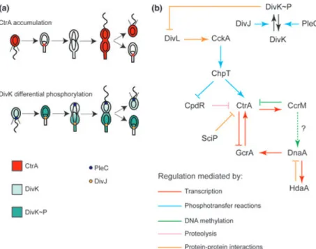
The C. crescentus genome contains a surprisingly high number of two-component signal transduction genes [105 of 3767 genes at the time of first annotation, (Nierman et al., 2001)], suggesting that these phospho-signaling proteins play a major role in the life cycle of this bacte-rium. DivK, an essential response regulator, acts as a cell-fate determinant and is regulated by phosphorylation. Phosphorylated DivK (DivK~P, phosphorylated on Asp 53) is found in the stalked cell, while dephosphorylated DivK prevails in the swarmer cell (Jacobs et al., 2001; Matroule et al., 2004). The histidine kinase DivJ that phosphorylates DivK is localized to the stalked pole and is therefore only inherited by the stalked daughter cell. Dephosphorylation of DivK~P is catalyzed by the phos-phatase PleC that is sequestered to the flagellar pole and partitions with the swarmer daughter cell. Thus, the daughter cell–specific inheritance of PleC or DivJ dictates which daughter has high levels of DivK~P and which one has low levels (Fig. 2a). Interestingly, DivK not only func-tions passively as a substrate in this phospho-transfer reaction, but also acts later in the cell cycle as an amplifi-cation device for the switch driving the swarmer to stalked transition by directly enhancing the kinase activity of DivJ and converting PleC into a kinase (Paul et al., 2008). PleC kinase activity drives polar remodeling (that is, ejection of the flagellum and development of the stalk and holdfast) through phosphorylation of the diguanylate cyclase PleD (Aldridge et al., 2003; Levi & Jenal, 2006), while increasing DivJ kinase activity rapidly boosts the levels of DivK~P in the cell. These allosteric activities of DivK likely accelerate its own changes in phosphorylation state and program genetic robustness into the system by the formation of a positive feedback loop.
The topology of the DivJ-DivK-PleC phospho-transfer reactions is also influenced by localization factors that direct DivJ and PleC to the appropriate pole. DivJ is recruited to the stalked pole by the muramidase homolog SpmX (Radhakrishnan et al., 2008), while PleC is directed to the swarmer pole by PodJ (Viollier et al., 2002a; Hinz et al., 2003; Lawler et al., 2006). The swarmer-to-stalked transition is accompanied by a sudden rise in DivK~P and a series of ordered polar remodeling events that act on PleC, PodJ, DivJ and SpmX. First, PleC is released from the flagellar pole and degraded along with PodJ (Viollier et al., 2002a, b; Chen et al., 2005). This
Fig. 1. Asymmetric cell division inCaulobacter. SW, swarmer cell; ST, stalked cell; PD, predivisional cell.
coincides with the acquisition of SpmX and its localiza-tion to the same pole. Localized SpmX then recruits and stimulates DivJ, leading to a ‘burst’ of DivK~P catalyzed by DivJ. Interestingly, the spmX gene is upregulated in a PleC-dependent manner prior to the transition, showing that this swarmer pole regulator signals forward to pre-pare the impending transition to the stalked cell pole (Radhakrishnan et al., 2008). PleC also regulates other developmental events by unknown mechanisms: the ejec-tion of the polar flagellum, the formaejec-tion of the holdfast and the elaboration of the stalk at the vacated pole, the switch in cell density during the swarmer (more dense) to stalked (less dense) cell transition, and its own release from the pole (Sommer & Newton, 1988, 1989; Wang et al., 1993; Viollier et al., 2002b; Aldridge et al., 2003; Biondi et al., 2006a; Radhakrishnan et al., 2008). The molecular events acting on and affected by DivK illustrate that the regulatory circuit is genetically imprinted to direct the development of the predivisional cell into swar-mer and stalked cells, and then differentiation of the swarmer progeny back into stalked cells, in the ensuing division cycle.
CtrA and CpdR: spatially regulated proteolysis How is the differential phosphorylation of DivK trans-lated into a downstream effect on the developmental cycle? The major transcriptional regulator of Caulobacter
development, CtrA, is a multifunctional DNA binding protein whose activity and abundance are indirectly influ-enced by the phosphorylation state of DivK. CtrA is regulated at several levels including transcription, phos-phorylation, localization, and proteolysis (Domian et al., 1997). In the swarmer cell, phosphorylated CtrA (CtrA~P) binds to sites near the chromosomal origin of replication (Quon et al., 1998). This interaction, presum-ably aided by other mechanisms (Cheng & Keiler, 2009; Collier & Shapiro, 2009), prevents premature initiation of DNA replication (Quon et al., 1998). CtrA is degraded at the swarmer-to-stalked transition (Domian et al., 1997), rendering the chromosomal origin of replication compe-tent to fire. CtrA is re-synthesised and (re)-phosphory-lated later in the stalked cell phase, and again binds to the replication origin. CtrA~P also binds to promoters of developmental genes to activate or repress their transcrip-tion (Laub et al., 2000), oscillating in-phase or out-of-phase, respectively, with CtrA activity over the cell cycle.
The phosphorylation and proteolysis of CtrA is regu-lated indirectly by DivK~P via the phosphotransfer path-way specified by the hybrid histidine kinase/phosphatase CckA and the histidine phosphotransferase protein ChpT (Biondi et al., 2006a, b). When DivK~P levels are low (in the swarmer cell), CckA is active and sequestered to the pole where it first autophosphorylates and then transfers the phosphate group to ChpT, which is used to phos-phorylate CtrA. When DivK~P levels are high (in the
Fig. 2. (a) Cell-type-dependent localization of the master regulator CtrA and cell-type dependent phosphorylation of the cell fate determinant DivK, the major events driving asymmetric development inCaulobacter. (b) The genetic circuit model of CtrA, GcrA, DnaA, and CcrM. Dotted lines indicate that the interaction is not fully elucidated; solid lines indicate that the link is confirmed but do not necessarily indicate a direct interaction.
nascent stalked cell), the phosphate flow is reversed: CckA is delocalized and its autokinase activity inhibited. Instead, CckA now acts as a phosphatase, ultimately draining the phosphate from CtrA (Biondi et al., 2006a, b; Chen et al., 2009). Remarkably, the same pathway regulates the phos-phorylation state of the single domain response regulator CpdR, which promotes proteolysis as an adaptor protein to the ClpXP protease (Abel et al., 2011) and is required for efficient degradation of CtrA and other proteins in vivo (Biondi et al., 2006b; Iniesta et al., 2006). In con-trast to CtrA, CpdR is inactive and dispersed when phos-phorylated. It is active when de-phosphorylated, localizing to the nascent stalked pole and recruiting ClpXP which degrades CtrA (Jenal & Fuchs, 1998). The ClpXP-dependent degradation of CtrA also seems to involve a second signal input in the form of cyclic-di-guanosine monophosphate (c-di-GMP) that interacts with a receptor protein, PopA, which facilitates CtrA degrada-tion in vivo (Duerig et al., 2009). Stalked polar localiza-tion of this protein is dependent on cyclic-di-GMP binding, and once localized, it recruits the CtrA-binding protein RcdA (McGrath et al., 2006) and the ClpXP pro-tease for CtrA degradation. Recent data show that the equilibrium between the activity of the DgcB diguanylate cyclase and that of the antagonistic PdeA phosphodiester-ase modulates this pathway (Abel et al., 2011).
Genetic circuits: CtrA and transcriptional regulation
The integrity and seamless function of transcriptional circuitry that drives the Caulobacter cell cycle and devel-opmental program is dependent on CtrA. CtrA defines a critical transcriptional node within this circuit and as such is essential for viability. As mentioned earlier, it reg-ulates many developmental genes, and the replication ori-gin, but it also tunes its own gradual accumulation over the cell cycle. Transcription of the ctrA gene is precisely regulated in space and time by two promoters, P1 and P2, both of which contain CtrA binding sites (Skerker & Laub, 2004). However, the response of each promoter to CtrA binding is different. The P1 promoter is activated, albeit weakly, in late stalked cells, triggering the synthesis of CtrA. This synthesis is self-reinforced with CtrA bind-ing and repressbind-ing the P1 promoter, while directly acti-vating the strong P2 promoter at the late predivisional stage to spark a pulse of CtrA production which leads to CtrA accumulation in the swarmer cell (Domian et al., 1999). Although the swarmer cell retains high levels of CtrA~P, this does not lead to continued activation of the P2 promoter after cell division (Quon et al., 1996), sug-gesting that other factors regulate ctrA transcription at other stages of the cell cycle.
One such factor is GcrA (Holtzendorff et al., 2004), a master regulatory protein that is essential for viability and that exhibits a cell cycle oscillation that is out-of-phase with that of CtrA. GcrA is responsible for the transcrip-tion of the ctrA gene from the P1 promoter in the late stalked cell (Holtzendorff et al., 2004). Another contribu-tor to the timing of ctrA transcription is the essential DNA methylase CcrM, which catalyzes methylation of adenine bases in the recognition site GANTC (Zweiger et al., 1994; Berdis et al., 1998). The P1 promoter of ctrA is active only in the hemimethylated state (Reisenauer & Shapiro, 2002), which occurs immediately after the DNA replication fork passes through the ctrA locus on the chromosome, leaving the DNA hemimethylated. At this stage, the ctrA P1 promoter is activated by GcrA, CtrA~P accumulates once again and activates transcription of several genes including ccrM. Upon its synthesis, CcrM re-methylates hemimethylated GANTC sites, inactivating the ctrA P1 promoter. Thus, CtrA activates transcription of its own negative transcriptional regulator.
The DnaA protein defines another critical node of the cell cycle circuitry (Gorbatyuk & Marczynski, 2001). DnaA is essential for the initiation of DNA replication, while also directly regulating the transcription of many cell cycle genes. As Caulobacter replicates its chromosome only once per cell cycle, it is vital that DnaA is tightly controlled in order to prevent re-initiation of a second round of replication before the cycle is completed and the daughter cells divide. DnaA activity is dependent on ATP binding, and hydrolysis of ATP renders DnaA inac-tive for replication initiation. DnaA is regulated at the post-translational level by the replisome-associated pro-tein HdaA, an inhibitor of DnaA activity (by stimulation of ATPase activity), as a replication initiator protein and perhaps also as a transcription factor (Collier & Shapiro, 2009). DnaA activates HdaA expression (directly or indi-rectly). Thus, after the peak in its activity, DnaA shuts itself down again by promoting the synthesis of its own inhibitor. DnaA also appears to be regulated at the level of proteolysis (Gorbatyuk & Marczynski, 2001; Grunenfel-der et al., 2001).
In addition to tight control of DnaA activity, dnaA transcription is cell cycle-regulated, accumulating prior to the onset of DNA replication (Zweiger & Shapiro, 1994; Laub et al., 2000). The dnaA gene is located relatively close to the origin of replication and therefore, is among the first genes to be replicated. After replication, the DNA is hemimethylated. It has been proposed that dnaA tran-scription is regulated by methylation of a CcrM-recogni-tion sequence (GANTC) located in the promoter (Collier et al., 2007). Indeed, mutation of the cytosine, although not the critical adenosine, impairs dnaA transcription (Cheng & Keiler, 2009). If CcrM-mediated adenosine
methylation directly regulates dnaA, then these findings suggest the simple transcriptional regulatory circuit of four sequentially acting master transcriptional regulators with the order: CcrM> DnaA > GcrA > CtrA > CcrM (Fig. 2b).
If regulation of dnaA by CcrM is indirect and does not involve adenine methylation of the dnaA promoter, then there are still missing links in the circuit. Perhaps dnaA is regulated by cytosine methylation, which could explain the observed effect of the key cytosine residue, while pre-serving the notion of the current model that the activity of the dnaA promoter is different in the methylated vs. hemi-methylated state and therefore dependent on DNA replication. Interestingly, at least two putative DNA cyto-sine methyltransferases, CC1033 and CC3626, are encoded in the C. crescentus genome. While their func-tions remain to be explored, CC1033 does contain one GANTC site in its promoter, suggesting the possibility of a link between adenosine and cytosine methylation. How-ever, the putative dependency of adenosine methylation on abundance or activity of CC1033 and CC3626 could also occur through an indirect route. Thus, chromosome methylation might function as a ‘ratchet’ to ensure that transcription of cell cycle genes proceeds in an ordered (forward) fashion (Collier et al., 2007). Together, these mechanisms act in a concerted fashion to restrict DnaA activity, to a short window during the swarmer-to-stalked cell transition when DNA replication initiates (Collier et al., 2006). Oscillations in DnaA activity dictate the temporal pattern of DNA replication during the cell divi-sion cycle that can act as a ‘pacemaker’ of DNA replica-tion even in the absence of CtrA, although the periodicity is apparently modulated by HdaA and tmRNA (Keiler & Shapiro, 2003; Collier & Shapiro, 2009; Jonas et al., 2011). The CtrA~P regulatory system is superimposed on the DnaA-controlled replication cycles to impart the spa-tial asymmetry of DNA replication at cell division (Jonas et al., 2011), ensuring the silencing of the origin of repli-cation in the progeny swarmer cell, while the origin in the progeny stalked cell can fire owing to the absence of CtrA~P.
In addition to its role as a DNA replication initiator, DnaA is a transcriptional regulator of gcrA (Collier et al., 2006). This regulation ensures that GcrA accumulates in the replicating stalked cell where the function of the GcrA target genes [encoding DNA replication factors such as RecJ, DnaQ, gyrase A and the ParE subunit of Topo IV, (Holtzendorff et al., 2004)] are needed.
Division plane establishment: MipZ and FtsZ
DnaA also appears to promote early events of cytokinesis by transcriptionally regulating the gene encoding FtsZ
(Hottes et al., 2005), a bacterial tubulin homolog that is a conserved mediator of cytokinesis in a wide range of bac-teria (Margolin, 2005). FtsZ monomers first polymerize into arcs or ring-like structures at the division plane of the cell. The FtsZ ring then recruits other components of the cell division machinery (the divisome) and is thought to contribute to the mechanical force which constricts the division plane and finally pinches off the two daughter cells from one another (Osawa et al., 2008). However, the regulatory mechanisms by which the division site is cho-sen and FtsZ positioned there are not so well conserved between bacteria. Two major regulatory mechanisms are the Min system and nucleoid occlusion (not mutually exclusive), where the Min proteins are localized to the cell poles and prevent GTP-dependent FtsZ polymerization there, so that the FtsZ ring only forms at mid-cell, while nucleoid occlusion prevents formation of the FtsZ ring in any region of the cell occupied by chromosomal DNA (Margolin, 2005). However, in Caulobacter, the Min system is not conserved, and the FtsZ ring has been observed to form at the division plane before chromo-some segregation is complete, implying that nucleoid occlusion is not operating either.
The mechanism employed by Caulobacter to regulate FtsZ positioning was identified by Thanbichler & Shapiro (2006) and involves the ParA-like ATPase MipZ. Like ParA, MipZ interacts with the ParB DNA-binding protein, but fulfills a different function. While ParA contributes to chromosome segregation by driving the ParB-bound origin region to the new pole, MipZ forms a bipolar gradient (through binding to ParB) with its max-ima at the ParB foci and a minimum at midcell. MipZ stimulates the GTPase activity of FtsZ and thus inhibits polymerization, permitting FtsZ assemblies only near the division plane. This inhibitory mechanism is distinct from that of MinC, the well-studied division inhibitor of E. coli which destabilises FtsZ protofilaments without affecting GTPase activity (Hu et al., 1999). At the ultrastructural level in vitro, MipZ converts straight protofilament bun-dles to curved structures, similar to those seen at the ends of eukaryotic microtubules (Tran et al., 1997), which may provide a physical explanation for the inhibition of FtsZ ring formation. Thus, MipZ provides a link between chromosome segregation, through the Par system, and FtsZ-mediated cytokinesis in Caulobacter.
Marking the new pole as the future flagellum assembly site: TipN, TipF and PflI
Polar flagellation in Caulobacter is intimately linked to cytokinesis as the flagellum is always constructed at the new pole, i.e. the one formed by the most recent division event. The reason for this consistent polarity was
unknown until recently, when the polarity factor TipN was identified (Huitema et al., 2006; Lam et al., 2006). This protein localizes to the pole opposite to the stalk or flagellum in stalked or swarmer cells, respectively, and during the development of the predivisional cell recruits flagellar assembly factors and structural proteins. At cyto-kinesis (once the flagellum has been assembled), TipN leaves the pole and relocalizes to the divisome through interaction with the Tol-Pal component of the divisome (Huitema et al., 2006; Lam et al., 2006; Yeh et al., 2010; Goley et al., 2011). It remains colocalized with FtsZ as the cell divides, so that it marks the newest pole after division and leads to the formation of the flagellum at the correct pole for the next round of division. TipN therefore acts as a ‘birth scar’ marker to identify the new pole of newly divided cells. Other factors modulating polar flagellum formation are the TipF assembly regulator (Huitema et al., 2006) and the PflI positioning factor (Obuchowski & Jacobs-Wagner, 2008). These proteins operate downstream of TipN, such that TipF relies on TipN for localization, and PflI in turn depends on TipF. Interestingly, TipF contains an EAL domain (named after the defining glutamate-alanine-leucine signature), which in other proteins can bind and/or hydrolyze the signaling molecule c-di-GMP (Jenal & Malone, 2006). In the case of TipF, the EAL domain acts essentially as a receptor protein as it is incompetent for c-di-GMP hydrolysis. Recent data show that c-di-GMP binding by TipF is a functional requirement for its own polar localization, recruitment of PflI and ultimately flagellum formation (N. J. Davis and P. H. Viollier, unpublished).
New insights into ‘hardwired’ developmental mechanisms
DivL and CckA: microdomains without membranes
Recent data implicate DNA replication as a trigger for the CckA-ChpT-CtrA/CpdR phosphorelay via the DivL histi-dine kinase. DivL is essential for viability and was origi-nally identified in a screen for motile suppressors of the pleC mutant phenotype which also led to the discovery of the genes encoding DivK and DivJ (Sommer & Newton, 1991). For some time, its role in cell division was myste-rious. DivL possesses a tyrosine residue (Y550) instead of a histidine at the catalytic site (Wu et al., 1999) and it appears that the critical functions of DivL in cell cycle control are not dependent on the kinase domain residing in the C-terminal part of the protein (Reisinger et al., 2007). Instead the N-terminal (signal sensing) domain appears to confer the essential activity and it was recently implied that DivL impinges on the CckA-ChpT-CtrA/
CpdR phosphorelay by acting on CckA. In an imaging-based screen for mutations which prevent the localization of CckA to the swarmer pole of the predivisional cell, it was found that DivL was required for the localization of CckA and that it stimulated its autophosphorylation. In the absence of DivL, CckA was not localized to the swar-mer pole and the phosphorelay was not activated, result-ing in the lack of phosphate transfer to CtrA. Again, the DivL kinase activity was dispensable for this function because a Y550F mutation had no effect on CckA locali-zation (Iniesta et al., 2010b). These authors also discov-ered that DivL and CckA localization to the pole was dependent on initiation of DNA replication (Iniesta et al., 2010b), implying that DivL may be part of a checkpoint which ensures that development of the predivisional cell does not proceed if chromosome replication cannot initi-ate. The role of DivL and DNA replication is particularly intriguing in light of the possibility that the kinase and phosphatase activities of CckA are confined to opposite poles. This notion has also been incorporated into recent cell cycle models to suggest the existence of phosphogra-dients of CtrA~P (Chen et al., 2010).
One issue with this model remained unclear until recently, namely why does CckA localize in a dynamic manner when it is neither asymmetrically inherited nor required for regulation of a polarly localized factor? This was resolved by the recent work of Tsokos et al. (2011) on the regulatory role of DivL. Here, it was confirmed that DivL is required to localize CckA at the swarmer pole of the predivisional cell, and that DivK is upstream of (and inhibits) DivL. Inhibition by DivK is mediated by direct binding of DivK~P to DivL, so that DivL is inactive and CckA is delocalized from the stalked pole once the DivK kinase DivJ is localized and active there (Fig. 2a). The lowest concentration of DivK~P is at the swarmer pole, because of the presence of the DivK phosphatase PleC, and this study found that PleC activity at the swar-mer pole was responsible for DivL and CckA activity there by keeping levels of the inhibitor DivK~P low. Hence, PleC provides a protective ‘microdomain’ at the swarmer pole in which CckA can activate its downstream phosphorelay, triggering the development of this pole into the swarmer daughter cell. This intricate mechanism provides a way of regulating development by localization to a functionally distinct part of the bacterial cell in the absence of membrane-limited internal compartments.
Yet another layer of regulation of the master regulator CtrA
With the identification of SciP, a small regulatory protein that inhibits CtrA activity and/or transcription of target genes (Gora et al., 2010; Tan et al., 2010), another additional
layer of regulation for CtrA was recently discovered. As described previously, it was not clear why the ctrA P2 promoter is inactive in the swarmer cell stage even though CtrA~P is present. SciP is present in the swarmer cell, is quickly degraded at the swarmer-to-stalked transi-tion and accumulates again as the predivisional cell is compartmentalized by the cytokinetic machinery. SciP binds directly to CtrA, disabling CtrA-mediated activation of transcription, while not affecting genes repressed by CtrA. While SciP does not affect DNA binding, phos-phorylation or degradation of CtrA, it appears to interfere with the recruitment of RNA polymerase. Consequently, many CtrA-dependent promoters that fire in the predivi-sional cell (for example those encoding the early flagellar structural proteins, components of the chemosensory apparatus and CtrA itself through the P2 promoter) are inhibited by the accumulation of SciP in the nascent swarmer cell compartment. The fact that pilA gene is acti-vated by CtrA in swarmer cells despite the presence of SciP, suggests that it is apparently immune to inhibition by SciP and/or that there are pockets from which SciP is excluded spatially. If this turns out to be true, the under-lying mechanism(s) remains to be determined.
The discovery of the SciP regulator provides another compelling example in the paradigm of fine tuning of two component systems by accessory factors. While ‘connec-tors’ which link two-component systems into networks have already been proposed (Mitrophanov & Groisman, 2008), it seems that SciP should rather be classed as a modulator because of its selective function on CtrA tran-scriptional activation. Notwithstanding the appropriate functional definition for SciP, it is clear that we can no longer consider cell cycle phospho-signaling systems of Alphaproteobacteria as simple two-state switches (usually phosphorylated= ON, dephosphorylated= OFF) but must take into account further layers of regulation permit-ting fine tuning akin to a dimmer switch, especially because SciP is conserved in all bacteria that possess a CtrA homolog (Gora et al., 2010). Further analysis of interconnection of transcriptional regulators, including SciP and CtrA, for example by chromatin immunoprecipi-tation – deep sequencing (ChIP-SEQ) may extend the model of the transcriptional circuit regulating develop-ment beyond its current two-dimensional state (Fig. 2).
Interaction of the par chromosome segregation system with the polarity factor TipN
It was recently elucidated how the replicated chromosome is directed poleward to coordinate chromosome partition-ing with the Caulobacter cell division cycle. The initial studies on TipN showed that in addition to a flagellar placement defect, TipN loss resulted in the misplacement
of the division septum to give a larger swarmer cell and smaller stalked cell than is normally observed (Lam et al., 2006), suggesting that TipN might also be involved in cytokinesis regulation. Indeed, it was recently found that TipN interacts genetically and biochemically with the Par chromosome segregation machinery (Ptacin et al., 2010; Schofield et al., 2010). Real-time analysis of FtsZ and MipZ dynamics showed that MipZ (and therefore, the origin of the newly replicated chromosome) travelled more slowly and erratically to the new pole in the TipN mutant, with occasional reverses back toward the old pole. This led to delayed formation and erroneous posi-tioning of the FtsZ ring at a position closer to the stalked pole than is usual, because the MipZ gradient extended further down the cell from the opposite end. Analysis of cells carrying fluorescent fusion derivatives of ParA showed that this effect on MipZ was mediated by the Par system. In wild-type cells, ParA formed a ‘cloud’ over the nucleoid, consistent with its DNA-binding activity (Ger-des et al., 2010) which retracted promptly to the new pole and remained there for the rest of the cell cycle. In TipN mutant cells, ParA did not retract smoothly to the new pole or accumulate there, and some remained at the old pole (which was never observed in wild type cells). Fluorescence resonance energy transfer (FRET) and pull-down experiments demonstrated that TipN and ParA interacted directly, leading to a model where TipN is pro-posed to bind and sequester ParA at the new pole as it is released from the DNA-bound ‘cloud’ thereby preventing it from returning behind the ParB-bound parS site and pulling it and the origin to the opposite pole (Schofield et al., 2010) where the ParB-parS complex is immobilized and captured by the PopZ polar matrix (Bowman et al., 2008; Ebersbach et al., 2008). Super-resolution fluores-cence microscopy recently revealed that the ParA ‘cloud’ seems to be composed of filamentous linear polymers (Ptacin et al., 2010), which were formed on (non-specific) binding to DNA and depolymerized by ParB. Interest-ingly, the ParA-mediated movement of the ParB-parS kinetochore-centromere (and the origin) is only one part of a recently proposed four-step poleward movement (Shebelut et al., 2010). The four stages are as follows: (1) release of both origins from PopZ (acts as a polar anchor for the chromosome) at the old pole (Bowman et al., 2010), (2) polar retraction of one origin back toward the old pole, (3) early translocation of the other origin (from pole to midcell), and (4) late translocation (from midcell to pole). The Par system was only required for late trans-location, which occurred at a significantly faster velocity than early translocation. These observations suggest that while initial origin separation may be by a relatively simple bulk separation mechanism, completion of chro-mosome segregation in Caulobacter is an active and
multi-phasic process with complex regulation similar to that seen in eukaryotes. They also indicate that a feedback loop consisting of the Par system, MipZ, FtsZ and TipN is an integral part of chromosome segregation and divi-sion control (Fig. 3).
New insights into metabolic and environmental influences on development
Extracellular DNA, a kin-specific dispersion
signal fromCaulobacter biofilms
In addition to planktonic growth, Caulobacter is capable of forming biofilms. In relatively nutrient-rich environ-ments, the swarmer cells do not disperse but tend to adhere to surfaces near their parents (Siegal-Gaskins & Crosson, 2008). As the swarmer cells go through the swarmer-to-stalked transition to obtain the ultra-adhesive holdfast, the nascent stalked cells bind firmly to the sur-face within a monolayer of cells that matures into a three-dimensional structure (Entcheva-Dimitrov & Spor-mann, 2004). The biofilm growth mode, while it enables the bacteria to profit from a readily available source of nutrients, imposes its own challenges on the cells buried in the core (e.g. the decreased availability of oxygen and nutrients). Not surprisingly, biofilm growth is regulated to balance these disadvantages against the advantages. An unprecedented mode of kin-specific biofilm regulation was recently discovered in Caulobacter by Berne et al. (2010). Unlike other biofilm-forming bacterial species, which incorporate macromolecules such as proteins and DNA into an extracellular matrix (Karatan & Watnick, 2009), and in some cases even require DNA as a struc-tural component of the matrix (Whitchurch et al., 2002), Caulobacter employs extracellular DNA (eDNA) as a bio-film dispersal signal. Low-molecular-mass eDNA inhibited the attachment of swarmer cells to the biofilm by binding to the holdfast and preventing its attachment to the bio-film-occupied surface, while it did not displace previously
attached stalked cells from the biofilm. eDNA concentra-tion correlated positively with cell death and negatively with biofilm formation, suggesting that the source of the eDNA is death and lysis of cells in the biofilm rather than secretion of DNA fragments from living cells. This hypothesis, however, does not exclude that cell death, induced for example by toxin-antitoxin systems, may be deliberately induced as part of a developmental program. Interestingly, the biofilm inhibitory effect was only observed for Caulobacter eDNA, as DNA from other spe-cies had no effect on Caulobacter biofilms (Berne et al., 2010). Therefore, the biofilm should be unaffected by the presence of unrelated bacteria, while modulating itself specifically according to the density of Caulobacter cells. These findings demonstrate that a hitherto unprecedented strategy can favor the motile stage of the cell cycle over the adhesive one.
Metabolic regulation of development
Caulobacter usually differentiates from a swarmer to a stalked cell after a fixed time in laboratory culture, sug-gesting that this differentiation process is ‘hard-wired’ and driven by an internal clock. While the constituents of such a potential ‘molecular clock’ remain to be identified, in the natural oligotrophic environment of Caulobacter environmental conditions are also likely to influence the relative length of the developmental stages. In support of this hypothesis, England et al. (2010) observed that growth in chemostatically nutrient-limited cultures caused global alterations in gene expression which led to changes in the developmental program. Specifically, nitrogen limi-tation prolonged the swarmer cell phase, consistent with the aforesaid hypothesis. Interestingly, carbon limitation lengthened the cell doubling time affecting each phase equally, suggesting that there are specific nutritional (metabolic) inputs into the developmental program. In another remarkable example of environmental signaling, Purcell et al. (2007) identified blue light as a physical stimulus that impacts the Caulobacter developmental pro-gram via the LovKR two-component system to fine-tune the adhesive properties of the cell. Carbon starvation was shown in two recent studies to feed into the core cell cycle circuitry driving development and cell division (Boutte & Crosson, 2011; Britos et al., 2011). The master regulator CtrA is downregulated in carbon-starved swar-mer cells in what appears to be a SigT-dependent man-ner, although the mechanism has not been identified (Britos et al., 2011). Meanwhile, the methylase CcrM was found to be under the control of SpoT, the Caulobacter ppGpp synthetase induced in response to starvation, and it is hypothesized that downregulation of CcrM under starvation conditions would lead to retention of high
Fig. 3. Feedback loop showing the interaction of the Par system, MipZ, FtsZ and TipN.
levels of CtrA and low levels of DnaA, instead of the antiphase fluctuations of these regulators which are nor-mally observed (Boutte & Crosson, 2011). Therefore, the core cell cycle regulatory circuit is susceptible to tuning by the availability of sufficient nutrients and, thus, the metabolic status of the cell.
The bifunctional regulatory protein KidO provides another illustrative example of how the cells might tune their developmental program according to their metabolic state (Radhakrishnan et al., 2010). An NAD(H)-binding oxidoreductase homolog, KidO modulates both CtrA~P levels and FtsZ function, contributing to the burst of DivK~P production in the stalked cell by activating DivJ kinase activity, while also regulating the assembly and/or stability of the cytokinetic FtsZ ring. Moreover, KidO abundance is cell-cycle regulated: it is present in the swar-mer and late predivisional cell, and is cleared from the cell in the stalked and early predivisional stages when the FtsZ ring forms. Remarkably, the degradation of KidO is catalyzed by the ClpXP protease via the same CckA/ ChpT/CpdR pathway that regulates the stability of CtrA.
In addition to regulation at the level of protein stabil-ity, there is evidence for another level of post-transla-tional regulation for KidO. While KidO can bind NADH, it lacks the catalytic residue required for NADH-depen-dent oxidation–reduction reactions. Mutations that dis-rupt the NADH-binding pocket of KidO prevent the FtsZ-inhibitory activity, while the DivJ positive regulation was unaffected. As the NADH-binding capacity of KidO is necessary for one of its functions (Radhakrishnan et al., 2010), the possibility exists that Caulobacter uses KidO to gauge cellular NADH levels to regulate cytokinesis depending on the energy level of the cell. Reminiscent of such a potential signaling role of NAD(H) in Caulobacter, eukaryotic cells are also thought to use metabolites such as NAD(P)H to signal cyclic processes such as the yeast cell cycle or the mammalian circadian clock (Tu & McKnight, 2006; Tu et al., 2007; Asher et al., 2008) even in the absence of transcription and translation (O’Neill & Reddy, 2011; O’Neill et al., 2011).
Regulated cell death through toxin–antitoxin
systems
Toxin–antitoxin (TA) systems, first discovered as plasmid-encoded genes, function as retention systems to kill off plasmid-free cells and ensure stable inheritance of the plasmid (Gerdes et al., 1986). However, with the advent of whole-genome sequencing, many chromosomally encoded TA systems have been discovered, frequently as multiple paralogous copies (Pandey & Gerdes, 2005). Three types of TA system have been characterized to date, which differ in the form and function of the antitoxin.
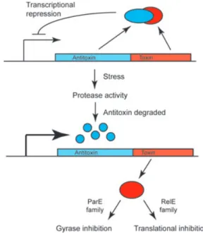
While the toxins of these systems are always proteins, type I antitoxins are cis-acting antisense mRNAs, while type II antitoxins are proteins. Type III TA systems have only recently been discovered and their antitoxins func-tion as protein-binding RNAs rather than antisense RNAs (Fineran et al., 2009). Type II TA systems (the others will not be further discussed here) are two-gene operons, usu-ally translationusu-ally coupled, with the antitoxin gene pre-ceding the toxin gene and often with the antitoxin acting as a repressor of its own transcription (Fig. 4). In unstressed cells, the antitoxin forms a complex with the toxin, preventing it from acting on its targets within the cell. Under stressful conditions, the antitoxin is degraded by proteases, freeing the toxin to act and relieving the transcriptional repression of the operon. In the case of TA systems for plasmid maintenance, the two daughter cells inherit the TA complex, but because the antitoxin protein is usually less stable than the toxin, cells can only replenish the antitoxin if they retain the plasmid. Plas-mid-free cells are killed upon release of the toxin, leading to stable maintenance of the plasmid in the population (Hayes, 2003). Killing is mediated through mRNA cleav-age at the ribosome by RelE-family toxins (Neubauer et al., 2009), or DNA gyrase inhibition by ParE-family toxins (Jiang et al., 2002).
The multiplicity of these inducible self-killing genes on bacterial chromosomes suggests that they may be used for executing controlled cell death as part of a developmental program (Engelberg-Kulka et al., 2006). Caulobacter pos-sesses 11 chromosomal type II TA systems, of which the
Fig. 4. The classical paradigm of type II TA systems of the Par and Rel family (other systems are not reviewed here).
functions are by and large not well understood. Recent work by Fiebig et al. (2010) on the RelBE and ParDE homologs of Caulobacter provides genetic evidence that these systems form insulated units so that each toxin interacts only with its co-encoded antitoxin and there is no cross-talk between systems, even when the antitoxin genes are artificially overexpressed, consistent with the idea that they are active under specialized conditions. Fur-thermore, transcription of the systems was differentially regulated in response to various environmental stressors such as oxidative stress and heat shock. Some of the ope-rons were also transcriptionally upregulated in mid log growth phase relative to early log growth, in the absence of any stress, implying a possible role for TA systems in the natural progression of the Caulobacter life cycle, although this is not yet confirmed. It might be interesting to investigate whether there is cross-talk between TA sys-tems and other developmental regulatory factors at the level of transcription (or elsewhere). Indeed, it has been observed that the promoters of some of the Caulobacter TA systems are bound by the SOS (DNA damage) response regulator LexA (da Rocha et al., 2008) (Radha-krishnan and Viollier, unpublished), suggesting that while they may be insulated from cross-talk with each other they can be integrated into genetic or developmental control circuits.
Conclusions
Studies of the bacterial cell cycle in Caulobacter have unmasked many regulatory mechanisms not observed in model systems with apparently symmetrical division. With the recent developments reviewed herein, additional levels of complexity have surfaced to an already intricate cell differentiation process in a so-called ‘simple’ bacterial cell. This progress is attributable in part to the rapid improvement of analytical methods that have fueled these discoveries, above all the methods for single-cell level flu-orescence imaging. FRET, fluflu-orescence loss in photoble-aching and fluorescence recovery after photoblephotoble-aching strategies especially have enabled in vivo confirmation of molecular interactions that could previously only be observed in vitro. Moreover, as the limits of resolution of fluorescence microscopy decrease by improved optical and computational methods, it is becoming possible to observe new processes at the submicron level. For exam-ple, high-resolution RNA localization experiments recently suggested that transcripts are immobile, localizing to the corresponding position in the cell where the gene is located, and that they capture the much larger ribo-somal particles that diffuse by (Montero Llopis et al., 2010), with tRNAs presumably posing an exception to this restricted diffusion of transcription. Moreover, a
single-cell-based FRET sensor (Christen et al., 2010) con-firmed the notion that the signaling molecule c-di-GMP is differentially partitioned at cell division with higher concentrations found in the Caulobacter swarmer cell than in the stalked cell (Paul et al., 2008). Future research using high-resolution microscopic methods will uncover new regulatory pathways that are confined in subcellular space and/or as a function of cell cycle as is the case in eukaryotes (Dehmelt & Bastiaens, 2010).
Acknowledgements
Funding support is from the Swiss National Science Foundation (Grant # 31003A_127287), the Human Fron-tier Science Program (Program Grant # RGP0051/2010), the Socie´te´ Acade´mique de Gene`ve (# 2011/70) and the University of Geneva.
References
Abel S, Chien P, Wassmann P, Schirmer T, Kaever V, Laub MT, Baker TA & Jenal U (2011) Regulatory cohesion of cell cycle and cell differentiation through interlinked
phosphorylation and second messenger networks. Mol Cell 43: 550–560.
Aldridge P, Paul R, Goymer P, Rainey P & Jenal U (2003) Role of the GGDEF regulator PleD in polar development of
Caulobacter crescentus. Mol Microbiol47: 1695–1708.
Asher G, Gatfield D, Stratmann M et al. (2008) SIRT1 regulates circadian clock gene expression through PER2
deacetylation. Cell134: 317–328.
Berdis AJ, Lee I, Coward JK, Stephens C, Wright R, Shapiro L & Benkovic SJ (1998) A cell cycle-regulated adenine DNA methyltransferase from Caulobacter crescentus processively methylates GANTC sites on hemimethylated DNA. P Natl
Acad Sci USA95: 2874–2879.
Berne C, Kysela DT & Brun YV (2010) A bacterial extracellular DNA inhibits settling of motile progeny cells within a
biofilm. Mol Microbiol77: 815–829.
Biondi EG, Skerker JM, Arif M, Prasol MS, Perchuk BS & Laub MT (2006a) A phosphorelay system controls stalk biogenesis during cell cycle progression in Caulobacter
crescentus. Mol Microbiol59: 386–401.
Biondi EG, Reisinger SJ, Skerker JM, Arif M, Perchuk BS, Ryan KR & Laub MT (2006b) Regulation of the bacterial
cell cycle by an integrated genetic circuit. Nature444: 899–
904.
Bodenmiller D, Toh E & Brun YV (2004) Development of
surface adhesion in Caulobacter crescentus. J Bacteriol186:
1438–1447.
Boutte CC & Crosson S (2011) The complex logic of stringent response regulation in Caulobacter crescentus: starvation
signalling in an oligotrophic environment. Mol Microbiol80:
Bowman GR, Comolli LR, Zhu J et al. (2008) A polymeric protein anchors the chromosomal origin/ParB complex at a
bacterial cell pole. Cell134: 945–955.
Bowman GR, Comolli LR, Gaietta GM et al. (2010) Caulobacter PopZ forms a polar subdomain dictating sequential changes in pole composition and function. Mol
Microbiol76: 173–189.
Britos L, Abeliuk E, Taverner T, Lipton M, McAdams H & Shapiro L (2011) Regulatory response to carbon starvation
in Caulobacter crescentus. PLoS ONE6: e18179.
Chen JC, Viollier PH & Shapiro L (2005) A membrane
metalloprotease participates in the sequential degradation of a
Caulobacter polarity determinant. Mol Microbiol55: 1085–
1103.
Chen YE, Tsokos CG, Biondi EG, Perchuk BS & Laub MT (2009) Dynamics of two phosphorelays controlling cell cycle
progression in Caulobacter crescentus. J Bacteriol191: 7417–
7429.
Chen YE, Tropini C, Jonas K, Tsokos CG, Huang KC & Laub MT (2010) Spatial gradient of protein phosphorylation underlies replicative asymmetry in a bacterium. P Natl Acad
Sci USA108: 1052–1057.
Cheng L & Keiler KC (2009) Correct timing of dnaA transcription and initiation of DNA replication requires
trans translation. J Bacteriol191: 4268–4275.
Christen M, Kulasekara HD, Christen B, Kulasekara BR, Hoffman LR & Miller SI (2010) Asymmetrical distribution of the second messenger c-di-GMP upon bacterial cell
division. Science328: 1295–1297.
Collier J & Shapiro L (2009) Feedback control of DnaA-mediated replication initiation by replisome-associated
HdaA protein in Caulobacter. J Bacteriol191: 5706–5716.
Collier J, Murray SR & Shapiro L (2006) DnaA couples DNA replication and the expression of two cell cycle master
regulators. EMBO J25: 346–356.
Collier J, McAdams HH & Shapiro L (2007) A DNA
methylation ratchet governs progression through a bacterial
cell cycle. P Natl Acad Sci USA104: 17111–17116.
Curtis PD & Brun YV (2010) Getting in the loop: regulation of development in Caulobacter crescentus. Microbiol Mol Biol
Rev74: 13–41.
Dehmelt L & Bastiaens PI (2010) Spatial organization of intracellular communication: insights from imaging. Nat
Rev Mol Cell Biol11: 440–452.
Domian IJ, Quon KC & Shapiro L (1997) Cell type-specific phosphorylation and proteolysis of a transcriptional regulator controls the G1-to-S transition in a bacterial cell
cycle. Cell90: 415–424.
Domian IJ, Reisenauer A & Shapiro L (1999) Feedback control of a master bacterial cell-cycle regulator. P Natl Acad Sci
USA96: 6648–6653.
Duerig A, Abel S, Folcher M et al. (2009) Second messenger-mediated spatiotemporal control of protein degradation
regulates bacterial cell cycle progression. Genes Dev23:
93–104.
Dworkin J (2009) Cellular polarity in prokaryotic organisms.
Cold Spring Harb Perspect Biol1: a003368.
Ebersbach G, Briegel A, Jensen GJ & Jacobs-Wagner C (2008) A self-associating protein critical for chromosome
attachment, division, and polar organization in Caulobacter.
Cell134: 956–968.
Engelberg-Kulka H, Amitai S, Kolodkin-Gal I & Hazan R (2006) Bacterial programmed cell death and multicellular
behavior in bacteria. PLoS Genet2: e135.
England JC, Perchuk BS, Laub MT & Gober JW (2010) Global regulation of gene expression and cell differentiation in Caulobacter crescentus in response to nutrient availability.
J Bacteriol192: 819–833.
Entcheva-Dimitrov P & Spormann AM (2004) Dynamics and control of biofilms of the oligotrophic bacterium
Caulobacter crescentus. J Bacteriol186: 8254–8266.
Errington J (2010) From spores to antibiotics via the cell cycle.
Microbiology156: 1–13.
Fiebig A, Rojas CM, Siegal-Gaskins D & Crosson S (2010) Interaction specificity, toxicity, and regulation of a paralogous set of ParE/RelE-family toxin-antitoxin systems.
Mol Microbiol77: 236–251.
Fineran PC, Blower TR, Foulds IJ, Humphreys DP, Lilley KS & Salmond GP (2009) The phage abortive infection system, ToxIN, functions as a protein-RNA toxin-antitoxin pair.
P Natl Acad Sci USA106: 894–899.
Gerdes K, Rasmussen PB & Molin S (1986) Unique type of plasmid maintenance function: postsegregational killing of
plasmid-free cells. P Natl Acad Sci USA83: 3116–3120.
Gerdes K, Howard M & Szardenings F (2010) Pushing and
pulling in prokaryotic DNA segregation. Cell141: 927–942.
Goley ED, Yeh YC, Hong SH, Fero MJ, Abeliuk E, McAdams HH & Shapiro L (2011) Assembly of the Caulobacter cell
division machine. Mol Microbiol80: 1680–1698.
Gora KG, Tsokos CG, Chen YE, Srinivasan BS, Perchuk BS & Laub MT (2010) A cell-type-specific protein-protein interaction modulates transcriptional activity of a master
regulator in Caulobacter crescentus. Mol Cell39: 455–467.
Gorbatyuk B & Marczynski GT (2001) Physiological consequences of blocked Caulobacter crescentus dnaA expression, an essential DNA replication gene. Mol Microbiol 40: 485–497.
Grunenfelder B, Rummel G, Vohradsky J, Roder D, Langen H & Jenal U (2001) Proteomic analysis of the bacterial cell
cycle. P Natl Acad Sci USA98: 4681–4686.
Hayes F (2003) Toxins-antitoxins: plasmid maintenance,
programmed cell death, and cell cycle arrest. Science301:
1496–1499.
Hinz AJ, Larson DE, Smith CS & Brun YV (2003) The Caulobacter crescentus polar organelle development protein PodJ is differentially localized and is required for polar targeting of the PleC development regulator. Mol Microbiol 47: 929–941.
Holtzendorff J, Hung D, Brende P, Reisenauer A, Viollier PH, McAdams HH & Shapiro L (2004) Oscillating global
regulators control the genetic circuit driving a bacterial cell
cycle. Science304: 983–987.
Hottes AK, Shapiro L & McAdams HH (2005) DnaA
coordinates replication initiation and cell cycle transcription
in Caulobacter crescentus. Mol Microbiol58: 1340–1353.
Hu Z, Mukherjee A, Pichoff S & Lutkenhaus J (1999) The MinC component of the division site selection system in Escherichia coli interacts with FtsZ to prevent
polymerization. P Natl Acad Sci USA96: 14819–14824.
Huitema E, Pritchard S, Matteson D, Radhakrishnan SK & Viollier PH (2006) Bacterial birth scar proteins mark future
flagellum assembly site. Cell124: 1025–1037.
Iniesta AA, McGrath PT, Reisenauer A, McAdams HH & Shapiro L (2006) A phospho-signaling pathway controls the localization and activity of a protease complex critical for
bacterial cell cycle progression. P Natl Acad Sci USA103:
10935–10940.
Iniesta AA, Hillson NJ & Shapiro L (2010a) Polar remodeling and histidine kinase activation, which is essential for Caulobacter cell cycle progression, are dependent on DNA
replication initiation. J Bacteriol192: 3893–3902.
Iniesta AA, Hillson NJ & Shapiro L (2010b) Cell pole-specific activation of a critical bacterial cell cycle kinase. P Natl
Acad Sci USA107: 7012–7017.
Jacobs C, Hung D & Shapiro L (2001) Dynamic localization of a cytoplasmic signal transduction response regulator controls morphogenesis during the Caulobacter cell cycle.
P Natl Acad Sci USA98: 4095–4100.
Jenal U & Fuchs T (1998) An essential protease involved in
bacterial cell-cycle control. EMBO J17: 5658–5669.
Jenal U & Malone J (2006) Mechanisms of cyclic-di-GMP
signaling in bacteria. Annu Rev Genet40: 385–407.
Jiang Y, Pogliano J, Helinski DR & Konieczny I (2002) ParE toxin encoded by the broad-host-range plasmid RK2 is
an inhibitor of Escherichia coli gyrase. Mol Microbiol44: 971–
979.
Jonas K, Chen YE & Laub MT (2011) Modularity of the bacterial cell cycle enables independent spatial and temporal
control of DNA replication. Curr Biol21: 1092–1101.
Kaiser D, Robinson M & Kroos L (2010) Myxobacteria, polarity, and multicellular morphogenesis. Cold Spring Harb
Perspect Biol2: a000380.
Karatan E & Watnick P (2009) Signals, regulatory networks, and materials that build and break bacterial biofilms.
Microbiol Mol Biol Rev73: 310–347.
Keiler KC & Shapiro L (2003) TmRNA is required for correct timing of DNA replication in Caulobacter crescentus.
J Bacteriol185: 573–580.
Lam H, Schofield WB & Jacobs-Wagner C (2006) A landmark protein essential for establishing and perpetuating the
polarity of a bacterial cell. Cell124: 1011–1023.
Laub MT, McAdams HH, Feldblyum T, Fraser CM & Shapiro L (2000) Global analysis of the genetic network controlling
a bacterial cell cycle. Science290: 2144–2148.
Lawler ML, Larson DE, Hinz AJ, Klein D & Brun YV (2006) Dissection of functional domains of the polar localization
factor PodJ in Caulobacter crescentus. Mol Microbiol59: 301–
316.
Levi A & Jenal U (2006) Holdfast formation in motile swarmer cells optimizes surface attachment during Caulobacter
crescentus development. J Bacteriol188: 5315–5318.
Lopez D, Vlamakis H & Kolter R (2009) Generation of multiple cell types in Bacillus subtilis. FEMS Microbiol Rev 33: 152–163.
Lopez D, Vlamakis H & Kolter R (2010) Biofilms. Cold Spring
Harb Perspect Biol2: a000398.
Maddock JR & Shapiro L (1993) Polar location of the chemoreceptor complex in the Escherichia coli cell. Science 259: 1717–1723.
Margolin W (2005) FtsZ and the division of prokaryotic cells
and organelles. Nat Rev Mol Cell Biol6: 862–871.
Matroule JY, Lam H, Burnette DT & Jacobs-Wagner C (2004) Cytokinesis monitoring during development; rapid pole-to-pole shuttling of a signaling protein by localized kinase and
phosphatase in Caulobacter. Cell118: 579–590.
McGrath PT, Iniesta AA, Ryan KR, Shapiro L & McAdams HH (2006) A dynamically localized protease complex and a polar specificity factor control a cell cycle master regulator.
Cell124: 535–547.
Mitrophanov AY & Groisman EA (2008) Signal integration in
bacterial two-component regulatory systems. Genes Dev22:
2601–2611.
Montero Llopis P, Jackson AF, Sliusarenko O, Surovtsev I, Heinritz J, Emonet T & Jacobs-Wagner C (2010) Spatial organization of the flow of genetic information in bacteria.
Nature466: 77–81.
Neubauer C, Gao YG, Andersen KR et al. (2009) The structural basis for mRNA recognition and cleavage by the
ribosome-dependent endonuclease RelE. Cell139: 1084–1095.
Nierman WC, Feldblyum TV, Laub MT et al. (2001) Complete genome sequence of Caulobacter crescentus. P Natl Acad Sci
USA98: 4136–4141.
Obuchowski PL & Jacobs-Wagner C (2008) PflI, a protein involved in flagellar positioning in Caulobacter crescentus.
J Bacteriol190: 1718–1729.
O’Neill JS & Reddy AB (2011) Circadian clocks in human red
blood cells. Nature469: 498–503.
O’Neill JS, van Ooijen G, Dixon LE et al. (2011) Circadian rhythms persist without transcription in a eukaryote. Nature 469: 554–558.
Osawa M, Anderson DE & Erickson HP (2008) Reconstitution
of contractile FtsZ rings in liposomes. Science320: 792–
794.
Pandey DP & Gerdes K (2005) Toxin-antitoxin loci are highly abundant in free-living but lost from host-associated
prokaryotes. Nucleic Acids Res33: 966–976.
Paul R, Jaeger T, Abel S et al. (2008) Allosteric regulation of histidine kinases by their cognate response regulator
determines cell fate. Cell133: 452–461.
Ptacin JL, Lee SF, Garner EC et al. (2010) A spindle-like apparatus guides bacterial chromosome segregation. Nat
Purcell EB, Siegal-Gaskins D, Rawling DC, Fiebig A & Crosson S (2007) A photosensory two-component system regulates
bacterial cell attachment. P Natl Acad Sci USA104: 18241–
18246.
Quon KC, Marczynski GT & Shapiro L (1996) Cell cycle control by an essential bacterial two-component signal
transduction protein. Cell84: 83–93.
Quon KC, Yang B, Domian IJ, Shapiro L & Marczynski GT (1998) Negative control of bacterial DNA replication by a cell cycle regulatory protein that binds at the chromosome
origin. P Natl Acad Sci USA95: 120–125.
Radhakrishnan SK, Thanbichler M & Viollier PH (2008) The dynamic interplay between a cell fate determinant and a lysozyme homolog drives the asymmetric division cycle of
Caulobacter crescentus. Genes Dev22: 212–225.
Radhakrishnan SK, Pritchard S & Viollier PH (2010) Coupling prokaryotic cell fate and division control with a bifunctional
and oscillating oxidoreductase homolog. Dev Cell18: 90–
101.
Reisenauer A & Shapiro L (2002) DNA methylation affects the cell cycle transcription of the CtrA global regulator in
Caulobacter. EMBO J21: 4969–4977.
Reisinger SJ, Huntwork S, Viollier PH & Ryan KR (2007) DivL performs critical cell cycle functions in Caulobacter
crescentus independent of kinase activity. J Bacteriol189:
8308–8320.
da Rocha RP, Paquola AC, Marques Mdo V, Menck CF & Galhardo RS (2008) Characterization of the SOS regulon of
Caulobacter crescentus. J Bacteriol190: 1209–1218.
Rudner DZ & Losick R (2010) Protein subcellular localization
in bacteria. Cold Spring Harb Perspect Biol2: a000307.
Schofield WB, Lim HC & Jacobs-Wagner C (2010) Cell cycle coordination and regulation of bacterial chromosome segregation dynamics by polarly localized proteins. EMBO J 29: 3068–3081.
Shapiro L, McAdams HH & Losick R (2009) Why and how
bacteria localize proteins. Science326: 1225–1228.
Shebelut CW, Guberman JM, van Teeffelen S, Yakhnina AA & Gitai Z (2010) Caulobacter chromosome segregation is an
ordered multistep process. P Natl Acad Sci USA107: 14194–
14198.
Siegal-Gaskins D & Crosson S (2008) Tightly regulated and heritable division control in single bacterial cells. Biophys J 95: 2063–2072.
Skerker JM & Laub MT (2004) Cell-cycle progression and the generation of asymmetry in Caulobacter crescentus. Nat Rev
Microbiol2: 325–337.
Sommer JM & Newton A (1988) Sequential regulation of developmental events during polar morphogenesis in Caulobacter crescentus: assembly of pili on swarmer cells
requires cell separation. J Bacteriol170: 409–415.
Sommer JM & Newton A (1989) Turning off flagellum rotation requires the pleiotropic gene pleD: pleA, pleC, and
pleD define two morphogenic pathways in Caulobacter
crescentus. J Bacteriol171: 392–401.
Sommer JM & Newton A (1991) Pseudoreversion analysis indicates a direct role of cell division genes in polar morphogenesis and differentiation in Caulobacter crescentus.
Genetics129: 623–630.
Tan MH, Kozdon JB, Shen X, Shapiro L & McAdams HH (2010) An essential transcription factor, SciP, enhances robustness of Caulobacter cell cycle regulation. P Natl Acad
Sci USA107: 18985–18990.
Thanbichler M & Shapiro L (2006) MipZ, a spatial regulator coordinating chromosome segregation with cell division in
Caulobacter. Cell126: 147–162.
Tran PT, Joshi P & Salmon ED (1997) How tubulin subunits are lost from the shortening ends of microtubules. J Struct
Biol118: 107–118.
Tsokos CG, Perchuk BS & Laub MT (2011) A dynamic complex of signaling proteins uses polar localization to regulate cell-fate asymmetry in Caulobacter crescentus. Dev
Cell20: 329–341.
Tu BP & McKnight SL (2006) Metabolic cycles as an underlying basis of biological oscillations. Nat Rev Mol Cell
Biol7: 696–701.
Tu BP, Mohler RE, Liu JC, Dombek KM, Young ET, Synovec RE & McKnight SL (2007) Cyclic changes in metabolic state
during the life of a yeast cell. P Natl Acad Sci USA104:
16886–16891.
Viollier PH, Sternheim N & Shapiro L (2002a) Identification of a localization factor for the polar positioning of bacterial
structural and regulatory proteins. P Natl Acad Sci USA99:
13831–13836.
Viollier PH, Sternheim N & Shapiro L (2002b) A dynamically localized histidine kinase controls the asymmetric
distribution of polar pili proteins. EMBO J21: 4420–4428.
Wang SP, Sharma PL, Schoenlein PV & Ely B (1993) A histidine protein kinase is involved in polar organelle development in Caulobacter crescentus. P Natl Acad Sci USA 90: 630–634.
Whitchurch CB, Tolker-Nielsen T, Ragas PC & Mattick JS (2002) Extracellular DNA required for bacterial biofilm
formation. Science295: 1487.
Wu J, Ohta N, Zhao JL & Newton A (1999) A novel bacterial tyrosine kinase essential for cell division and differentiation.
P Natl Acad Sci USA96: 13068–13073.
Yeh YC, Comolli LR, Downing KH, Shapiro L & McAdams HH (2010) The Caulobacter Tol-Pal complex is essential for outer membrane integrity and the positioning of a polar
localization factor. J Bacteriol192: 4847–4858.
Zweiger G & Shapiro L (1994) Expression of Caulobacter dnaA
as a function of the cell cycle. J Bacteriol176: 401–408.
Zweiger G, Marczynski G & Shapiro L (1994) A Caulobacter DNA methyltransferase that functions only in the