© 1997 Kluwer Academic Publishers. Primed in the Netherlands.
Original article
Breast cancer: Pretreatment drug resistance parameters (GSH-system,
ATase, P-glycoprotein) in tumor tissue and their correlation with clinical and
prognostic characteristics
K. Buser,
1F. Joncourt,
2H.-J. Altermatt,
3M. Bacchi,
4A. Oberli
2& Th. Cerny
1institute for Medical Oncology. Inselspital: 2 Institute of Clinical and Experimental Cancer Research; } Institute of Pathology. University of Bern. 4SIAKCoordination Center. Bern, Switzerland
Summary
Background: The identification of new factors predicting
relapse, outcome and response to systemic therapy in breast cancer is warranted. The measurement of biological markers such as drug resistance parameters (DRPs), which are part of the phenotype of malignant cells and contribute to resistance to anti-cancer drugs may be a possibility, which may ultimately lead to improvement of therapeutic results.
Patients and methods: The level of glutathione (GSH),
activities of glutathione-S-transferase (GST), glutathione-per-oxidase (GPx), 06-alkylguanine-DNA-alkyltransferase (ATase), and P-glycoprotein (PGP) were measured in tumor and adja-cent tumor free tissue samples from 89 consecutive, untreated females with breast cancer and correlated with clinical and prognostic factors. Early breast cancer (EBC) was diagnosed in 56 patients, 22 patients had locally advanced (LABC) and 11 patients metastatic breast cancer.
Results: All DRPs showed significantly higher expression
in tumor than in tumor free tissues. GPx was positively cor-related with GST (r = 0.3, P = 0.0048) and with GSH (r = 0.5,
P = 0.0001) in tumor as well as in normal tissue. GST activity
was significantly higher in EBC than in LABC or metastatic breast cancer (P = 0.02). GSH level was significantly higher in grade 1 than in grade 2 or grade 3 tumors {P = 0.01). When clinical characteristics were related to the level of DRP, 'high' GSH was associated with age > 60 years (P = 0.01) in EBC, and with grade 1-2 tumors (P = 0.05) in LABC. No differences in OS were apparent between groups of 'high' and 'low' DRP-expression. However, the four-year estimated disease-free sur-vival of EBC tended to be higher in patients with 'high' GST
(P = 0.10) and of LABC in patients with 'high' GPx levels (P
-0.06).
Conclusion. We conclude that 'high' levels of DRP in tumor
tissue of breast cancer patients are part of the initial phenotype of the malignant cells. Due to its high prevalence (83% in EBC, 100% in primarily metastatic breast cancer), PGP did not add to prognostic information. High levels of GSH, GST and and GPx were associated with favorable clinical characteristics and good prognosis, whereas low levels of GSH and GST activity were associated with more aggressive or more advanced disease.
Key words: ATase, breast cancer, drug resistance, PGP, GPx,
GSH, GST, prognosis
Introduction
Breast cancer has become the second leading cause of cancer deaths in women after lung cancer [1]. Currently, none of the known prognostic factors in breast cancer are capable of fully defining the patients with highest risk for relapsing disease [2]. Therefore, the identification of new factors predicting relapse, outcome and response to systemic therapy is warranted. The measurement of biological markers, which are part of the phenotype of malignant cells and contribute to resistance to anti-cancer drugs, may be a possibility, which may ultimately lead to improvement of therapeutic results.
Current clinical investigations try to circumvent resistance pathways, by adding resistance modulators, previously identified as modulators in vitro, to chemo-therapeutic regimens. So far, results remain behind expectations [3], suggesting that therapy response in most cases may depend on more than one parameter
and that the modulators may not be specific and/or effective enough. While most studies focus on one parameter, the aim of this study was to measure P-glyco-protein (PGP), glutathione (GSH), GSH-S-transferases (GST), glutathione-peroxydase (GPx) and O6 -alkyl-guanine-DNA alkyltransferase (ATase) simultaneously in tumor samples of patients with newly diagnosed early, locally advanced and metastatic breast cancer as well as normal breast tissue of the same patients.
The best understood drug resistance parameter is PGP, a 170-kd protein coded for by the MDR1 gene, which works as an energy-dependent efflux pump to decrease intracellular accumulation of a number of cytostatic drugs [4]. PGP expression was found in a majority of tumor specimens in patients with breast cancer. Although high PGP expression appears to cor-relate with poor response to chemotherapy and short disease-free survival in some instances [5], its role is still controversial. Some evidence suggests that PGP may
also be a marker of 'biological maliciousness' of cancers [6].
Subsequently, other biochemical parameters, which play an important role in the detoxification or cell repair systems, were identified. Although many of them were shown to contribute to drug resistance in vitro, a direct connection, between these parameters and drug resist-ance in vivo has not yet been established. GSH plays an important role in detoxification and repair of cellular injury caused by cyclophosphamide, nitrosoureas and quinone antibiotics [7]. Elevated levels of GSH have been described in different human tumor tissue samples when compared with normal tissue [8, 9].
GSTs are a family of enzymes which catalyse the conjugation of electrophilic substrates with GSH, there-by detoxifying them. Increased levels of these enzymes have been found in human breast cancer when com-pared with benign lesions [10]. GST-7T. expression, one of the four different classes of GST known in humans, was reported to be inversely correlated with hormone receptor status in breast cancer [11]. GPx, an enzyme removing toxic oxygen intermediates, is also associated with GSH. GPx activity was found at increased levels in breast cancer when compared to normal tissue [12].
ATase is an enzyme involved in DNA repair and is present in all human tissues [13]. Many human tumor tissues show higher activity than corresponding normal tissue. However, so far no differences in ATase activity between breast cancer and corresponding normal breast tissue have been reported [14].
Despite the great amount of information about GSH, GST, GPx, and ATase, and their involvement in the cellular defense against toxic substances, association with either clinical behavior or prognosis has yet to be established.
We attempted to evaluate whether the simultaneous assessment of these parameters would allow detection of a distinct pattern of distribution in tumor tissues and whether an association between any of these parameters and established prognostic factors and/or clinical out-come could be found.
Patients and methods
Patient characteristics
From May 1988 until November 1991. 89 nonsdected Caucasian female patients with histologically proven breast cancer, but no other cancer, were evaluated. None of the patients had received any systemic treat-ment. The patients characteristics are presented in Table 1 The median follow-up from diagnosis is 45 months. Relapse or disease progression was observed in 34 patients, 30 patients died during follow-up and of these six patients died without relapse or progression (one cerebro-vascular insult, three cardiocerebro-vascular disease, one influenza, one pneu-monia).
Tissue collection
Tumor tissue and tumor-free tissue samples were obtained from the
operating theater at first surgery. Tissue samples were snap frozen and maintained at - 7 0 "C until assayed.
Pathologic examination
The pathology reports and the histological slides of the tumors were evaluated by a board-certified pathologist. The histological determina-tion of the tumor type was performed according to the WHO clas-sification. Histological tumor grading was performed according to Elston and Ellis [15]. Tissue samples from tumor and tumor-free tissue samples were used for pathologic review It was confirmed by the pathologist that the tissue samples examined for histology were directly adjacent to the sample used for the biochemichal assays.
The tumor mass, estimated on the corresponding histological slides, ranged between 15% and 90%, the remaining tissue consisted of desmoplastic stromal fibrosis and fat tissue. None of the tumor samples contained relevant necrosis and none of the samples from tumor-free breast tissue revealed any breast cancer cells. For the steroid hormone receptor determination the dextrane coated charcoal method (DCC) was used [16]. Patients were designated as hormone-receptor positive (progesterone hormone-receptor > 20 fmol/mg protein and/or estrogen receptor >10 fmol/mg protein) or hormone-receptor nega-tive (progesterone receptor <20 fmol/mg protein and/or estrogen receptor < 10 fmol/mg protein).
Tissue preparation
Techniques of sample preparation and assays have been described in a previous report [8]
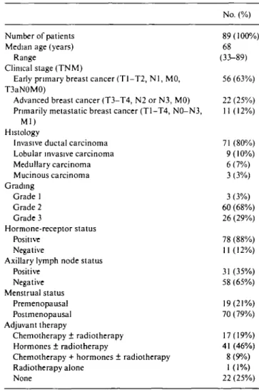
Table 1. Patient characteristics.
Number of patients Median age (years)
Range
Clinical stage (TNM)
Early primary breast cancer (TI-T2, N l , M0, T3aN0M0)
Advanced breast cancer (T3-T4, N2 or N3, M0) Primarily metastatic breast cancer (T1-T4, N0-N3,
Ml) Histology
Invasive ductal carcinoma Lobular invasive carcinoma Medullary carcinoma Mucinous carcinoma Grading Grade 1 Grade 2 Grade 3 Hormone-receptor status Positive Negative
Axillary lymph node status Positive Negative Menstrual status Premenopausal Postmenopausal Adjuvant therapy Chemotherapy ± radiotherapy Hormones ± radiotherapy
Chemotherapy + hormones ± radiotherapy Radiotherapy alone None No. (%) 89(100%) 68 (33-89) 56 (63%) 22 (25%) 11 (12%) 71 (80%) 9(10%) 6 (7%) 3 (3%) 3 (3%) 60 (68%) 26 (29%) 78 (88%) 11(12%) 31 (35%) 58 (65%) 19(21%) 70 (79%) 17(19%) 41 (46%) 8 (9%) 1(1%) 22 (25%)
337
Enzyme assays and Western blotting
GSH content was measured according to Tietze's recycling assay. Overall GST activity was measured according to the method of Habig. GPx activity was assayed with the improved method of Gunzler. For enzyme assays several data points in the linear response range were used and the results were calculated per mg of protein in the cytoplas-matic fraction as determined using Bradford's reagent with bovine serum albumin as standard.
ATase was measured according to the method of Morten and Margison and calculated as fmol methyl transferred to protein per ug DNA.
PGP was determined semi-quantitatively by Western blot after separation of membrane proteins by SDS-PAGE [8]. The antibody used was a polyclonal rabbit antiserum raised against amino acids 1205-1224 of the human mdr protein. This peptide (ALDTESEKVV-QEALDKAREG) was made for us by Multiple Peptide Systems, Inc., San Diego, CA The antibody used recognizes the gene products of both MDR1 and MDR3. In the tissue samples analyzed, however, MDR3 is not ecpected to contribute the the signals detected, since no MDR3 expression has been found in human breast tissue. Positive and negative controls were crude membrane pellets from the doxorubicin-resistant lung cancer cell line SW 1573 IR 500-0 and its drug sensitive parent SW 1573, generously provided by Dr. H. Joenje, Amsterdam. All gels included internal standards consisting of three different amounts of membrane protein from the positive control. The signals produced by PGP were compared to the internal standards and three categories were arbitrarily defined: 1) no detectable signal = 0 (nega-tive); 2) weakly positive signal = + (PGP-signal weaker than one produced by 1.25 ug protein of the positive control) and 3) strongly positive signal = ++ (PGP-signal equal to or stronger than the one produced by 1.25 ug protein of the positive control). For practical reasons, in the result presentation, we grouped together weakly and strongly positive PGP (labeled as 'positive').
All analyses were performed with the investigators unaware of the patients' characteristics and outcome.
Statistical methods
Differences between expression/activities of GSH, ATase, GST, and GPx in normal breast and tumor tissue were calculated for each patient, and the 'paired' Wilcoxon signed rank test was used to test the hypothesis of no difference. Correlation between different DRPs was measured with the Spearmann rank correlation coefficient The Wilcoxon rank-sum test or the Kruskal-Wallis test were used to compare the distribution of the DRPs according to clinical and prognostic parameters. In case of ordered groups (clinical stage and grade) a non parametric test for trend was performed [17]. The relationship between PGP levels and other parameters was determined with the chi-square or Fisher's exact test where appropriate Values of DRPs except for PGP expression/activity were divided into 2 groups taking the median values as cutting point ( ^ median:'low', > median: 'high'). This decision was made a priori, before examination of the results for outcome and prognosis. The Kaplan-Meier method was used to estimate distributions of disease-free survival (DFS), time to progression (TTP), and overall survival (OS) [18]. The estimates are reported with standard errors. Differences in time distributions were evaluated by the log-rank test [19]. P-values < 0.05 were considered to be statistically significant. All F-values were derived from two-sided tests for significance. No adjustment for multiple comparison was performed.
Results
DRP expression in normal breast and tumor tissue Significantly higher levels of all tested DRPs were found
in tumor tissue compared to normal breast tissue. More-over, in normal tissue GPx activity significantly cor-related with GST activity (r = 0.48, P - 0.0001) and with GSH levels (r = 0.51, P = 0.0001). Similarly, in tumor tissue a significant correlation of GPx activity with GST (r = 0.3, P = 0.0048) and GSH (r = 0.52, P = 0.0001) was
observed.
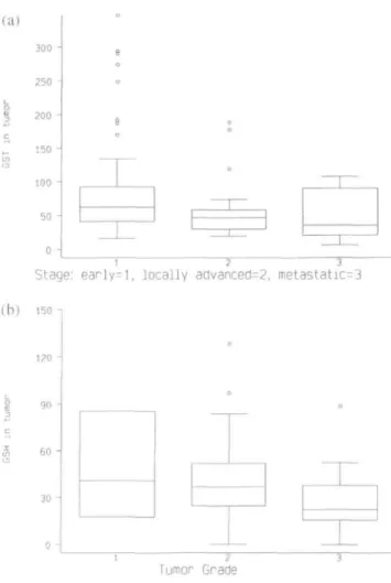
Biological behavior and outcome is different in pa-tients with EBC as compared to papa-tients with LABC or primary metastatic breast cancer. These three groups of patients were therefore separated for further analyses. GST activities were high in early and decreased in locally advanced and metastatic breast cancer (median values: 62.6 vs. 46.9 vs. 35.9 nmol/min/mg protein re-spectively; test for trend P-value = 0.02; Figure la). Similarly, GSH-levels were higher in grade 1 than in grade 2 and 3 tumors (median values: 40.9 vs. 36.9 vs. 22.4 nmol/mg protein, respectively; test for trend P-value = 0.01; Figure lb). (a) 300 -250 -200 -150 J 100 50 o -e o 8 o 0 o —r—
1 '
—
J—
1 2 " 3Stage: early=l, locally advanced=2, metastatic=3
120
60
30
o
-Tumor- Grade
Figure 1 (a) Box plots of GST activity in early, locally advanced and
metastatic breast cancer. The line in the middle of the box represents the median. The box extends from the 25th to the 75th percentile. The lines emerging from the box extend to the upper and lower 'adjacent values' Points more extreme are individually plotted, (b) Box plots of GSH activity in grade 1, 2 and 3 breast cancer. The line in the middle of the box represents the median. The box extends from the 25th to the 75th percentile. The lines emerging from the box extend to the upper and lower 'adjacent values'. Points more extreme are individually plotted.
DRP expression and clinical characteristics in early breast cancer
The median levels of DRPs in tumor tissue of EBC-patients calculated separately for the different clinical and pathological characteristics (age, histology, grading, hormone-receptor status, axillary node involvement and menopausal status) are shown in Table 2a. With one exception none of the DRPs appeared to be associated with any of these characteristics. Only the median GSH level was significantly elevated in patients older than 60 years as compared to younger patients (P - 0.01). PGP was expressed in the tumors of 44 of 53 patients (83%) and the frequency of expression was similar for all tested characteristics (Table 2a). We were interested to see whether any of the DRPs analyzed influenced OS or DFS in these patients. No significant differences in OS or DFS were apparent. However, a trend was found for GST, the four-year estimated DFS being 74% (SE 8%) for 'high' and 57% (SE 9%) for 'low' GST activity (log-rank P = 0.10).
DRP expression and clinical characteristics in locally advanced breast cancer
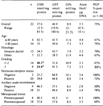
Median levels of GSH, ATase, GST, GPx and PGP activity/expression in tumor tissue of patients with LABC for the different clinical and pathological charac-teristics are shown in Table 2b. Again with one exception none of the DRPs appeared to be associated with any of these characteristics. Only the median GSH level was higher in patients with low-grade tumors (P - 0.05) compared to high grade. PGP expression was found in 15 of 20 patients (75%). The analysis of OS and DFS in this subgroup of patients revealed no significant differ-ences but a clear trend for GPx, the four-year estimated DFS being 75% (SE 16%) for 'high'and 46% (SE 16%) for 'low' GPx activity (log-rank P = 0.06).
DRP expression in primarily metastatic breast cancer Eleven of our patients had primarily metastatic breast cancer. This group was therefore too small to allow a meaningful correlation between DRPs and prognostic characteristics. Median values in this group were 29.2 nmol/mg protein for GSH (range 7.16-62.62), 35.9 nmol/min/mg protein for GST (range 6.5-108.2), 8.38 mU/mg protein for GPx (range 1.07-50.5) and 3.85 fmol/ug DNA for ATase (range 0.74-14.76). All 11 patients expressed PGP, without any difference in TTP for levels of positivity. All patients with metastatic dis-ease progressed, and all, except one, died during the observation time.
Discussion
Breast cancer is a disease, which can be treated with variable success. More valid criteria for defining patients
Table 2a Drug resistance parameters in tumor tissue of early breast
cancer by prognostic characteristics. Median values (% positive for PGP). Overall Range Age < 60 years > 60 years Histology Invasive ductal Other Grading 1-2 3 n 56 20 36 48 8 43 13 Hormone-receptor status Negative Positive 7 49 Axillary node involvement
Negative Positive Menopausal status Premenopausal Postmenopausal 26 30 12 44 GSH nmol/mg protein 35 3 (0-128 6) 23.7" 38.1" 36 8 28 7 36 8 22.9 21.4 36.8 34.9 35 3 26 1 36.9 GST nmol/ min/mg protein 62.6 ( 1 6 5 -347.2) 75 3 60.3 62.6 83 1 63 2 57 3 73 1 62.1 65 61 7 73 1 61.2 GPx mU/mg protein 9.3 (3.5-32 2) 7.9 9 9 9.4 7.3 9.1 109 9.1 9 3 7 7 10.1 8.1 9.3 Atase fmol/ ME DNA 4.6 (0.9-11 7) 3.3 5.5 4.4 6 4 4 5 5.4 5 7 4.5 4.7 4.4 3 7 5 1 PGP % posi-tive (n = 53) 83% 89% 79% 80% 100% 78% 100% 100% 80% 78% 87% 100% 78%
1 Significantly different values are in bold (/"-value = 0 01)
Table 2b Drug resistance parameters in tumor tissue of locally
advanced breast cancer by prognostic characteristics. Median values (% positive for PgP). Overall Range Age < 60 years > 60 years Histology Invasive ductal Other Grading 1-2 3 n 22 6 16 15 7 14 8 Hormone-receptor status Negative Positive 2 20 Axillary node involvement
Negative Positive Menopausal status Premenopausal Postmenopausal 4 18 4 18 GSH nmol/mg protein 37 6 (2 5 -85 5) 42 3 35 34.3 52.7 40.1" 24.6" 25.2 39.8 46.5 35 40.3 37.6 GST nmol/ min/mg protein 46 9 (19.9-188 6) 41 5 49.4 43.7 58 5 51.4 41 5 84.9 448 37.1 49.4 41.5 37 6 GPx mU/mg protein 8.9 (2-21.5) 11 6 7 3 7 9 11 9 109 7 2 10 1 8.9 6.1 8 9 11 8.5 Atase fmol/ DNA 3 3 (0 5 -10 1) 4.4 3.3 3 2 4 6 3 3 3.3 3.6 3 4 2 8 3.4 4 3 3.3 PGP % posi-tive (n = 20) 75% 67% 79% 79% 67% 67% 88% 100% 72% 50% 78% 100% 69%
a Significantly different values are in bold (/"-value = 0.05)
at risk of poor response to chemotherapy would be helpful. Our study was an attempt to define additional criteria by measuring the level of expression of several pretreatment parameters thought to be involved in cyto-static drug resistance. GSH, GST, GPx and ATase were all found to be expressed at significantly higher levels
339 and PGP was significantly more frequently expressed in
tumor, when compared to tumor-free tissue. Similar observations have been made for other cancer types [20] and support the hypothesis that these biological markers are part of the intrinsic, metabolic pattern of malignant cells.
The comparison of DRPs in tumor tissue and corre-sponding normal tissue by biochemical methods has to be interpreted with caution because the amount of epithelial cells within the normal tissue fragments is about 5%-10% whereas the amount of neoplastic cells in tumor tissue fragments vary from 10%—90%. The biochemical measurements in normal tissue should only give an impression on the natural arsenal of DRP in normal breast tissue. Therefore, the results of the DRP measurements of normal tissue fragments has not been included for the correlation with clinical and prognostic characteristics of the investigate breast cancer.
While PGP is the best studied of all putative DRPs, its role as prognostic factor is still controversial. Most authors agree that PGP is present in many cases of untreated breast cancer. It is not clear, however, whether its expression may influence treatment outcome. Eighty-three percent of our patients had detectable levels of PGP in their tumor tissue. No association with prognos-tic factors was apparent, a finding that matches the results of other investigators [21, 22]. Interestingly, how-ever, all patients with primarily metastatic disease expressed PGP. This is more extreme than in Linn et al. [23] where 58% of the samples were PGP positive. PGP was highly prevalent (83% in EBC, 100% in primarily metastatic breast cancer), and therefore not adding further to prognostic information. The semiquantitative assessment of PGP did not improve its prognostic value. There was no apparent influence of the level of PGP-expression on OS, DFS or TTP. Recently, a similar lack of correlation between PGP-expression and chemother-apy response or survival was reported by Decker et al. [24]. Other authors, however, found high PGP-expres-sion to be associated with a poor response to chemo-therapy and short DFS [5]. These discrepancies suggest that more studies are needed in order to clarify the role of PGP in breast cancer. A major obstacle in comparing the results from different studies is the use of variable techniques and reference standards. Thus, while many of the recently published studies used histochemistry to detect PGP, we have chosen to assess PGP expression by Western blot. This was done so as to be able to perform the measurement of all DRPs on material derived from the same sample. With this method, how-ever, it was not possible to localize PGP within the tissue. Nevertheless, contamination of analyzed tissue samples e.g., with white blood cells expressing MDR1 and/or MDR3, which are both recognized by our anti-body, can be neglected, because the level of expression of PGP in these cells is below the detection limit of our assay [25].
The overexpression of GSH, GST, GPx and ATase in the tumor tissue show, that potential drug resistance
mechanisms other than PGP are present in tumor cells of untreated patients. The high interindividual variabil-ity of expression/activvariabil-ity indicate, that the measured parameters may contribute to resistance to variable degrees in different tissues. The relationship between DRPs and clinical as well as pathological characteristics in breast cancer has been studied by several authors, so far with contradictory results [11, 22, 23, 26, 27]. In the present study ATase activity was increased in tumor tissue, a finding which to our knowledge is new for breast cancer, but matches the findings reported in non small cell lung cancer [9]. An association of ATase activity with any of the other characteristics considered was not apparent in our untreated patients. We think, however, that the role of ATase in breast cancer merits further investigation, e.g., it would be interesting to know whether ATase is upregulated in tumor tissue of women treated with DNA-damaging agents. This would, however, require follow-up tumor samples, which is clinically rarely justified.
Most authors agree that GSH and its associated enzymes are overexpressed in human breast cancer in comparison with tumor-free breast tissue [28, 12]. Fur-thermore we observed a correlation of GPx with GSH and GST in tumor as well as in tumor-free tissue. The significant correlations between these three related parameters may point out the presence of common regulatory elements [29].
In our patients GSH levels were higher in grade 1 when compared to grade 2 or 3 tumors. In the subgroup of EBC 'high' GSH levels were associated with age > 60 years and in LABC with grade 1 and 2 tumors. GST was significantly higher in EBC when compared to LABC or metastatic disease. In contrast, low levels of GSH or GST activity seemed to be associated with a more malig-nant phenotype of tumor cells or more advanced dis-ease. When analyzed for prognosis, 'high' GST in EBC and 'high' GPx in LABC showed a borderline associa-tion with longer disease-free survival. This finding was unexpected but similar to observations made in leuke-mias [30]. So far 'high' GST-levels in tumor tissue have been assumed to be associated with poor prognosis, although a significant correlation has been rarely found [11]. One reason why our observations differ from those of Gilbert et al. may be that our population consisted mainly of elderly women (median age: 68 years). Fur-thermore it may be that the expression of GSH and its associated enzymes is hormone dependent as it has been shown in endometrium cancer [31]. Such a hormone dependent modulation, which is not taken into account in any of these studies, may influence the interpretation of the results [32].
We conclude that high levels of potential DRPs such as PGP, GSH, GST, GPx and ATase in tumor tissue of newly diagnosed, untreated breast cancer are part of the phenotype of the malignant cells and are a reflection of their constitutive characteristics. Overexpression/activ-ity of potential DRPs does not necessarily explain if and how they function as DRP modulators in clinical drug
resistance. In addition EBC and LABC as well as pri-marily metastatic disease may have their own specific pattern of potential DRP composition. Our findings illustrate the difficulty to associate single biological parameters with prognosis. DRPs appear to be a com-plex system of partly interdependent parameters, most likely with none of them being exclusively responsible for treatment outcome. Studies which aim at circum-venting drug resistance by adding only one modulating agent to standard chemotherapy may therefore ulti-mately fail [3, 33]. If significant impacts on clinical out-come are expected from future studies on human tumor tissue samples so that 'bench work' can be translated into therapy recommendations for the patient, it will be necessary to standardize laboratory assays and to study a representative variety of markers prospectively in larger and more homogeneous patient groups.
Acknowledgements
This study was supported by the Swiss Cancer League, Zentralschweizerische Krebsliga, Aargauische Krebs-liga, Bernische Stiftung fur Klinische Krebsforschung and the Cancer Research Campaign as well as the SAKK (Schweizerische Arbeitsgemeinschaft fur Klini-sche Krebsforschung). The authors thank Monique Nef and Marlcus Stadler for their excellent technical assis-tance and Heidi Gusset for the English translation.
References
1. Boring CC, Squires TS, Tung T. Cancer statistics 1988. CA 1991; 1: 19-36
2. Elledge RM, MC Guire WL, Osborne CK. Prognostic factors in breast cancer. Semin Oncol 1992; 19. 244-53.
3. Mross K, Bohn C, Edler L et al. Randomized phase II study of single agent epirubicin + / - verapamil in patients with advanced breast cancer. Ann Oncol 1993, 4: 45-50.
4. D'lncalci M, Broxterman HJ, Van Kalken CK. Membrane trans-port multidrug resistance, development and disease. Ann Oncol
1991; 2-635-9.
5. Botti G, Chiappetta G, D'Aiuto G et al. PCNA/Cyclin and P-glycoprotein as prognostic factors in locally advanced breast cancer. An immunohistochemical, retrospective study. Tumon
1993; 79. 214-8.
6. Gottesman NM. How cancer cells evade chemotherapy: Sixteenth Richard and Minda Rosenthal Foundation Award lecture. Can-cer Res 1993; 53: 747-54.
7. Murray GI, Burke MD, Ewen SWB. Glutathione localization in benign and malignant human breast lesions. Br J Cancer 1987, 55: 605-9.
8. Redmond SMS, Joncourt F, Buser K et al Assessment of P-gly-coprotein, glutathione-based detoxifying enzymes and 06-alkyl-guanine-DNA alkyltransferase as potential indicators of constit-utive drug resistance in human colorectal tumors. Cancer Res 1991; 51. 2092-7.
9. Oberli-Schraemmli AE, Joncourt F, Stadler M et al. Parallel assessment of glutathione-based detoxifying enzymes, 06 -alkyl-guanine-DNA alkyltransferase and P-glycoprotein as indicators ofdrug resistance in tumor and normal lung of patients with lung cancer. Int J Cancer 1994, 59: 629-36.
10. Colovai Al, Stoica A, Cinca SA et al. Glutathione S-transferase
activity in human breast cancer tumors. Neoplasma 1992; 39. 249-54.
11. Gilbert L, Elwood LJ, Merino M et al. A pilot study of pi-class glutathione S-transferase expression in breast cancer: Correlation with estrogen receptor expression and prognosis. J Clin Oncol
1993, 1:49-58.
12. Singh SV, Brunnert SR, Roberts B, Krishan A. Differential expression of glutathione S-transferase, glutathione peroxidase and glutathione reductase in normal and malignant human breast tissue. Cancer Lett 1990; 51. 43-8.
13. D'lncalci M, Citti L, Taverna P, Catapano CV. Importance of the DNA repair enzyme 06-alkylguanine alkyltransferase (AT) in cancer chemotherapy. Cancer Treat Rev 1988; 15: 279-92. 14. Myrnes B, Norstrand K, Giercksky KE et al. A simplified assay
for 06-methylguanine-DNA methyltransferase activity and its application to human neoplastic and nonneoplastic tissues. Car-cinogenesis 1984; 5: 1061-4.
15. Elston CW, Ellis IO Pathological prognostic factors in breast cancer. I. The value of histological grade in breast cancer: Experi-ence from a large study with long-term follow-up. Histopathology 1991; 19.403-10.
16. Korenmann SG, Dukes BA. Specific estrogen binding by the cytoplasm of human breast carcinoma. J Clin Endocrinol Metabol 1970; 30. 639-45.
17. Cuzick J. A Wilcoxon-type test for trend. Stat Med 1985; 4: 87-90. 18. Kaplan EL, Meier P. Nonparametric estimation from incomplete
observations J Am Stat Assoc 1958, 457-81.
19. Mantel N. Evaluation of survival data and two new rank order statistics arising in its consideration. Cancer Chemother Rep 1966; 50: 163-7.
20. Joncourt F, Oberli AE, Stadler M et al Patterns of drug resistance parameters in adult leukemia. Leuk Lymph 1995; 17: 101-9
21. Wishart GC, Plump JA, Going JJ et al. P-glycoprotein expression in primary breast cancer detected by immuno-cytochemistry with two monoclonal antibodies. Br J Cancer 1990; 62: 758-61. 22. Wallner J, Depisch D, Hopfner M et al. MDR1 gene expression
and prognostic factors in primary breast cancer Eur J Cancer 1991; 27: 1352-5.
23. Linn SC, Giaccone G, Van Diest PJ et al. Prognostic relevance of P-glycoprotein expression in breast cancer. Ann Oncol 1995; 6: 679-85.
24. Decker DA, Morris LW, Levine AJ et al. Immunohistochemical analysis of P-glycoprotein expression in breast cancer: Clinical correlations. Ann Clin Lab Sci 1995; 25; 52-9.
25. Joncourt F, Oberli A, Redmond SMS et al. Cytostatic drug resistance' Parallel assessment of glutathione-based detoxifying enzymes, 06-alkylguanine-DNA-alkyltransferase and P-glyco-protein in adult patients with leukemia. Br J Haematol 1993, 85; 103-11.
26. Verelle P, Meissonnier F, Fonck Y et al. Clinical relevance of immunohistochemical detection of multidrug resistance P-glyco-protein in breast carcinoma. J Natl Cancer Inst 1991; 83: 111-6. 27. Perry RR, Mazetta JA, Levin M, Barranco SC. Glutathione
levels and variability in breast tumors and normal tissue. Cancer 1993; 72: 783-7.
28. Cairns J, Wright C, Cattan AR et al. Immunohistochemical demonstration of glutathione S-transferases in primary human breast carcinomas. J Pathol 1992; 166: 19-25.
29. Efferth T, Volm M. Immunocytochemical detection of onco-proteins in animal and human tumor lines with acquiredor inherent multidrug-resistance. Cancer Detect Prev 1992; 16: 2 3 7 ^ 3 .
30. Lohri A, Van Hille B, Bacchi M et al. Simultaneous assessment of five putative drug resistance (DR) parameters (MDR1/P-glyco-protein [PGP], MDR-associated (MDR1/P-glyco-protein [MRP], Glutathione-S-Transferase [GST], bcl-2 and topoisomerase I la [Topo I la]) in 57 newly diagnosed acute myeloid leukemia's (AML). ASCO Proc 1995; 14. 179
341 activity in endometrium: Effects of sex hormones and cancer.
Gynecol Oncol 1996, 60: 277-82.
32. Chang BD, Beatti CW, Hussain RA, Anderson CH. Estrous cycle modulation of 06-alkyiguanine-DNA alkyltransferase expression in rat mammary epithelial cells. Cancer Lett 1993; 75: 11-8 33. Linn SC, Van Kalken CK, Van Tellingen O et al. Clinical and
pharmacologic study of multidrug resistance reversal with vin-blastine and bepridil. J Clin Oncol 1994; 12: 812-9
Received 16 November 1994, accepted 29 January 1997
Correspondence w
K. Buser, MD Clinic Beau-Site Oberweg 14