Occurrence of Localized Intense Cation Uptake Sites in
the Vascular Rings of Red Beet Storage Tissue during
Ageing
A. MOZAFAR AND J. J. OERTLI
Swiss Federal Institute of Technology (ETH), Crop Science Institute, 8092 Zurich, Switzerland
Received 11 July 1983
A B S T R A C T
Development and decline of cation uptake capacity in discs taken from the vascular and parenchyma rings of storage tissue of red table beet (Beta vulgaris L.) were observed during 12 d of ageing. Uptake capacity for Na+ and Rb+ showed a steady rise reaching maximums by the fourth to fifth days of
ageing. Thereafter, there was a steady decline in the uptake rates. Vascular ring tissues were able to develop a greater uptake capacity for both Na+ and Rb+ than the tissues of parenchyma rings. This
difference, which was more pronounced for Rb+ than for Na+ uptake, is attributed to a combination of
variations in cell density and differences in the acquisition and retention of the cation uptake capacity. Respiration of tissue discs showed no significant rise during ageing, nor were there significant differences in the respiration of vascular and parenchyma tissues. Vascular tissues contained significantly more betacyanin than parenchyma tissues; and they retained their pigment, as well as their acquired cation uptake capacity, for a longer period during the ageing process.
Key words: Cation uptake; Red beet; Vascular rings; Ageing. I N T R O D U C T I O N
Slices of storage tissue from red table beet (Beta vulgaris L.) subjected to a period of washing, usually referred to as ageing, are often used as model tissue for ion uptake studies (Sutcliffe, 1957; Briggs, Hope, and Pitman, 1958; Van Steveninck, 1975; Poole, 1976; Francois, Bogemans, and Neirinckx, 1982). A common feature of beet storage tissues is the presence of a series of concentric rings of small vascular cells (xylem and phloem) that are separated from one another by broader bands of highly vacuolated thin-walled parenchyma cells (Artschwager, 1926; Hayward, 1938).
In our studies of ion uptake kinetics, we have encountered great variability in the uptake of Na+ and Rb+ by aged discs taken randomly from different regions of beet storage tissue.
Although transport properties of vascular and parenchyma cells have been reported to be similar (Poole, 1976), we suspected that the differences in development of cation transport capacity in vascular and parenchyma cells are the source of the variability observed. Our findings are described in this report.
M A T E R I A L S A N D M E T H O D S
Storage tissue of red table beet (Beta vulgaris L. cv. Feuerkugel) was produced at the experiment station of the ETH Crop Science Institute in Eschikon and stored in peat until use. Beets were cut into
198
1 0 mm thick slices perpendicular to their vertical axis with the aid of an all-purpose slicing machine. These slices were then placed over a back-lighted glass panel and 4-2 mm diameter discs were cut from the centre of the vascular and parenchyma rings with a corkborer. Although vascular and parenchyma rings are not strictly composed of one type of cell (Artschwager, 1926). disc samples taken from the ring regions will be referred to as vascular and parenchyma, respectively, in this discussion. About one thousand each of vascular and parenchyma discs were aged separately by washing them at 20 °C in several hundred times their volume of continuously aerated 0-1 mol m~3 CaCl2 solution which was renewed every 24 h. At different ageing periods from 1-12 d, ten discs each of the tissue types were randomly selected, placed into 150 ml of labelled treatment solution, and their uptake of Na+ was measured for a 24 h period. This uptake procedure was repeated for Rb+. The Na+ and Rb+ treatment solutions contained 0-5 mol m ' CaSO4 plus 0 1 mol m"3 NaCl labelled with 22Na (45-4 x 10" Bq mol"1) or 0-5 mol m~3 CaSO4 plus 0 1 mol m"3 RbCl labelled with 86Rb (138-1 x 10' Bq mol '), respectively. At the end of the uptake period (24 h, 20 °C and continuous shaking and aeration), the discs were rinsed sequentially for 15 s in tap water, 50 mol m '3 CaCl2. and distilled water. Their absorbed radioactivity was measured by a solid scintillation counter Gammazint 5300. Auto-radiograms of beet slices, aged for 4 d. were produced by first loading the slices in 22Na- or 86Rb-labelled solutions (8 x 10⁷ Bq mol"1) for 24 h. The slices were then dried slowly between filter blotters held by wire screens and paper clamps at 30 °C in a vacuum oven at 1 -5 kPa pressure. Kodak X-OMAT AR no-screen autoradiography film was exposed in a deep freezer at —15 °C for 5 d and developed according to the manufacturer's recommendation.
Oxygen uptake of single discs was measured polarographically in 5-0 cm3 of air-saturated Rb+ treatment solution (as described above but not labelled) by following the change in oxygen
1 500 1000 5 0 0 0 vaacular rings f
/A
-
/ / L A
V y ring* P -C-^r i i i i i i d \ "2N aI
\
o 2 4 e e 1 0 1 2 Duration of ageing (day)1500 7 1000 5 0 0 vascular ringt 8 6 Rb 2 4 S 8 Duration of agalng (day)
1 0 1 2
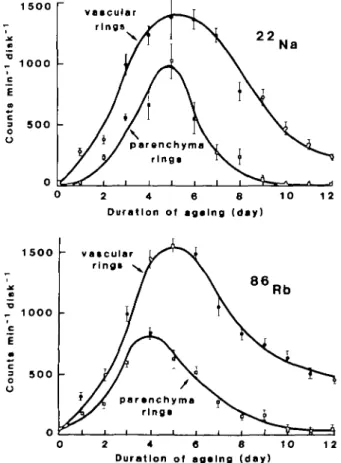
FIG. I. 22Na and " R b activity in the tissue discs taken from vascular and parenchyma rings of beet
storage tissue after incubation for 24 h in the treatment solution. Values are means of 10 replicates: vertical lines represent + I s.e.
199 concentration with a Biological Oxygen Monitor (Yellow Springs Instruments, model 53). The content of betacyanin and betaxanthin in the discs was measured by homogenizing 10 discs in 10 cm3 of
100 mol ITT3 Mcllvaine's citrate-phosphate buffer at pH 5-0. The homogenate was filtered through
Whatman No. 1 paper and centrifuged at 15 000 rev. min~' for 20 min to remove cell debris. A 0-5 cm' aliquot of supernatant was diluted to 5-0 cm3 and its optical density was measured at 476, 537 and
600 nm. The betacyanin and betaxanthin contents were calculated by Nilsson's formula as presented by Wiley and Lee (1978).
R E S U L T S A N D D I S C U S S I O N
Ageing beet discs increased their uptake capacity for both Na+ and Rb+ by more than one
thousand times (Fig. 1). Although this uptake capacity reaches its maximum on about the fifth day of ageing, there are pronounced differences in its course in the vascular and parenchyma tissues. Autoradiograms of beet slices confirm more intense uptake of both Na+
and Rb+ in the vascular regions (Plate 1). Replicate autoradiograms of freeze-dried (but often
cracked) beet slices were identical to those of vacuum-oven-dried preparations. This was taken as evidence that no significant movement of tracers took place at 30 °C within the vacuum-oven-dried tissues, contrary to reported tracer movements when plant leaves were dried at 80 °C in a conventional oven (Levi, 1962).
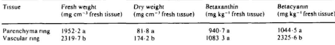
Vascular rings have higher dry and fresh weights than parenchyma rings per unit tissue volume (Table 1). Thus, when the uptake data is presented on the dry weight basis, the development of Rb+ and Na+ uptake capacity is nearly identical up to the third and fifth days,
respectively, in the vascular and parenchyma ring cells (Fig. 2). However, the decline of cation uptake capacity, which sets in after the maximum has been reached, results in significant differences in uptake capacities of the two tissues (Fig. 2). The degree of
PLATE 1. Autoradiograms of perpendicular (left) and longitudinal (right) cross sections of beet storage tissue showing sites of intense uptake of Rb+ and Na+. respectively, in the vascular rings.
T A B L E 1. Density and pigment content of parenchyma and vascular rings of unaged red beet storage tissue
Means in each column followed by different letters differ significantly at the 5% probability level.
Tissue Parenchyma nng Vascular ring Fresh weight (mg cm"1 fresh tissue) 1952-2 a 2319-7 b 1 . 5 * 1.0 u mo l * 0.5 m a. 3 m 2 0 Na -i ^ i i Dry weight (mg c m " ' fresh tissue) 8l-8a 174-2 b
H )
(
parenchyma ring*V
Y
• i i i i Betajcanthin (mg kg"1 fresh ussue) 940-7 a 1083 3 a vascular ' ring*x
\
Betacyamn (mgkg-1 fresh tissue) 1044-5 a 2325-6 b 2 4 S 8 10Duration o( ageing (day)
1 2 2 . 4 2.0 1 . 0 0 . 5 8 6 Rb vascularring* 1 0 1 2
Duration of ageing (day)
FIG. 2. Time course of development of Na+ and Rb+ uptake capacity in the tissues of vascular and
parenchyma rings of beet storage tissue. Values are means of 10 replicates; vertical lines represent ± I s.e.
heterogeneity in the development and decline of cation uptake capacity, expressed as percent coefficient of variability between discs, is presented in Fig. 3. Heterogeneity in the cation uptake capacity is very high in the freshly cut beet slices. This decreases with ageing and reaches a minimum by about the fifth day when uptake is at its maximum. Thereafter, parenchyma cells undergo an even greater heterogeneous decline in their cation uptake capacity, which results in a sharp increase in their coefficient of variability.
Respiration measurements during the ageing process showed that, on the dry weight basis, vascular ring cells consistently had lower rates. Variability between tissue samples was very
Mozafar and Oertli—Localized Uptake Sites in Red Beet
o 5 10 Duration of ageing (day)
FIG. 3. Heterogeneity in development and decline of Na+ uptake capacity in the vascular and
parenchyma ring tissues of beet storage tissue, expressed as the coefficient of variation (CV). from random tissue samples during the course of ageing.
- 50 4 0 3 0 j « 20 a 3 S 1 0 v a s c u l a r r i n g 0 2 4 6 8 10 12 Duration of ageing (day)
FIG. 4. Oxygen uptake by the vascular and parenchyma tissues of beet storage tissue as measured by polarography. Values are means of a minimum of four replicates; vertical lines represent + I s.e.
high, and differences between the vascular and parenchyma ring cells during the 12 d ageing period were not significant at the 5% level (Fig. 4). Comparison of the oxygen consumption and the development of ion uptake capacity reveals no apparent relationship between the two processes.
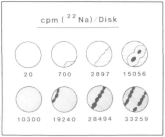
Vascular ring cells contain a significantly higher amount of betacyanin than parenchyma ring cells (Table 1) and therefore look dark red when lit from the back. So far, no physiological function has been attributed to betalain pigments (Piatelli, 1976). We do not wish to imply a direct cause and effect relation between the higher betacyanin content and the development of higher cation uptake capacities in the vascular ring cells. However, there seems to be some relation between the degree of retention of betacyanin pigments and that of cation uptake capacities. For example, during the decline phase of cation uptake capacity (from the fifth to twelfth day), some discs (but not all), become partially discoloured due to loss of their betacyanin. This discolouration, which first'becomes visible in the parenchyma ring cells, is accompanied by large variations in the tissue cation uptake capacity. Therefore, if larger discs of, for example, 12 mm diameter containing parts of both parenchyma and
2 2
c p m ( " N a ) D i s k
7 0 0 2 8 9 7 1 5056
o<z>®
1 0 3 0 0 1 9 2 4 0 2 8 4 9 4 3 3 2 5 9
FIG. 5. Sodium uptake by 12 mm diameter beet discs at the twelfth day of ageing. Shading intensities signify different extents of betacyanin retention. White areas denote complete loss of betacyanin from the tissue: solid areas indicate the presence of vascular bundles in the discs. Uptake took place between
the seventh and twelfth days of ageing (5 d).
vascular rings are used in uptake studies, a pronounced experimental (sample) variation can occur, especially if the discs are aged for more than 5 d (Fig. 5).
Diffusion is believed to be an important element in the ageing (washing) phenomenon, controlling the loss from the tissue of key regulatory compounds (Van Steveninck, 1975). The rate of diffusion may be different in parenchyma and vascular tissues because of dissimilar cell densities with different consequences for the course of the ageing process in the two tissues.
In conclusion, the lack of development of a constant level cation uptake capacity and the rapid increase in the heterogeneity in the rate of cation uptake by the parenchyma cells after the maximum capacity was reached warrant certain experimental precautions. These include experimental designs with short durations, use of parenchyma or vascular tissues of exactly the same age (preferably less than 5 d old), and expression of the uptake data on the dry weight basis. Furthermore, in cases where, due to experimental design, larger diameter discs containing both tissue types are required, care should be taken to select discs with the same ratio of vascular to parenchyma cells in order to minimize experimental error.
A C K N O W L E D G E M E N T
The financial support of the Swiss National Science Foundation (Grant No. 3.096-0.81) is greatly appreciated.
LITERATURE CITED
ARTSCHWAGER. E.. 1926. Anatomy of the vegetable organs of the sugar beet. Journal of Agricultural Research. 33, 143-76.
BRIGGS. G. E., HOPE, A. B.. and PITMAN, M. G., 1958. Exchangeable ions in beet discs at low temperature. Journal of Experimental Botany. 25. 128—41.
FRANCOIS, G., BOGEMANS. J.. and NEIRINCKX. L.. 1982. Undirectional sodium fluxes in red beet
storage tissue (Beta vulgaris L. cv. Platronde Egyptische): effects of the phytohormones indol-3yl-acetic acid and abscisic acid. Plant, Cell, and Environment. 5, 5-8.
HAYWARD, H., 1938. The structure of economic plants. MacMillan. New York. LEVI, I.. 1962. An artifact in plant autoradiography. Science. 137, 343—4.
PIATTELLI, M., 1976. Betalains. In Chemistry and biochemistry of plant pigments. Volume 1. Ed. T. W.
POOLE, R. J., 1976. Transport in cells of storage tissues. In Encyclopedia of plant physiology (New Series). Volume 2, Section A. Eds U. Liittge and M. G. Pitman. Springer-Verlag, Berlin. Pp. SUTCUFFE, J. F., 1957. The selective uptake of alkali cations by red beet tissue. Journal of
Experimental Botany, 8, 36—9.
VAN STEVENINCK, R. F. M., 1975. The 'washing' or 'ageing' phenomenon in plant tissues. Annual Review of Plant Physiology, 26, 237-58.
WILEY, R. C , and LEE, Y., 1978. Recovery of betalines from red beets by a diffusion extraction procedure. Journal of Food Science, 43, 1056-8.