Journal of Dairy Research (1989) 56, 603-611 Printed in Great Britain 6 0 3
Tryptic phosphopeptides from whole casein
I. Preparation and analysis by fast protein liquid chromatography BY MARCEL A. JUILLERAT, ROBERT BAECHLER,
RAPHAEL BERROCAL, SERGE CHANTON, JEAN-CLAUDE SCHERZ AND ROLF JOST
Nestle Research Centre, Nestec Ltd, Vers-chez-les-Blanc, CH-1000 Lausanne 26, Switzerland
(Received 29 April 1988 and accepted for publication 25 January 1989)
SUMMARY. Tryptic phosphopeptides were obtained from whole bovine casein by chromatography on the anion exchange resin QAE-Sephadex A 25. Salt gradient clution of the column allowed separation of non-phosphorylated peptidcs from phosphorylated species. The preparations obtained contained at least seven distinct phosphopeptides of which the following casein fragments were identified: asl(43-58):2P, asl(59-79): 5P, as2(46-70): 4P, /?(1-28):4P, /?(2-28):4P, and
/?(33-48): IP. Fast protein liquid chromatography (FPLC) on Mono Q HR 5/5 resin showed that the phosphopeptides were eluted in the same order as from the QAE-Sephadex resin. However, on the analytical column HR 5/5 the fragments asl(59-79): 5P and /?(2-28): 4P, having the same net charge under the conditions of
chromatography, co-eluted, whereas they were at least partly separated on the preparative column HR 16/10. Following enzymic dephosphorylation, the peptides eluted at lower salt strength in the gradient. FPLC on Mono Q resin thus permitted dephosphorylation to be monitored and intermediates between the parent species and the fully dephosphorylated peptide to be identified.
Casein phosphopeptides have interesting physicochemical properties such as the formation of soluble complexes with bi- or trivalent metals (Oesterberg, 1966; Manson & Cannon, 1978), or the stabilization of Ca phosphate solutions against precipitation of tricalcium phosphate (Reeves & Latour, 1958). This latter property led to speculation that phosphopeptides might favourably influence intestinal Ca solubility (Naito el al. 1972; Lee el al. 1980). To enable their properties to be investigated in more detail, procedures by which casein phosphopeptides can be prepared in large amounts from whole cows' milk casein are necessary. Such procedures must include a selective process by which phosphorylated peptidcs are preferentially extracted from a casein hydrolysate. An interesting possible way of achieving this is offered by the occurrence of Ca-induced aggregation of phos-phorylated species in a pancreatic hydrolysate of casein. Non-phosphos-phorylated peptides which do not participate in the aggregation can be removed by ultrafiltration/diafiltration (Brule el al. 1982). An alternative procedure is discussed in this paper. Reversible binding to an ion exchange resin allows easy removal of most non-phosphorylated species and subsequent stepwise elution of phospho-peptides. Fast protein liquid chromatography (FPLC) on Mono Q resin was used to monitor such a preparative ion exchange process.
MATERIALS AND METHODS
Na caseinate was prepared from skimmed, unhcatcd bulk milk. Crystalline porcine trypsin (EC 3 . 4 . 2 1 . 4 ) was obtained from Novo Industri A/S, Bagsvaerd, Denmark. It had a strength of 2800 USP units/mg. Acid phosphatase from potato (EC 3 . 1 . 3 . 2 ) , grade 11, was purchased from Boehringer Mannheim GmbH, FRG, and QAE-Sephadex A 25, capacity 3 mequiv./g, was a product of Pharmacia LKB Biotechnology, S-751 82, Uppsala, Sweden.
Analytical procedures Dry mineralization for ash content and cations
Lyophilizcd samples (~ 1 g, exact weight known) were burned for 3 h in a Biichi 430 digester (Biichi AG, CH-9230 Flawil, Switzerland) followed by overnight min-eralization in a Lindborg oven at 550 °C. After cooling and backweighing. the ash was dissolved in hot 2 M-HCI (3 ml) and the volume of each solution adjusted to 50 ml
with distilled water.
Wet mineralization for determination of P
Samples (0-1-0-2 g powder) were heated with concentrated H2SO4 (10 ml), K2SO4
(1-7 g) and HgO (0-08 g) for 4 h in Kjeldahl tubes in a Biichi 425 digestion apparatus. The mineralized samples were adjusted to 50 ml with distilled water.
Atomic absorption spectroscopy
This was performed on a Perkin Elmer 603 instrument (Porkin Elmer, Kiisnacht, Switzerland). The following diluents were used: 1% (w/v) lanthanc in H2O for Ca and Mg; 2 M-HCI for Na and K.
Total and free P
Wet ashed samples were subjected to the Fiske-Subbarow procedure for colorimctric P determination (Fiske & Subbarow, 1925). Inorganic phosphorus (P,) was measured by ion chromatography. The peptides, 50 mg/ml of dry solids in distilled water, were filtered through a Millipore 0-45 fim membrane (Milliporc AG, Kloten, Switzerland) and were injected into a Varian chromatograph (Varian International AG, Basle, Switzerland) fitted with an ORH 801 organic acids column purchased from Interaction Chemicals Inc., Basle, Switzerland. The eluant used was 0-01 M-HCI at a flow rate of 0-3 ml/min and ions were detected by conductimetry.
Procedure for producing phosphopeptides from Na caseinate
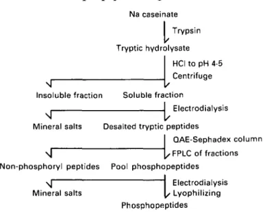
The procedure is shown schematically in Fig. 1 and described in detail below.
Step J, limited hydrolysis of Na caseinate. Na caseinate (5 kg) was dispersed in
dcmincralizcd water (78 kg) and the dispersion stirred at 25 °C. Casein was
precipitated by addition of 2 M-HCI (~1\5 1) at pH 4 5 and was separated by
centrifugation. It was redispersed in demineralized water (60 kg), with the aid of a polytron. The pH was raised to 8-0 by addition of 2 M-NaOH with stirring. The dispersion was poured into a double-walled hydrolysis reactor equipped with a pH-stat and heated to 30 °C. The pH-pH-stat unit was equipped with a burette containing 2 M-NaOH as titrant and was set to maintain the pH at 7-9. Hydrolysis was initiated by addition of crystalline trypsin (150 g), predispersed in 0-01 M-HCI, and was allowed to proceed for exactly 1 h, after which the pH was lowered to 4-5 by addition of 2 M-HCI (3-1 1). Flocculation was completed on standing at ambient temperature
Phosphopeptides separation 605
Na caseinate Trypsin Tryptic hydrolysate HCI to pH 4-5 Centrifuge Insoluble fraction Soluble fractionL
Electrodialysis Mineral salts Desalted tryptic peptidesQAE-Sephadex column
l;
\ | [, FPLC of fractions Non-phosphoryl peptides Pool phosphopeptides
\J Electrodialysis Mineral salts [, Lyophilizing
Phosphopeptides
Fig. 1. Flow sheet for the preparation of tryptic phosphopeptides from whole bovine casein.
for 2 h. The flocculated material was then removed by centrifugation and a clear peptide solution (76 kg) having a total solids content of 4-5% (w/v) was obtained.
Step 2, desalting by electrodialysis. The peptide solution was electrodialysed in an
Ionics Stackpack pilot unit (Ionics Inc., Watertown, USA) fitted with cationic membranes, type 61-AZL-386, and anionic membranes, type 103-QZL-386. The total
membrane area was 4180 cm2. Electrolyte used in the concentration compartment
was 0-01 M-KCI (initial concentration) and in the electrode compartment was 01
M-Na2SO4 adjusted to pH 25 with cone. H2SO4. The voltage applied was 55 V at a
maximum current of 9-5 A. The flow rate was 1-5-041/min. Progress of de-mineralization was monitored continuously by a Radiometer CDM 80 conductimeter equipped with a CDC 114 flow cell, both from Radiometer A/S, Copenhagen, Denmark.
Step 3, chromatography on QAE-Sephadex. QAE-Sephadex A 25 (400 g) was
equilibrated in 0-05 M-phosphate buffer, pH 6-5, and the slurry poured into a Pharmacia LKB chromatography column, type K100/45. The column was washed with the same buffer containing 20 mg chlorhexidine/1 at 500 ml/h for 48 h at 10-15 °C.
A 10% solution (10 kg) of the demineralized peptide solution was loaded on to the column at pH 4-5 with a flow rate of 1-15 1/h, after which demineralized water (20 kg) was pumped through the column to complete adsorption. Peptide fractions were obtained by stepwisc elution with 0-1 M-KCI (17 1) followed by 0-2 M-KCI (2-1 1) and 0-5 M-KCI (8-2 1). The eluatc was continuously monitored for absorbance at 214 and 280 nm. During each elution step, numerous fractions were assayed by FPLC. The first phosphopeptides emerged from the column very shortly after application of
the 0-5 M-KCI buffer had started. The peptides were eluted from the column in
groups, enabling us to collect fractions containing one or two peptide species. When the last phosphopeptides had emerged from the column, material remaining on the column was removed by elution with 1 M-NaCl. A pool of all phosphopeptide-containing fractions was created (Fig. 2, profile B).
Step 4, demineralization by electrodialysis. The pooled fractions were
606 0 2 4 6 8 10 12 14 16 18 20 007 005 003 001 -007 005 -003 } L 001 0 2 4 6 8 10 12 14 16 18 20 0 2 4 6 8 10 12 14 16 18 20 -007 •0-05 • 0 0 3 001
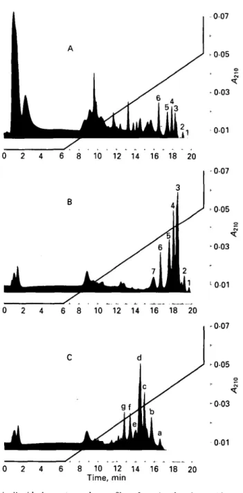
Fig. 2. Fast protein liquid chromatography profiles of casein phosphopeptides on Mono Q resin, column HR 5/5; other conditions described in the Materials and Methods section. Profile A, pH 4-5-soluble fraction of tryptic digest of Na caseinate; profile 13, phosphopeptide preparation as obtained by the procedure outlined in Fig. 1; profile C, preparation as in profile B following dephosphorylation with potato acid phosphatase.
was concentrated under reduced pressure and lyophilized. A clean white powder (120 g) (Table 1) was obtained.
FPLC Mono Q resin
FPLC was performed using equipment obtained from Pharmacia LKB which contained either an HR 5/5 column (analytical scale) or a 16/10 column (preparative scale). For analytical chromatography two solutions were used. Solution A consisted
Phosphopeptides separation 607
Table 1. Percentage composition of casein phosphopeptide preparations produced by
ion exchange chromatography on QAE-Sephadex Product Na casemate Preparation I Preparation 11 Preparation 111 N 14-7 11-2 111 120 ND, P 0-72 2-8 3-2 3 0 Not P. <001 <002 <002 <001 Ash 3-5 16-9 19-4 12-5 determined. Na ND 6-2 5 0 0-4 K ND ND ND 4-7 Ca ND 005 005 0-05
of 0-02 M-Tris-HCl buffer (pH 8-0) and solution B which contained 1 M-NaCl in the same buffer. These were applied to the column in the following sequence. From zero time to 6 min, solution A was applied. This was followed from 7 to 21 min by a linear gradient of J3 in A up to 50% (v/v) of solution B and from 22 to 25 min by solution B and finally from 26 to 30 min by solution A. The flow rate was 1 ml/min and detection was based on absorbance at 214 nm. Sample load was 1-2 mg dry solids/run.
For preparative chromatography, 0-02 M-Na borate buffer, pH 8-0, was used in place of Tris buffer to allow ninhydrin detection of peptides. The flow rate was 5 ml/min and the sample load was 30 mg/run. The gradient of NaCl was developed within 65 min after which the salt concentration was 0-5 M (50% solution B in A). Fractions of 2 ml were collected and analysed for peptide content by reaction with ninhydrin and for P content by the Fiske-Subbarow analysis of wct-ashod samples. Following the establishment of a complete peptide profile, pure fractions were pooled and desalted on Sephadex G-10 in 5 % (w/v) acetic acid as solvent. Desalted peptides were concentrated in vacuo and lyophilized. The purity of the isolates was assayed by analytical FPLC. Following purity assessment, amino acid composition of the isolates was established after total acid hydrolysis under standard conditions.
Dephosphorylation by potato acid phosphatase
Each isolate (50-100 mg portions) was dissolved individually in 0-05 M-Na
acetate buffer, pH 5-5. The peptides were dephosphorylated by treatment with
phosphatase at a concentration of 0-l mg enzyme/ml for 1 h at 37 °C, after which the reaction was stopped by heating briefly in a boiling water bath. After cooling, the peptide solution was adjusted to pH 8-0 using 1-0 M-Tris solution. The peptides were then examined by FPLC to assess the extent of dephosphorylation.
RESULTS Characterization of the peptides
The peptide preparation (Fig. 1) had an average P content of 3 % based on total dry solids which represents a 4-2-fold average enrichment over the starting material when the P content of Na caseinate is taken as 0-8%. As indicated by mineral analysis (Table 1), the peptides were present in the form of a potassium salt, KC1 having been used in the desorption step. A typical yield of the ion exchange procedure was 120 g phosphopeptides from 1 kg desalted acid-soluble tryptic peptides.
Chromatography on Mono Q resin (Fig. 2, profile B) showed a selective increase of strongly adsorbed peptides and depletion of peptides eluting at low salt strength;
Tabl e 2 . Amino acid composition of phosphopeptides isolated. Experimental values are shown as molar equivalents of amino acids with theoretical values for the assigned peptide, where known, given in parentheses © 0 Pea k n o Asparti c aci d Threonin e Serin e Glutami c aci d Pr o lin e Glycin e Alanin e Cystin e Valin e Methionin e Isoleucin e Leucin e Tyrosin e Phenylalanin e Lysin e Histidin e Arginin e L residue s Assignmen t P 6 2-1 (2 ) 1-5(1 ) 1-2(1 ) 7-4 (9 ) - (0 ) (0 ) -(0 ) — (0 ) — (0 ) (0 ) 0-6 (0 ) 0-9(1 ) — (0 ) 0-7(1 ) 1-0(1 ) --(0 ) - (0 ) 15-4 (16 ) A(33-48 ) P 5 3-0 (3 ) 1-4(1 ) 2-2 (2 ) 4-8 (4 ) -(0 ) 1-0(1 ) 1-1 (1 ) -(0 ) 0-6 (0 ) 1-0(1 ) 2-3 (2 ) -(0 ) 0-6 (0 ) — (0 ) 1-3(1 ) -(0 ) -(0 ) 19-3 (16 ) asl (43-58 ) P5 ' 2 0 0-9 3 0 5-3 0-6 0-9 0-6 — 13 0-5 17 01 — — 10 -- 0-6 19-4 ? P 4 1-6(2 ) 1-4(1 ) 5-3 (5 ) 8-4 (7 ) 1 1 (1 ) 0-9(1 ) (0 ) — (0 ) 2-4 (2 ) 0-6 (0 ) 2-5 (3 ) 2-5 (3 ) — (0 ) - (0 ) 0-8(1 ) "(0 ) 1-5(2 ) 2 9 (28 ) A C -28 ) P 3 1-7(2 ) 0-9(1 ) 5-3 (5 ) 9-4 (7 ) 1-4(1 ) 0-9(1 ) 0-7 (0 ) — (0 ) 2-5 (2 ) 0-6 (0 ) 2-4 (3 ) 2-4 (3 ) -(0 ) - (0 ) 0-7(1 ) -(0 ) 0-9(1 ) 29-8 (27 ) AC-25 ) - . No t detected . P3 ' 1-4(1 ) (0 ) 4-4 (5 ) 7-9 (7 ) 1-2(1 ) (0 ) 1-4(1 ) (0 ) 2-2 (2 ) 1-0(1 ) 2-1 (2 ) -(0 ) - (0 ) (0 ) 1-3(1 ) (0 ) - (0 ) 22-9 (21 ) asl (59-79 ) P3 " 3 0 10 7-8 14-5 1 6 12 3-3 3-6 1-2 3 1 - - 0-7 2 1 43 1 ? P 2 2-0 (2 ) 1-0(1 ) 4-5 (5 ) 7-2 (8 ) (0 ) 0-9(1 ) 2-5 (3 ) - (0 ) 2-1 (2 ) (0 ) 1-5(1 ) — (0 ) 1-7(1 ) - (0 ) 1 1 (1 ) (0 ) --(0 ) 24-5 (25 ) as2 (46-70 ) P I 1-9 0-9 4-3 5-2 0-6 2-8 2 0 1-3 1-2 0-5 1 1 1-0 - 22-i ? o H Tabl e 3 . Pattern of dephosphorylation indicated by fast protein liquid chromatography analysis Retentio n time , min , o n H R 5/ 5 Pea k HR16/1 0 2 3' 3 4 5' 5 6 Peptid e assigne d a s as2 (46-70 ) as l (59-79 ) AC-25 ) AC-28 ) ? as l (43-58 ) A(33-48 ) Ser-P/mo l (theory ) 4 5 4 4 2 1 Initia l p e 19-2 18-5 18-4 18 0 17-6 17-5 16-6 Initia l peptid e Product s (pea k height ) 19-2 (±) ; 18-4 ( + + +) ; 17-4(++) ; 15-7 ( + ) 18-5 (±) ; 18-1 (++) ; 17-5(++) ; 16-4 ( + ) ; 15-8 ( + ) 18-4 (-) ; 17-4 (±) ; 1 6 2 ( + +) ; 14-8 ( + ) ; 14-5 ( + ) 18-0 (-) ; 16-2 (±) ; 15-3 ( + ) ; 14-6 ( + +) ; 13-6 (++ ) 17-6 (±) ; 15-6 ( + +) ; 12-0 ( + ) 17-5 (—) : 16-7 ( + ) ; 14i)( + + + ) 16-6 ( + ) ; 15-1 ( + + + )
Phosphopeptides separation 609
Table 4. Assignment and properties of tryptic phosphopeptides from whole casein
Peak on HR 5/5 1 2 3 4 5 6 HR 16/10 1 2 3 " 3' 3 4 5' 5 6 Assignment Not identified «xs2(46-70) Not identified a8l (59-79) £(1-25) ? £(1-28) Not identified asl (43-58) £(33-48) Calculated net charge at pH 7 - 1 1 - 9 - 9 - 8 - 7 - 6 Calculated P content, % 41 5-7 3 9 3-6 3-2 1-5 Calculated MT 3009 2721 3123 3469 1928 2062
the latter were well represented in the acid extract of the tryptic digest (Fig. 2. profile A). In the profile B, seven distinct peptide fractions were eluted in the concentration range > 0-3 < 0-5 M of the linear NaCl gradient.
Following treatment with potato acid phosphatase, the peptides showed strikingly different retention characteristics (Fig. 2, profile C) due to the loss of negative net charge. As one might expect, the partly or completely dephosphorylated peptides were eluted at lower salt concentrations.
Identification of individual phosphorylated species
The peptide profile obtained from the preparative column HR 16/10 was very similar to that from the analytical column, but the larger column provided additional separation of phosphopeptides. Thus, peak 3 from the smaller column gave rise to peaks 3, 3' and 3", and peak 5 was split into 5 and 5'. For six out of nine fractions, amino acid composition allowed the assignment of the isolate to a particular tryptic casein fragment (Table 2). This tentative identification was further supported by clectrophoretic mobility at pH 2 in high-voltage paper electrophoresis of the isolate (data not shown). Additional support for our assignment of a given fragment was obtained from the dephosphorylation pattern observed after incubation of the peptides with phosphatase. Enzymic dephosphorylation leading to the loss of one negative charge for each of the phosphate groups removed resulted in lower retention on the Mono Q column.
Thus, characteristic dephosphorylation patterns were obtained with each of the fractions (Table 3). The number of peptide peaks observed was apparently related to the number of phosphoserine residues of the phosphopeptide in question. This study indicated that dephosphorylation had occurred stepwise and resulted in the formation of different intermediate products representing all possible stages of dephosphorylation.
The main characteristics of phosphopeptides so far identified are compiled in Table 4. The strength of binding to Mono Q resin increased with the net negative charge of the peptide at the pH of chromatography. According to this rule, the phosphopeptides with the lowest net negative charge should elute first in the salt gradient and those with the highest net charge last. The analytical column (HR 5/5) is capable of separating phosphopeptides _which differ by one unit of net charge, whereas separation of different peptides having an identical net charge does not occur. This is seen with fragments asl(59-79) and /?(2-28). On the preparative
Some expected tryptic phosphopeptides such as the fragment as2(l-21): 4P were
not found and appear to be missing in our preparation. According to its net charge of —6, this peptide should have been eluted near to, or with, phosphopeptide 6. It is not understood why this large and highly phosphorylated fragment could not be identified (e.g. on the basis of its histidine content). Three smaller peptides with a low degree of phosphorylation, asl(106-119): IP, as2(126-136): 2P, as2(137 149): IP,
were likewise missing. It is possible that these peptides were associated with the pH 4-5 insoluble fraction of the tryptic digest. It is also conceivable that, owing to their relatively low net negative charge, they co-eluted with acid non-phosphoryl-ated peptides from the ion exchange column and escaped our attention for this reason.
DISCUSSION
Tryptic cleavage of pure asl-casein has been shown to yield three phosphorylated
peptides which are soluble at pH 4-5 (Grosclaude et al. 1970). These peptides were fragments 42-79, 43-58 and 59-79. Tryptic cleavage of sodium cascinate yielded fragments 43-58 (peptide 5), and 59-79 (peptide 3') of asl-casein both of which arc
soluble at pH 4-5. The overlapping large peptide 42(43?)—79 observed by Grosclaude
et al. (1970) was missing in our preparation.
In the case of /?-casein, tryptic cleavage of the purified protein has been reported to give fragment 1-25 (Manson & Annan, 1971), while our peptide 4 had an amino acid composition compatible with the first 28 residues from the N-terminus of ft-casein. Peptide 3 appears to be residues 2-28 of the same protein, based on its amino acid composition and the higher negative net charge ( — 9). However, this implies the tryptic cleavage of N-terminal arginine, which is at least surprising and not in agreement with the strict endopeptidase character of the trypsin. As peptides 3 and 3' are incompletely separated on the resin, we may assume contamination of fraction 3 with peptide 3'. This would suggest that peptide 3 does not contain lysine on its own but that the lysine measured results from the contamination from 3' and the same would be true for the small amount of methionine found in 3.
The most likely assignment for peptide 3 would then be ft (1-25), instead of 2 28. The missing arginine residue is the problem for the definitive assignment as ft (1-25). With some reservation, tryptic hydrolysis of whole casein would thus produce a mixture of/? (1-28) and ft (1-25). Fragment 33-48, described by Ribadcau-Dumas
et al. (1971), corresponds to peptide 6 of our preparation.
The order of elution of tryptic phosphopeptides from Mono Q resin was the same as that obtained with the QAE-Sephadex column. Since the elution of phos-phopeptides from the ion exchange column was monitored by FPLC, it was possible to collect fractions with a single phosphopeptide species or with mixtures of two peptides only. By adequate manipulation of the salt gradient, it should be possible to isolate pure phosphopeptides in gram quantities. By using a highly specific enzyme such as trypsin and a short digestion time, the cleavage of the casein substrate was limited to the most sensitive tryptic cleavage sites. This avoided the formation of peptides too small to be separated from excess salt. Apparently desalting was not a problem with tryptic phosphopeptides, when electrodialysis was used for this purpose, as may be seen from the negligible losses in N and P.
Peptide 3 was subjected to three steps of Edman degradation and the following sequence was observed: Glu.Leu.Glu. No N-terminal Arg could be detected. The most likely assignment for peptide 3 would then be ft (2-25). The exopeptidase activity of our trypsin preparation is surprising but could explain our results.
Phosphopeptides separation 611
We gratefully acknowledge the able technical assistance of Blaise Pavillard in P and mineral analysis.
REFERENCES
BRULE, G., ROGER, L., FAUQUANT, J, & PIOT, M. 1982 Phosphopeptides from casein based material. United States Patent 4358405
FISKG, C. H. & SUBBAROW, Y. 1925 The eolorimetric determination of phosphorus. Journal of Biological Chemistry 66 375-400
GROSCLAUDB, F\, MERCIER, J. C. & RIBADEAU-DUMAS, B. 1970 [Primary structure of bovine asl-casein. Location of tryptic peptides in fragments obtained by tryptic hydrolysis of maleyl casein. | European Journal of Biochemistry 14 98-107
LEE, Y. S., NOGUCHI, T. & NAITO, H. 1980 Phosphopeptides and soluble calcium in the small intestine of rats given a casein diet. British Journal of Nutrition 43 457-467
MANSON, \V. & ANNAN, W. D. 1971 The structure of a phosphopeptide derived from /?-casein. Archires of Biochemistry and Biophysics 145 16-26
MANSON, \V. & CANNON, J. 1978 The reaction of as l-and /?-casein with ferrous ions in the presence of oxygen. Journal of Dairy Hesearch 45 59-67
NAITO, H., KAWAKAMI, A. & IMAMURA, T. 1972 In vivo formation of phosphopeptide with calcium-binding property in the small intestinal tract of the rat fed on casein. Agricultural and. Biological Chemistry 36 409-415 OESTBRBERO, R. 1966 Phosphorylated peptides, study of their primary structure and metal complexity.
Thesis. Uppsala: Almqvist and Wiksell
REBVBS, R. E. & LATOUR, N. G. 1958 Calcium phosphate sequestering phosphopeptide from casein. Science
128 472
RIBADEAU-DL'MAS, R , GROSCLAUDE, F. & MERCIER, J. C. 1971 [Primary structure of bovine /?-casein.| European Journal of Biochemistry 18 252-257
![Amber-green light-emitting diodes using order-disorder Al[subscript x]In[subscript 1−x]P heterostructures](data:image/gif;base64,R0lGODlhAQABAIAAAP///wAAACH5BAEAAAAALAAAAAABAAEAAAICRAEAOw==)