Advance Access publication 2 January 2014
Colour improvement and stability of white spot lesions following
infiltration, micro-abrasion, or fluoride treatments in vitro
Enver Yetkiner*, Florian Wegehaupt**, Annette Wiegand***, Rengin Attin**** and
Thomas Attin**
*Department of Orthodontics, University of Ege, Izmir, Turkey, **Clinic for Preventive Dentistry, Periodontology and Cariology, University of Zurich, Switzerland, ***Department of Preventive Dentistry, Periodontology and Cariology, University of Göttingen, Germany and ****Clinic for Orthodontics and Pediatric Dentistry, University of Zurich, Switzerland
Correspondence to: Enver Yetkiner, Faculty of Dentistry, Department of Orthodontics, University of Ege, 35100,
Bornova, Izmir, Turkey. E-mail: eyetkiner@hotmail.com
summary
BACkGROUnD/OBjECTIvES: White spot lesions (WSLs) are unwelcome side effects of fixed appliances that
compromise the treatment outcome. Recently, infiltration of WSLs has been introduced as a viable treat-ment alternative. The objective was to evaluate the colour improvetreat-ment of WSLs and their stability against discolouration following infiltration, fluoride, or micro-abrasion treatments in vitro.
MATERIALS/METhODS: Artificial WSLs were created in bovine enamel (N = 96) using acidic buffer solution
(ph 5, 10 days) and were randomly allocated to four groups. Specimens were treated with infiltration (Icon, DMG), fluoride (Elmex Caries Protection, GABA), and micro-abrasion (Opalustre, Ultradent) or remained untreated (control). Groups were discoloured for 24 hours in tea or tea + citric acid. Colour components and visible colour change (L*, a*, b*, ΔE) were measured spectrophotometrically on follow-ing time points: baseline, after WSL formation, after treatment, and durfollow-ing discolouration (8, 16, and 24 hours). Data were analysed using kruskal–Wallis and Mann–Whitney tests.
RESULTS: WSL formation increased (L*) in all groups. Only infiltration reduced this effect to baseline.
highest ΔE improvement was obtained by infiltration and micro-abrasion followed by fluoride. This improvement was stable only for infiltration during discolouration. L*, a*, and b* changed significantly during discolouration in all groups except infiltration. Within the same treatment group, discolouration solutions did not differ significantly.
LIMITATIOnS: In vitro testing cannot replicate the actual mode of colour improvement or stability but can be
used for ranking materials and techniques.
COnCLUSIOnS/IMPLICATIOnS: Infiltration and micro-abrasion treatments were capable of diminishing the
whitish appearance of WSLs. Only infiltrated WSLs were stable following discolouration challenge.
Introduction
Subsurface enamel demineralizations are known as white spot lesions (WSLs), and they represent the early phase of caries formation (Derks et al., 2004; Bergstrand and Twetman, 2011). Prevalence of WSLs is relatively high, affecting more than 25 per cent of the patients receiving orthodontic treatment, acquiring at least one new lesion during treatment (Hadler-Olsen et al., 2012; Lucchese and Gherlone, 2013). Demineralization may take place rapidly, as fast as within 4 weeks after the placement of brackets and can stay present even years after treatment (Bergstrand and Twetman, 2011). Clinically, surfaces are intact when gently probed in early phases. However, cavi-tation may occur if the cariogenic challenge is ongoing, which might lead to the necessity of invasive restorative treatments (Derks et al., 2004; Bergstrand and Twetman, 2011).
As light refraction through enamel is directly related to the level of mineralization, WSLs manifest themselves as white opacities visually (Derks et al., 2004; Bergstrand and Twetman, 2011). The most superficial layer is richer in cal-cium, and the inner structure is more porous due to mineral loss. In the presence of cariogenic environment, deminerali-zation progresses and the appearance may get more opaque (Derks et al., 2004; Bergstrand and Twetman, 2011). WSLs might become even more perceptible when extrinsic stain-ing occurs, which may compromise the aesthetic outcome of orthodontic treatment (Addy and Moran, 1995; Watts and Addy, 2001).
Although there is no golden standard for WSL treatment, three treatment modalities are more frequently preferred, depending on the degree and activity level of the lesion. Because WSL is a form of demineralization, remineraliza-tion is the most conservative method to be tried primarily
(Derks et al., 2004; Bergstrand and Twetman, 2011; Hamdan
et al., 2012). Remineralization can be obtained through increasing the calcium and phosphate content in the oral environment and forming more stable compounds such as calcium fluoride (Marinho et al., 2003; Beerens et al., 2010; Hamdan et al., 2012). Numerous studies have been performed aiming at the regression of early demineraliza-tions by means of fluoride application. It was reported that regular topical fluoride application is an efficient method for preventing and remineralizing early enamel caries (Marinho
et al., 2003; Beerens et al., 2010). Low concentrations of fluoride application have been advocated in order not to hyper-mineralize the outer surface, which might obstruct further remineralization of deeper enamel lesions (Marinho
et al., 2003; Hamdan et al., 2012). Another treatment option is micro-abrasion, performed either by using hydrochloric acid (HCl) containing abrasive slurry or abrasive powders applied with high-pressurized air (Murphy et al., 2007;
Neuhaus et al., 2010). Micro-abrasion mainly aims to remove the discoloured enamel mechanically. The prompt improvement in the appearance of the lesion and clinical results has made this technique a feasible treatment option (Murphy et al., 2007; Neuhaus et al., 2010; Pliska et al., 2012). Because micro-abrasion is comparably more inva-sive in nature, delayed application was thought to be benefi-cial considering the spontaneous improvements of the lesion via saliva-based remineralization and spontaneous surface
abrasion following debonding (Bergstrand and Twetman,
2011; Hamdan et al., 2012). More recently, a minimally invasive treatment approach was introduced, in which the WSL is infiltrated using a low-viscosity resin (Paris et al., 2007a,b; Kielbassa et al., 2009). In this technique, the outer surface is transformed into a more permeable layer with the help of HCl etching, and the porous structure beneath is infiltrated using a triethyleneglycol dimethacrylate-based resin (Paris et al., 2007a,b; Kielbassa et al., 2009). It is note-worthy that this resin has a light refraction index similar to sound enamel, which improves the appearance of the lesion besides reinforcing the weakened enamel prism structure (Paris et al., 2007b; Kielbassa et al., 2009).
Assessment of the therapeutic effect provided by these methods has been made under various settings. Micro-abrasion (Murphy et al., 2007; Neuhaus et al., 2010; Pliska
et al., 2012) and infiltration (Kielbassa et al., 2009; Torres
et al., 2011; Hammad et al., 2012; Kim et al., 2013; Paris
et al., 2013; Soviero et al., 2013) methods were reported to produce satisfactory results compared with remineralization using fluoride or amorphous calcium phosphate derivatives. However, to the authors’ best knowledge, comparison of the colour outcome achieved by micro-abrasion, infiltration, and fluoride remineralization has not been made previously. Furthermore, no information is available on how these treated surfaces will show resistance against discolouration that might also be subjected to extrinsic discolouration later.
Therefore, the aims of this study were 1. to compare the colour-masking effect of infiltration, fluoride reminer-alization, and micro-abrasion treatments of WSLs and 2. to compare the resistance of these treated surfaces against discolouration. The null hypotheses tested were that 1. the WSL treatment modalities would not produce differences in colour masking compared with non-treated WSLs and that 2. the resistance of treated WSLs would not be better than non-treated WSLs against discolouration.
Materials and methods Study design
Artificial WSLs created on bovine enamel (N = 96, n = 12 per test group) were treated forming the following groups: infiltration (Icon; DMG, Hamburg, Germany), fluoride rem-ineralization (Elmex Caries Protection; GABA, Therwil, Switzerland), micro-abrasion (Opalustre; Ultradent, Utah, USA), and control (remain untreated). All specimens were subjected to discolouration in tea or tea + citric acid solu-tions for 24 hours. Colour components were measured at baseline (prior to demineralization), after WSL formation, after treatment, and after 8-, 16-, and 24-hour discoloura-tion. Chemical compositions and respective manufacturer information of the materials are summarized in Table 1.
Table 1 Composition of the low-viscosity caries infiltrant, enamel micro-abrasion slurry, and the fluoride rinse according to the manufacturers’ information.
Product Chemical composition Manufacturer Icon TEGDMA-based resin matrix,
Initiators—additives
DMG, Hamburg, Germany, Batch no. 634902 Opalustre Hydrochloric acid 6%; Silicon carbide <45% Ultradent Products, Inc., South Jordan, Utah, USA,
Batch no. B6JHJ Elmex Aqua, PEG-40 hydrogenated castor oil, olaflur
(Amine fluoride 100 ppm F-), aroma, potassium acesulfame, sodium fluoride (150 ppm F-), polyaminopropyl biguanide, hydrochloric acid
GABA International AG, Therwil, Switzerland, Batch no. 10073018
Specimen preparation
Bovine incisors stored in 0.5 per cent chloramine solution at 4°C no longer than 6 months were initially cut from their roots. Four discs of enamel with a diameter of 3 mm were cut from the labial aspect of each tooth using a
custom-made diamond-coated trephine bur (80 μm, Intensiv SA,
Lugano-Grancia, Switzerland). The discs were then flat-tened from the bottom to approximately 3 mm in height (Struers, Birmensdorf, Switzerland). Each piece was ran-domly assigned to four groups assuring equal distribution of incisal and gingival sections per group. They were embed-ded with their labial surfaces exposed in auto-polymeriz-ing acrylic resin (Palapress; Heraeus Kulzer, Wehrheim, Germany) in cylindrical moulds (6 mm diameter and 3 mm thickness). Embedded specimens were ground flat and pol-ished with water-cooled carborundum discs (1200, 2400, and 4000 grit; Struers, Erkrat, Germany) and stored in tap water until demineralization.
Demineralization procedure
Demineralization was achieved by immersing the specimens in acidic buffer solution (pH 5, 37°C, 10 days) following the formulation given by Buskes et al. (1985). The solution was renewed each second day to keep the pH constant.
WSL treatments
Following demineralization, the specimens were treated forming the following groups:
1. Infiltration: 15 per cent HCl (Icon etch; DMG, Hamburg, Germany) was applied for 120 seconds. Substrates were rinsed with water for 30 seconds and air-dried. They were treated with 99 per cent ethanol (Icon Dry) for 30 seconds and air-dried. Infiltrant (Icon) was applied in one coat with a micro-brush, let set for 180 seconds, light-cured for 60 seconds, a second layer was applied, let set for 60 seconds, and light-cured for 40 seconds. The surfaces were polished with fine and superfine alu-minium oxide discs for 20 seconds each (Sof-Lex; 3M, Neuss, Germany).
2. Fluoride: Specimens were immersed in 2 ml of fluoride solution (Elmex Caries Protection) for 1 minute daily for 30 days. In between, they were stored in artificial saliva as described below.
3. Micro-abrasion: Specimens were treated with rubber polishing cups (Produits Dentaires; Vevey, Switzerland) using HCl containing abrasive slurry (Opalustre) at 300 rpm for 1 minute. The surfaces were polished with rubber cups afterwards for 20 seconds.
4. Control: Specimens remained untreated.
All specimens were stored in artificial saliva (3 speci-men/25 ml, 37°C) under dark conditions for 30 days prior to discolouration. Artificial saliva was prepared according to
the formula given by Klimek et al. (1982) and was renewed every 2 days. Measured pH ranged between 6.5 and 6.8.
Discolouration procedure
Black tea and black tea + citric acid were used as discol-ouration solutions. Black tea was prepared by steeping of 5 teabags (3.125 g/bag, extra strong, Marks and Spencer, Chester, UK) in 1 l of boiling distilled water for 10 minutes. Following this, the tea bags were removed, and the solution was left for cooling. Finally, the pH was measured, which ranged between 4.2 and 4.4. For the preparation of tea + cit-ric acid, the same procedure was repeated, and 0.1 M citcit-ric acid was added until pH 4.0 was obtained.
During discolouration, specimens were fixed to the base of boxes containing the discolouration solution using mould-ing dough (Plastilin; Pelikan, Hannover, Germany). The boxes were then placed in a water bath (3 specimens/25 ml, 37°C, under constant motion). The solution was renewed every 8 hours.
Colour measurements
Colour measurements were assessed at standardized ambi-ent conditions using a spectrophotometer (CM-2600d, Konica Minolta; Osaka, Japan), which was set to standard illuminant D65, 3 mm reading area and 6 mm lighting area. Observer angle was set to 2 degree, and specular component was included. Colour and spectral distributions were meas-ured according to Commission International de l’Eclariage (CIE) L*a*b* system (CIE Colorimetry Publication, 1986), using Spectra Magic NX Version 1.9 colour data software (Konica Minolta, Osaka, Japan). The L* axis represents the degree of lightness within a sample and ranges from 0 (black) to 100 (white). The a* value is the red/green axis where an increase indicates a higher red colour component. The b* value is the yellow/blue axis where an increase indi-cates higher yellow colour. The visible colour change (ΔE) was calculated as follows (CIE Colorimetry Publication, 1986):
∆E L1 L2 (a1 a )2 2 b b
1 2
2 1 2
=[( *− *)2+ *− * +( *− *) ]/ ΔE was accepted clinically detectable when it exceeded 3.7 units (Johnston and Kao, 1989). The spectrophotometer was calibrated before each measurement. Prior to each colour measurement, the specimens were taken out from the rel-evant storage solution regarding the time point, and 300 brushing strokes (Paro M43; medium bristle stiffness, Esro, Kilchberg, Switzerland) were administered in order to elim-inate the superficial staining (Attin et al., 2003) using a two-axis brushing machine (Willytec, Feldkirchen-Westerham,
Germany) with 2.5 N force (Wiegand and Attin, 2011).
The toothpaste slurry consisted of 85 per cent glycerine (10 per cent), 1.62 per cent sodium bicarbonate (10.3 per cent), and carboxymethylcellulose (Göhring et al., 2004).
Two millilitres of slurry for each brushing session was used assuring that the specimens were sufficiently covered. Following brushing, the specimens were rinsed under run-ning water to remove toothpaste remnants and finally rinsed with distilled water. Each specimen was dried using drying paper and immediately placed into the 2 mm diameter frame for colour reading. The reading frame allowed precise repo-sitioning of each specimen at each time point.
Statistical analysis
A sample size of 12 in each group was calculated to have 90 per cent power to detect a difference in means of 3.7 ΔE. This assumes that in one group (control) the standard devia-tion is 1.4, and in the other group (infiltradevia-tion) the standard deviation is 3.5 using a two-group Satterthwaite t-test with a 0.05 two-sided significance level. Kolmogorov–Smirnov and Shapiro–Wilk tests were used to test normal distribu-tion of the data. As the data were not normally distributed, Kruskal–Wallis test was applied to analyse possible dif-ferences between the groups at the same time points and the differences between the time points within each group. This was followed by Mann–Whitney test, separately for all combinations of two group comparisons. Level for signifi-cance was set at P < 0.0083 for comparisons at the same time points between groups and at P < 0.0033 for compari-sons between time points within each group according to Bonferroni correction.
Results
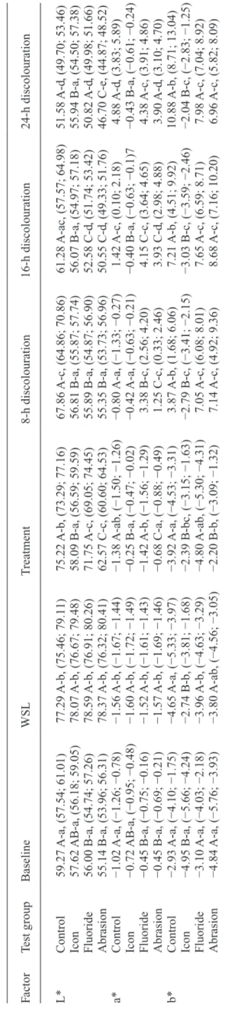
Median values, confidence intervals, and groups presenting significant colour changes at baseline, after WSL forma-tion, after treatment, and after discolouration cycles of 8, 16, and 24 hours are presented in Tables 2 and 3.
Lightness (L* value)
Formation of WSL increased the lightness (L* value) signif-icantly in all groups. Only the infiltration treatment dimin-ished the whitish appearance back to the baseline level. Micro-abrasion reduced L* value significantly better than the control and fluoride rinse, but this value was still sig-nificantly higher than the baseline measurement. L* value presented a decreasing trend throughout the whole discol-ouration procedure for all groups except infiltration.
Red–green chromaticity (a* value)
The a* value presented a decrease in all groups after the formation of WSL, indicating a shift to the green compo-nent. Following WSL treatment, a* value increased signifi-cantly in infiltration and micro-abrasion groups. Starting with the discolouration procedure, a* value increased in all groups, indicating a shift to the red component except for
infiltration. Tab
le 2
Median v
alues and confidence inter
vals of colour measurements at baseline, after
WSL for
mation, after treatment, and after discolouration c
ycles (b
lack tea) of 8, 16, and 24
h. Factor Test g roup Baseline WSL T reatment 8-h discolouration 16-h discolouration 24-h discolouration L* Control 58.10 A-a, (57.03; 60.91) 79.23 A-b, (75.49; 80.48) 76.74 A-b, (73.62; 78.74) 60.54 A-a, (59.03; 63.86) 59.16 A-a, (57.42; 62.51) 53.82 A-c, (51.82; 56.46) Icon 54.09 B-a, (52.17; 55.49) 76.18 A-b, (72.29; 78.73) 58.57 B-a, (56.04; 59.45) 57.14 B-a, (55.89; 58.24) 57.45 A-a, (54.99; 57.86) 55.3 A-a, (53.26; 56.00) Fluoride 55.34 AB-a, (53.43; 58.31) 77.60 A-b, (74.42; 79.84) 72.10 A-b, (68.63; 74.97) 53.22 C-ac, (51.86; 55.46) 48.43 B-cd , (47.41; 51.53) 47.36 B-d , (45.88; 48.47) Abrasion 55.15 AB-a, (53.25; 57.56) 78.87 A-b, (76.02; 80.53) 59.72 B-c, (58.45; 62.52) 54.99 BC-ad , (52.97; 56.04) 52.45 B-de, (49.25; 54.21) 48.90 B-e, (46.25; 50.05) a* Control −1.27 A-a, (−1.45; −0.82) −1.50 A-a, (−1.64; −1.36) −1.44 A-a, (−1.56; −1.27) 1.27 A-b, (0.11; 1.58) 2.32 A-bc, (1.79; 4.84) 5.64 A-c, (4.48; 7.07) Icon −0.54 B-a, (−0.68; −0.18) −1.68 A-b, (−1.79; −1.45) −0.28 B-a, (−0.33; −0.05) −0.40 A-a, (−0.45; −0.17) −0.31 B-a, (−0.47; −0.3) −0.27 B-a, (−0.55; 0.18) Fluoride −0.72 AB-a, (−0.93; −0.53) −1.56 A-b, (−1.69; −1.43) −1.41 A-b, (−1.60; −1.32) 6.00 B-c, (3.00; 6.70) 4.54 A-c, (2.80; 5.19) 4.48 A C-c, (3.53; 4.92) Abrasion −0.64 AB-a, (−0.82; −0.33) −1.52 A-b, (−1.63; −1.42) −0.65 C-ac, (−0.81; −0.40) 0.35 A-c, (−0.39; 1.66) 1.27 B-d , (0.54; 2.91) 2.73 C-d , (2.23; 4.40) b* Control −3.42 A-a, (−4.60; −2.81) −4.08 A-a, (−5.05; −3.47) −1.86 A-b, (−2.93; −1.33) 10.17 A-c, (6.52; 10.71) 10.19 A-cd , (9.06; 12.65) 13.24 A-d , (11.03; 14.41) Icon −4.07 B-a, (−5.47; −2.43) −5.89 A-b, (−6.32; −4.63) −3.12 A-a, (−4.09; −2.24) −2.29 B-a, (−3.30; −1.11) −2.21 B-a, (−3.46; −0.69) −1.33 B-a, (−2.84; 0.45) Fluoride −4.17 A-a, (−5.54; −3.13) −5.26 A-a, (−5.81;−4.44) −4.73 B-a, (−5.61; −4.11) 10.28 A-b, (7.91; 11.63) 5.61 C-c, (4.94; 6.88) 5.97 C-bc, (4.80; 7.94) Abrasion −4.03 A-ab, (−4.82; −2.94) −4.35 A-a, (−4.92; −3.45) −2.92 A-b, (−3.65; −1.88) 3.85 C-c, (3.21; 7.18) 5.74 C-c, (3.82; 7.00) 7.90 C-c, (6.62; 8.89) WSL, w
hite spot lesion. Comparisons of the treatments at each time point of measurement that are not significantl
y different are mark
ed with the same capital letters within L, a, or b v
alues (read
ver
ticall
y). Comparisons of the measurements for each treatment at different time points that are not significantl
y different are mark
ed with the same lo
w
er
-case letters (read horizontall
Yellow–blue chromaticity (b* value)
Formation of WSL created a slight decrease in the b* component for infiltration groups in both discolouration specimens and the fluoride group in the tea + citric acid specimens. Infiltration produced an increase causing the b* value to return to baseline, whereas micro-abrasion and arti-ficial saliva treatments (control) caused a decrease for the tea discolouration specimens. None of the treatments induced a change in the b* component in the tea+ citric acid discol-ouration specimens. A significant shift to the yellow compo-nent (increase in b* value) was seen immediately after the first 8-hour discolouration in all groups except infiltration, and this increase was sustained throughout the whole dis-colouration process for all groups except infiltration.
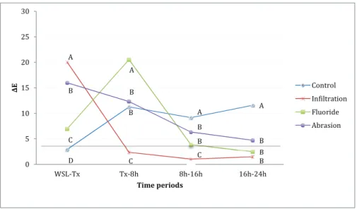
Delta E
Highest colour change obtained by the treatment of WSL was in infiltration and micro-abrasion groups followed by fluoride. The least affected group by the discolouration pro-cess was infiltration. Within each treatment group, the two used discolouration solutions did not differ significantly. Colour change obtained between time points and significant differences between groups at each time point are shown in
Figures 1 and 2.
Discussion
The colour improvement of WSLs following infiltration, micro-abrasion, or fluoride treatments and their resistance against two discolouration solutions were investigated in this study. Based on the obtained results, infiltration and micro-abrasion treatments performed better in diminishing the opaque WSL appearance compared with the fluoride treatment and control. This effect was stable only for the infiltration treatment under discolouring effects. Both null hypotheses are rejected.
In this study, bovine enamel was chosen as the test sub-strate in order to facilitate homogenous allocation of speci-mens from the same crown to the four test groups. Despite the fact that use of human enamel would be more prefer-able in dental material testing, it has been stated that bovine enamel could be safely used as a substitute for human enamel, particularly when a large crown size for preparing samples from the same crown was necessary (Wiegand and Attin, 2011).
Effects of different treatments in terms of improving the appearance of WSL and the resistance of the treated sur-face against discolouration were evaluated by the change in colour components (L*, a*, and b*). The standard quan-tification of colour change was performed using a spectro-photometer in the present set-up. The reproducibility of the measurements and the reliability of this method have led to its frequent use in such studies (Torres et al., 2011, Kim
et al., 2013).
T
ab
le 3
Median v
alues and confidence inter
vals of colour measurements at baseline, after
WSL for
mation, after treatment, and after discolouration c
ycles (b
lack tea + citric acid)
of 8, 16, and 24 h. Factor Test g roup Baseline WSL T reatment 8-h discolouration 16-h discolouration 24-h discolouration L* Control 59.27 A-a, (57.54; 61.01) 77.29 A-b, (75.46; 79.11) 75.22 A-b, (73.29; 77.16) 67.86 A-c, (64.86; 70.86) 61.28 A-ac, (57.57; 64.98) 51.58 A-d , (49.70; 53.46) Icon 57.62 AB-a, (56.18; 59.05) 78.07 A-b, (76.67; 79.48) 58.09 B-a, (56.59; 59.59) 56.81 B-a, (55.87; 57.74) 56.07 B-a, (54.97; 57.18) 55.94 B-a, (54.50; 57.38) Fluoride 56.00 B-a, (54.74; 57.26) 78.59 A-b, (76.91; 80.26) 71.75 A-c, (69.05; 74.45) 55.89 B-a, (54.87; 56.90) 52.58 C-d , (51.74; 53.42) 50.82 A-d , (49.98; 51.66) Abrasion 55.14 B-a, (53.96; 56.31) 78.37 A-b, (76.32; 80.41) 62.57 C-c, (60.60; 64.53) 55.35 B-a, (53.73; 56.96) 50.55 C-d , (49.33; 51.76) 46.70 C-e, (44.87; 48.52) a* Control −1.02 A-a, (−1.26; −0.78) −1.56 A-b, (−1.67; −1.44) −1.38 A-ab, (−1.50; −1.26) −0.80 A-a, (−1.33; −0.27) 1.42 A-c, (0.10; 2.18) 4.88 A-d , (3.83; 5.89) Icon −0.72 AB-a, (−0.95; −0.48) −1.60 A-b, (−1.72; −1.49) −0.25 B-a, (−0.47; −0.02) −0.42 A-a, (−0.63; −0.21) −0.40 B-a, (−0.63; −0.1)7 −0.43 B-a, (−0.61; −0.24) Fluoride −0.45 B-a, (−0.75; −0.16) −1.52 A-b, (−1.61; −1.43) −1.42 A-b, (−1.56; −1.29) 3.38 B-c, (2.56; 4.20) 4.15 C-c, (3.64; 4.65) 4.38 A-c, (3.91; 4.86) Abrasion −0.45 B-a, (−0.69; −0.21) −1.57 A-b, (−1.69; −1.46) −0.68 C-a, (−0.88; −0.49) 1.25 C-c, (0.33; 2.46) 3.93 C-d , (2.98; 4.88) 3.90 A-d , (3.10; 4.70) b* Control −2.93 A-a, (−4.10; −1.75) −4.65 A-a, (−5.33; −3.97) −3.92 A-a, (−4.53; −3.31) 3.87 A-b, (1.68; 6.06) 7.21 A-b, (4.51; 9.92) 10.88 A-b, (8.71; 13.04) Icon −4.95 B-a, (−5.66; −4.24) −2.74 B-b, (−3.81; −1.68) −2.39 B-bc, (−3.15; −1.63) −2.79 B-c, (−3.41; −2.15) −3.03 B-c, (−3.59; −2.46) −2.04 B-c, (−2.83; −1.25) Fluoride −3.10 A-a, (−4.03; −2.18) −3.96 A-b, (−4.63; −3.29) −4.80 A-ab, (−5.30; −4.31) 7.05 A-c, (6.08; 8.01) 7.65 A-c, (6.59; 8.71) 7.98 A-c, (7.04; 8.92) Abrasion −4.84 A-a, (−5.76; −3.93) −3.80 A-ab, (−4.56; −3.05) −2.20 B-b, (−3.09; −1.32) 7.14 A-c, (4.92; 9.36) 8.68 A-c, (7.16; 10.20) 6.96 A-c, (5.82; 8.09) WSL, w
hite spot lesion. Comparisons of the treatments at each time point of measurement that are not significantl
y different are mark
ed with the same capital letters within L, a, or b v
alues (read
ver
ticall
y). Comparisons of the measurements for each treatment at different time points that are not significantl
y different are mark
ed with the same lo
w
er
-case letters (read horizontall
Extrinsic enamel discolouration is frequently seen due to effects such as smoking, consumption of tannin-rich
foods, and long-term use of cationic agents (Addy and
Moran, 1995; Watts and Addy, 2001). The mechanism of these negative side effects is associated to nonenzymatic browning, protein denaturation, formation of pigmented metal sulphides, and precipitation of dietary chromogens (Addy and Moran, 1995; Watts and Addy, 2001). In this study, regular and citric acid added black tea was cho-sen as discolouration solutions. The rationale behind this approach was twofold. Black tea had been reported as one of the most powerful discolouring agents previously (Addy and Moran, 1995; Watts and Addy, 2001). However, because it is a natural product and the final solution may
vary in terms of pH depending on the processing of the tea plant (Kumar et al., 2013), citric acid was used to obtain a second discolouration solution with standard pH. Enhancement of discolouration by increasing the perme-ability of the superficial WSL surface as well as imitat-ing the consumption of acidic soft drinks was also aimed secondarily (Paris et al., 2007a; Neuhaus et al., 2013). Nevertheless, the discolouration results showed no differ-ence between the two solutions in this study. The lack of this effect was attributed to the slight difference between the pH values. The black tea solution already had a pH of 4.2–4.4, and this value was adjusted to pH 4 in the second group. A more acidic pH was avoided with respect to pos-sible erosive effects and considering the buffering effect
Figure 1 Colour change (ΔE) between each time period (WSL-Tx: WSL formation to treatment; Tx-8h: treatment to 8-h discolouration) for the black tea specimens. The horizontal line represents ΔE 3.7, which is the clinical detection limit (minimum) by naked eye. Comparisons of measurements that are not significantly different at each time period are marked with same capital letters (read vertically).
Figure 2 Colour change (ΔE) between each time period (WSL-Tx: WSL formation to treatment; Tx-8h: treatment to 8-h discolouration) for the black tea + citric acid specimens. The horizontal line represents ΔE 3.7, which is the clinical detection limit (minimum) by naked eye. Comparisons of measurements that are not significantly different at each time period are marked with same capital letters (read vertically).
of saliva avoiding lower pH levels in vivo (Simpson et al., 2001; Wiegand and Attin, 2011).
It is generally accepted that WSLs tend to regress in the presence of remineralizing agents, when the cariogenic attack is avoided (Marinho et al., 2003; Derks et al., 2004;
Beerens et al., 2010; Bergstrand and Twetman, 2011). Treatment of these surfaces with fluoride was shown to enhance subsurface remineralization (Marinho et al., 2003;
Derks et al., 2004; Beerens et al., 2010). In this study, a low-concentration fluoride rinse (250 ppm) contain-ing sodium and amine fluoride was used for this purpose (Marinho et al., 2003; Hamdan et al., 2012). The ration-ale for applying low doses of fluoride was to avoid hyper-mineralization of the lesion surface, which might obstruct further regression (Marinho et al., 2003; Hamdan et al.,
2012). Colour improvement of the WSLs was achieved
with the effect of fluoride to some extent, which was supe-rior compared with the control. However, this effect did not bring the colour components back to the initial levels. Previously, it was reported that a similar demineraliza-tion soludemineraliza-tion used on bovine enamel (pH 4.55) presented a lesion depth of 95 ± 32 µm even after the application of low-dose fluoride (250 ppm) for 28 days (Chin et al., 2009). This implied an incomplete remineralization of the porous enamel; thus, the susceptibility to discolouration was still increased. Similarly, this might be the reason for the limited improvement and instability of the WSL colour in this study. However, verifying this claim microscopically was not performed in this study.
Infiltration and micro-abrasion were the two effective treatment modalities significantly improving the whitish appearance of WSLs as well as creating significant visible colour improvement. Infiltration resulted in regression of all colour components back to the baseline values except b* value in tea + citric acid specimens, whereas micro-abrasion could not revert lightness to the initial levels. These two find-ings were in accordance with previous in vitro and in vivo results in which the treatment efficacy of these two methods was compared with fluoride therapy or saliva remineraliza-tion (Torres et al., 2011; Kim et al., 2013; Neuhaus et al., 2013; Paris et al., 2013). However, effects of infiltration and micro-abrasion treatments on improving WSL appearance were not compared previously. The main difference between these two treatment methods is that micro-abrasion removes the demineralized enamel, whereas infiltration stabilizes the lesion and reinforces the weakened prism structure within the lesion (Paris et al., 2007b; Kielbassa et al., 2009). Previously, the infiltrant was shown to penetrate subsur-face demineralized areas up to 400 µm (Paris et al., 2007a;
Neuhaus et al., 2013). The deep penetration of the resin infiltrating leading to the plugging of porosities within the WSLs might be the factor increasing the resistance against discolouration and improving the colour by having a similar light refraction index as shown in the present results (Paris
et al., 2013).
Previously, HCl micro-abrasion was shown to remove demineralized enamel up to 134 ± 35 microns (Schmidlin
et al., 2003). In this study set-up, micro-abrasion was applied for 1 minute, but the amount of enamel loss was not measured. The treatment was successful in improving the whitish appearance but was more prone to discolouration compared with infiltrated specimens. This can be explained in two ways; micro-abrasion with 6.6 per cent HCl slurry for 60 seconds might not have been enough to remove all the porous structure, which might have caused these speci-mens to be more prone to discolouration. Second, although the surface was polished following micro-abrasion, the sur-face might have been rough still (Paic et al., 2008), thus presenting an increased susceptibility to discolouration.
Conclusion
Within the limitations of this study, the following could be concluded:
• Infiltration of WSLs can treat the white opaque appear-ance, and this outcome is stable under discolouring effects. • Micro-abrasion reduces the white opaque appearance of
WSL considerably; however, this outcome is not resistant to discolouration.
• Low-concentration fluoride treatment improves the WSL appearance more than the clinical detectable limit, but the stability is not different than the effect of saliva remineralization.
Funding
DMG, Hamburg, Germany. The funders had no role in study design, data collection and analysis, decision to publish, or preparation of the manuscript.
References
Addy M, Moran J 1995 Mechanisms of stain formation on teeth, in par-ticular associated with metal ions and antiseptics. Advances in Dental Research 9: 450–456
Attin T, Manolakis A, Buchalla W, Hannig C 2003 Influence of tea on intrin-sic colour of previously bleached enamel. Journal of Oral Rehabilitation 30: 488–494
Beerens M W, van der Veen M H, van Beek H, ten Cate J M 2010 Effects of casein phosphopeptide amorphous calcium fluoride phosphate paste on white spot lesions and dental plaque after orthodontic treatment: a 3-month follow-up. European Journal of Oral Sciences 118: 610–617 Bergstrand F, Twetman S 2011 A review on prevention and treatment of
post-orthodontic white spot lesions - evidence-based methods and emerging technologies. The Open Dentistry Journal 5: 158–162 Buskes J A, Christoffersen J, Arends J 1985 Lesion formation and lesion
remineralization in enamel under constant composition conditions. A new technique with applications. Caries Research 19: 490–496 Chin M Y, Sandham A, Rumachik E N, Ruben J L, Huysmans M C 2009
Fluoride release and cariostatic potential of orthodontic adhesives with and without daily fluoride rinsing. American Journal of Orthodontics and Dentofacial Orthopedics 136: 547–553
Commission Internationale de l’Eclairage 1986 Colorimetry Publication No.15, Supplement 2
Derks A, Katsaros C, Frencken J E, van’t Hof M A, Kuijpers-Jagtman A M 2004 Caries-inhibiting effect of preventive measures during orthodontic treatment with fixed appliances. A systematic review. Caries Research 38: 413–420
Göhring T N, Zehnder M, Sener B, Schmidlin P R 2004 In vitro microle-akage of adhesive-sealed dentin with lactic acid and saliva exposure: a radio-isotope analysis. Journal of Dentistry 32: 235–240
Hadler-Olsen S, Sandvik K, El-Agroudi M A, Øgaard B 2012 The inci-dence of caries and white spot lesions in orthodontically treated adoles-cents with a comprehensive caries prophylactic regimen–a prospective study. European Journal of Orthodontics 34: 633–639
Hamdan A M, Maxfield B J, Tüfekçi E, Shroff B, Lindauer S J 2012 Preventing and treating white-spot lesions associated with orthodontic treatment: a survey of general dentists and orthodontists. Journal of the American Dental Association (1939) 143: 777–783
Hammad S M, El Banna M, El Zayat I, Mohsen M A 2012 Effect of resin infiltration on white spot lesions after debonding orthodontic brackets. American Journal of Dentistry 25: 3–8
Johnston W M, Kao E C 1989 Assessment of appearance match by visual observation and clinical colorimetry. Journal of Dental Research 68: 819–822
Kielbassa A M, Muller J, Gernhardt C R 2009 Closing the gap between oral hygiene and minimally invasive dentistry: a review on the resin infil-tration technique of incipient (proximal) enamel lesions. Quintessence International (Berlin, Germany: 1985) 40: 663–681
Kim Y, Son H H, Yi K, Kim H Y, Ahn J, Chang J 2013 The color change in artificial white spot lesions measured using a spectroradiometer. Clinical Oral Investigations 17: 139–146
Klimek J, Hellwig E, Ahrens G 1982 Fluoride taken up by plaque, by the underlying enamel and by clean enamel from three fluoride compounds in vitro. Caries Research 16: 156–161
Kumar C S, Subramanian R, Rao L J 2013 Application of enzymes in the production of RTD black tea beverages: a review. Critical Reviews in Food Science and Nutrition 53: 180–197
Lucchese A, Gherlone E 2013 Prevalence of white-spot lesions before and during orthodontic treatment with fixed appliances. European Journal of Orthodontics 35: 664–668
Marinho V C, Higgins J P, Logan S, Sheiham A 2003 Topical fluoride (toothpastes, mouthrinses, gels or varnishes) for preventing dental car-ies in children and adolescents. Cochrane Database Systematic Reviews 4: CD002782
Murphy T C, Willmot D R, Rodd H D 2007 Management of postortho-dontic demineralized white lesions with microabrasion: a quantita-tive assessment. American Journal of Orthodontics and Dentofacial Orthopedics 131: 27–33
Neuhaus K W, Ciucchi P, Donnet M, Lussi A 2010 Removal of enamel caries with an air abrasion powder. Operative Dentistry 35: 538–546 Neuhaus S, Schlafer A, Lussi B, Nyvad K W 2013 Infiltration of natural
caries lesions in relation to their activity status and acid pretreatment in vitro. Caries Research 47: 203–210
Paic M, Sener B, Schug J, Schmidlin P R 2008 Effects of microabrasion on substance loss, surface roughness, and colorimetric changes on enamel in vitro. Quintessence International (Berlin, Germany: 1985) 39: 517–522
Paris S, Meyer-Lueckel H, Cölfen H, Kielbassa A M 2007a Penetration coefficients of commercially available and experimental composites intended to infiltrate enamel carious lesions. Dental Materials: Official Publication of the Academy of Dental Materials 23: 742–748
Paris S, Meyer-Lueckel H, Cölfen H, Kielbassa A M 2007b Resin infiltra-tion of artificial enamel caries lesions with experimental light curing resins. Dental Materials Journal 26: 582–588
Paris S, Schwendicke F, Keltsch J, Dörfer C, Meyer-Lueckel H 2013 Masking of white spot lesions by resin infiltration in vitro. Journal of Dentistry 41 (Suppl 5): e28–e34
Pliska B T, Warner G A, Tantbirojn D, Larson B E 2012 Treatment of white spot lesions with ACP paste and microabrasion. The Angle Orthodontist 82: 765–769
Schmidlin P R, Göhring T N, Schug J, Lutz F 2003 Histological, morpho-logical, profilometric and optical changes of human tooth enamel after microabrasion. American Journal of Dentistry 16 Spec No: 4A–8A Simpson A, Shaw L, Smith A J 2001 Tooth surface pH during drinking of
black tea. British Dental Journal 190: 374–376
Soviero V M, Paris S, Leal S C, Azevedo R B, Meyer-Lueckel H 2013 Ex vivo evaluation of caries infiltration after different application times in primary molars. Caries Research 47: 110–116
Torres CRG, Borges A B, Torres L M, Gomes I S, de Oliveira R S 2011 Effect of caries infiltration technique and fluoride therapy on the colour masking of white spot lesions. Journal of Dentistry 39: 202–207 Watts A, Addy M 2001 Tooth discolouration and staining: a review of the
literature. British Dental Journal 190: 309–316
Wiegand A, Attin T 2011 Design of erosion/abrasion studies–insights and rational concepts. Caries Research 45 (Suppl 1): 53–59