Advance Access Publication 1 June 2009
Introduction
The introduction of the straightwire appliance ( Andrews,
1976a ) provided new treatment possibilities for the
orthodontist. Although the straightwire philosophy has a number of advantages, it also has certain limitations. Less than ideal fi nal treatment results may occur if some of these issues are not taken into account. In-built bracket prescriptions allow the orthodontist to focus on important treatment goals rather than the time-consuming in-out, vertical, and mesio-distal considerations for each tooth. Indeed the attractiveness of the straightwire philosophy is that a fully engaged archwire should express the in-out, inclination, angulation, and rotation prescription of each bracket.
However, clinical experience shows that wire bending is still required to achieve ideal results with the straightwire system ( Miethke and Melsen, 1999 ; Armstrong et al. , 2007 ), even with the proliferation of new prescriptions which are available ( Creekmore and Kunik, 1993 ). Clinicians have also recommended the use of different bracket prescriptions depending on the space-closing mechanics to be used and whether or not extractions have been performed ( Andrews, 1976b ; Roth, 1987 ).
When brackets are not ideally placed, positional discrepancies may arise. The same bracket prescription can lead to variable expression if it is bonded in different positions, for example along the vertical axis ( Thickett
et al. , 2007 ). Such discrepancies can be addressed by
replacing the bracket in its correct position or compensating the bracket placement error with a bend in the archwire. Over the years, different bracket placement protocols have
been recommended for the straightwire system ( Roth, 1987 ; Andrews, 1989 ; McLaughlin and Bennett, 1995 ) and this is still the subject of some debate.
These limitations are small compared with the overall advantages of the straightwire appliance but may be responsible for some of the treatment diffi culties encountered with the straightwire approach.
Modern orthodontics has also taken advantage of the three-dimensional digitization of plaster casts ( Kuroda et al. , 1996 ; Hayasaki et al. , 2005 ; Hildebrand et al. , 2008 ). With the appropriate software, the digitized model can be virtually modifi ed in order to obtain a set-up of the case and undertake treatment planning, considering different strategies. Accurate space measurement can be undertaken by the computer in order to manage tooth alignment, levelling, rotation, tip, and torque. Software packages also allow different appliance set-ups and prediction of tooth movements. This tool allows the infl uence of bracket positioning on the end treatment results to be considered prior to starting treatment.
To determine the result of orthodontic treatment, different assessment methods have been proposed. Many indices have been introduced ( Eismann, 1974 , 1980 ; Berg, 1975 ; Gottlieb,
1975 ) including the Peer Assessment Rating Index
( Richmond, 1990 ) and the American Board of Orthodontics (ABO) grading system ( Afsharpanah et al. , 1995 Feghali et al. , 1996 ; Hassanein et al. , 1996 ). The ABO evaluation system is based on eight criteria that are individually assessed ( Casko et al. , 1998 ): alignment, marginal ridge height, buccolingual inclination, occlusal relationship, occlusal contact, overjet, interproximal contact, and root angulation.
The effect of constant height bracket placement on marginal
ridge levelling using digitized models
Carlos Suárez * and Teresa Vilar **
* Department of Orthodontics, Université de Genève, Switzerland and ** Department of Odontostomatology, Universitat de Barcelona, Spain
SUMMARY Bracket placement is an important phase of orthodontic treatment. Final compensatory archwire
bends or bracket repositioning may be avoided if brackets are accurately positioned at the outset, so as to correctly express their built-in prescription. The purpose of this study was to investigate the levelling of marginal ridges when a bracket placement protocol, with fi xed values from the incisal edges and occlusal surfaces, was used on digitized models. A computerized tool, OrthoCAD®, was used to predict the end result using virtual set-up software. The appliances used for digital simulation were 3M MBT Victory Series 0.022 inch with a 0.019 × 0.025 inch stainless steel fi nal archwire on 42 digitized models. A paired t -test was used to investigate differences between the means of the pre- (T1) and post- (T2) treatment marginal ridge heights.
The results showed that most of the marginal ridge points studied deteriorated during digitized treatment prediction compared with T1. Statistical and clinically signifi cant changes ( P < 0.05) were found for upper premolar and lower molar marginal ridge points. Variability in the facial contour of the teeth seemed to play an important role.
The fi nal criterion, root angulation, is evaluated by means of a panoramic radiograph. Measurements can be carried out directly on the plaster model using special gauges, or by a computer-aided system on digitized plaster models.
The aim of the present investigation was to determine the effect of a constant vertical height bracket-bonding protocol by measuring the changes at the marginal ridge using the levelling criterion of the ABO grading system. The aim was to assess to what extent ideal levelling can be attained. For that purpose, measurements of pre-treatment (T1) values of marginal ridge heights were compared with the post-treatment (T2) values after computerized prediction. Materials and methods
Forty-seven digitized models were randomly selected for the study (supplied by OrthoCAD© software development centre, Cadent Ltd, Or Yehuda, Israel). All models were of Caucasian patients seeking orthodontic treatment for Class I, Class II division 1, or Class II division 2 malocclusions. Five models were discarded: two due to damage and three because they did not fulfi l the inclusion criteria for the present study. The OrthoCAD® software was downloaded from the offi cial website www.orthocad.com and installed on a conventional laptop computer (Toshiba, Tokyo, Japan).
Virtual set-ups were created for all models in order to perform marginal ridge levelling. Marginal ridge heights were measured according to the ABO (2008) criterion with the ABO software tool. Differences were measured digitally in millimetres.
All points ( Figures 1 and 2 ) were identifi ed by the same author (CS). These show the interproximal points as described by the ABO grading system and also how the height was measured. The points are described according to the interproximal point to which they refer using the Federation Dentaire Internationale nomenclature. For example, the interproximal point between the upper right premolars was labelled as 1514 and the interproximal point between the upper left fi rst molar and second premolar as 2526. The digital model was rotated in three dimensions in order to identify the correct marginal ridge points. Given that bracket placement should be performed exactly as a one-off task, no error study was undertaken.
The set-up models were treated virtually with MBT Victory Series 0.022 inch brackets (3M Unitek Dental Products, Monrovia, California, USA). Brackets were placed as recommended by McLaughlin et al. (2001) ,
measuring from the incisal or occlusal edges of the upper (U) and lower (L) teeth in millimetres: U 7 = 2.0, U 6 = 3.0,
U 5 = 4.0, U 4 = 4.5, U 3 = 5.0, U 2 = 4.5, U 1 = 5.0, L 7 = 2.5,
L 6 = 2.5, L 5 = 3.5, L 4 = 4.0, L 3 = 4.5, L 2 = 4.0, and L 1 = 4.0.
Bracket placement was carried out using the digital height window in the software, marking the exact measurement recommended in the protocol. The fi nal archwire was 0.019 × 0.025 inch stainless steel, as recommended by the MBT philosophy and because good engagement with suffi cient torque expression should be achieved with the 0.022 inch slot brackets. Marginal ridge heights were measured again on the T2 virtual set-up view in order to study the change achieved by levelling during computerized prediction.
Descriptive statistical analysis was performed in order to describe T1 and T2 measurements and to compare the changes after simulation. A paired t -test was used to investigate differences between the means of the T1 and T2 marginal ridge heights ( P < 0.05).
Results
The mean, standard deviation, and ranges for T1 and T2 marginal ridge heights are shown in Table 1 . The mean measurements increased for all points, except for 4645 and 3536 for which there was a slight decrease. According to the ABO criteria, values above 0.5 mm require correction. Therefore, values above 0.5 mm were set as clinically important and requiring correction. The means of all T2 points were above 0.5 mm at the end of simulation.
Probability plots for T1 and T2 values show that the data were normally distributed. The plot of 1716 is shown as an example in Figure 3 .
At T1, all points except those for initial values of 1514 and 2425 had a large proportion of marginal ridge heights above the 0.5 mm limit ( Figure 4 ). It can also be seen that all points appeared to deteriorate after simulation (T2), increasing the height values for the marginal ridge relationship.
Table 2 shows the percentages of values outside the clinically acceptable range at T1 and T2. All points, with the exception of 3536, deteriorated, with a tendency for a poorer marginal ridge relationship at T2. In the upper arch, 13.5 – 33.4 per cent of marginal ridges worsened compared with T1. The lower arch showed smaller values, ranging from 5.1 to 24.3 per cent of the marginal ridge points. There was an improvement in marginal ridge values for 3536 as shown by the negative difference. In all, 2.6 per cent of ridges that were initially above the 0.5 mm limit became clinically acceptable resulting in values below 0.5 mm. Statistically signifi cant differences ( P < 0.05) were found for 1514, 2425, 4746, and 3637 ( Table 3 ), for which the changes showed a deterioration.
The marginal ridge points were allocated to one of three categories: improved, no change, and worsened ( Figure 5 ). Marginal ridges deteriorated between 41 and 71.4 per cent
Table 1 Descriptive values of marginal ridge heights at the start (T1) and following (T2) virtual treatment (mm).
Point T1 T2
Mean SD Range Mean SD Range
1716 0.67 0.65 0 – 2.2 0.75 0.58 0 – 2.1 1615 0.5 0.44 0 – 1.7 0.64 0.52 0 – 2.3 1514 0.41 0.4 0 – 2.4 0.63 0.45 0 – 2.1 2425 0.35 0.34 0 – 1.9 0.58 0.49 0 – 2.1 2526 0.48 0.44 0 – 1.7 0.73 0.62 0 – 3.5 2627 0.74 0.71 0 – 3.8 0.97 0.63 0 – 2.8 4645 0.57 0.67 0 – 2.9 0.51 0.36 0 – 1.5 3536 0.67 0.52 0 – 2.3 0.62 0.48 0 – 1.6 3637 0.5 0.49 0 – 2.3 0.84 0.75 0 – 3.7
Figure 3 Probability plot for the initial (T1) and fi nal (T2) values of
point 1716 at T1 and T2.
Figure 4 Changes in the descriptive statistics of marginal ridges from the
start (T1) to following (T2) virtual treatment.
Table 2 Percentage of points with a marginal ridge relationship greater than 0.5 mm at the start (T1) and following (T2) virtual treatment. Point T1 T2 Change 1716 40.5 54 13.5 1615 29.7 45.9 16.2 1514 20 48.5 28.5 2425 11 44.4 33.4 2526 30.5 50 19.5 2627 42.8 68.5 25.7 4647 48.6 62.1 13.5 4645 30.7 35.8 5.1 3536 48.7 46.1 − 2.6 3637 35.1 59.1 24.3
able to accept that marginal ridge relationships remain unaltered, but it is more diffi cult to accept that marginal ridges may deteriorate. The results show that brackets placed according to the fi xed vertical position lead to poorer marginal ridge relationships compared with T1 for between 5.1 and 33.4 per cent of cases ( Table 2 ).
All brackets were placed at fi xed vertical positions measured from the incisal or occlusal reference. This does not take into account two important factors: the total length of the clinical crown and the convexity in the vertical and horizontal axes of the tooth. These two factors are likely to be responsible for the different expression of the bracket prescriptions. Therefore, no matter what vertical height bracket placement protocol is used, the same problem will arise if the reference is taken from the incisal or occlusal edge.
It should be noted that, in the upper arch, the points that initially showed the best marginal ridge relationship (1514 and 2425, Table 2 ) experienced the greatest deterioration compared with the other points. It should also be noted that although changes are clearly seen when the clinical limit of 0.5 mm is set, the statistical analysis of the means at T1 and T2 showed statistically signifi cant changes ( P < 0.05) only for 1514, 2425, 4746, and 3637 ( Table 3 ). These fi ndings are in agreement with the ABO experience for points 4647 and 3637. The ABO state that the most diffi cult points to obtain a good marginal ridge post-treatment are 1716, 2627, 4647, and 3637 ( ABO, 2008 ).
An important uncontrolled factor that should be considered is anatomical variability. The fi ndings of studies on facial contour variation have reported large intra-individual variations in tooth morphology and this may explain the fi ndings of the present study ( Germane et al. , 1989 ; Miethke and Melsen, 1999 ). Germane et al. (1989) found that facial surface contours were not consistent among teeth of the same type. Standard deviations in a sample of 600 maxillary and mandibular teeth ranged from ± 2.6 to ± 6.4 degree for the points studied. Those authors also noted that variability in facio-lingual contours increased
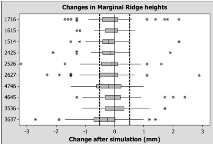
Figure 6 Changes in marginal ridges heights (values in mm).
of cases and improved in 22.8 – 48.7 per cent. Greater improvements in marginal ridge values were recorded for 4645 and 3536, at 48.7 and 46.1 per cent, respectively. The marginal ridge heights remained unaltered in 5.2 – 16.3 per cent of all cases.
Figure 6 illustrates the change in marginal ridge heights which occurred during simulation. A negative value implies a worsening in the marginal ridges, while a positive value implies that the relationship improved. The majority of values were negative. Changes appeared to be in equilibrium for 3536 and 4645. The differences which occurred as a result of treatment simulation remained under the threshold of 0.5 mm for all marginal ridges (1716, 1615, 1514, 2425, 2526, 2627, 4746, 4645, 3536, and 3637).
Discussion
The results of the present study show a tendency for marginal ridge values to deteriorate after levelling using computer prediction, when brackets are positioned at fi xed heights from the incisal or occlusal edges.
Correction of marginal ridges following the protocol used in the present study is far from ideal. The clinician may be
Figure 5 Changes in marginal ridge points (values in %).
Table 3 Statistically signifi cant changes from the start (T1) to following (T2) virtual treatment .
Point P value 1716 0.571 1615 0.077 1514 0.033* 2425 0.039* 2526 0.078 2627 0.159 4647 0.017* 4645 0.611 3536 0.62 3637 0.019* * P < 0.05.
progressively between teeth from anterior to posterior in both the upper and lower arches. This is in agreement with ABO (2008) results regarding diffi culties in achieving marginal ridge levelling interproximally for 1716, 2627, 4647, and 3637 and also, to some extent, with the results obtained in the present study. The third conclusion reached by Germane et al. (1989) was that vertical bracket placement errors of 1 mm were found to alter torque values by up to 10 degrees and this may also contribute to problems in marginal ridge levelling.
The straightwire philosophy and the resulting pre-adjusted appliance has been a great advance that most orthodontists acknowledge. However, pre-adjusted appliances cannot assume responsibility for nature’s variability and asymmetry and appliances will never be responsible for an optimal orthodontic treatment by themselves.
A recent study ( Armstrong et al. , 2007 ) focused on the accuracy of bracket placement when comparing two techniques. The authors concluded that using distances from incisal edges lead to more accurate bracket placement in the vertical dimension for the upper and lower teeth. However, they also noted that the extent of error in bracket placement, regardless of the placement technique, necessitates either bends being placed in the archwire or sometimes bracket repositioning. It is this point which was the focus of the present study. Prior to giving advice on bracket placement protocols, an initial and more fundamental question should be addressed: will all teeth move in the expected way when a bracket placement protocol is followed? The results of this study suggest that even though bracket placement errors exist, anatomical variation acts as an additional and fundamental factor whose effects will need correction by arch bending or readjustment of the bracket position. Therefore, further computerized studies may assist in fi nding both new bracket placement and new prescription values that, taking into account anatomical variation, will lead the pre-adjusted straightwire philosophy to come closer to the ideal occlusal outcome. Therefore, variations in facial surface contours may have affected the results obtained in this study, but this fact should be proven.
Both the validity and the reliability of the software used in this study have been investigated previously. According to Zilberman et al. (2003) , the accuracy of OrthoCAD is clinically acceptable and Santoro et al. (2003) also concluded that differences were suffi ciently small to be considered clinically acceptable. A study by Costalos et al. (2005) concluded that measurements which were undertaken on study models were not signifi cantly different between plaster and digital models.
In contrast with these studies, Okunami et al. (2007) found signifi cant differences between measurements taken on plaster and digital models for some of the variables they measured, but they did not fi nd signifi cant differences when comparing alignment and marginal ridge heights
(which are similar to the variables measured in the current study). Hildebrand et al. (2008) also noted statistically signifi cant differences for alignment, occlusal contact, and overjet measurements but not for marginal ridge height measurements, which again suggests that measurement of this variable with OrthoCAD is reliable when compared with plaster models.
Although the present study was based on a computerized treatment planning tool, it has some advantages over ‘ real-life ’ studies in that inter- and intra-operator variability are minimized as the position of the brackets is performed automatically by the software.
The marginal ridge parameter was chosen for the present study because it is clearly related to the vertical bracket position, although other parameters are also corrected during treatment. It is well known that the occlusion has to be adjusted towards the end of treatment and marginal ridge compensations and corrections may take place for instance using elastics. This is beyond the scope of the present study, but it should be borne in mind.
Conclusions
The clinically relevant difference for marginal ridge heights was set at 0.5 mm, in accordance with ABO standards. Points 1514, 2425, 4746, and 3637 showed both statistically signifi cant and clinically relevant deterioration in marginal ridge relationships.
Vertical placement bracket protocols which ignore individual labial crown convexities and crown lengths may introduce an initial bracket placement error which may lead to poor marginal ridge levelling at the end of treatment.
Computerized simulations adjusting bracket heights to perfect marginal ridge relationships are possible with this type of software and may lead to new height bracket placement protocols in the future.
Address for correspondence Dr Carlos Suárez
Université de Genève Faculté de Medecine
Section de Médecine Dentaire (Division d’Orthodontie) Rue Barthélemy-Menn 19
1205 Genève Switzerland
E-mail: [email protected] Acknowledgement
We acknowledge the counselling of Drs Carlos Vicente, Vicente Sada, Silvia Geron, and Rafi Romano regarding discussion of the present research. We also acknowledge Nir Danai (OrthoCAD®) for supplying the digitized models for the study.
References
American Board of Orthodontics 2008 Grading system for dental casts and panoramic radiographs . ( http://www.americanboardortho.com/professionals/ downloads/Grading%20System%20Casts-Radiographs.pdf )
Afsharpanah A , Nelson S , Feghali R , Hans M G 1995 Assessment of orthodontically untreated sample using the PAR Index . Journal of Dental Research 74 : 139 (abstract)
Andrews L F 1976a The straight wire appliance, origin, controversy, commentary . Journal of Clinical Orthodontics 10 : 99 – 114
Andrews L F 1976b The straight wire appliance. Extraction series brackets . Journal of Clinical Orthodontics 10 : 425 – 441
Andrews L F 1989 Straight wire: the concept and appliance . L A Wells , San Diego
Armstrong D , Shen G , Petocz P , Darendeliler M A 2007 A comparison of accuracy in bracket positioning between two techniques — localizing the centre of the incisal crown and measuring the distance from the incisal edge . European Journal of Orthodontics 29 : 430 – 436
Berg R 1975 Postretention analysis of treatment problems and failures in 264 consecutively treated cases . European Journal of Orthodontics 1 : 55 – 68
Casko J S et al. 1998 Objective grading system for dental casts and panoramic radiographs . American Journal of Orthodontics and Dentofacial Orthopedics 114 : 589 – 599
Costalos P A , Sarraf K , Cangialosi T J , Efstratiadis S 2005 Evaluation of the accuracy of digital model analysis for the American Board of Orthodontics objective grading system for dental casts . American Journal of Orthodontics and Dentofacial Orthopedics 128 : 624 – 629 Creekmore T D , Kunik R L 1993 Straightwire: the next generation . American
Journal of Orthodontics and Dentofacial Orthopedics 104 : 8 – 20 Eismann D 1974 A method of evaluating effi ciency of orthodontic treatment .
Transactions of the European Orthodontic Society , pp. 223 – 232 Eismann D 1980 Reliable assessment of morphological changes resulting
from orthodontic treatment . European Journal of Orthodontics 2 : 19 – 25
Feghali R , Afsharpanah A , Hans M G , Nelson S , Hassanein R 1996 Assessing orthodontic treatment outcome from 1980-1985 using the PAR Index . Journal of Dental Research 75 : 363 (abstract)
Germane N , Bentley B E , Isaacson R J 1989 Three biologic variables modifying faciolingual tooth angulation by straightwire appliances . American Journal of Orthodontics and Dentofacial Orthopedics 96 : 312 – 319
Gottlieb E 1975 Grading your orthodontic treatment results . Journal of Clinical Orthodontics 9 : 156 – 161
Hassanein R , Nelson S , Hans M G , Feghali R 1996 Assessment of orthodontic treatment using the PAR Index . Journal of Dental Research 75 : 338 (abstract)
Hayasaki H , Martins R , Gandini Jr L ., Saitoh K , Nonaka K 2005 A new way of analysing occlusion 3 dimensionally . American Journal of Orthodontics and Dentofacial Orthopedics 128 : 128 – 132
Hildebrand J C , Palomo J M , Palomo L , Sivik M , Hans M 2008 Evaluation of a software program for applying the American Board of Orthodontics objective grading system to digital casts . American Journal of Orthodontics and Dentofacial Orthopedics 133 : 283 – 289
Kuroda T , Motohashi N , Tominaga R , Iwata K 1996 Three-dimensional dental cast analysing system using laser scanning . American Journal of Orthodontics and Dentofacial Orthopedics 110 : 365 – 369
McLaughlin R P , Bennett J C 1995 Bracket placement with the pre-adjusted appliance . Journal of Clinical Orthodontics 29 : 302 – 311
McLaughlin R P , Bennett J C , Trevisi H J 2001 Systemised orthodontic treatment mechanics . Mosby Inc ., Edinburgh
Miethke R R , Melsen B 1999 Effect of variation in tooth morphology and bracket position on fi rst and third order correction morphology with pre-adjusted appliances . American Journal of Orthodontics and Dentofacial Orthopedics 116 : 329 – 335
Okunami T R , Kusnoto B , BeGole E , Evans C A , Sadowsky C , Fadavi S 2007 Assessing the American Board of Orthodontics objective grading system: digital vs plaster dental casts . American Journal of Orthodontics and Dentofacial Orthopedics 131 : 51 – 56
Richmond S 1990 A critical evaluation of orthodontic treatment in the General Dental Services in England and Wales . Thesis , University of Manchester
Roth R H 1987 The straight wire appliance 17 years later . Journal of Clinical Orthodontics 21 : 632 – 642
Santoro M , Galkin S , Teredesai M , Nicolay O F , Cangialosi T J 2003 Comparison of measurements made on digital and plaster models . American Journal of Orthodontics and Dentofacial Orthopedics 124 : 101 – 105
Thickett E , Taylor N G , Hodge T 2007 Choosing a pre-adjusted orthodontic appliance prescription for anterior teeth . Journal of Orthodontics 34 : 95 – 100 Zilberman O , Huggare J A , Parikakis K A 2003 Evaluation of the validity of tooth size and arch width measurements using conventional and three-dimensional virtual orthodontic models . The Angle Orthodontist 73 : 301 – 306