British Journal of Rheumatology 1996;35:542-547
THE VALUE OF ISOTYPE DETERMINATION OF SERUM ANTIBODIES
AGAINST CHLAMYDIA FOR THE DIAGNOSIS OF CHLAMYDIA
REACTIVE ARTHRITIS
S. BAS, T. CUNNINGHAM, T. K. KVIEN,* A. GLENNAS,* K. MELBYf and T. L. VISCHER Division of Rheumatology, University Hospital, Geneva, Switzerland, *Oslo City Department of Rheumatology, Norwegian Lutheran Hospital, Oslo and ^Department of Microbiology, Ullevil University Hospital, Oslo, Norway
SUMMARY
In clinical rheumatology, the diagnosis of Chlamydia reactive arthritis is difficult because an incomplete form of the disease can closely resemble an undifferentiated seronegative mono/oligoarthritis. We investigated whether measuring specific isotypes of anti-Chlamydia antibodies in serum can improve the diagnosis, by comparing such antibody concentrations in the serum of patients with well-defined disease, i.e. Chlamydia trachomatis sexually acquired reactive arthritis (CT-SARA), with other arthritides. Antibody levels were determined by enzyme-linked immunosorbent assay (ELISA). When considering two different isotypes and their combination, the best sensitivity (63%) was obtained for IgM and/or IgA results with a specificity of 81%. The patients with CT-SARA and SARA had the highest levels of antibodies of all isotypes tested. It is concluded that, in our experimental conditions, only very high values of specific isotypes could indicate a diagnosis of Chlamydia reactive arthritis.
KEY WORDS: Sexually acquired reactive arthritis, Chlamydia, Antibodies, Enzyme-linked immunoassay.
CHLAMYDIA has been shown to induce arthritis in man mostly after sexually acquired genitourinary infection [1]. Several studies [2-6] have shown that an acute Chlamydia trachomatis (CT) infection was present in 36-69% of patients, immediately before the onset of sexually acquired reactive arthritis (SARA), even in the absence of clinical symptoms of urethritis. An incomplete form of reactive arthritis without a history of an initial infectious event can closely resemble an undifferentiated seronegative mono/oligoarthritis. Kingsley and Sieper [7] reported that reactive arthritis with a subclinical rather than a clinical triggering infection is common, but is not often recognized as such. It is well known that some chlamydial infections in humans can be inapparent [8] and CT has been found in urogenital samples of patients without appropriate symptoms and mono/oligoarthritis [9-11]. Chlamydia pneumoniae may also trigger undifferenti-ated oligoarthritis [12].
In current practice, the diagnosis of Chlamydia reactive arthritis is difficult and based on clinical suspicion, the demonstration of organisms in genito-urinary specimens and/or the detection of serum antibodies. The role of serological testing in diagnosis remains controversial because antibodies against Chlamydia are frequent in the general population. Epidemiological studies have revealed a prevalence of positive antibody tests for this organism in ~ 5 % of the normal population [13, 14] and in 10-45% of patients attending sexually transmitted disease clinics in Western Europe and North America [8].
Submitted 17 August 1995; revised version accepted 4 January 1996.
Correspondence to: S. Bas, Research Laboratory, Division of Rheumatology, University Hospital, 1211 Geneva 14, Switzerland.
Wollenhaupt et al. [15] found that the determination of C/i/a/ny^/a-specific serum antibodies of the IgA class may yield further diagnostic information in IgG-pos-itive patients with suspected chlamydial-induced arthritis. However, Sieper et al. [16] determined the value of specific antibody detection in the diagnosis of reactive arthritis and concluded that no test had a significant positive predictive value (PPV) for reactive arthritis in the general population and, even in the rheumatology clinic, the PPV was low.
We therefore investigated whether measuring specific isotypes of anti-Chlamydia antibodies in serum could improve the diagnosis of Chlamydia reactive' arthritis by comparing such antibody concentrations in the serum of patients with well-defined disease, i.e. CT-SARA, with other arthritides. We have determined the concentrations of serum IgG, IgM and IgA antibodies against Chlamydia by an enzyme-linked immunosorbent assay (ELISA) method in 103 patients with different forms of arthritis (30 having a well-characterized CT-SARA), and have compared the results obtained for one, two or three isotypes in CT-SARA with those in other forms of arthritis and in healthy blood donors.
PATIENTS AND METHODS Patients
Serum samples came from our serum collection and have been kept for various times at — 70°C. The diagnosis was taken from the chart at the time of sample collection. All the patients were questioned about possible genitourinary infection (urethritis, dysuria, discharge?).
Patients were divided into the following groups. 1. CT-SARA (/i = 30): asymmetrical mono/oligoarthritis with urethritis and evidence of C. trachomatis infection [five had a positive urethral/endocervical Chlamydia
© 1996 British Society for Rheumatology
antigen detection by direct immunofluorescence (IF), 24 had a positive urethral/endocervical Chlamydia culture and had been included in a study on reactive arthritis [11], one had a positive urethral Chlamydia DNA amplification with the Amplicor test of Roche Diagnostic Systems Inc. (Branchburg, NJ)]; median age (yr): 25, range: 18-58; 20% were female patients; HLA-B27: 12 positive, 13 negative patients, five not determined. 2. SARA (n = 23): asymmetrical mono/ oligoarthritis with urethritis, but without proof or evidence for CT infection (15 had a negative urethral/endocervical Chlamydia detection, for eight patients the detection was not performed); median age (yr): 36, range: 19-62; 13% were female patients; HLA-B27: seven positive, eight negative patients, eight not determined. 3. Undifferentiated rheumatoid factor-negative mono/oligoarthritis (n = 56): 1-4 joints, asymmetrical, mainly lower extremities; absence of evidence for another denned rheumatic disease; median age (yr): 40, range: 16-77; 46% were female patients; HLA-B27: six positive, 25 negative patients, 25 not determined. 4. Rheumatoid factor-positive rheumatoid arthritis (RA) (n = 37): median age (yr): 63, range: 24-83; 70% were female patients. 5. Crystal-induced arthritis (n = 17): gout (8), chondrocalcinosis (9); median age (yr): 51, range: 33-88; 24% were female patients. 6. Mechanical arthropathies (n = 19): osteoarthritis (9), femoropatellar chondropathies (3), post-traumatic arthropathies (3), meniscus lesion (3), fracture (1); median age (yr): 58, range: 22-80; 63% were female patients.
The urethral/endocervical Chlamydia detection was not performed for the negative disease groups (4, 5 and 6) and was presumed to be negative.
The healthy control group was composed of 100 blood donors; median age (yr): 44, range: 22-68; 37% were female individuals.
Materials
Flat-well microtitre plates (Immunoplate I, catalogue no. 439454) were purchased from Nunc (Life Tech-nologies, Basel, Switzerland), CT LGV-1 + Chlamydia psittaci 6BC antigen preparation from Virion (Cham, Switzerland), fetal calf serum (FCS) from Gibco (Life Technologies, Basel, Switzerland) and F(ab02 fragment of polyclonal goat IgG anti-human IgG, IgM or IgA from Cappel (Embrach, Switzerland).
Measurements of anti-Chlamydia antibodies by ELISA The strains used for the Chlamydia antigen preparation were CT LGV-1 and C. psittaci 6BC, both obtained from the American Type Culture Collection. They were grown in the Vero cell line, disrupted by homogenization and an elementary body-enriched fraction separated by sucrose density-gradient differential centrifugation. The control antigen was prepared in parallel using non-infected cells. Flat-well microtitre plates were used as the solid phase and 'coated' overnight at 4°C with 100 fi I/well of Chlamydia antigen or control antigen protein (1.5/ig/ml), dissolved in carbonate 'coating' buffer (pH 9.6). FCS
(1%) was used to saturate the microplates after 'coating' (100/iI/well for l h at room temperature). Dilutions (usually 1/20, 1/40, 1/80, more if necessary) of the same sample in phosphate-buffered saline (PBS) with 0.05% Tween 20 were tested for Chlamydia antibodies and for anticellular antibodies (100 /il/well for 2 h at room temperature). Antibody was detected with alkaline phosphatase-labelled (conjugation according to Voller et al. [17]) Ffcb^ fragment of polyclonal goat IgG anti-human IgG (Fc specific), IgM (ji chain specific) or IgA (a chain specific) (100 /il/well for 2 h at room temperature). The working dilutions of the alkaline phosphatase-labelled antibodies were determined after calibration with the previous homemade preparation and were usually 1/600. Substrate (100 /il/well of 1 mg/ml p-nitrophenyl phosphate in 10% diethanolamine) was added for 1 h at room temperature. The colour development was stopped by addition of 25 /il/well of 3 M NaOH.
The optical density (OD) value obtained for the control antigen was subtracted from the OD value obtained for chlamydial antigen. A plot of the OD versus sample dilutions for high-positive sera used as standard and different patient sera showed parallel curves. Antibodies were quantified by comparing the OD at 410 nm of three serial 2-fold dilutions with standard curves using a Dynatech MR 5000 Microplate reader linked to a Macintosh computer equipped with the Biocalc program [18]. Antibody concentrations were expressed as relative units (1 unit is the value obtained from a 10"4 dilution of a reference serum). Antibody titres of sera from 100 healthy blood donors were measured, and values at least 3 s.D. higher than the mean antibody concentration of these sera were defined as positive.
Calculations
Sensitivity, specificity, positive predictive value, negative predictive value, false positives and false negatives were calculated as described by Griner et al. [19]. A sample was designated 'true positive' if it came from a patient with CT-SARA and antibodies of the isotype concerned (level greater than the mean of donor 4- 3 s.D.). A sample was designated 'true negative' if it was obtained from a patient who had a diagnosis of RA, crystal-induced arthritis or mechanical arthropathy and had no antibody of the isotype concerned (level less than the mean of donor + 3 s.D.). Thus, sensitivity = true positives/true positives + false negatives, specificity = true negatives/ true negatives + false positives, positive predictive value = true positives/true positives + false positives, negative predictive value = true negatives/true neg-atives + false negneg-atives, false positive rate = false positives/total patients without disease, false negative rate = false negatives/total patients with disease.
RESULTS
Serum concentrations of antibodies against Chlamydia The distributions obtained for IgG, IgM and IgA in
544 BRITISH JOURNAL OF RHEUMATOLOGY VOL. 35 NO. 6 tgQ and-Chtamydta antfbodtes n p r a c M d In untttrtn) of Mfisn 60000; 55000 50000; 45000; 40000! 35000! 30000; 25000 20000J 15000: 10000; 1 - 5000J • • • • • • • • • • • • • -•- i . * • • 65000 = E c 600001 Illll l B m m 40000= t z 35000 = uni i 30000j X 25000| ni u 20000 = nnnn n 10000J •II I 5000l
h untaAnl of Mrun npresMd tn unfts/rcJ ot scrum
0= 0 1 2 3 4 5 6 7 18000-16000 = 14000 = 12000 = 10000-8000 : 0000' 4000; 2 3 4 5 6 7
Fio. 1.—Distribution of serum anti-Chlamydia antibody concentrations expressed in units/ml. 1. Chlamydla trachomatis sexually acquired reactive arthritis (CT-SARA), n •» 30; 2. SARA (negative or not performed urethral/endocervical Chlamydia detection), n =- 23; 3. undifferentiated seronegative mono/oligoarthritis, n = 56; 4. rheumatoid factor-positive rheumatoid arthritis, n = 37; 5. crystal-induced arthritis, n = 17; 6. mechanical arthropathies, n <= 19; 7. healthy blood donors, n — 100.
the six diagnostic groups of patients and healthy blood donors are given in Fig. 1. We observed that the groups of patients with CT-SARA and with SARA had the highest levels of antibodies of all isotypes tested. They were the only groups which had positive mean antibody concentrations (greater than the mean + 3 S.D. of the 100 blood donors' serum values) for all isotypes, except for the IgM of CT-SARA which was just below the cut-off. For the group of patients with undifferentiated mono/oligoarthritis, the means were lower than the mean + 3 S.D. of the 100 blood donors' serum values, but were always higher than those obtained for the control groups. Furthermore, we
observed that with higher cut-off (greater than the mean of the 100 blood donors' serum values + 4 S.D. for IgG, H-6S.D. for IgM, +9 S.D. for IgA), except for one IgA value from group 4, positive values would be only found in the CT-SARA, SARA and undifferentiated mono/oligoarthritis groups of patients.
For individual or isotype combinations, the percent-ages of samples with antibody concentration greater than the mean + 3 S.D. of antibody concentration obtained for donors are given in Table I. Patients and donors with a value higher than the mean + 3 S.D. of antibody concentration obtained for donors,
TABLE I
Chlamydia-specific antibody isotype distribution in the six diagnostic groups of patients and in healthy blood donors (% of samples with values higher than the mean of donors + 3 S.D.)
Group
IgG + IgM IgG or IgM IgG only IgM only IgA only IgG + IgM IgG + IgA IgM + IgA + IgA or IgA 1. Chlamydia trachomatis sexually
acquired reactive arthritis (n •= 30) 2. Sexually acquired reactive arthritis
(n = 23) 3. Undifferentiated seronegative mono/oligoarthritis (n = 56) 4. Rheumatoid arthritis (n — 37) 5. Crystal-induced arthritis (n = 17) 6. Mechanical arthropathies (n «= 19) 7. Controls (blood donors) (n - 100)
10 17 13 23 22 10 10 73 71 27 29 7 0 6 5 2 13 0 12 11 1 13 5 12 16 2 2 0 0 0 0 5 0 6 5 0 5 3 0 0 0 2 0 0 0 0 47 8 36 37 5 53 92 64 63 95
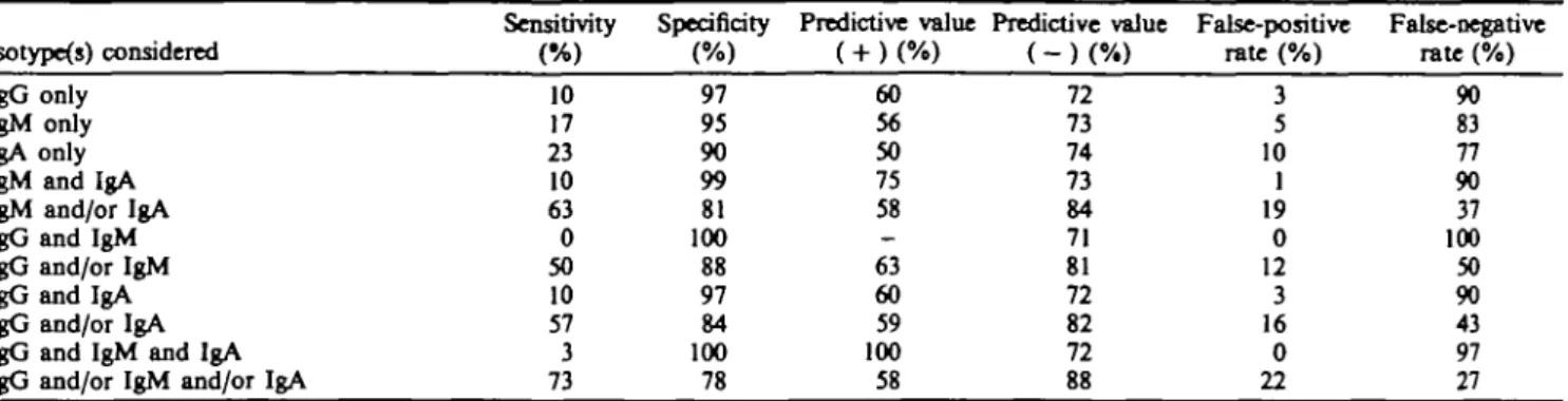
TABLE II
Sensitivity, specificity, positive predictive value, negative predictive value, false-positive and false-negative values obtained for the different isotypes and their combinations
Sensitivity Specificity Predictive value Predictive value False-positive False-negative (%) (%) ( + ) ( % ) ( - ) ( % ) rate(%) rate (%) Isotype(s) considered
IgG only IgM only IgA only IgM and IgA IgM and/or IgA IgG and IgM IgG and/or IgM IgG and IgA IgG and/or IgA IgG and IgM and IgA IgG and/or IgM and/or IgA
10 17 23 10 63 0 50 10 57 3 73 97 95 90 99 81 100 88 97 84 100 78 60 56 50 75 58 -63 60 59 100 58 72 73 74 73 84 71 81 72 82 72 88 3 5 10 1 19 0 12 3 16 0 22 90 83 77 90 37 100 50 90 43 97 27 Positive patients were 30 patients from group 1 (C. trachomatis sexually acquired reactive arthritis) and negative patients were 37 patients with rheumatoid arthritis, 17 patients with crystal-induced arthritis and 19 patients with mechanical arthropathies.
considered positive for Chlamydia antibodies, were observed in all groups tested. The prevalence of positive values was highest in the group of patients with CT-SARA: 73% had at least one positive isotype. In the group of patients with SARA, 71% had at least one positive isotype and in the group of patients with undifferentiated seronegative mono/oligoarthritis, 53%. In all other groups, positive patients were found for any of the three isotypes, reaching 36% for the group of patients with crystal-induced arthritis and 37% for the group of mechanical arthropathies. As shown in Fig. 1, most of the positive values obtained with groups 4-6, used as controls, were just slightly higher than the cut-off limit.
No difference was observed between the HLA-B27-positive and -negative patients, either for the mean antibody concentration of the different isotypes or for the prevalence of positive values.
Sensitivity, specificity, positive predictive value, negative predictive value, false positives and false negatives obtained for the different isotypes and their combinations These values were calculated as described by Griner et al. [19] and are presented in Table II. With two different isotypes and their combination, the best sensitivity (63%) was obtained when considering IgM and/or IgA results with a specificity of 81%. The negative predictive value was 84%.
DISCUSSION
In the current study, antibody responses against Chlamydia were detected in sera of mainly two groups of patients. One group is composed of patients with definite, suspected or possible Chlamydia-related rheumatic disease, the other of patients with well-defined rheumatic diseases unrelated to Chlamydia. Since it is well known that antibodies to C. trachomatis are present in a large number of cases with no clinical or microbiological evidence of chlamydial infection [13, 14], it was important to examine the antibody levels in healthy blood donors. We considered the results obtained for a group of 100 blood donors to provide a baseline for the positive antibody levels. In
order to improve specificity, we applied a relatively high limit for a positive antibody level (3 S.D. above the mean value for blood donors). In these conditions, the percentage of individuals with positive antibody levels in our blood donor population was 5%, a percentage which is in agreement with previous studies [13,14]. In our control groups with defined non-Chlamydia rheumatic diseases (RA, crystal-induced arthritis and mechanical arthropathies), the percentage of patients with positive antibody levels was 22%. Therefore, these patients have been exposed to Chlamydia at a higher rate than our blood donor population. The exposure to C. pneumoniae could be responsible for some of the positive results since the proportion of normal individuals with anti-C. pneumoniae antibodies increases with age [20-22]. Indeed, the median ages of the RA, crystal-induced arthritis and mechanical arthropathy groups of patients (63, 51, 58) were higher than that of the healthy control group (44) which was, however, at middle distance between both groups of patients, the median age of the CT-SARA group being 25. This points out the difficulty of getting an age-matched control group for this type of study, since most of the rheumatic diseases with no Chlamydia implication, chosen for the control group of patients, occur in elderly population.
In addition to the presence of positive anti-Chlamydia antibody levels in the general population, another difficulty is the absence of antibodies in some of the patients with well-defined Chlamydia-'mvolvcd rheumatic disease, i.e. CT-SARA. Our results showed that 27% of these patients had no aaXi-Chlamydia antibodies. Other authors have made similar observations [3, 9, 23]. Several explanations could be proposed. It is possible that antibodies are synthesized, but bind to antigen with high affinity and are not free for detection. It could also be hypothesized that, in some instances, chlamydial antigens are not processed or not presented in the context of HLA class II antigens or not recognized by CD4+ T cells and could thus escape immune surveillance. This absence of an immune response could be influenced by genetic factors of the host or by the state of the bacteria. Indeed,
546 BRITISH JOURNAL OF RHEUMATOLOGY VOL. 35 NO. 6
in Chlamydia-assodated reactive arthritis, atypical chlamydial forms have been observed [24]. They appear identical to persisting chlamydiae described in vitro, known to have a decrease in the levels of the major structural constituents [25].
In spite of these observations, which lower both the sensitivity and specificity of any serological test used for the diagnosis of Chlamydia reactive arthritis, we determined the sensitivity, specificity, positive predictive value and negative predictive value for each isotype and their combination. It has been suggested that the consideration of particular isotypes, mainly IgM and IgA, may be diagnostically useful. As IgG might remain in the circulation for periods in excess of 1 yr after cessation of apparent infection [3], the significance of raised IgG titres seems uncertain [3], because it is difficult to differentiate between a current or previous infection. The short duration of the IgM antibody response should indicate a recent or current chlamydial infection [3]. The detection of IgA indicates prolonged B-cell stimulation by a persistent agent since their half-life is only 5-6 days in normal subjects [26]. For our calculations, we took positive patients as those from group 1 (CT-SARA), and negative patients from groups 5 (RA), 6 (crystal-induced arthritis) and 7 (mechanical arthropathies). The main problem was the low sensitivity of this type of determination (between 0 and 73%). The specificity was always >78%. With two different isotypes and their combination, the best sensitivity (63%) was obtained when considering IgM and/or IgA results, with a specificity of 81%. The negative predictive value was 84%; therefore the absence of IgM and/or IgA could be helpful in order to eliminate suspicion, but is not sufficient since there would be 19% false positives and 37% false negatives. Thus, a negative serological test would not provide reasonable evidence that subclinical infection is not present. Our results agree with those of Sieper et al. [16].
With a higher cut-off (greater than the mean of the 100 blood donors' serum values + 4 S.D. for IgG, + 6 S.D. for IgM, + 9 S.D. for IgA), it was possible to get a specificity of 100% for IgG and IgM, and of 99% for IgA. The sensitivity was decreased slightly for IgG and by about hah" for IgM and IgA. However, in these conditions of analysis, most of the positive values (except one) were found in groups of patients with proven or possible CT-SARA. In the group of patients with an undifferentiated mono/oligoarthritis, some individual values were very high and suggest the presence of Chlamydia infection in some of these patients. High antibody titres are thus useful for diagnosis.
To summarize, our results suggest that, in our experimental conditions, only very high values of specific isotypes of zntl-Chlamydia antibodies in serum could indicate a diagnosis of Chlamydia reactive arthritis.
It is possible that a better antigen preparation could increase both sensitivity and specificity. Arthritogen proteins or peptides could be used instead of
elementary or reticulate body preparations, but recent results [27] are not promising for the determination of these target proteins or peptides.
ACKNOWLEDGEMENT
The technical assistance of Ursula Spenato is gratefully acknowledged.
REFERENCES
1. Kcat AC, Thomas BJ, Taylor-Robinson D. Chlamydial infection in the aetiology of arthritis. Br Med Bull 1983;3<hl68-74.
2. Kousa M, Saikku P, Richmond S, Lassus A. Frequent association of chlamydial infection with Rciter's syndrome. Sex Trans Dis 1978;5:57-61.
3. Keat AC, Thomas BJ, Taylor-Robinson D, Pegnim GD, Maini RN, Scott JT. Evidence of Chlamydia trachomatis infection in sexually acquired reactive arthritis. Ann
Rheum Dis 1980;39:431-7.
4. Vilppula AH, Yli-Kerttula UI, Ahlroos AX, Terho PE. Chlamydial isolations and serology in Reiter's syndrome.
Scand J Rheumatol 1981;10:181-5.
5. Amor B. Chlamydia and Reiter's syndrome. Br J
Rheumatol 1983;22(suppl. 2): 156.
6. Martin DH, Pollack S, Kuo C-C, Wang S-P, Brunham RC, Holmes KK. Chlamydia trachomatis infections in men with Reiter's syndrome. Ann Intern Med 1984;10O:207-13.
7. Kingsley G, Sieper J. Current perspectives in reactive arthritis. Immunol Today 1993;14:387-91.
8. 0stcrgaard L, Traulscn J, Birkelund S, Christiansen G. Evaluation of urogenital Chlamydia trachomatis infec-tions by cell culture and the polymerase chain reaction using a closed system. Eur J Clin Microbiol Infect Dis 1991:10:1057-61.
9. Taylor-Robinson D, Thomas BJ, Dixey J, Osborn MF, Furr PM, Keat A. Evidence that Chlamydia trachomatis causes seronegative arthritis in women. Ann Rheum Dis 1988:47:295-9.
10. Sieper J, Braun J, Brandt J et al. Pathogenetic role of Chlamydia, Yersinia and Bon-elia in undifferentiated oligoarthritis. J Rheumatol 1992;19:1236-42.
11. Kvien TK, Glennas A, Melby K et al. Reactive arthritis: incidence, triggering agents and clinical presentation.
J Rheumatol 1994^1:115-22.
12. Braun J, Laitko S, Treharne J et al. Chlamydia
pneumoniae—a new causative agent of reactive arthritis
and undifferentiated oligoarthritis. Ann Rheum Dis 1994;53:100-5.
13. Meijer CJ, Calame JJ, de Windt EJ et al. Prevalence of Chlamydia trachomatis infection in a population of asymptomatic women in a screening program for cervical cancer. Eur J Clin Microbiol Infect Dis 1989;8:127-30. 14. Schachter J. Chlamydial infections. N Engl J Med
1978:298:428-35.
15. Wollenhaupt HJ, Krech T, Schneider C, Zeidler H. Specific serum IgA-antibodies in Chlamydial-induced arthritis. 2 Rheumatol 1989;48:86-8.
16. Sieper J, Braun J, Reichardt M, Eggens U. The value of specific antibody detection and culture in the diagnosis of reactive arthritis. Clin Rheumatol 1993;12:245-52. 17. Voller A, Bidwell D, Bartlett A. Microplate enzyme
immunoassays for the immunodiagnosis of virus infections. In; Rose NR, Friedman H, eds. Manual of
clinical immunology. Washington: American Society for
18. Dudley RA, Edwards P, Ekins RP et al. Guidelines for immunoassay data processing. Clin Chem 1985^31:1264. 19. Griner PF, Mayewski RJ, Mushlin AI, Greenland P. Selection and interpretation of diagnostic tests and procedures. Ann Intern Med 1981;94:553-600.
20. Moss TR, Darougar S, Woodland RM, Nathan M, Dines RJ, Cathrine V. Antibodies to Chlamydia species in patients attending a genitourinary clinic and the impact of antibodies to C. pneumoniae and C. psittaci on the sensitivity and the specificity of C. trachomatis serology tests. Sex Trans Dis 1993^0:61-5.
21. Saikku P. Diagnosis of acute and chronic Chlamydia
pneumoniae infections. In: Orfila J, Byrne GI, Chemesky
MA et al., eds. Chlamydial infections. Bologna: Societa Editrice Esculapio, 1994:163-72.
22. Gnarpe H, Gnarpe J. Increasing prevalence of specific antibodies to Chlamydia pneumoniae in Sweden. Lancet 19930:381.
23. Kihlstrom E, Gromberg A, Bengtsson A. Immunoblot analysis of antibody response to Chlamydia trachomatis in patients with reactive arthritis and ankylosing spondylitis. Scand J Rheumatol 1989;18:377-83. 24. Coles AM, Reynolds DJ, Harper A, Devitt A, Pearcc JH.
Low-nutrient induction of abnormal chlamydial develop-ment: a novel component of chlamydial pathogenesis?
FEMS Microbiol Lett 1993;106:193-200.
25. Beatty WL, Morrison RP, Byrne GI. Reactivation of persistent Chlamydia trachomatis infection in cell culture.
Infect Immun 1995;63:199-205.
26. Tomasi TB, Grey HM. Structure and function of immunoglobulin A. Prog Allergy 1972;16:81-113. 27. Larsen B, Birkelund S, Mordhorst CH, Ejstrup L,
Andersen LS, Christiansen G. The humoral immune response to Chlamydia trachomatis in patients with acute reactive arthritis. Br J Rheumatol 1994;33: 534-40.