M A J O R A R T I C L E
Expression in Yeast Links Field Polymorphisms
in PfATP6 to in Vitro Artemisinin Resistance
and Identi
fies New Inhibitor Classes
Serena Pulcini,1Henry M. Staines,1Jon K. Pittman,2Ksenija Slavic,1Christian Doerig,3,4Jean Halbert,4Rita Tewari,5 Falgun Shah,6Mitchell A. Avery,6Richard K. Haynes,7and Sanjeev Krishna1
1
Centre for Infection and Immunity, Division of Clinical Sciences, St. George’s, University of London, United Kingdom;2Faculty of Life Sciences, University of Manchester, United Kingdom;3Department of Microbiology, Monash University, Clayton, Australia;4INSERM-EPFL Joint Laboratory, Global Health Institute, Lausanne, Switzerland;5Centre for Genetics and Genomics, School of Biology, Queen’s Medical Centre, University of Nottingham, United Kingdom;6Department of Medicinal Chemistry, University of Mississippi, Oxford; and7Department of Chemistry, Institute of Molecular Technology for Drug Discovery and Synthesis, Hong Kong University of Science and Technology, Kowloon, (PR China)
Background. The mechanism of action of artemisinins against malaria is unclear, despite their widespread use in combination therapies and the emergence of resistance.
Results. Here, we report expression of PfATP6 (a SERCA pump) in yeast and demonstrate its inhibition by arte-misinins. Mutations in PfATP6 identified in field isolates (such as S769N) and in laboratory clones (such as L263E) decrease susceptibility to artemisinins, whereas they increase susceptibility to unrelated inhibitors such as cyclopia-zonic acid. As predicted from the yeast model, Plasmodium falciparum with the L263E mutation is also more sus-ceptible to cyclopiazonic acid. An inability to knockout parasite SERCA pumps provides genetic evidence that they are essential in asexual stages of development. Thaperoxides are a new class of potent antimalarial designed to act by inhibiting PfATP6. Results in yeast confirm this inhibition.
Conclusions. The identification of inhibitors effective against mutated PfATP6 suggests ways in which artemisi-nin resistance may be overcome.
Keywords. Artemisinins; PfATP6; yeast; malaria; Plasmodium falciparum; drug resistance; thaperoxides; cyclo-piazonic acid; desferioxamine.
Schatzmann’s observation that cardiac glycosides in-hibited active exchange of Na+ and K+across red cell membranes was critical to the discovery of Na+/K+ ATPase as their target [1]. Subsequently, activated benzimidazoles were found to inhibit parietal cell H+/ K+ATPases [2], thereby providing effective treatments for inappropriately elevated gastric acid. These studies established the value of P-type ATPases as targets for distinct chemical classes used to treat a variety of unre-lated diseases. Artemisinins are key components of most
commonly used antimalarial combination therapies. They were hypothesized to act by inhibiting a parasite-encoded P-Type Ca2+ATPase (PfATP6) of the SERCA family [3]. Consistent with this suggestion, PfATP6 was inhibited after expression in Xenopus laevis oocytes. Mutations in PfATP6 (eg, L263E) were predicted and then found to decrease sensitivity to artemisinins in this assay [4], in which approximately 1% of membrane-associated protein may be PfATP6 [5]. Amino acid polymorphisms at other positions in PfATP6 were sub-sequently associated with decreased susceptibility of field parasites to artemisinins (reviewed in [6–8]). However, introducing mutations in PfATP6 into cloned parasite lines generates phenotypes that vary in sensitivity to artemisinins [9,10]. Also, highly purified PfATP6 reconstituted in artificial membranes is not in-hibited by artemisinins and only poorly by thapsigar-gin, a specific SERCA inhibitor [11] that is predicted in modeling studies to inhibit PfATP6 [12]. To resolve Received 8 January 2013; accepted 30 January 2013; electronically published 18
April 2013.
Correspondence: Sanjeev Krishna, Centre for Infection and Immunity, Division of Clinical Sciences, St. George’s, University of London, Cranmer Terrace, London SW17 0RE, UK (s.krishna@sgul.ac.uk).
The Journal of Infectious Diseases 2013;208:468–78
© The Author 2013. Published by Oxford University Press on behalf of the Infectious Diseases Society of America. All rights reserved. For Permissions, please e-mail: journals.permissions@oup.com.
these discrepancies, we developed a whole cell yeast expression system to study PfATP6. This model conveniently allows as-sessments of how polymorphisms in PfATP6 in field isolates relate to the emerging problem of artemisinin resistance. It also clarifies mechanisms of antiparasitic action of this class of drug. Results associate polymorphisms in PfATP6 linked to arte-misinin resistance in vitro in French Guiana and Sénégal [13] with decreased sensitivity to artemisinins of yeast-expressed PfATP6 sequences. They also independently validate PfATP6 as a drug target by genetic experiments and identify new inhibi-tors as leads for antimalarials.
RESULTS
Genetic Studies of PfATP6
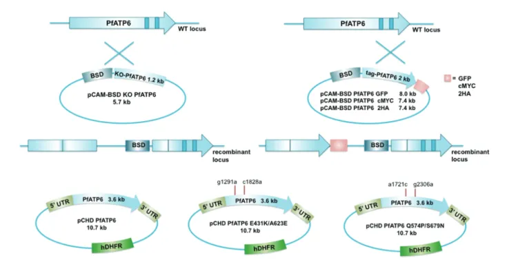
Genetic validation of PfATP6 as a drug target has not been reported to our knowledge. We carried out knockout (KO)/ complementation experiments in the reference line 3D7 with a single crossover homologous recombination strategy (Figure1A), as with the glucose transporter, PfHT [14]. Two attempts were unsuccessful, including complementation with wild-type or double mutated full length pfatp6 (Figure1C) and tagging the 3′ end of the gene with green fluorescent protein (GFP), myc, or double HA epitopes (Figure 1B). In all cases,
blasticidin-resistant parasites were obtained with episomes. Similarly, a double recombination construct failed to KO pbserca in the more tractable Plasmodium berghei system, and C-terminal tagging was also unsuccessful (data not shown). Thus, pfatp6 is accessible for crossovers introducing single mutations, support-ing the idea that it cannot be knocked out [9,10] or tagged at the C-terminus. We developed an alternative expression system to study the function of PfATP6 and mutations including those infield isolates.
Yeast Expression
To avoid extensive purification and reconstitution after expres-sion in yeast [11] or laborious assays of membrane preparations of Xenopus oocytes [3], we adapted a yeast functional assay used to express several exogenous eukaryotic Ca2+ATPases [15]. This assay has the advantage of assessing drug action in intact cells. The host strain (K667) lacks one of its 2 Ca2+ ATPase genes PMC1, whereas the other gene, PMR1, is inactive as calci-neurin is also missing. A codon-optimized version of pfatp6 was required for yeast expression [11].
PfATP6 was localized to cytosolic structures in yeast (Figure 2A, upper panel) with a similar pattern in parasites (Figure2A, lower panel) that is consistent with distribution of endoplasmic reticulum (ER). To verify this localization, PfATP6 was compared in ER-enriched microsomal membranes and
Figure 1. Strategy for knockout (KO), complementation and tagging of pfatp6 gene in Plasmodium falciparum 3D7. A, Single cross-over homologous recombination of the KO ( pCAM-BSD KOA6) plasmid and the endogenous gene (WT locus) should result in 2 truncated nonfunctional copies of the gene (recombinant locus). B, Tagging of the 3′ end of PfATP6 locus with green fluorescent protein (GFP), double HA or cmyc tags. Wild-type (WT) and recombi-nant loci are shown. Arrows indicate the location of polymerase chain reaction (PCR) primers. C, Complementation constructs, carrying WT and mutated PfATP6 ( pCHD-A6) would allow PfATP6 expression under the Pfhsp86 promoter. Complementation of KO parasites without a tagged pfatp6 construct was not successful, and neither was episomal expression only of PfATP6.
whole cell extracts from yeast by Western analysis (Figure2B). Endogenous yeast non-SERCA P type Ca2+ ATPases (Pmr1p and Pmc1p) in reference strain BY4741 are not detected by anti-PfATP6 anti-peptide polyclonal reagent (Figure 2B, lane a). PfATP6 is detected in membrane extracts from both trans-genic yeast and parasites (P. falciparum 3D7; Figure2B, lane b and right panel).
Yeast Assays
To confirm function, we first established conditions for yeast growth that depend on rescue by PfATP6. A reference strain (BY4741) with 2 Ca2+ATPases can grow on solid (YPD) me-dium supplemented with CaCl2(0–100 mM, Figure3A, upper row). As expected, strain K667 cannot grow when stressed with ambient calcium concentrations >25 mM (Figure3A, 2nd row), and the addition of an empty vector (K667::pUGpd; Figure3A, 3rd row) does not alter this calcium sensitivity. Growth of K667 is restored toward that of the reference strain by expressing PfATP6, albeit not to wild-type levels (K667::pfatp6; Figure3A, last row).
Liquid cultures allow quantitation of growth and show that PfATP6 expressing yeast (OD620nm) grow significantly better
in media supplemented with≥50 mM CaCl2 compared with yeast strains (K667 and K667::pUGpd) without Ca2+ATPases (Figure3B; P < .001). PfATP6 cannot restore growth in increas-ing manganese concentrations, confirmincreas-ing that (as with recon-stituted protein [11]) it acts specifically as a Ca2+ATPase. This manganese sensitivity also confirms that the Ca2+
/Mn2+ ATPase PMR1 is inactive in K667 (Figure3C). Growth curves for yeast in different calcium concentrations were used to select 42 hours as the time point for assessing effects of drugs ( Sup-plementary Figure 1). This allows discrimination of growth rates at the plateau phase (see Methods). The mean ± standard deviation (SD) interassay coefficient of variation (CV) for pUGpd was 1.85% ± 0.017% (calculated as an overall mean (n = 28) using the mean CV of 3 biological replicates after assays with artemisinins and cyclopiazonic acid [CPA] at all re-ported concentrations) and for K667::pfatp6 was 6.65% ± 0.07% (calculated as for pUGpd with 40 mM CaCl2in medium, and including assays for chloroquine and thapsigargin (n = 36)).
BY4741 growth without calcium is inhibited by artemisinins perhaps by inhibition of yeast mitochondrial function [16] and through effects on its own Ca2+ATPases [17]. Strain K667 can
Figure 2. PfATP6 in yeast and parasites. A, Upper panel: Indirect immunofluorescence with anti-PfATP6 polyclonal antibody on Saccharomyces cerevi-siae K667::pfatp6 confirms expression in cytosolic structures (green) with nuclei stained with DAPI (blue). Lower panel: A similar distribution is apparent in asexual stage parasites (large trophozoites of Plasmodium falciparum strain 3D7). B, Upper panel: Western blot of total protein extract of yeast (10 µg, left) and parasites (10 µg, right). Reference strain BY4741 (lane a) is a negative control, whereas K667::pfatp6 shows a band of predicted mass (upper arrow). A similar band is detected on parasite extracts (3D7). Lower panel: Reference yeast strain (BY4741, 10 µg lane a) and K667::pfatp6 were used to make ER-enriched preparations (Methods) and assayed with anti-PfATP6 antibody. Only K667::pfatp6 (10 µg lane b) shows PfATP6.
only be grown without calcium, so artemisinins can still inhibit growth by acting on mitochondria but not on yeast Ca2+ ATPases. We therefore used 40 mM CaCl2, in liquid cultures to screen artemisinins for their ability to inhibit rescue of K667 by PfATP6. These conditions do not permit K667 to grow without PfATP6, allowing the conclusion that inhibition is of PfATP6′s ability to rescue yeast. All artemisinins inhibited yeast growth significantly (Figure4A, P < .001).
CPA inhibits SERCAs at a different binding site from thapsi-gargin [18] (where we hypothesized that artemisinins may act), and significant inhibition is also observed with CPA (Figure4A). Several experiments confirmed the specificity of these inhibi-tors. Growth of K667 is not inhibited by dimethyl sulfoxide solvent (1% maximum, not shown) and CPA (up to 100 µM, Figure4B) did not inhibit any strain without PfATP6 (BY4741, K667, K667::pUGpd).
Table1A includes results with different concentrations of in-hibitors of K667::pfatp6. 2-deoxy artemisinin lacks an endoper-oxide bridge and is therefore inactive as an antimalarial and
interestingly does not inhibit growth. Chloroquine, an antima-larial that acts in the parasite’s food vacuole, also does not inhibit K667::pfatp6. As is conventional, we are careful not to translate concentrations that inhibit K667::pfatp6 to those re-quired to kill parasites. Higher concentrations of ATPase inhib-itors are needed to inhibit yeast growth compared with potencies in biochemical assays [18].
Results from K667::pUGpd (vector control) are not directly comparable to those with K667::pfatp6, as the former have been grown without supplementary calcium.Supplementary Table 1A presents the effects of inhibitors on pUGpd, representing inhi-bition that is not dependent on PfATP6. Most inhibitors have little or no effect. Thapsigargin and CPA inhibit growth in K667::pfatp6 but not in K667::pUGpd, consistent with a specif-ic interaction with PfATP6 (Table 1A and Supplementary Table 1A). Some artemisinins inhibit K667::pUGpd at higher concentrations (for example, artemisone (100 µM) inhibits by approximately 25%) but all Ca2+ATPase inhibitors (including artemisinins) inhibit K667::pfatp6 significantly more.
Figure 3. Functional assay of PfATP6 in growing yeast. A, Saturated liquid cultures of yeast cells were diluted (as indicated by OD600values) and
spotted onto yeast peptone dextrose (YPD) plates supplemented with 25, 50, or 100 mM CaCl2. BY4741 (upper row) tolerates all calcium concentrations
whereas K667 and K667::pUGpd (bearing empty vector; middle rows) cannot grow in calcium≥50 mM. PfATP6 (K667::pfatp6) restores growth in higher ambient calcium (lower row). B, K667 (bars), K667::pUGpd (red bars) and K667::pfatp6 (blue bars) were diluted to a cell density of 0.5 and inoculated into YPD medium containing CaCl2at indicated concentrations. Yeast cell density was determined after growth at 30°C for 18 h. Data are mean ± SEM values
of at least 9 replicates (3 independent biological experiments). PfATP6 rescue increases growth of K667 significantly (***P < .0001). C, K667 (bars), K667:: pUGpd (blue bars), and K667::pfatp6 (red bars) were diluted to a cell density of 0.5 and inoculated into YPD medium containing MnSO4at indicated
concen-trations. Data are mean ± SEM values of at least 9 replicates (3 independent biological experiments). There are no significant differences between groups (P > .05).
To confirm that mitochondrial inhibition by artemisinins is not relevant to inhibition of K667::pfatp6, we assessed the effects of adding deferoxamine (DFO). Previously, the artemisi-nin susceptibility of yeast mitochondria was antagonized by DFO [16]. In contrast, DFO increased the inhibition of yeast growth by artemisinin (10 µM, Supplementary Table 1B) and had no effect on actions of artemether or artemisone. At the highest concentrations of artesunate there was some antago-nism with DFO.
Mutations in PfATP6
Next we tested how mutations in PfATP6 influence artemisinin activity. These assays (in extracellular calcium between 10 and 30 mM) examined mutations (L263E, A623E, S769N A623E/ S769N) by comparing results with wild-type PfATP6 and vector only controls (Figure5A, lower panel).
All pfatp6 constructs rescued K667 growth in 10- and 20-mM calcium, although S769N and A623E/S769N mutants were significantly less efficient in 20-mM calcium. In 30-mM calcium, all mutants of PfATP6 failed to rescue yeast in contrast to wild-type PfATP6 (Figure5A, lower panel). Western analysis
confirmed expression of mutant PfATP6 proteins (Figure5A, upper panel). Mutations in PfATP6 reduce the efficiency of yeast rescue, presumably by interfering with function. We then tested sensitivity of mutant PfATP6 sequences to artemisinins and compared them with wild-type PfATP6 (Table 1B and Supplementary Table 2for 95% confidence intervals [CIs] of mean differences between groups). S769N attenuated artemisi-nins’ inhibition (except for artesunate 1 µM and artemisone 100 µM). For a double mutant (PfATP6 A623E/S769N) there was also significant attenuation of inhibition by all artemisinins (10 µM). In contrast, higher concentrations of artemisone (100 µM) [19] produced even greater inhibition on all mutants. The A623E mutant had less marked effects, failing to attenuate arte-sunate’s inhibition.
In Xenopus oocyte membrane preparations, there was abolition of sensitivity of PfATP6 to artemisinins [4]. We confirmed in yeast that L263E PfATP6 significantly reduces but does not abolish sensitivity to all artemisinins (Table1B), perhaps re-flecting differences in 3 amino acid residues in PfATP6 sequence expressed in Xenopus compared with the wild-type (3D7) PfATP6 sequence expressed in yeast (sequence differences
Figure 4. Sensitivity of K667 with and without rescue by PfATP6 to artemisinins and cyclopiazonic acid. A, Inhibition of K667::pUGpd (blue bars) and K667::pfatp6 (red bars) by different antimalarials expressed as percentage of control cultures (growth of each strain in absence of the drug). Yeast strains were diluted to a cell density of 0.1 and inoculated into YPD medium (with 40 mM CaCl2for experiments with K667::pfatp6) and 10 µM of drug as
indicat-ed. Yeast cell density was determined after growth at 30°C for 42 h ( plateau phase,Supplementary Figure 4). Data are mean ± SEM values of at least 9 replicates (3 independent biological experiments) with significant differences between growth of yeast without PfATP6 and supplementary Ca2+and yeast with PfATP6 and supplementary Ca2+(***P < .001). B, CPA does not inhibit growth of yeast strains not expressing PfATP6 (BY4741, K667 and K667:: pUGpd). Yeast strains were diluted to a cell density of 0.1 and inoculated into YPD medium containing various concentration of drug as indicated (bars, no drug, light green, 1 µM, medium green, 10 µM, dark green, 100 µM). CPA structure is reported. Abbreviations: AM, artemether; ART, artemisinin; AS, artesunate; ATS, artemisone; CPA, cyclopiazonic acid; DHA, dihydroartemisinin.
Table 1. Sensitivity of K667 Expressing Wild Type and Mutated PfATP6 to Antimalarials (µM) Growth (% control) PfATP6 (A) artemisinin 1 100.2 ± 1.1 10 87.3 ± 1.2* 100 76.4 ± 1.2* artesunate 1 90.1 ± 0.8 10 82.8 ± 1.3* 100 72.6 ± 0.8* artemether 1 94.1 ± 2.7 10 80.7 ± 3.0** 100 77.1 ± 2.8** artemisone 1 90.0 ± 0.3* 10 80.2 ± 0.9* 100 56.3 ± 1.2* DHA 1 80.9 ± 0.9* 10 69.1 ± 1.4* 100 65.8 ± 3.0* CPA 1 79.5 ± 6.7** 10 31.7 ± 2.4** 100 24.0 ± 1.7** thapsigargin 1 100.5 ± 0.5 10 99.5 ± 0.6 100 86.9 ± 0.8* chloroquine 1 101.6 ± 1.4 10 102.3 ± 1.0 100 100.4 ± 1.2 2de-QHS 1 100.9 ± 1.4 10 103.0 ± 1.5 100 98.1 ± 0.9
(µM) PfATP6 wt A623E/S769N A623E S769N L263E (B) artemisinin 1 101.9 ± 1.0 104.7 ± 1.2 104.3 ± 1.7 105.3 ± 1.7 104.4 ± 2.1 10 91.7 ± 0.6 101.7 ± 1.1*** 100.5 ± 1.6*** 105.3 ± 1.8*** 104.5 ± 1.9*** 100 71.8 ± 2.9 91.8 ± 0.5*** 73.6 ± 1.8 82.9 ± 0.5**** 75.4 ± 2.2 artesunate 1 96.6 ± 0.6 97.9 ± 2.0 96.6 ± 0.9 91.9 ± 1.1#### 98.8 ± 0.9 10 85.1 ± 0.9 91.1 ± 1.1*** 86.9 ± 0.6 87.8 ± 1.1 89.0 ± 0.6**** 100 70.5 ± 2.3 85.4 ± 0.9*** 70.2 ± 2.4 78.5 ± 0.8**** 72.2 ± 2.2 artemether 1 100.1 ± 0.6 104.1 ± 1.5***** 100.0 ± 1.1 100.9 ± 1.5 103.5 ± 1.4***** 10 87.9 ± 1.5 95.7 ± 1.8**** 99.7 ± 1.6*** 103.0 ± 1.2*** 104.2 ± 1.3*** 100 65.7 ± 2.4 59.0 ± 4.1 64.6 ± 1.4 74.8 ± 1.6**** 69.3 ± 2.4
between PfATP6 in oocytes and the 3D7 sequence can be com-pared with accession numbers PF3D7_0106300 and the original sequence in [3]). Results (Table1B) suggest that >1 mutation in PfATP6 may synergize to reduce sensitivity to artemisinins.
It is striking that despite reduced capacity of mutant PfATP6 sequences to rescue yeast grown in calcium, they show de-creased rather than inde-creased sensitivity to artemisinins. To confirm this effect is specific for artemisinins, we tested sensi-tivity of mutants to CPA and found it was increased (Figure5B) compared with wild-type PfATP6. We then examined parasites with the L263E mutation to determine if they had increased sensitivity to CPA, as predicted by results in yeast. Figure5C demonstrates that the IC50 for CPA approximately halved, compared with an isogenic control (mean ± SEM for L263 = 10.3 ± 2.7 compared with 263E = 4.7 ± 1.1 µM; n = 5 P = .011). There was no change in sensitivity to chloroquine (0.12 ± 0.01 compared with 0.14 ± 0.01 µM respectively).
New Chemical Classes
Figure6A shows how a new chemical class introducing an en-doperoxide bridge into the sesquiterpene scaffold of thapsigargin (guaianolide-endoperoxides [12]) inhibits K667::pfatp6 without inhibiting control yeast strains (eg, growth of strain BY4741 with or without calcium supplementation (Figure6B)). DISCUSSION
Whole cell expression in yeast provides new insights into PfATP6 as a drug target as well as a screening tool for discovery
of inhibitors. Results confirm findings from Xenopus oocytes that all endoperoxides tested inhibit PfATP6 [3]. Yeast assays have also elucidated functions of a Schistosoma mansoni Ca2+ ATPase [20], a Fasciola hepatica plasma membrane Ca2+ ATPase and mammalian SERCA1a. SERCA1a is sensitive to thapsigargin [18] as well as to artemisinin [21], suggesting that experiments with purified and relipidated PfATP6 may compro-mise assessment of different inhibitor classes [11,22]. Reconstitu-tion may not achieve full physiological funcReconstitu-tionality as PfATP6 operates at one third the rate of mammalian SERCA [11].
Confounding by off-target effects of artemisinins in this yeast model [16] has been minimized because assays have been designed to rely on function of PfATP6, by selecting fermenta-tive conditions when mitochondria are metabolically inacfermenta-tive. Also, the lack of antagonism (and sometimes synergism) by DFO of artemisinin inhibition of PfATP6 contrasts with an-tagonism in yeast in nonfermentative conditions [16] when artemisinins cause mitochondrial depolarization. Transgenic parasites that no longer rely on mitochondria for asexual stage sur-vival do not show increased resistance to artemisinins, as would be predicted if mitochondria were important targets in vivo [23]. Artemisinins also inhibit the SERCAs of Toxoplasma [24,25] and cancer cell lines in membrane preparations [26] supporting results from PfATP6 [3]. Alternative hypotheses for mechanisms of action of artemisinins continue to be investigated to increase un-derstanding of this important class of drug [8].
In modeling studies, thapsigargin, artemisinins, and thaper-oxide [4, 12, 27, 28] may share a common binding site in PfATP6 different from that which binds CPA [29]. Inhibition
Table 1 continued. (µM) Growth (% control) PfATP6 artemisone 1 91.9 ± 0.7 100.3 ± 0.6*** 98.4 ± 1.7**** 96.4 ± 1.8***** 101.0 ± 2.0*** 10 83.1 ± 0.7 96.5 ± 1.8*** 86.9 ± 1.2***** 91.9 ± 1.3*** 89.6 ± 1.7**** 100 45.2 ± 4.2 56.1 ± 1.9 34.5 ± 2.2##### 30.9 ± 1.7#### 34.0 ± 2.8##### DHA 1 90.7 ± 0.7 92.0 ± 1.7 94.9 ± 0.9**** 91.8 ± 2.1 95.8 ± 1.1**** 10 82.7 ± 0.6 88.5 ± 1.7**** 82.2 ± 1.1 82.1 ± 2.3 80.1 ± 1.1 100 61.7 ± 3.0 72.6 ± 2.7***** 63.9 ± 3.1 60.7 ± 6.5 62.8 ± 3.4
(A) Sensitivity of K667 expressing PfATP6 to antimalarials. Results (mean±SEM) are expressed as percentage of control (growth in absence of drug). Data are the mean of at least 9 replicates in 3 independent experiments. Growth in presence of drug was compared to growth without drug (in 40 mM CaCl2) with ANOVA. (B)
Sensitivity of K667 expressing PfATP6 (wild-type or mutated, as shown) to artemisinin derivatives. Growth is described as percentage of control (growth in YPD medium supplemented with 20 mM CaCl2) Data are mean ± SEM values of at least 9 replicates (3 independent experiments). Studentt test was used to compare
growth of mutated PfATP6 to wild-type. (Supplementary Table 2summarizes 95% CAs of mean difference.) *indicates decreased sensitivity when compared to PfATP6 wild-type, whereas # indicates increased sensitivity.
Abbreviations: CPA, cyclopiazonic acid; DHA, dihydroartemisinin. *P < .0001.
**P < .0005. ***P < .0009. ****P < .0061. *****P < .0385.
by CPA of PfATP6 is demonstrable in 3 systems [3]: after ex-pression in membranes of Xenopus oocytes, in yeast assays (this study), and after reconstitution [11]. When CPA inhibition is assayed with the L263E mutation in PfATP6, a compromise in function in yeast increases sensitivity. This is paralleled by in-creased sensitivity of parasites with the L263E mutation, pro-viding an estimate of the degree of functional compromise (halving of IC50values) caused by such mutations for CPA.
This may explain why transfection of P. falciparum with mutated PfATP6, or why field isolates with mutations, yield variable results for susceptibility to artemisinins. Mutations in PfATP6 reduce sensitivity to artemisinins, but because PfATP6 function is compromised, this may not translate to large in-creases in IC50. A“corrected” IC50value for artemisinins may be obtainable by normalizing results with simultaneous IC50 values for CPA.
The L263E mutation may also show less artemisinin resistance in parasites than in oocytes because sequences assayed in oocytes
and parasites were not identical and also because of differences between whole cell assays (parasites or yeast) and in Xenopus membrane preparations. In parasites, other transport proteins such as PfMDR1 may modulate the artemisinin resistance phe-notype [9,30]. The proposedfitness cost of mutations in PfATP6 may then cause parasites to be outgrown in culture when adapted from patients [31]. Linking mutations in PfATP6 infield isolates to artemisinin resistance could then depend on genetic background, including compensatory mutations in proteins in-volved in calcium homeostasis. Background is critical to assessing the impact of other resistance genes such as pfcrt [32,33].
The yeast model may therefore allow rapid assessment of functional consequences of mutations in PfATP6, by eliminat-ing variables in parasite assays and overcomeliminat-ing the difficulty of establishing long-term cultures in parasites harboring pfatp6 field mutations. These mutations may modify the propensity of peroxides to cleave SERCAs through several mechanisms [34]. The increased susceptibility of mutant PfATP6 to CPA (Figure5),
Figure 5. Effect of mutations in K667::pfatp6 and susceptibility of mutated P. falciparum to chloroquine and cyclopiazonic acid. A, Upper panel: Western blot of total yeast protein. Anti-PfATP6 polyclonal antibody does not detect a band from yeast without PfATP6 (K667::pUGpd), whereas K667::pfatp6 and mutants express protein. Growth of K667 (assayed as in Figure3) with vector only or PfATP6 sequences was examined at different concentrations of calcium in yeast peptone dextrose (YPD) and expressed as a percentage of control values after growth without calcium. Data are mean ± SEM values (at least 9 replicates and 3 independent experiments). S769N PfATP6 and A623E/S769N PfATP6 impair growth rescue of yeast in higher Ca2+(≥20 mM) con-centrations (PfATP6 vs A623E/S769N **P = .0079 PfATP6 vs S769N **P = .0024; Student t test). Ten outliers identified by Grubbs test were removed from a total of 504 replicates. B, Sensitivity of K667::pfatp6 wild-type (dark gray bars) and pfatp6 mutants (light-gray bars) to CPA (in 20 mM CaCl2). Yeast were
assayed and analyzed as in 4a with indicated concentrations of CPA. Inhibition by CPA of PfATP6 wild-type and individual mutants *** P < .0001. C, IC50
values for chloroquine (CQ) and CPA were estimated with [3H]hypoxanthine uptake in growth assays using synchronized ring-stage cultures of Plasmodium falciparum (L263 7G8 and L263E 7G8). Growth was measured at 48 h as described by Desjardin et al [50] IC50s ± SEM are the mean of 5 independent
in contrast to artemisinins encourages pursuit of CPA deriva-tives as new leads.
A novel thaperoxide class of antimalarials was developed to test the hypothesis that PfATP6 is targeted by artemisinins. In-troduction of a peroxide bridge into the sesquiterpene heart of thapsigargin increases antimalarial potency of thapsigargin by approximately 100-fold. The most potent derivative (thaperox-ide 22) also inhibits K667::pfatp6, confirming that it remains on target and providing assays for lead optimization (Figure6). Modeling studies of thaperoxides with PfATP6 and mutants provide testable predictions [35].
Genetic validation of PfATP6 as a drug target should stimu-late this further and has been attempted using KO and comple-mentation methods, twice in 2 species of Plasmodium (as with PfHT [14]), suggesting that SERCA function is essential for blood stages of infection. Tagging the 3′ end of pfatp6 and pbserca has not been possible despite accessibility of pfatp6 for single site recombination experiments (Figure1) [10].
Resistance to artemisinins can be defined as for other classes of antimalarial by a decrease in susceptibility of cultured para-sites to one or more derivatives. As resistance increases, it may increase treatment failures of some artemisinin combination therapies. The magnitude of increase in resistance in vitro that is associated with treatment failures in vivo has not been defined [36] although it is noteworthy that some parasites with S769N PfATP6 have IC50values >100 nM for artemether [13,
37]. A requirement for compensatory mutations may have limited the spread of resistance associated with polymorphisms in PfATP6 at present.
An in vivo definition of artesunate resistance that manifests itself in prolonged parasite clearance estimators has been reported with association of this phenotype to parasite’s chromo-some 13 [38]. This phenotype includes important contributions from host [39] as well as parasite factors in assessment (parasite heritability of approximately 60%), thereby adding to complexi-ties of understanding underlying mechanisms. The contribution of parasite dormancy also needs to be ascertained [40].
Almost 2 decades after P-type cation ATPases were hypothe-sized to be important drug targets in parasites [41], evidence continues to accumulate in support of SERCAs as targets for artemisinins [8]. Other calcium pumps [5] are now being ex-ploited as targets for new classes of drug such as spiroindo-lones [42], and the assays presented here may also be useful for their study.
METHODS
Reagents
All reagents were purchased from Sigma Aldrich Chemical Co except for artemisone (made by R. K. H.) and thaperoxide 22 (made by F. S. and M. A.).
Plasmid Construction
An open reading frame of PfATP6 was codon-optimized for ex-pression in S. cerevisiae (GeneScript USA Inc). A plasmid con-taining the wild-type pfatp6 gene regulated by the strong constitutive glyceraldehyde-3-phosphate dehydrogenase (GPD) promoter TDH3, was constructed by subcloning the BamHI-XbaI fragment from commercial vector pcc1 into the expres-sion vector pUGpd (Ura selectable marker) [43]. Vector pUGpd is a yeast centromere plasmid containing a yeast cen-tromere sequence (CEN) and autonomously replicating se-quence (ARS), which confers mitotic and meiotic stability. Mutants of PfATP6 were generated using QuikChange Light-ning Site-Directed Mutagenesis Kit (Stratagene).
Yeast Transformation
The K667 yeast (Saccharomyces cerevisiae) strain (MATa; pmc1::TRP1; cnb1::LEU2; vcx1Δ; ura3-1) [44] was transformed with pUGpd-pfatp6, L263E,
pUGpd-pfatp6-Figure 6. Sensitivity of K667::pfatp6 to different concentration of tha-peroxide 22. A, K667::pfatp6 was diluted to a cell density of 0.1 and inocu-lated into yeast peptone dextrose (YPD) medium supplemented with 40 mM CaCl2and thaperoxide 22 as indicated. Yeast cell density was
deter-mined after growth at 30°C for 42 h. Data are mean ± SEM of 6 replicates (2 independent experiments). Growth in presence of the drug was com-pared to controls without drug using Student unpaired t test. Inhibition at 100 µM is significant (*** P < .0001). The chemical structure of thaperox-ide 22 is reported. B, BY4741 grown at normal or higher Ca2+ concentra-tion and K667 expressing pUGpd are not inhibited by thaperoxide 22 (black bars, no drug; blue bars, 100 µM thaperoxide 22.
A623E, pUGpd-pfatp6-S769N and pUGpd-pfatp6-A623E/ S769N plasmids, or pUGpd vector only using a Li-acetate method [45]. Transformants were selected on SD medium lacking uracil to yield strains K667::pfatp6, K667::pfatp6-L263E, K667::pfatp6-A623E, K667::pfatp6-S769N, K667::pfatp6-A623E/S769N, and K667::pUGpd.
Protein Extraction, SDS-PAGE and Western Blotting
SDS gel electrophoresis and Western blots were performed on yeast total cell lysates prepared from equivalent numbers of log phase cells grown in SC-ura medium. Cells were pelleted and lysed by bead beating in SUMEB buffer supplemented with cOmplete ETDA-free Cocktail tablets protease inhibitor (Roche). ER-enriched microsomal membranes were prepared as described elsewhere [46,47]; 10 µg of protein was used to load gels. Blots were probed with goat anti-PfATP6 antibody, followed by mouse rabbit anti-goat immunoglobulin G (IgG) (H + L)-HRP conjugate secondary (BioRad). Parasite total protein was obtained from saponin freed 3D7 parasites, treated with 0.5% Triton X-100 [48].
Indirect Immunofluorescence of Yeast and Parasites
Polyclonal anti-PfATP6 reagent generated in goat from the peptide CQSSNKKDKSPRGINK (the sequence from Q to K corresponds to the 574–588 region of PfATP6) was used [11]. Immunofluorescence assay (IFA) on K667::pfatp6 and K667:: pUGpd was with anti-PfATP6 antibody as described else-where [49]. IFA on P. falciparum 3D7 was performed in solu-tion, by fixing infected red cells with 4% paraformaldehyde/ 0.0075% glutaraldehyde for 30 minutes. Cells were washed in PBS, permeabilized for 10 minutes (0.1% Triton X-100 in phos-phate-buffered saline [PBS]) blocked with PBS/3% BSA, and incubated with anti-PfATP6 antibody for 1 hour and then with rabbit anti-goat IgG FITC for 30 minutes. Images were taken using a Nikon TE2000 inverted microscope.
Growth Assays
BY4741 (MATa; his3Δ1; leu2Δ0; met15Δ0; ura3Δ0) yeast and K667 transformed and untransformed strains were used for growth assays. For spot assays cells were grown in liquid culture overnight at 30°C (in synthetic complete [SC] without uracil and containing 20 g L-1 glucose). After adjusting to OD600= 0.5, 0.1, 0.02, 0.004, aliquots were inoculated onto agar 2% with yeast peptone dextrose (YPD) medium and various additions and incubated at 30°C for 3–5 days. To determine growth rate in liquid CaCl2 solutions, yeast cultures were inoculated in YPD medium and grown at 30°C, shaking for 42 hours in 96-wellflat bottomed plates, with absorbance measured at 620 nm. Drug assays were at specified concentrations and growth re-corded at plateau phase (18 hours at basal Ca2+concentration and 42 hours at higher Ca2+ concentrations). Concentrations
between 20 and 40 mM calcium were assayed before perform-ing experiments.
Statistical Analysis
Growth was measured as OD620 or as percentage of control (growth in YPD medium only) at different conditions and compared by Student t-test (with 2 alpha considered significant at P < .05). When groups of 3 drug concentrations were com-pared with growth without drug (Table1A andSupplementary Table 1A), then ANOVA with Dunnett test was applied. Possi-ble outliers were assessed and removed after applying Grubbs test where indicated in results. All analyses were in GraphPad (Prism v6.0a).
Supplementary Data
Supplementary materialsare available at The Journal of Infectious Diseases online (http://jid.oxfordjournals.org/). Supplementary materials consist of data provided by the author that are published to benefit the reader. The posted materials are not copyedited. The contents of all supplementary data are the sole responsibility of the authors. Questions or messages regarding errors should be addressed to the author.
Notes
Acknowledgments. We thank U. Straschil (Imperial College) and D. Guttery (Nottingham University) for essential help with P. berghei infec-tions.
Funding. This work was supported by EU FP7 Marie Curie-funded Initial Training Network InterMal (Grant Agreement No. 215281-2). Work in the SK and CD laboratories was supported in part by the EU-FP7 MALSIG programme and some research leading to these results has re-ceived funding from the European Union Seventh Framework Programme ([FP7/2007-2013]) under grant agreement n° 304948 - NANOMAL.
Author contribution. S. K. and H. S. conceived experiments carried out and analyzed by S. P. and K. S., J. K. P. supervised work with yeast, C. D. and J. H. supervised transfection studies with P. falciparum and R. T. with P. berghei, F. S. and M. A. synthesized thaperoxide, and R. K .H. the artemisinins. All authors contributed to writing the article that was drafted by S. K.
Potential conflicts of interest. S. K. has acted as an advisor to GSK for early drug discovery in the past and S. K. and H. S. are share holders in QuantuMDx. All other authors: No reported conflicts.
All authors have submitted the ICMJE Form for Disclosure of Potential Conflicts of Interest. Conflicts that the editors consider relevant to the content of the manuscript have been disclosed.
References
1. Skou JC. Nobel Lecture. The identification of the sodium pump. Biosci Rep 1998; 18:155–69.
2. Olbe L, Carlsson E, Lindberg P. A proton-pump inhibitor expedition: the case histories of omeprazole and esomeprazole. Nat Rev Drug Discov 2003; 2:132–9.
3. Eckstein-Ludwig U, Webb RJ, Van Goethem ID, et al. Artemisinins target the SERCA of Plasmodium falciparum. Nature 2003; 424:957–61. 4. Uhlemann AC, Cameron A, Eckstein-Ludwig U, et al. A single amino acid residue can determine the sensitivity of SERCAs to artemisinins. Nat Struct Mol Biol 2005; 12:628–9.
5. Krishna S, Woodrow C, Webb R, et al. Expression and functional char-acterization of a Plasmodium falciparum Ca2+-ATPase (PfATP4) be-longing to a subclass unique to apicomplexan organisms. J Biol Chem 2001; 276:10782–7.
6. Krishna S, Pulcini S, Fatih F, Staines H. Artemisinins and the biological basis for the PfATP6/SERCA hypothesis. Trends Parasitol 2010; 26:517–23. 7. Tilley L, Davis TM, Bray PG. Prospects for the treatment of
drug-resistant malaria parasites. Future Microbiol 2006; 1:127–41.
8. Golenser J, Waknine JH, Krugliak M, Hunt NH, Grau GE. Current per-spectives on the mechanism of action of artemisinins. Int J Parasitol 2006; 36:1427–41.
9. Cui L, Wang Z, Jiang H, Parker D, Wang H, Su XZ. Lack of association of the S769N mutation in Plasmodium falciparum SERCA (PfATP6) with resistance to artemisinins. Antimicrob Agents Chemother 2012; 56:2546–52.
10. Valderramos SG, Scanfeld D, Uhlemann AC, Fidock DA, Krishna S. In-vestigations into the role of the Plasmodium falciparum SERCA (PfATP6) L263E mutation in artemisinin action and resistance. Anti-microb Agents Chemother 2010; 54:3842–52.
11. Cardi D, Pozza A, Arnou B, et al. Purified E255L mutant SERCA1a and purified PFATP6 are sensitive to SERCA-type inhibitors but insensitive to artemisinins. J Biol Chem 2010.
12. Sun L, Shah F, Helal MA, et al. Design, synthesis, and development of novel guaianolide-endoperoxides as potential antimalarial agents. J Med Chem 2010; 53:7864–8.
13. Jambou R, Legrand E, Niang M, et al. Resistance of Plasmodium falcip-arumfield isolates to in- vitro artemether and point mutations of the SERCA-type PfATPase6. Lancet 2005; 366:1960–3.
14. Slavic K, Straschil U, Reininger L, et al. Life cycle studies of the hexose transporter of Plasmodium species and genetic validation of their essen-tiality. Mol Microbiol 2010; 75:1402–13.
15. Pittman JK. Vacuolar Ca2+uptake. Cell Calcium 2011; 50:139–46. 16. Wang J, Huang L, Li J, et al. Artemisinin directly targets malarial
mito-chondria through its specific mitomito-chondrial activation. PLoS One 2010; 5:e9582.
17. Moore CM, Hoey EM, Trudgett A, Timson DJ. Artemisinins act through at least two targets in a yeast model. FEMS Yeast Res 2011; 11:233–7. 18. Johnson NA, Liu F, Weeks PD, et al. A tomato ER-type Ca2+-ATPase,
LCA1, has a low thapsigargin-sensitivity and can transport manganese. Arch Biochem and Biophys 2009; 481:157–68.
19. Pooley S, Fatih FA, Krishna S, et al. Artemisone uptake in Plasmodium falciparum-infected erythrocytes. Antimicrob Agents Chemother 2011; 55:550–6.
20. Talla E, de Mendonca RL, Degand I, Goffeau A, Ghislain M. Schistoso-ma Schistoso-mansoni Ca2+-ATPase SMA2 restores viability to yeast Ca2+ -ATPase-deficient strains and functions in calcineurin-mediated Ca2+
tolerance. J Biol Chem 1998; 273:27831–40.
21. Moore CM, Hoey EM, Trudgett A, Timson DJ. A plasma membrane Ca2+-ATPase (PMCA) from the liverfluke, Fasciola hepatica. Int J Para-sitol 2012; 42:851–8.
22. Racker E. Reconstitutions of transporters, receptors and pathological states. London, UK: Academic Press, 1985.
23. Vaidya A. Naphthoquinones: atovaquone, and other antimalarials tar-geting mitochondrial function. In: Staines H, Krishna S, eds. Treatment and preventions of malaria Antimalarial drug chemistry, action and use. 1st. ed. Basel: Springer, 2012:315.
24. Nagamune K, Beatty WL, Sibley LD. Artemisinin induces calcium-de-pendent protein secretion in the protozoan parasite Toxoplasma gondii. Eukaryot Cell 2007; 6:2147–56.
25. Nagamune K, Moreno SN, Chini EN, Sibley LD. Calcium regulation and signaling in apicomplexan parasites. Subcell Biochem 2008; 47:70–81. 26. Riganti C, Doublier S, Viarisio D, et al. Artemisinin induces
doxorubi-cin resistance in human colon cancer cells via calcium-dependent acti-vation of HIF-1alpha and P-glycoprotein overexpression. Br J Pharmacol 2009; 156:1054–66.
27. Naik PK, Srivastava M, Bajaj P, et al. The binding modes and binding affinities of artemisinin derivatives with Plasmodium falciparum Ca2+
-ATPase (PfATP6). J Mol Model 2010.
28. Lepore R, Simeoni S, Raimondo D, Caroli A, Tramontano A, Via A. Identification of the Schistosoma mansoni molecular target for the anti-malarial drug artemether. J Chem Inf Model 2011; 51:3005–16.
29. Yao S, Gallenkamp D, Wolfel K, Luke B, Schindler M, Scherkenbeck J. Synthesis and SERCA activities of structurally simplified cyclopiazonic acid analogues. Bioorg Med Chem 2011; 19:4669–78.
30. Bustamante C, Folarin OA, Gbotosho GO, et al. In vitro-reduced sus-ceptibility to artemether in P. falciparum and its association with poly-morphisms on transporter genes. J Infect Dis 2012.
31. Legrand E, Volney B, Meynard JB, Mercereau-Puijalon O, Esterre P. In Vitro monitoring of Plasmodium falciparum drug resistance in French Guiana: a synopsis of continuous assessment from 1994 to 2005. Anti-microb Agents Chemother 2008; 52:288–98.
32. Valderramos SG, Valderramos JC, Musset L, et al. Identification of a mutant PfCRT-mediated chloroquine tolerance phenotype in Plasmo-dium falciparum. PLoS Pathog 2010; 6:e1000887.
33. Klonis N, Crespo-Ortiz MP, Bottova I, et al. Artemisinin activity against Plasmodium falciparum requires hemoglobin uptake and diges-tion. Proc Natl Acad Sci USA 2011; 108:11405–10.
34. Moreau VH, Castilho RF, Ferreira ST, Carvalho-Alves PC. Oxidative damage to sarcoplasmic reticulum Ca2+-ATPase AT submicromolar iron concentrations: evidence for metal-catalyzed oxidation. Free Radic Biol Med 1998; 25:554–60.
35. Helal MA, Avery MA. Combined receptor-based and ligand-based ap-proach to delineate the mode of binding of guaianolide-endoperoxides to PfATP6. Bioorg Med Chem Lett 2012; 22:5410–4.
36. Noedl H, Se Y, Schaecher K, Smith BL, Socheat D, Fukuda MM. Evi-dence of artemisinin-resistant malaria in western Cambodia. N Engl J Med 2008; 359:2619–20.
37. Jambou R, Martinelli A, Pinto J, et al. Geographic Structuring of the Plasmodium falciparum Sarco(endo)plasmic Reticulum Ca2+ATPase (PfSERCA) gene diversity. PLoS One 2010; 5:e9424.
38. Phyo AP, Nkhoma S, Stepniewska K, et al. Emergence of artemisinin-resistant malaria on the western border of Thailand: a longitudinal study. Lancet 2012; 379:1960–6.
39. Amaratunga C, Sreng S, Suon S, et al. Artemisinin-resistant Plasmodi-um falciparPlasmodi-um in Pursat province, western Cambodia: a parasite clear-ance rate study. Lclear-ancet Infect Dis 2012; 12:851–8.
40. Codd A, Teuscher F, Kyle DE, Cheng Q, Gatton ML. Artemisinin-induced parasite dormancy: a plausible mechanism for treatment failure. Malar J 2011; 10:56.
41. Krishna S, Cowan G, Meade JC, Wells RA, Stringer JR, Robson KJ. A Family of cation ATPase-like molecules from Plasmodium falciparum. J Cell Bio 1993; 120:385–98.
42. Rottmann M, McNamara C, Yeung BK, et al. Spiroindolones, a potent compound class for the treatment of malaria. Science 2010; 329:1175–80. 43. Zhao J, Connorton JM, Guo Y, et al. Functional studies of split
Arabi-dopsis Ca2+/H+exchangers. J Biol Chem 2009; 284:34075–83. 44. Cunningham KW, Fink GR. Calcineurin inhibits VCX1-dependent
H+/Ca2+exchange and induces Ca2+ATPases in Saccharomyces cerevi-siae. Mol Cell Biol 1996; 16:2226–37.
45. Gietz D, St Jean A, Woods RA, Schiestl RH. Improved method for high efficiency transformation of intact yeast cells. Nucleic Acids Res 1992; 20:1425.
46. Hwang I, Harper JF, Liang F, Sze H. Calmodulin activation of an endo-plasmic reticulum-located calcium pump involves an interaction with the N-terminal autoinhibitory domain. Plant Physiol 2000; 122:157–68. 47. Funato K, Riezman H. Vesicular and nonvesicular transport of
cer-amide from ER to the Golgi apparatus in yeast. J Cell Biol 2001; 155:949–59.
48. Howard RJ, Lyon JA, Uni S, et al. Transport of an Mr approximately 300,000 Plasmodium falciparum protein (Pf EMP 2) from the intraery-throcytic asexual parasite to the cytoplasmic face of the host cell mem-brane. J Cell Biol 1987; 104:1269–80.
49. Kilmartin JV, Adams AE. Structural rearrangements of tubulin and actin during the cell cycle of the yeast Saccharomyces. J Cell Biol 1984; 98:922–33.
50. Desjardins RE, Canfield CJ, Haynes JD, Chulay JD. Quantitative assess-ment of antimalarial activity in vitro by a semiautomated microdilution technique. Antimicrob Agents Chemother 1979; 16:710–8.


![Figure 6A shows how a new chemical class introducing an en- en-doperoxide bridge into the sesquiterpene scaffold of thapsigargin (guaianolide-endoperoxides [12]) inhibits K667::pfatp6 without inhibiting control yeast strains (eg, growth of strain BY4741 wi](https://thumb-eu.123doks.com/thumbv2/123doknet/14908001.657077/7.890.75.816.96.278/chemical-introducing-doperoxide-sesquiterpene-thapsigargin-guaianolide-endoperoxides-inhibiting.webp)
