The Impact of Penicillinase on Cefamandole Treatment and Prophylaxis of
Experimental Endocarditis Due to Methicillin-Resistant Staphylococcus aureus
Yok-Ai Que, Jose´-M. Entenza, Patrick Francioli, Division of Infectious Diseases, Department of Internal Medicine,Centre Hospitalier Universitaire Vaudois, Lausanne, Switzerland and Philippe Moreillon
b-lactams active against methicillin-resistant Staphylococcus aureus (MRSA) must resist penicil-linase hydrolysis and bind penicillin-binding protein 2A (PBP 2A). Cefamandole might share these properties. When tested against 2 isogenic pairs of MRSA that produced or did not produce penicil-linase, MICs of cefamandole (8 – 32 mg/L) were not affected by penicilpenicil-linase, and cefamandole had a§40 times greater PBP 2A affinity than did methicillin. In rats, constant serum levels of 100 mg/ L cefamandole successfully treated experimental endocarditis due to penicillinase-negative isolates but failed against penicillinase-producing organisms. This suggested that penicillinase produced in infected vegetations might hydrolyze the drug. Indeed, cefamandole was slowly degraded by penicil-linase in vitro. Moreover, its efficacy was restored by combination with sulbactam in vivo. Cefaman-dole also uniformly prevented MRSA endocarditis in prophylaxis experiments, a setting in which bacteria were not yet clustered in the vegetations. Thus, while cefamandole treatment was limited by penicillinase, the drug was still successful for prophylaxis of experimental MRSA endocarditis.
Most methicillin-resistant staphylococci produce both peni- therapy with vancomycin in both rat and rabbit experiments. Against penicillinase-producing isolates, however, successful cillinase and a new, low-affinity penicillin-binding protein
called PBP 2A [1]. This additional membrane polypeptide con- treatment required the addition of critical amounts of penicil-linase inhibitors, such as sulbactam or clavulanate, to protect fers intrinsic resistance to virtually all b-lactam antibiotics and
is thought to ensure peptidoglycan assembly when normal the drugs from penicillinase-induced hydrolysis [5, 9]. Thus, anti-MRSA b-lactams should combine the abilities both to bind staphylococcal PBPs are blocked by b-lactam drugs. Indirect
evidence for this hypothesis was provided both by cell wall to PBP 2A and to resist penicillinase-mediated degradation if they were to be considered for clinical use.
analysis of methicillin-susceptible and -resistant staphylococci
[2] and by site-specific mutation of the PBP 2A active serine An experimental carbapenem (L-695,256) sharing these properties has recently demonstrated good in vivo activity in residue [3]. These experiments suggested that PBP 2A was a
transpeptidase and thus resembled other bacterial PBPs. More- the rabbit model of MRSA endocarditis [7]. However, such molecules are not yet available for treatment in humans. Among over, when methicillin-resistant staphylococci were grown in
the presence of high methicillin concentrations, they switched nonexperimental b-lactams, on the other hand, the cephalospo-rin cefamandole might possess these characteristics. Cefaman-from the synthesis of a normal peptidoglycan (in the absence
of the drug) to the production of a poorly cross-linked and dole has a good penicillinase stability and an excellent activity against methicillin-susceptible staphylococci [10]. In addition, structurally distinct cell wall (in the presence of methicillin)
that was likely to result from the uninhibited activity of PBP cefamandole has relatively low MIC for MRSA and was re-ported to be effective in the treatment of MRSA soft tissue 2A [4]. Therefore, it is not astonishing that certain b-lactams
with relatively good PBP 2A affinity have a demonstrable activ- infections in humans [11, 12]. In the present experiments, we further investigated the efficacy of cefamandole against a series ity against methicillin-resistant staphylococci [5 – 9].
The potential usefulness of such compounds was first ob- of isogenic pairs of MRSA that produced or did not produce penicillinase. Investigations included in vitro determinations of served with ‘‘old-fashioned’’ molecules such as penicillin G,
ampicillin, and amoxicillin [5 – 9]. These molecules appeared drug susceptibility and PBP 2A affinity as well as in vivo therapeutic and prophylactic studies in the rat model of experi-to be effective in the treatment of experimental endocarditis
due to penicillinase-negative isolates of methicillin-resistant mental endocarditis.
Staphylococcus aureus (MRSA) and were equivalent to control
Materials and Methods
Microorganisms and growth conditions. The bacterial isolates Received 24 January 1997; revised 21 July 1997.
Grant support: Eli Lilly, Geneva. used in this study are described in table 1. COL and P8 were 2 Reprints or correspondence: Dr. Philippe Moreillon, Division of Infectious clinical isolates of MRSA expressing, respectively, homogeneous Diseases, Dept. of Internal Medicine, Centre Hospitalier Universitaire Vaudois,
and heterogeneous resistance to methicillin [5, 13]. COL was peni-1011 Lausanne, Switzerland.
cillinase-negative, while P8 was penicillinase-positive. A
penicil-The Journal of Infectious Diseases 1998; 177:146 – 54
linase-producing transformant of COL was obtained by DNA
q 1998 by The University of Chicago. All rights reserved.
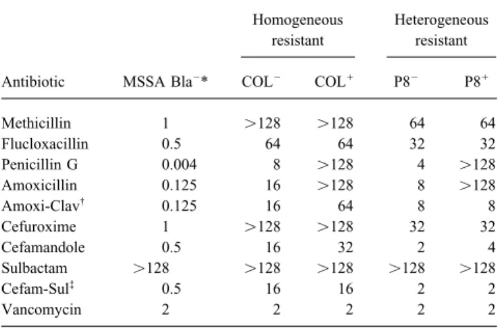
Table 1. Phenotypes of methicillin resistance and penicillinase (Bla) the plates against the concentrations of antibiotic in the plates. In production and MICs of various antibiotics for test organisms. vitro time-kill curves were made as described [6].
Titration of PBP 2A. The presence of PBP 2A was determined Homogeneous Heterogeneous in membrane fractions of bacterial lysates of the penicillinase-resistant resistant negative COL0, as previously described [13]. In brief, 75-mL
por-tions of membrane suspensions containing 4 mg/mL protein were Antibiotic MSSA Bla0* COL0 COL/ P80 P8/
incubated for 10 min at 377C with 25 mL of a 1:10 (wt/wt) solution of [3
H]penicillin (77 mCi/mg) and cold penicillin to a final
concen-Methicillin 1 ú128 ú128 64 64
tration of 2.5 mg/L [3
H]penicillin/mL of membrane suspension.
Flucloxacillin 0.5 64 64 32 32
The reaction was stopped by the addition of a 100-fold excess of
Penicillin G 0.004 8 ú128 4 ú128
Amoxicillin 0.125 16 ú128 8 ú128 cold penicillin, and the membranes were dissolved in the detergent
Amoxi-Clav† 0.125 16 64 8 8
sarkosyl. The membrane proteins were separated by SDS-PAGE,
Cefuroxime 1 ú128 ú128 32 32 and the [3
H]penicillin-labeled PBPs were visualized by fluorogra-Cefamandole 0.5 16 32 2 4 phy [5]. The binding affinities of methicillin, cefamandole, and Sulbactam ú128 ú128 ú128 ú128 ú128 amoxicillin for PBP 2A were assessed by measuring their ability Cefam-Sul‡
0.5 16 16 2 2
to compete for the binding of [3
H]penicillin to PBPs [5]. Aliquots
Vancomycin 2 2 2 2 2
of membrane suspensions were distributed into series of tubes containing 2-fold serial dilutions of the competitor and incubated * Methicillin-susceptible S. aureus (MSSA) strain RN 2677 used as control.
†
Amoxicillin and clavulanate combined in 5/1 (wt/wt) ratio. for 10 min at 377C before [3H]penicillin was added. The tubes were ‡For MIC determination, cefamandole was combined with constant
concen-incubated for another 10 min at 377C and processed as described. tration of 4 mg/L sulbactam. Table indicates MICs of cefamandole. Intensities of the PBP 2A bands were quantified by densitometry by use of the ImageQuant version 3.3 software (Molecular Dynam-ics, Sunnyvale, CA). Binding affinities of the competing drugs were derived from densitometry quantification and expressed as penicillinase-negative derivative of P8 was obtained by tempera- the drug concentration inhibiting binding of [3
H]penicillin by 50% ture-induced loss of the penicillinase-encoding plasmid during and 90% (IC
50and IC90).
growth at 437C [5]. For the sake of clarity, these pairs of bacteria Penicillinase stability of antibiotics in vitro. The ability of are referred to as COL0/COL/ and P80/P8/ (for
cefamandole, methicillin, and amoxicillin to resist penicillinase-negative and penicillinase-positive isolates, respectively). RN2677 induced degradation was measured in broth cultures by use of a is a methicillin-susceptible laboratory isolate of S. aureus used as described bioassay [15]. Both pairs of MRSA P8 and COL, produc-a control in in vitro experiments [5, 13]. Unless otherwise stproduc-ated, ing or not producing penicillinase, were used in this test. bacteria were grown at 357C in tryptic soy broth (TSB; Difco, Production of endocarditis and infusion pump installation. Detroit) with aeration or on tryptic soy agar (TSA; Difco) supple- Catheter-induced sterile aortic vegetations were produced in rats mented with 2% NaCl. Plates were routinely supplemented with as previously described [16]. At the same time, an intravenous (iv) penicillinase (Bacto-Penase concentrate; Difco; 2000 U/mL final catheter was inserted via the jugular vein into the superior vena concentration) to avoid antibiotic carryover. Stocks were kept at cava and connected to a programmable infusion pump (Pump 44; 0707C in TSB supplemented with 10% (vol/vol) glycerol. Harvard Apparatus, South Natick, MA) to deliver the antibiotics
Antibiotics and chemicals. Cefamandole and vancomycin [6]. The pump was set to deliver a volume of 0.2 mL of saline/h were obtained from Eli Lilly (Indianapolis); cefuroxime was ob- to keep the catheter open until the onset of therapy. No iv catheters tained from Glaxo Pharmaceuticals (London); penicillin G was were placed in the control animals.
obtained from Hoechst-Pharma (Zurich); [3
H]penicillin (9 mCi/ Bacterial endocarditis was induced 24 h after catheterization by mL; 117 mg/L) was provided by Merck Sharp & Dohme (Rahway, iv challenge of the animals with 0.5 mL of saline containing 105
NJ); sulbactam was obtained from Pfizer (Orsay, France); and cfu of either of the test organisms. This inoculum was 10 times methicillin, flucloxacillin, amoxicillin, and amoxicillin-clavulanate larger than the minimum inoculum producing endocarditis in 90% were obtained from Beecham Research Laboratories (Brockham of the untreated rats.
Park, UK). Treatment of experimental endocarditis. Antibiotic therapy
Antibiotic susceptibility, population analysis profile, and in vitro was started 12 h after bacterial challenge and lasted for 3 days. time-kill curves. MICs of antibiotics were determined by a pre- The drugs were administrated at changing flow rates with the pump viously described broth macrodilution method [14] in Mueller- to produce either of the following kinetics in the serum of rats: Hinton broth (Difco) supplemented with 2% NaCl; 105
cfu/mL was simulation of sequential iv administration of 3 g of cefamandole used as inoculum. The MIC was defined as the lowest antibiotic given 4 times a day to humans [17, 18]; continuous iv infusion of concentration inhibiting visible bacterial growth after 24 h of incu- cefamandole producing constant serum concentrations of 100 mg/ bation at 357C. The phenotypic expression of b-lactam resistance L (this concentration was chosen because it inhibited at least 90% was determined by spreading large bacterial inocula (109
cfu) as of PBP 2A in vitro; see Results); and simulation of sequential iv well as smaller inocula (106
, 105
, and 103
cfu) onto TSA plates administration of 1 g of vancomycin given twice daily to humans containing 2% NaCl and 2-fold serial dilutions of antibiotics [5]. [6]. This required total amounts of antibiotics of, respectively, The numbers of colonies growing on the plates were enumerated cefamandole at 250 mg/kg of body weight/24 h (for the two cefa-after 48 h of incubation at 357C. Population analysis profile curves mandole regimens) and vancomycin at 23.2 mg/kg of body weight/ 12 h. In certain experiments, cefamandole was combined with were generated by plotting the numbers of colonies growing on
sulbactam, which was administered via a separate pump and which Kruskal-Wallis one-way analysis of variance on ranks with subse-quent pairwise testing by the Dunn’s method. Overall, differences simulated in rats the human pharmacokinetics produced by
admin-istration of 1 g of the drug given 4 times a day [9]. This required were considered significant at P£ .05 by use of two-tailed signifi-cance levels.
the administration of sulbactam at 50 mg/kg of body weight/6 h.
Prophylaxis of experimental endocarditis. Antibiotic prophy-laxis was started 1 h before bacterial challenge and lasted for 1 day [19]. Kinetics of antibiotics in the serum of rats were as
Results follows: simulation of sequential iv administration of 1 g of
flucloxacillin given 4 times a day to humans [20], simulation of
Antibiotic susceptibility, population analysis profile, and
sequential iv administration of 3 g of cefamandole given 4 times
time-kill curves. Table 1 shows the MICs of various antibiot-a dantibiot-ay to humantibiot-ans (antibiot-as antibiot-above), continuous iv infusion of cefantibiot-amantibiot-andole
producing constant serum levels of 100 mg/L (as above), or simula- ics for the 4 test organisms. All of the organisms were highly tion of sequential iv administration of 1 g of vancomycin given resistant to methicillin and flucloxacillin. Amoxicillin had a twice daily to humans (as above). Total amount of drug given to relatively good activity against the penicillinase-negative deriv-the animals was 42.3 mg of flucloxacillin/6 h. Amounts of drug atives but required the addition of clavulanate to be active for the other regimens were as described above. against the penicillinase-producing organisms. In contrast, the
Antibiotic concentrations in serum. Concentrations of
antibi-MIC of cefamandole remained essentially unaffected by peni-otic in serum were determined in groups of 4 – 9 uninfected or
cillinase production or by combination with sulbactam. infected rats. Serum levels in infected animals came from internal
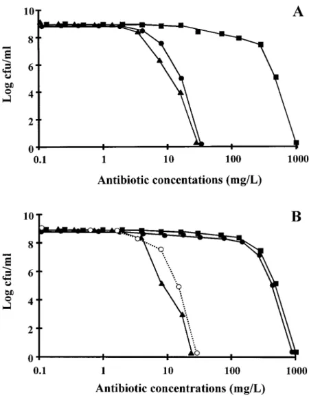
Figure 1 shows the population analysis profile of the homo-controls for adequate drug delivery in therapeutic experiments.
geneously resistant COL0 and its penicillinase-producing
de-Blood was drawn by puncturing the periorbital sinuses of the
ani-rivative COL/. As in the MIC determinations, amoxicillin was
mals at several time points during and after antibiotic
administra-relatively effective against the penicillinase-negative version tion. Antibiotic concentrations were determined by an agar
diffu-sion assay with antibiotic medium 1 (Difco) and Bacillus subtilis of the organisms but lost its activity in the presence of penicil-ATCC 6633 as the indicator organism for cefamandole, flucloxa- linase. In contrast, cefamandole was active against both penicil-cillin, and vancomycin and Acinetobacter calcoaceticus as the linase-negative and penicillinase-positive isolates. The same indicator organism for sulbactam. The diluent was pooled rat se- observation was made with the penicillinase-negative and peni-rum. The limits of detection of the assays were 0.3 mg/L for cillinase-positive versions of the heterogeneously resistant P8. cefamandole, 0.3 mg/L for flucloxacillin, 0.6 mg/L for
vancomy-Moreover, when an additional panel of 10 unrelated clinical cin, and 3.12 mg/L for sulbactam. The linearity of the standard
isolates of penicillinase-producing MRSA were tested, none of curves was assessed by a regression coefficient of§0.995, and
them grew on agar plates containingú32 mg/L cefamandole, intraplate and interplate variations were£10%.
while they grew on plates containing up to 1000 mg/L
methicil-Evaluation of infection. In therapeutic experiments, the control
lin, flucloxacillin, or amoxicillin (data not shown). This shows rats were sacrificed at the time of treatment onset (i.e., 12 h after
that the relatively good anti-MRSA activity of cefamandole inoculation) to measure both the frequency and the severity of
valvular infection at the start of therapy. Treated rats were sacri- could be extended beyond the limit of the test bacteria used in ficed 18 – 24 h after the last antibiotic dose, at least 12 h after the the present experiments.
trough level of drug in serum was reached. At that time, no residual Time-kill experiments were done with antibiotic concentra-antibiotic could be detected in the blood. In prophylaxis experi- tions readily achieved in human serum. Cefamandole was bac-ments, control rats were killed 24 h after bacterial challenge, tericidal against all 4 test organisms, as shown by viability whereas rats receiving prophylaxis were killed after 3 days (i.e.,
losses of§3 log10 cfu/mL after 24 h of exposure to drug at
2 days after the end of antibiotic administration). The valvular
100 mg/L. Amoxicillin (40 mg/L) was bactericidal only against vegetations were sterilely dissected, weighed, homogenized in 1
the penicillinase-negative derivatives and required the addition mL of saline, and serially diluted before being spread on
penicil-of clavulanate to kill penicillinase-producing isolates. In con-linase-containing plates for colony counts. Quantitative blood
cul-trast, neither methicillin nor flucloxacillin killed or inhibited tures and spleen cultures were done in parallel. The numbers of
any of the isolates when used at peak concentrations achievable colonies growing on the plates were determined after 48 h of
incubation at 357C. Bacterial densities in the vegetations were in human serum during iv therapy (i.e., 100 mg/L).
expressed as log10colony-forming units per gram of tissue. The Determination of PBP 2A affinity. The drug concentrations
dilution technique permitted the detection of§2 log10cfu/g of inhibiting 50% and 90% of [3H]penicillin labeling of PBP 2A
vegetation. For statistical comparisons of differences between the were determined in membrane fractions of the penicillinase-median vegetation bacterial densities of various treatment groups, negative strain COL0. The IC
50 and IC90 of methicillin were
culture-negative vegetations were considered to contain 2 log10
350 and 4000 mg/L, respectively. In comparison, these values cfu/g.
were§40-fold lower for cefamandole and amoxicillin: 8 and
Statistical analysis. Fisher’s exact test was used to compare
100 mg/L for cefamandole and 4 and 90 mg/L for amoxicillin. the rates of valvular infection. Bonferroni’s correction was used
These results were reproducible on repeating the experiments for multiple-group comparison. Median bacterial densities in the
several times with independent batches of cell membranes. vegetations of various treatment groups were compared by the
Figure 1. Population analysis profile of penicil-linase-negative and homogeneously methicillin-resis-tant strain COL0
(A) and its penicillinase-producing transformant COL/
(B). Various sizes of bacterial in-ocula were spread on agar plates containing increasing concentrations of either methicillin (j), cefamandole (m), or amoxicillin (l). B also shows results with amoxicillin-clavulanate (s) against penicillinase-pro-ducing strain COL/
. Points indicate no. of colonies growing on plates after 48 h of incubation at 357C.
amoxicillin against MRSA correlated with a greater PBP 2A successfully cured valvular infections and was equivalent to or better than control treatment with vancomycin. Therefore, affinity of these compounds compared with that of methicillin.
Treatment of experimental endocarditis due to penicillinase- continuous infusion of cefamandole was used in the next series of experiments.
negative MRSA. The homogeneously methicillin-resistant but
penicillinase-negative strain COL0was tested in these experi- Treatment of experimental endocarditis due to
penicillinase-producing MRSA. A second series of experiments investi-ments. Two cefamandole regimens mimicking drug
concentra-tions achievable in the sera of humans were tested, one simulat- gated the impact of bacterial penicillinase production on the outcome of cefamandole therapy in vivo. Both the penicil-ing standard 4 times daily administration of 3 g of cefamandole,
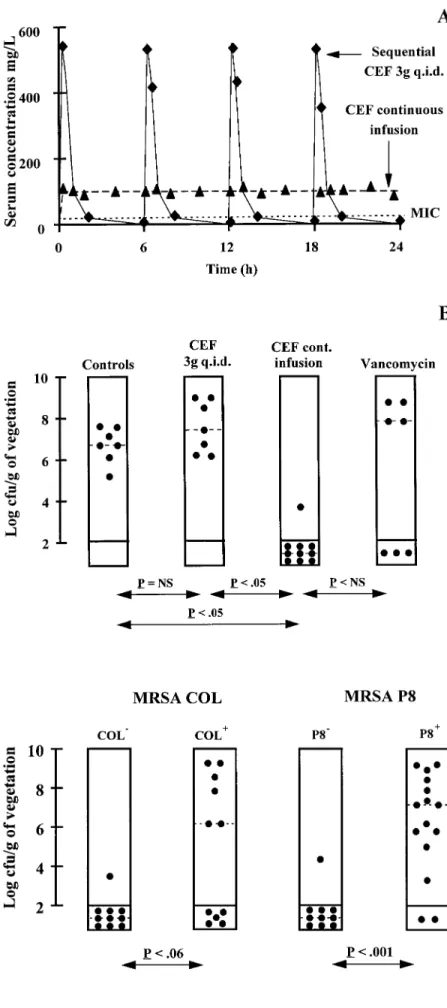
and one consisting of continuous infusion of the drug. Figure linase-negative and -positive versions of strains COL and P8 were tested in parallel. Figure 3 shows that continuous infusion 2 depicts the serum kinetics of these cefamandole regimens
(figure 2A) and their therapeutic results after 3 days of treat- of cefamandole successfully treated animals infected with the b-lactamase – negative organisms. However, although cefa-ment (figure 2B). Human-like kinetics produced by sequential
treatment failed to cure the animals, in spite of repeated high mandole was not affected by penicillinase in susceptibility tests in vitro, the antibiotic was significantly less effective against peak serum levels of the drug (Ç600 mg/L). Since cefamandole
had a short serum half-life, it was possible that transient antibi- the penicillinase-producing version of the strains in vivo. This observation raised questions about the stability of cefamandole otic peaks might not compensate for the prolonged periods of
drug levels below the MIC and that continuous drug infusion to penicillinase in this condition.
Penicillinase stability of cefamandole. One reason for the might perform better than sequential administration. Indeed,
figure 2 shows that continuous infusion producing steady serum discrepancy between in vitro and in vivo results might be re-lated to an inoculum effect. As illustrated in figure 4, penicil-concentrations of cefamandole of Ç100 mg/L (dashed line)
Figure 2. Kinetics of cefamandole in serum of rats (A) and results of treatment of experimental endocar-ditis due to penicillinase-negative strain COL0
(B). A depicts cefamandole kinetics that either simulated human pharmacokinetics produced by sequential ad-ministration of 3 g of drug 4 times daily (CEF 3g q.i.d.) or produced constant antibiotic levels of 100 mg/L (CEF continuous infusion). Dotted line in A indicates MIC of cefamandole for test organism. In B, treatment groups are indicated at tops of columns. Each dot indicates bacterial density in vegetations of single animal. Dashed lines indicate median value in each treatment group. Statistical differences between groups were determined by Kruskal-Wallis 1-way analysis of variance on ranks. Differences in median values among treatment groups were significant (Põ .001). Differences between specific groups, as deter-mined by Dunn’s method, are indicated at bottom. NS Å not significant.
Figure 3. Treatment of experimental endocarditis with cefamandole given in continuous infusion (pro-ducing constant serum levels of 100 mg/L) in rats infected with negative and penicillinase-positive versions of strains COL and P8. Untreated controls were all heavily infected and are not shown. Each dot in columns indicates bacterial density in veg-etation of single animal; dashed lines indicate median value in each treatment group. Statistical differences between pairs of groups were determined by Mann-Whitney-Wilcoxon unpaired test and Fisher’s exact test. Both tests gave concordant results. Therefore, only highest P values given by either test are shown at bottom.
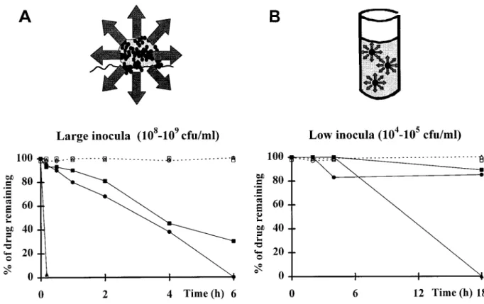
Figure 4. Model for inoculum-dependent degradation ofb-lactams operating in infected vegetations (A) compared with situation in MIC test tubes (B). Upper part of A illustrates bacterial clusters packed in infected vegetations, surrounded by large concentrations of penicillinase (arrows). Graph in A shows in vitro degradation of amoxicillin (triangles), cefamandole (circles), and methicillin (squares) exposed to large bacterial inocula of either strain COL/
(penicillinase-producer; closed symbols) or COL0
(penicillinase-negative; open symbols). B shows results of similar experiments with lower inoculum sizes, to mimic situation in MIC test tubes. Model is similar to that proposed by Goldman and Petersdorf [15].
linase concentrations might be higher around bacterial clusters this supports the possibility that large bacterial densities and penicillinase concentrations in the vegetations might result in packed in infected vegetations (figure 4A) than around single
organisms suspended in broth cultures or spread on agar plates significant degradation of cefamandole at the infected site. This hypothesis was further tested by using the penicillinase inhibi-(figure 4B). Since cefamandole is not entirely immune to
peni-cillin-induced hydrolysis [21], degradation of cefamandole tor sulbactam in the next experiments.
Prevention of penicillinase-induced degradation of
cefaman-might become an issue when switching from the in vitro
sus-ceptibility tests to deep-seated infections in vivo. A possible dole by sulbactam. If the above assumption is correct, then combination of cefamandole with a penicillinase inhibitor, such inoculum effect was investigated in vitro by testing the stability
of cefamandole and other antibiotics exposed to broth cultures as sulbactam, might restore its antibacterial efficacy. This was tested both in vitro and in vivo. First, large concentrations of containing various bacterial counts of either strain COL/ or
strain COL0. In the presence of large bacterial numbers (i.e., sulbactam (400 mg/L) successfully prevented degradation of
cefamandole in vitro, as tested by an assay similar to that 108– 109cfu/mL; figure 4A, graph), cefamandole and
methicil-lin lost almost 50% of their original activity within 3 h of presented in figure 4 that used the penicillinase-producing strain COL/as test bacterium. In a typical experiment, the residual
exposure. Accordingly, amoxicillin barely resisted a few
min-utes in this experiment. In contrast, the three antibiotics were activity of cefamandole exposed alone to the culture was 76% after 2 h of incubation and 0% after 4 h and 6 h of incubation. perfectly stable when exposed to broth cultures of the
penicil-linase-negative strain COL0. In the presence of lower bacterial In the presence of sulbactam, in comparison, the residual
activity of cefamandole was 100% at 2 h, 60% at 4 h, and numbers (i.e., 105– 106cfu/mL; figure 4B, graph), on the other
hand, both cefamandole and methicillin were quite stable for 33% at 6 h.
Second, combination of cefamandole with sulbactam in vivo up to 18 h, whereas amoxicillin was progressively degraded
over this time. Similar results were obtained when the pair of also restored the drug efficacy. Sulbactam administration pro-duced a peak concentration in the serum of rats (mean{ SD penicillinase-producing and -nonproducing MRSA P8/ and
concentrations of 30.8{ 1 mg/L at 1 h, 9.8 { 0.1 mg/L at 2 h, and undetectable values at 6 h. Figure 5 indicates that administration of sulbactam at this dose 4 times a day reestab-lished the therapeutic efficacy of continuous infusion of cefa-mandole, as if it were used against the penicillinase-negative version of the organism (see figure 3). Thus, taken together, these experiments strongly argue in favor of a direct role of in situ production of penicillinase as a cause of cefamandole treatment failure against experimental endocarditis due to peni-cillinase-producing MRSA.
Prophylaxis of experimental endocarditis. Although cefa-mandole was ineffective against experimental endocarditis due to penicillinase-producing MRSA, it might be successful for prophylaxis of such infection. Indeed, prophylaxis more closely resembles the test tube situation than does established infection,
Figure 6. Cefamandole prophylaxis of experimental endocarditis because prophylactic drugs are given while bacteria are
circu-due to penicillinase-producing strain COL/
. Prophylaxis regimens are lating in the blood and only beginning to colonize the cardiac indicated. No. of animals in each group are shown at bottom. lesions [22, 23]. Figure 6 shows that cefamandole successfully
prevented endocarditis due to the penicillinase-producing strain COL/, whether it was administered sequentially as in humans
centration of penicillinase around the bacteria might be a criti-or in continuous infusion, producing constant serum levels of
cal factor determining antibiotic efficacy. drug of 100 mg/L. Vancomycin prophylaxis was also effective,
whereas flucloxacillin completely failed to prevent infection.
These results further support the hypothesis that the local con- Discussion
The present studies highlight two pharmacodynamic limita-tions of cefamandole in the treatment of experimental endocar-ditis: one relatively trivial, which relates to the antibiotic dosing regimen, and a second, more fundamental, that points to the essential role of penicillinase in b-lactam resistance of MRSA. First, treatment of experimental endocarditis due to penicil-linase-negative MRSA could not be achieved by sequential drug administration, because repeated transient peaks of cefa-mandole in the serum were unable to ensure prolonged supra-MIC drug concentrations in vivo. It is well established that b-lactam drugs must be maintained above growth-inhibiting concentrations to be effective [24, 25], and this limitation was easily overcome by using continuous infusion of the drug.
Second, treatment of endocarditis due to penicillinase-posi-tive MRSA could not be achieved even by continuous drug infusion, presumably because cefamandole was inactivated in situ by bacterial penicillinase produced in the vegetations. This limitation was not expected, because large doses of the rela-Figure 5. Treatment of experimental endocarditis with cefaman- tively penicillinase-stable cefamandole were assumed to over-dole given to rats in continuous infusion (producing constant serum come penicillinase-induced hydrolysis in vivo. However, com-levels of 100 mg/L) either alone (CEF) or in combination with sulbac- plementary experiments clearly demonstrated an inoculum-tam (CEF/ SUL) at doses simulating human pharmacokinetics
pro-dependent degradation of cefamandole by penicillinase, which duced by administration of 1 g of drug 4 times a day. Rats were
could be prevented by coadministration of a penicillinase inhib-infected with penicillinase-producing, homogeneous
methicillin-resis-tant strain COL/
. Each dot indicates bacterial density in vegetation itor such as sulbactam. This supports the possibility that large of single animal. Dashed lines indicate median value in each treatment amounts of the enzyme surrounding bacterial clusters in the group. Statistical differences between groups were determined by vegetations could protect the microorganisms from the drug Kruskal-Wallis 1-way analysis of variance on ranks (Põ .014) with
and confirms a previous hypothesis by Goldman and Petersdorf pairwise comparison between specific groups by Dunn’s method and
[15], who postulated that the poor activity of cefazolin against Fisher’s exact test with Bonferroni’s correction. Both tests gave
strain of S. aureus could be due to degradation of the drug at enzyme are crucial for the survival of the organisms. On the side of the antibiotic, slow diffusion into the vegetation, strong the infected site. Moreover, it was further supported by the fact
that any of the cefamandole regimens that failed in therapeutic penicillinase-inducing capacity, and low penicillinase stability are playing against the drug. Some of these characteristics can experiments were highly effective in the prophylaxis studies,
a setting more closely resembling the test tube than does the be defined, at least on the antibiotic side. However, the interplay of these factors in vivo may be difficult to predict. The repro-vegetation situation (see figure 4). Therefore, while the slow
inactivation of cefamandole by penicillinase did not affect the ducible therapeutic success of certain b-lactams against penicil-linase-negative MRSA, on the other hand, supports the fact results of in vitro susceptibility tests, it was clearly responsible
for treatment failure in vivo. that PBP 2A can be blocked in vivo. Nevertheless, b-lactams with improved PBP 2A affinity would certainly gain in anti-The observation is important because it underlines the fact
that in vitro susceptibility tests might not be predictive of in MRSA efficacy and act in conjunction with their stability to penicillinase against these organisms. Therefore, both better vivo results. Moreover, the lack of cefamandole efficacy against
penicillinase-producing strains also highlights the everlasting PBP 2A affinity and penicillinase stability are important in the development of future b-lactams active against MRSA. role of penicillinase in b-lactam resistance of staphylococci.
Besides MRSA, penicillinase may also adversely affect the Taken together, the present observations show that while cefamandole demonstrates both relatively good PBP 2A affinity efficacy of anti-staphylococcal b-lactams against
methicillin-susceptible S. aureus. For example, combination of cefopera- and anti-MRSA activity in vitro, it may fail against MRSA infections in vivo because of production of bacterial penicil-zone with sulbactam was more effective than cefoperapenicil-zone
alone against experimental endocarditis due to methicillin-sus- linase. This is in contradiction to a report suggesting that cefa-mandole might be effective for treatment of soft tissue infec-ceptible S. aureus in rabbits, presumably because sulbactam
protected cefoperazone from penicillinase-induced degradation tions due to MRSA in humans [11] and raises caution concerning this indication. On the other hand, however, 1-[26]. Another example is borderline methicillin-resistant S.
aureus, which have increased methicillin MICs because of pen- day prophylaxis with cefamandole could effectively prevent experimental MRSA endocarditis, presumably because individ-icillinase overproduction [27]. Although these organisms are
not clinically relevant, they were able to decrease the efficacy ual production of penicillinase by circulating bacteria is low. Cefamandole was known to be effective for prophylaxis in of ampicillin-sulbactam treatment of experimental endocarditis
due to borderline methicillin-resistant S. aureus in rats [28]. cardiovascular surgery before the methicillin resistance era [31, 32]. It might now be considered for prophylaxis against MRSA Therefore, production of penicillinase is not harmless, even
when supposedly penicillinase-stable drugs are used to treat and methicillin-resistant S. epidermidis as well, especially when the use of vancomycin is prohibited to avoid the selection methicillin-susceptible S. aureus.
In the case of MRSA, penicillinase may represent a primary of vancomycin-resistant enterococci. Additional studies are warranted to further assess this prophylactic efficacy. restriction to the possible use of existing b-lactams against
infections due to such bacteria. Indeed, several studies have validated the fact that b-lactams with a relatively good PBP 2A
Acknowledgments affinity, such as penicillin, ampicillin, and amoxicillin, could
successfully treat experimental MRSA infections, provided that
We thank Marlyse Giddey, Jacques Vouillamoz, and Oscar Mar-they could escape penicillinase-induced degradation [5 – 9].
chetti for outstanding technical assistance. These observations were recently extended to
methicillin-resis-tant Staphylococcus epidermidis, which also produce PBP 2A
[6, 29]. However, although human-like kinetics of amoxicillin- References clavulanate could successfully treat experimental MRSA
endo-1. Hartman BJ, Tomasz A. Low-affinity penicillin-binding protein associated carditis in rats [5], experiments in rabbits showed that combina- with beta-lactam resistance in Staphylococcus aureus. J Bacteriol1984; tions of ampicillin-sulbactam or penicillin-sulbactam tended to 158:513 – 6.
2. Gaisford WC, Reynolds PE. Methicillin resistance in Staphylococcus epi-be less effective against infections due to
penicillinase-produc-dermidis. Relationship between the additional penicillin-binding protein
ing than -nonproducing MRSA [9, 30]. Therefore, these results
and an attachment transpeptidase. Eur J Biochem1989; 185:211 – 8.
raised concern about the potential safety of penicillin –
penicil-3. Wu CYE, Alborne WE, Flokowitsch JA, et al. Site-directed mutagenesis linase inhibitor combinations for therapeutic use against peni- of the mecA gene from a methicillin-resistant strain of Staphylococcus cillinase-producing MRSA in humans. aureus. J Bacteriol1994; 176:442 – 9.
4. de Jonge BL, Tomasz A. Abnormal peptidoglycan produced in a methicil-The present experiments with cefamandole further
empha-lin-resistant strain of Staphylococcus aureus grown in the presence of size the crucial role of penicillinase as a troublemaker in the
methicillin: functional role for penicillin-binding protein 2A in cell wall system. Figure 4 is an oversimplified illustration of the subtle
synthesis. Antimicrob Agents Chemother1993; 37:342 – 6.
confrontation between bacterial factors and antibiotics in the 5. Franciolli M, Bille J, Glauser MP, Moreillon P. b-lactam resistance mecha-vegetation environment. On the side of the bacteria, rapid in- nisms of methicillin-resistant Staphylococcus aureus. J Infect Dis1991;
163:514 – 23. duction of penicillinase and production of large amounts of the
6. Entenza JM, Fluckiger U, Glauser MP, Moreillon P. Antibiotic treatment 20. Frank U, Schmidt-Eisenlohr E, Schlosser V, Spillner G, Schindler M, of experimental endocarditis due to methicillin-resistant Staphylococcus Daschner FD. Concentrations of flucloxacillin in heart valves and subcu-epidermidis. J Infect Dis1994; 170:100 – 9. taneous and muscle tissues of patients undergoing open-heart surgery. 7. Chambers HF. In vitro and in vivo antistaphylococcal activities of L- Antimicrob Agents Chemother1988; 32:930 – 1.
695,256, a carbapenem with high affinity for the penicillin-binding 21. Farrar WE, O’Dell NM. Beta-lactamase resistance of newer cephalosporins protein PBP 2A. Antimicrob Agents Chemother1995; 39:462 – 6. and antimicrobial effectiveness against gram-negative bacilli. Infection 8. Hirano L, Bayer AS. Beta-lactam – beta-lactamase-inhibitor combinations 1977; 5:224 – 7.
are active in experimental endocarditis caused by beta-lactamase – pro- 22. Hall G, Hedstrom SA, Heimdahl A, Nord CE. Prophylactic administration ducing oxacillin-resistant staphylococci. Antimicrob Agents Chemother of penicillins for endocarditis does not reduce the incidence of
postex-1991; 35:685 – 90. traction bacteremia. Clin Infect Dis1993; 17:188 – 94.
9. Fantin B, Pierre J, Castela-Papin N, et al. Importance of penicillinase 23. Moreillon P, Overholser CD, Malinverni R, Bille J, Glauser MP. Predictors production for activity of penicillin alone or in combination with sulbac- of endocarditis in isolates from cultures of blood following dental ex-tam in experimental endocarditis due to methicillin-resistant Staphylo- tractions in rats with periodontal disease. J Infect Dis 1988; 157: coccus aureus. Antimicrob Agents Chemother1996; 40:1219 – 24. 990 – 5.
10. Azimi PH. Clinical and laboratory investigation of cefamandole therapy 24. Joly V, Pangon B, Vallois JM, et al. Value of antibiotic levels in serum of infections in infants and children. J Infect Dis1978; 137(suppl): and cardiac vegetations for predicting antibacterial effect of ceftriaxone S155 – 60. in experimental Escherichia coli endocarditis. Antimicrob Agents Che-11. Frongillo RF, Donati L, Federico G, et al. Clinical comparative study on mother1987; 31:1632 – 9.
the activity of cefamandole in the treatment of serious staphylococcal 25. Craig WA, Ebert SC. Continuous infusion of beta-lactam antibiotics. Anti-infections caused by methicillin-susceptible and methicillin-resistant microb Agents Chemother1992; 36:2577 – 83.
strains. Antimicrob Agents Chemother1986; 29:789 – 96.
26. Chambers HF, Fournier MA. Efficacy of cefoperazone in combination 12. Frongillo RF, Bianchi P, Moretti A, Pasticci MB, Ripa S, Pauluzzi S.
with sulbactam in experimental Staphylococcus aureus endocarditis in Cross-resistance between methicillin and cephalosporins for
staphylo-rabbits. J Antimicrob Chemother1993; 32:453 – 8. cocci: a general assumption not true for cefamandole. Antimicrob
27. McDougal LK, Thornsberry C. The role of beta-lactamase in staphylococ-Agents Chemother1984; 25:666 – 8.
cal resistance to penicillinase-resistant penicillins and cephalosporins. 13. Murakami K, Tomasz A. Involvement of multiple genetic determinants in
J Clin Microbiol1986; 23:832 – 9. high-level methicillin resistance in Staphylococcus aureus. J Bacteriol
28. Pefanis A, Thauvin-Eliopoulos C, Eliopoulos GM, Moellering RC Jr. Ac-1989; 171:874 – 9.
tivity of ampicillin-sulbactam and oxacillin in experimental endocarditis 14. Sahm DF, Washington JA. Antibacterial susceptibility test: dilution
meth-caused by beta-lactamase – hyperproducing Staphylococcus aureus. ods. In: Balows A, Hausler WJ, Herrmann KL, Isemberg HD, Shadomy
Antimicrob Agents Chemother1993; 37:507 – 11. HJ, eds. Manual of clinical microbiology. 5th ed. Washington, DC:
29. Ramos MC, Ing M, Kim E, Witt MD, Bayer AS. Ampicillin-sulbactam is American Society for Microbiology,1991:1109 – 16.
effective in prevention and therapy of experimental endocarditis caused 15. Goldman PL, Petersdorf RG. Importance of b-lactamase inactivation in
by beta-lactamase – producing coagulase-negative staphylococci. Anti-treatment of experimental endocarditis caused by Staphylococcus
microb Agents Chemother1996; 40:97 – 101. aureus. J Infect Dis1980; 141:331 – 7.
30. Chambers HF, Sachdeva M, Kennedy S. Binding affinity for penicillin-16. Heraief E, Glauser MP, Freedman LR. Natural history of aortic valve
binding protein 2a correlates with in vivo activity of b-lactam antibiotics endocarditis in rats. Infect Immun1982; 37:127 – 31.
against methicillin-resistant Staphylococcus aureus. J Infect Dis1990; 17. Colaizzi PA, Goodwin SD, Poynor WJ, Karnes HT, Polk RE. Single-dose
162:705 – 10. pharmacokinetics of cefuroxime and cefamandole in healthy subjects.
31. Scher KS, Jones CW. Which cephalosporin for wound prophylaxis? Clin Pharmacy1987; 6:894 – 9.
An experimental comparison of three drugs. Surgery 1985; 98: 18. Griffith RS, Black HR, Brier GL, Wolny JD. Cefamandole: in vitro and
30 – 4. clinical pharmacokinetics. Antimicrob Agents Chemother 1976; 10:
32. Gatell JM, Riba J, Lozano ML, Mana J, Ramon R, Garcia SanMiguel J. 814 – 23.
Prophylactic cefamandole in orthopaedic surgery. J Bone Joint Surg 19. Parry GW, Holden SR, Shabbo FP. Antibiotic prophylaxis for cardiac