Frataxin promotes antioxidant defense in a thiol-dependent manner resulting in diminished malignant transformation in vitro
8
0
0
Texte intégral
(2) 816. Human Molecular Genetics, 2002, Vol. 11, No. 7. Figure 1. Stable transgenic expression of human frataxin in murine fibroblasts. Immunoblot against hemagglutinin-tagged human frataxin in whole-cell extracts from mock-transfected (left lane) and frataxin-transfected (right lane) 3T3L1 cells gives two specific bands at 29 kDa (native frataxin) and 18 kDa (mature frataxin) plus a set of non-specific bands in the upper molecular range indicating that equal amounts of protein are loaded on the gel.. which indicated an ∼2-fold increase of the overall frataxin expression (12). To confirm the integrity of the protein, we compared cells overexpressing human frataxin with vectortransfected control cells (mock-transfected) by immunoblotting. Figure 1 depicts a typical western blot after loading equal amounts of protein, as indicated by the intensities of the nonspecific upper bands. In the lower range of the gel, the mocktransfected cells (Fig. 1, left lane) show no signal, while the cells overexpressing human frataxin (Fig. 1, right lane) show a typical double band consisting of the native cytosolic frataxin (29 kDa) and the processed mitochondrial frataxin (18 kDa). These findings together with our previously published observations (12) suggest appropriate processing of the transfected native frataxin towards a functional protein. Frataxin increases cellular resistance to oxidative stress Cells overexpressing frataxin and mock-transfected cells were grown to a sub-confluent state and subsequently exposed to various concentrations of the ROS tert-butyl-hydrogen peroxide to determine putative differences in the resistance to oxidative stress. Survival in the absence of tert-butyl-hydrogen peroxide was defined as 100%. A final concentration of 600 µM tert-butyl-hydrogen peroxide induced cell death within 16 h in all cells (both frataxin-overexpressing and mock-transfected) (Fig. 2). Concentrations from 100 to 500 µM (increment: 100 µM) caused a corresponding incremental increase of cell death in both cell lines; however, cells overexpressing frataxin exhibited an increased resistance to tert-butyl-hydrogen peroxide when compared to mock-transfected cells (Fig. 2). This difference was significant for all concentrations of tert-butyl-hydrogen peroxide evaluated within this range (P < 0.05), where concentrations of 200, 300 and 400 µM showed a highly significant difference (P < 0.005) (Fig. 2). Thus, frataxin appears to increase resistance against exogenous oxidative stress. Frataxin decreases intracellular levels of free radicals To determine whether frataxin has an effect on endogenous levels of ROS, a confocal laser scanning technique was. Figure 2. Frataxin increases resistance to exogenous reactive oxygen species. Cell survival assay comparing relative numbers of cells surviving a 16 h exposure to tert-butyl-hydrogen peroxide at various concentrations: black bars indicate mock-transfected cells; grey bars indicate frataxin overexpressing cells; error bars indicate SD.. employed. To detect intracellular free radical production, fluorescence of DCF was quantified. Its precursor H2DCF-DA is a non-polar compound that readily diffuses into cells, where it is hydrolyzed to the non-fluorescent polar derivative H2DCF and thereby trapped within the cell. In the presence of an appropriate oxidant, H2DCF is oxidized to the highly fluorescent DCF, which is then detected by microscopic techniques. Employing this method, frataxin-overexpressing cells appear to show lower levels of ROS when compared to mock-transfected cells (Fig. 3A). When quantifying the fluorescence activities in a minimum of 100 cells per experiment, a highly significant difference in light emission, proportional to free radical content, was observed (P < 0.005) (Fig. 3B). Thus, frataxin appears to decrease levels of intracellular oxidative stress. Frataxin promotes antioxidant defense via glutathione and its peroxidase To determine whether this antioxidant effect of frataxin can be attributed to the protein itself, in vitro assays were performed. Firstly, purified recombinant human frataxin was used to determine putative activities by employing assays for the major antioxidant enzyme complexes: superoxide dismutase (SOD), catalase (CAT) and glutathione peroxidase (GPX). No activities were detected with the purified frataxin (Table 1). Functionality of each assay was confirmed using purified CAT (bovine liver) and SOD (bovine erythrocytes) (both from Calbiochem, Schwalbach, Germany) as positive controls. Thus, frataxin itself appears not to function as an antioxidant. To further determine whether the antioxidant effect of frataxin might be explained by indirect activation of known ROS-detoxifying enzymes, total activity of SOD, CAT and GPX was measured in cell extracts, and activity in cells overexpressing frataxin was compared to that in mock-transfected controls. While quantification of SOD and CAT showed no relative increase of activities in comparison to control cells, activity of total glutathione peroxidase was found to be increased by >50%. This difference was found to be highly significant (P < 0.001; Table 1). Since GPX activity is.
(3) Human Molecular Genetics, 2002, Vol. 11, No. 7. 817. Figure 3. Frataxin reduces endogenous levels of reactive oxygen species. (A) Confocal laser scanning evoked fluorescence of 2 ′,7′-dichlorofluorescein within mock-transfected (left) and frataxin overexpressing (right) cells, where intensity of fluorescence is proportional to the amount of intracellular reactive oxygen species. (B) Results of computerized quantification of fluorescence intensity, where the black bar indicates mock-transfected cells and the grey bar represents frataxin overexpressing cells. Table 1. Enzymatic activities of purified frataxin and cellular extracts from cells overexpressing frataxin relative to mock-transfected cells Recombinant frataxin. Cellular extracts. Superoxide dismutase. No activity. No difference. Catalase. No activity. No difference. Glutathione peroxidase. No activity. +51.9 (±10.1)%a. Thiol content. Not applicable. +38.8 (±3.9)%b. aP. < 0.001; bP = 0.010.. dependent on the presence of reduced thioles, the content of this cofactor was determined as well. The content for thioles in the cellular extracts evaluated was found to be elevated by more than a third in comparison to mock-transfected cells (P = 0.010; Table 1). Thus, the antioxidant properties of frataxin might be explained by its effects on glutathione peroxidase activity and thiol content. Frataxin inhibits colony formation after exposure to reactive oxygen species Malignant transformation is characterized by repeated occurrence of mutations in DNA, cumulatively leading to loss of cell cycle control. Multiple mutagenic agents, specifically ROS, including singlet oxygen, have been described. Based on the findings above, we asked whether frataxin might inhibit ROS-induced malignant transformation. To answer this question, cells overexpressing frataxin were compared with mocktransfected cells by continuously passaging them in media containing 2 µM menadione (Sigma), a vitamin K derivative known to cause perpetual generation of ROS. A subset of mock-transfected cells (a total of four out of a total population of 30) exhibited loss of anchorage-dependent growth as determined by crystal-violet staining of colonies (Fig. 4A) and so-called soft agar assays (Fig. 4B), putatively indicating occurrence of cell cycle transformation. None of the cells. overexpressing frataxin (0 out of a total of 30) formed colonies, i.e. they remained apparently untransformed (P = 0.043) (Figs. 4A and B). Thus, frataxin appears to inhibit loss of anchoragedependent growth subsequent to continuous exposure to ROS. Since loss of anchorage-dependent growth suggests previous occurrence of malignant transformation, putative tumourigenicity in vivo was determined. Those mock-transfected cells which lost anchorage-dependent growth characteristics, as well as cells with normal growth characteristics, were evaluated. Cells of normal growth consisted of frataxin-transfected cells as well as cells which were mock-transfected, but did not transform. These three groups were compared concerning their ability to generate tumours in immune-compromised mice. Suspensions of cells were injected into the right hind and evaluated for tumour growth over a time period of 6 weeks. As shown in Figure 4C, only cells which exhibited a loss of anchoragedependent growth generated tumours, while apparently normal cells did not exhibit tumour formation in nude mice. Histological analyses of tumours revealed carcinoma-like characteristics without evidence for invasive growth (data not shown). Together, these data suggest frataxin inhibits malignant transformation subsequent to continuous exposure to ROS. DISCUSSION FA, the most common hereditary ataxia, leads to premature death within the third decade of life (1). Based on its progressively degenerative nature, attempts to treat FA have employed synthetic antioxidants, including idebenone, a coenzyme Q derivative (25–28). This therapeutic approach is supported by several findings, suggesting a state of increased oxidative stress in FA, as indicated in the introductory section (14–21). Our present findings indicate that frataxin itself does not exhibit antioxidant properties, at least not in a classical manner, i.e. it does not resemble SOD, CAT or GPX in standard enzyme activity assays. Overexpression of the protein causes a specific activation of GPX activity in whole cell extracts, accompanied by an elevation of cellular thiol levels..
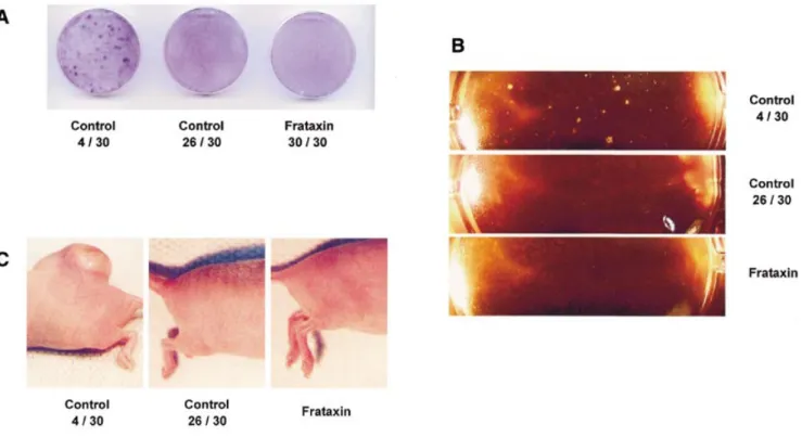
(4) 818. Human Molecular Genetics, 2002, Vol. 11, No. 7. Figure 4. Frataxin inhibits loss of anchorage-dependent growth subsequent to exposure to oxidative stress. (A) Growth characteristics of cells continuously exposed to menadione, a generator of oxidative stress, indicating colony forming growth in a subset of mock-transfected cells (left dish), while cell lines overexpressing frataxin never formed colonies (right dish). (B) Growth characteristics of identical cells after plating into agar-supplemented media, consistent with loss of anchorage-dependent growth subsequent to exposure to oxidative stress. (C) Hind region of nude mice 6 weeks after injection of cell lines shown in (A), indicating tumour formation (left) by those cells with loss of anchorage-dependent growth.. These effects lead to an increased resistance to exogenous oxidative stress induced by tert-butyl-hydrogen peroxide. We also demonstrate here that metabolically active cells exhibit decreased endogenous levels of ROS when overexpressing frataxin. These findings are consistent with previously published data, specifically on increased sensitivity of fibroblasts from FA patients (showing reduced expression levels of frataxin) to exogenous ROS (15) as well as decreased levels of free glutathione in blood of such individuals (29). With regard to the mechanism, recent data from Rustin’s group (30) suggest an impaired activation of SOD in FA fibroblasts relative to controls subsequent to exposure to oligomycin. While these data were obtained in cells with reduced levels of frataxin (as found in FA), our data suggest that the opposite approach, i.e. overexpression of frataxin, does not affect the activity of SOD, at least not in the basal state. Furthermore, overexpression of frataxin selectively activates GPX in our experimental setup, while Chantrel-Groussard et al. (30) did not evaluate the activity of this enzyme, nor did they quantify CAT activity. These differences in the experimental setup prohibit any direct comparison of the present data with those from Rustin’s group, since opposite models were employed; however, our findings combined with those of Chantrel-Groussard et al. (30) provide a consistent model for how frataxin exerts its effects on antioxidant defense: the previously demonstrated effects on activity of SOD (30), together with a specific regulation of GPX activity suggested by the present study, would lead to a loss of iron-sulfur-proteins, as observed in the corresponding. knock-out models (20), which may be caused by increased amounts of toxic ROS in cells lacking frataxin. While the occurrence of clinically observed cancer in patients suffering from FA appears to be anecdotal (4–6), the occurrence of malignant transformations in FA might be significantly higher. Firstly, people with FA have a severely reduced life expectancy (1). Secondly, development of clinically observed cancer is thought to usually occur several decades after its molecular initiation (31). We therefore hypothesized that the increased antioxidant defense observed in cells overexpressing frataxin might protect from ROS-induced malignant transformation (32), at least in vitro. Indeed, tumour-forming growth characteristics subsequent to exposure to menadione were not observed in cells protected by frataxin. These observations are consistent with data suggesting protection from cancer development by supplementation with exogenous antioxidants (33,34). While our findings concerning the protective effect of frataxin should be considered very preliminary, they might inversely suggest that regulation of frataxin expression could be of relevance in the induction of malignancies not only in FA patients, but also in the general population (35). Further studies, specifically in a haplo-deficient animal model of FA (36), will be required to support the in vitro observations presented here. In summary, frataxin exerts its antioxidant properties by induction of a classic antioxidant enzyme, thereby protecting cells from induction of malignant transformation in vitro. Consequences for the induction of cancer in humans are possible, but remain to be evaluated..
(5) Human Molecular Genetics, 2002, Vol. 11, No. 7. MATERIALS AND METHODS. 819. measurements from samples consisting of not less than 100 cells.. Cell lines The cell lines evaluated were engineered as described by Ristow et al. (12), briefly by retrovirus-mediated transfection of hemagglutinin-tagged human frataxin into murine 3T3L1 cells (ATCC, Manassas, VA). In addition to our previously published data where transfection of frataxin was demonstrated at the RNA level by northern blotting (12), transgenic expression of the protein was confirmed by immunoblotting (see Results). Immunoblotting Western blotting was performed on protein extracts after polyacrylamide gel electrophoresis (16%) using a primary monoclonal anti-hemagglutinin antibody (clone 12CA5, Roche, Mannheim, Germany) followed by the standard peroxidase method (37) on whole cell lysates protected by the protease inhibitor Complete (Roche). Cell survival assay Subconfluent cells were maintained in standard media supplemented for 16 h with the appropriate amounts of tertbutyl-hydrogen peroxide (Sigma, Munich, Germany). Supernatant media including non-adherent cells were removed and stored aside. Adhesive cells were trypsinized and added to previously removed media. Cells were pelleted and resuspended in phosphate buffered saline, then stained in trypan blue solution (final concentration: 0.1%) (Sigma). Numbers of surviving (unstained) cells were determined by employing a modified Neubauer chamber, and calculated relative to the total number of cells (stained plus unstained).. Generation of recombinant human frataxin Expression of frataxin was performed as described previously (10), briefly by cloning a fragment corresponding to mature human frataxin (amino acids 88–210) into pET28a (Novagen, Madison, WI), subsequent expression in Escherichia coli (BL21DE3), and purification in cobalt affinity medium. After employing bovine thrombin to remove the His-tag, protein was further purified by an anion-exchange chromatography (Mono-Q FPLC). Protein used to obtain crystals (10) was from the same batch as used in the present study. Two-dimensional gel electrophoresis of the protein followed by silver staining revealed highly purified protein (S.Lehr, M.Schiller, J.Kotzka, D.Müller-Wieland, unpublished data). Superoxide dismutase activity SOD was quantified using the nitroblue tetrazolium assay as described previously (39) and modified (40), using crude cell homogenates after sonication in potassium phosphate buffer (0.05 M, pH 7.8), and spectroscopic quantification of nitroblue tetrazolium, which is an indicator of the superoxide anion (02–) generated by a xanthine–xanthine oxidase system in competition to the relative amount of superoxide dismutase present. Catalase activity CAT was quantified by direct photometric visualization of hydrogen peroxide decomposition at 240 nm as described previously (41) and modified (40), using crude cell homogenates (potassium phosphate 0.05 M pH 7.0, EDTA 1 mM).. Confocal laser scanning microscopy. Glutathione peroxidase activity. Intracellular ROS production was measured by 2′,7′-dichlorofluorescein (DCF) fluorescence (38) using confocal laser scanning microscopy techniques. Dishes of sub-confluent cells were washed in assay buffer (containing NaCl 135 mmol/l, KCl 5.4 mmol/l, CaCl2 1.8 mmol/l, MgCl2 1 mmol/l, glucose 10 mmol/l pH 7.5) and incubated in the dark for 30 min in the same buffer containing 10 mmol/l 2′,7′-dichloro-dihydro-fluorescein-diacetate (H 2DCF-DA, Molecular Probes, Eugene, OR). H2DCF-DA is a non-polar compound that readily diffuses into cells, where it is hydrolyzed to the non-fluorescent polar derivative H2DCF and thereby trapped within the cell. In the presence of a proper oxidant, H2DCF is oxidized to the highly fluorescent DCF. Culture dishes were transferred to a Zeiss Axiovert 135 inverted microscope (Carl Zeiss, Jena, Germany), equipped with a 25×, numerical aperture 0.8, oilimmersion objective (Plan-Neofluar, Carl Zeiss) and Zeiss LSM 410 confocal attachment, and ROS generation was detected as a result of the oxidation of H 2DCF (excitation, 488 nm; emission longpass LP515 nm filter set). 512 × 512 pixel images were collected by single rapid scans, using identical parameters, such as contrast and brightness, for all samples. In three separate experiments, five groups of 25 cells each were randomly selected from the image, and fluorescence intensity was measured. The relative fluorescence intensities are average values of all experiments, and each value reflects. GPX was quantified by an indirect method as described previously (42) and modified (40), using crude cell homogenates (potassium phosphate 0.05 M pH 7.0, EDTA 1 mM) photometrically measuring a decrease in NADPH due to recycling of oxidized glutathione (GSSG) in the presence of excess glutathione reductase. Thiol content Thiol content was determined by photometric quantification of the conversion of 5,5′-dithiobis-2-nitrobenzoic acid into 5-thio-2-nitrobenzoic acid at 512 nm as described previously (43) and modified (40), using crude cell homogenates (potassium phosphate 0.05 M pH 7.5, EDTA 1 mM). Normalization for protein content Protein content of crude cell homogenates was determined using the Bradford method (44), and enzyme activities and glutathione content were normalized per milligram of protein. Two-dimensional colony formation assay Master plates were trypsinized, and cells were counted as described above. 20 000 cells were plated into each dish, and cells were grown in standard media to confluence. Cells were.
(6) 820. Human Molecular Genetics, 2002, Vol. 11, No. 7. fixed in phosphate-buffered formalin (10%, Sigma), and stained with water-based crystal violet (Sigma) solution (0.5%).. 10.. Three-dimensional colony formation assay (‘soft agar’) Master plates were trypsinized and cells were counted as described above. Noble Agar (Sigma) was dissolved in a 3% stock solution and diluted with DMEM media (including standard antibiotics and 10% fetal bovine serum) to a final concentration of 0.3% and sterile filtered. 20 000 cells were added per ml of media, and suspension was plated and stored in an incubator (5% CO2) for 10 days before counting of colonies (45).. 11.. 12.. 13. 14.. Nude mice tumour formation assay Nude mice (female Crl:CD1-nu, 6 weeks of age) were obtained from Charles River Laboratories (Kisslegg, Germany). 100 000 cells were resuspended in 200 µl of phosphate-buffered saline and injected into the right hind using an insulin syringe (Becton-Dickinson, Heidelberg, Germany). Tumour sizes, where applicable, were evaluated weekly, and mice were killed after 6 weeks. Tumours were removed and fixed in phosphatebuffered formalin (10%), then subjected to standard hemotoxylin and eosin staining and light microscopy followed by microscopic photography (Nikon, Tokyo, Japan). ACKNOWLEDGEMENTS The authors thank S.Dhe-Paganon and S.Shoelson for participating in the generation of recombinant human frataxin, J.Hescheler for use of his facilities, M.F.Pfister for essential suggestions, and P.Steinberg for providing nude mice. This work was supported by grants from the Deutsche Forschungsgemeinschaft (Ri 1076/1-1), the Deutsche Diabetes Stiftung (103/03/2001) and the Fritz-Thyssen-Stiftung (10.01.2.102) (all to M.R.).. 15.. 16.. 17.. 18.. 19.. 20.. 21.. REFERENCES 1. McKusick,V.A. (2001) Friedreich Ataxia. www.ncbi.nlm.nih. gov/htbin-post/Omim/dispmim?229300. 2. Lodi,R., Cooper,J.M., Bradley,J.L., Manners,D., Styles,P., Taylor,D.J. and Schapira,A.H. (1999) Deficit of in vivo mitochondrial ATP production in patients with Friedreich ataxia. Proc. Natl Acad. Sci. USA, 96, 11492–11495. 3. Vorgerd,M., Schöls,L., Hardt,C., Ristow,M., Epplen,J.T. and Zange,J. (2000) Mitochondrial impairment of human muscle in Friedreich ataxia in vivo. Neuromusc. Disord., 10, 430–435. 4. Barr,H., Page,R. and Taylor,W. (1986) Primary small bowel ganglioneuroblastoma and Friedreich’s ataxia. J. R. Soc. Med., 79, 612–613. 5. Ackroyd,R., Shorthouse,A.J. and Stephenson,T.J. (1996) Gastric carcinoma in siblings with Friedreich’s ataxia. Eur. J. Surg. Oncol., 22, 301–303. 6. Kidd,A., Coleman,R., Whiteford,M., Barron,L.H., Simpson,S.A. and Haites,N.E. (2001) Breast cancer in two sisters with Friedreich’s ataxia. Eur. J. Surg. Oncol., 27, 512–514. 7. Campuzano,V., Montermini,L., Molto,M.D., Pianese,L., Cossee,M., Cavalcanti,F., Monros,E., Rodius,F., Duclos,F., Monticelli,A. et al. (1996) Friedreich’s ataxia: autosomal recessive disease caused by an intronic GAA triplet repeat expansion. Science, 271, 1423–1427. 8. Koutnikova,H., Campuzano,V., Foury,F., Dolle,P., Cazzalini,O. and Koenig,M. (1997) Studies of human, mouse and yeast homologues indicate a mitochondrial function for frataxin. Nat. Genet., 16, 345–351. 9. Musco,G., Stier,G., Kolmerer,B., Adinolfi,S., Martin,S., Frenkiel,T., Gibson,T. and Pastore,A. (2000) Towards a structural understanding of. 22.. 23.. 24.. 25.. 26.. 27.. 28.. 29.. Friedreich’s ataxia: the solution structure of frataxin. Struct. Fold Des., 8, 695–707. Dhe-Paganon,S., Shigeta,R., Chi,Y.I., Ristow,M. and Shoelson,S.E. (2000) Crystal structure of human frataxin. J. Biol. Chem., 275, 30753–30756. Wilson,R.B. and Roof,D.M. (1997) Respiratory deficiency due to loss of mitochondrial DNA in yeast lacking the frataxin homologue. Nat. Genet., 16, 352–357. Ristow,M., Pfister,M.F., Yee,A.J., Schubert,M., Michael,L., Zhang,C.Y., Ueki,K., Michael,M.D.,II, Lowell,B.B. and Kahn,C.R. (2000) Frataxin activates mitochondrial energy conversion and oxidative phosphorylation. Proc. Natl Acad. Sci. USA, 97, 12239–12243. McKusick,V.A. (2001) Familial isolated deficiency of vitamin E. www.ncbi.nlm.nih.gov/htbin-post/Omim/dispmim?277460. Lewis,P.D., Corr,J.B., Arlett,C.F. and Harcourt,S.A. (1979) Increased sensitivity to gamma irradiation of skin fibroblasts in Friedreich’s ataxia. Lancet, 2, 474–475. Wong,A., Yang,J., Cavadini,P., Gellera,C., Lonnerdal,B., Taroni,F. and Cortopassi,G. (1999) The Friedreich’s ataxia mutation confers cellular sensitivity to oxidant stress which is rescued by chelators of iron and calcium and inhibitors of apoptosis. Hum. Mol. Genet., 8, 425–430. Tan,G., Chen,L.S., Lonnerdal,B., Gellera,C., Taroni,F.A. and Cortopassi,G.A. (2001) Frataxin expression rescues mitochondrial dysfunctions in FRDA cells. Hum. Mol. Genet., 10, 2099–2107. Bidichandani,S.I., Purandare,S.M., Taylor,E.E., Gumin,G., Machkhas,H., Harati,Y., Gibbs,R.A., Ashizawa,T. and Patel,P.I. (1999) Somatic sequence variation at the Friedreich ataxia locus includes complete contraction of the expanded GAA triplet repeat, significant length variation in serially passaged lymphoblasts and enhanced mutagenesis in the flanking sequence. Hum. Mol. Genet., 8, 2425–2436. Schulz,J.B., Dehmer,T., Schöls,L., Mende,H., Hardt,C., Vorgerd,M., Burk,K., Matson,W., Dichgans,J., Beal,M.F. and Bogdanov,M.B. (2000) Oxidative stress in patients with friedreich ataxia. Neurology, 55, 1719–1721. Emond,M., Lepage,G., Vanasse,M. and Pandolfo,M. (2000) Increased levels of plasma malondialdehyde in friedreich ataxia. Neurology, 55, 1752–1753. Puccio,H., Simon,D., Cossee,M., Criqui-Filipe,P., Tiziano,F., Melki,J., Hindelang,C., Matyas,R., Rustin,P. and Koenig,M. (2001) Mouse models for Friedreich ataxia exhibit cardiomyopathy, sensory nerve defect and Fe-S enzyme deficiency followed by intramitochondrial iron deposits. Nat. Genet., 27, 181–186. Rötig,A., de Lonlay,P., Chretien,D., Foury,F., Koenig,M., Sidi,D., Munnich,A. and Rustin,P. (1997) Aconitase and mitochondrial iron-sulphur protein deficiency in Friedreich ataxia. Nat. Genet., 17, 215–217. Adamec,J., Rusnak,F., Owen,W.G., Naylor,S., Benson,L.M., Gacy,A.M. and Isaya,G. (2000) Iron-dependent self-assembly of recombinant yeast frataxin: implications for Friedreich ataxia. Am. J. Hum. Genet., 67, 549–562. Lutz,T., Westermann,B., Neupert,W. and Herrmann,J.M. (2001) The mitochondrial proteins Ssq1 and Jac1 are required for the assembly of iron sulfur clusters in mitochondria. J. Mol. Biol., 307, 815–825. Huynen,M.A., Snel,B., Bork,P. and Gibson,T.J. (2001) The phylogenetic distribution of frataxin indicates a role in iron-sulfur cluster protein assembly. Hum. Mol. Genet., 10, 2463–2468. Rustin,P., von Kleist-Retzow,J.C., Chantrel-Groussard,K., Sidi,D., Munnich,A. and Rötig,A. (1999) Effect of idebenone on cardiomyopathy in Friedreich’s ataxia: a preliminary study. Lancet, 354, 477–479. Lodi,R., Hart,P.E., Rajagopalan,B., Taylor,D.J., Crilley,J.G., Bradley,J.L., Blamire,A.M., Manners,D., Styles,P., Schapira,A.H. and Cooper,J.M. (2001) Antioxidant treatment improves in vivo cardiac and skeletal muscle bioenergetics in patients with Friedreich’s ataxia. Ann. Neurol., 49, 590–596. Schöls,L., Vorgerd,M., Schillings,M., Skipka,G. and Zange,J. (2001) Idebenone in patients with Friedreich ataxia. Neurosci. Lett., 306, 169–172. Lerman-Sagie,T., Rustin,P., Lev,D., Yanoov,M., Leshinsky-Silver,E., Sagie,A., Ben-Gal,T. and Munnich,A. (2001) Dramatic improvement in mitochondrial cardiomyopathy following treatment with idebenone. J. Inherit. Metab. Dis., 24, 28–34. Piemonte,F., Pastore,A., Tozzi,G., Tagliacozzi,D., Santorelli,F.M., Carrozzo,R., Casali,C., Damiano,M., Federici,G. and Bertini,E. (2001).
(7) Human Molecular Genetics, 2002, Vol. 11, No. 7. 30. 31. 32.. 33. 34. 35. 36.. 37. 38.. Glutathione in blood of patients with Friedreich’s Ataxia. Eur. J. Clin. Invest., 31, 1007–1011. Chantrel-Groussard,K., Geromel,V., Puccio,H., Koenig,M., Munnich,A., Rötig,A. and Rustin,P. (2001) Disabled early recruitment of antioxidant defenses in Friedreich’s ataxia. Hum. Mol. Genet., 10, 2061–2067. Vogelstein,B. and Kinzler,K.W. (1993) The multistep nature of cancer. Trends Genet., 9, 138–141. Ravanat,J.L., Di Mascio,P., Martinez,G.R. and Medeiros,M.H. (2001) Singlet oxygen induces oxidation of cellular DNA. J. Biol. Chem., 276, 40601–40604. Weisburger,J.H. (2001) Antimutagenesis and anticarcinogenesis, from the past to the future. Mutat. Res., 480–481, 23–35. Cerutti,P.A. (1994) Oxy-radicals and cancer. Lancet, 344, 862–863. DePinho,R.A. (2000) The age of cancer. Nature, 408, 248–254. Cossee,M., Puccio,H., Gansmuller,A., Koutnikova,H., Dierich,A., LeMeur,M., Fischbeck,K., Dolle,P. and Koenig,M. (2000) Inactivation of the Friedreich ataxia mouse gene leads to early embryonic lethality without iron accumulation. Hum. Mol. Genet., 9, 1219–1226. Ausubel,F.M., Brent,R., Kingston,R.E., Moore,D.D., Seidman,J.G., Smith,J.A. and Struhl.,K. (1997) Short Protocols in Molecular Biology, 3rd edn. Jon Wiley & Sons, New York. Frenkel,K. and Gleichauf,C. (1991) Hydrogen peroxide formation by cells treated with a tumor promotor. Free Radic. Res. Commun., 12–13, 783–794.. 821. 39. Beauchamp,C. and Fridovich,I. (1971) Superoxide dismutase: improved assays and an assay applicable to acrylamide gels. Anal. Biochem., 44, 276–287. 40. Greenwald,R.A. (1985) CRC Handbook of Methods for Oxygen Radical Research. CRC Press, Inc., Boca Raton, FL, USA, p. 447. 41. Beers,R.F. and Sizer,I.W. (1952) A spectrophotometric method for measuring the breakdown of hydrogen peroxide by catalase. J. Biol. Chem., 195, 133. 42. Paglia,D.E. and Valentine,W.N. (1967) Studies on the quantitative and qualitative characterization of erythrocyte glutathione peroxidase. J. Lab. Clin. Med., 70, 158. 43. Tietze,F. (1969) Enzymic method for quantitative determination of nanogram amounts of total and oxidized glutathione: applications to mammalian blood and other tissues. Anal. Biochem., 27, 502–522. 44. Bradford,M.M. (1976) A rapid and sensitive method for the quantitation of microgram quantities of protein utilizing the principle of protein-dye binding. Anal. Biochem., 72, 248–254. 45. Visonneau,S., Cesano,A. and Santoli,D. (1995) Growth and activation of human leukaemic cells in vitro and their growth in SCID mouse model. In Studzinski,G.P. (ed.), Cell Growth and Apoptosis. IRL Press at Oxford University Press, Oxford, UK. pp. 205–226..
(8) 822. Human Molecular Genetics, 2002, Vol. 11, No. 7.
(9)
Figure


Documents relatifs