Original article
Long-term follow-up and residual sequelae after treatment for
intracerebral germ-cell tumour in children and adolescents
M. Schmugge, E. Boltshauser, H. J. Pliiss & F. K. Niggli
University Children's Hospital, Zurich, Switzerland Summary
Background: Information on long-term follow-up of children and adolescents treated for intracerebral germ-cell tumour is scant. We report on the results of a small series of patients treated at a single institution.
Patients and methods: Hospital records from 15 patients treated between 1980 and 1998 were reviewed. An attempt was made to correlate sequelae to tumour location and treatment modalities.
Results: This cohort constitutes 5.5% of all brain tumours diagnosed at our institution. Histology: 10 germinomas, 2 be-nign teratomas, 2 malignant teratomas, and one mixed germ-cell tumour. Overall survival was 87%, with a mean follow-up time of 7 years and 8 months. The majority of patients have long-term sequelae involving one or several organ systems. In 66% endocrine, in 47% ophthalmologic, in 60% neuropsycho-logical defects were observed. Endocrine and ophthalmologic sequelae show a correlation to tumour location. Neuropsycho-logical long-term abnormalities are frequent and are associated with cranial irradiation in particular at young age, but less with tumour location, irradiation dose or surgery.
Conclusions: Our preliminary data suggest that today intra-cerebral germinomas and mature teratomas have a good prog-nosis even when a relapse occurs. The outcome for mixed germ-cell tumours and malignant teratomas is less favourable. Although long-term sequelae are present in the majority of patients, there is some evidence that patients treated after 1990 suffer fewer severe long-term defects, thereby indicating that recent treatment protocols may result in a reduction of sequelae.
Key words: endocrine, germ-cell tumour, germinoma,
long-term sequelae, neuropsychological, ophthalmologic defects
Abbreviations: CNS - central nervous system; CSF -
cerebro-spinal fluid; GCT - germ-cell tumour; GCTC - germ-cell tumours of the CNS; AFP - alpha fetoprotein; PHCG - beta human chorionic gonadotropin; TRH - thyrotropin releasing hormone; FT4 - free and total thyroxine; T3 - total trijodothyronin; FSH follicle stimulating hormone; LH -luteinizing hormone; ACTH - adrenocorticotropin hormone; GH - growth hormone; IGF - insulin-like growth factor.
Introduction
Primary germ-cell tumours of the CNS (GCTC) are rare, representing only l%-3.4% of all paediatric brain tumours in western countries [1-4]. A higher incidence is reported from Japan, where GCTC account for 5%-15% of paediatric brain tumours [2, 5].
Approximately 90% of GCTC afflict patients younger than 20 years, with a peak incidence occurring between the ages of 10-12 years [1, 5]. According to the WHO classification [6], GCTC can be divided into five histological subtypes: Germinoma (or dysgerminoma), teratoma, yolk sac tumour, embryonal carcinoma and choriocarcinoma. GCT in general have a preferential midline location and GCTC occur mainly in the pineal or suprasellar region. Their location explains the charac-teristic clinical manifestations: patients with suprasellar tumour typically present with diabetes insipidus, hypo-pituitarism and visual symptoms while hydrocephalus, Parinaud syndrome and precocious puberty are typically seen in patients with pineal tumours.
Oncofetal antigens are of some limited value as markers for histological differential diagnosis in pineal or suprasellar tumours. AFP and (3HCG levels in serum and CSF are useful to determine histological subtypes and therapeutic response but are of restricted value in predicting prognosis [3, 4, 7].
In the last decade advances in neurosurgical proce-dures, craniospinal radiotherapy and adjuvant chemo-therapy have made paediatric GCTC a potentially curable disease. In patients with germinoma a five-year survival rate of over 90% has been reported with irradiation and adjuvant chemotherapy [2-4, 8]. Yolk sac tumours, embryonal carcinomas and choriocarcinomas are less sensitive to irradiation and had a poor prognosis in the past. However, new treatment strategies including cisplatin or carboplatin based chemotherapy have re-sulted in either longer remission duration or increased cure rate [2, 3].
We have studied the outcome and long-term sequelae in patients with GCTC treated in our institution, aspects which have only been sparsely reported in children
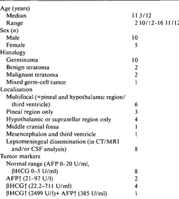
Table 1. Characteristics of 15 patients with GCTC. Age (years) Median 113/12 Range 2 10/12-16 11/12 Sex («) Male 10 Female 5 Histology Germinoma 10 Benign teratoma 2 Malignant teratoma 2 Mixed germ-cell tumor 1 Localisation
Multifocal (=pineal and hypothalamic region/ third ventricle) 6 Pineal region only 3 Hypothalamic or suprasellar region only 4 Middle cranial fossa 1 Mesencephalon and third ventricle 1 Leptomeningeal dissemination (in CT/MRI
and/or CSFanalysis) 8 Tumor markers
Normal range (AFP 0-20 U/ml,
(3HCG 0-5 U/ml) 8 AFPf (21-97 U/l) 2 (3HCGT (22.2-711 U/ml) 4 PHCGT (2499 U/l)+ AFPf (385 U/ml) 1
treated with modern protocols. In addition we tried to evaluate the effects of newer protocols that are stratified more specifically against different histological types and which better respect potential radiotoxicity.
Table 2. Symptoms at presentation.
Symptom Affected patients (n = 15)
Nausea and vomiting Severe headache Diabetes insipidus Chronic fatigue Chronic weight loss Symptoms of hemiparesis Ophthalmologic symptoms
Abnormal eye movements Diplopia
Complete Pannaud syndrome Decreased visual acuity Visual field defect Ptosis Pilloedema 10 9 8 5 4 3
repeated measuring of specific weight of urine, serum and urine electrolytes. As several patients required hormone substitution after therapy end, we did not suspend substitution to perform endocrino-logical follow-up testing.
Neurological and neuropsychological evaluation
Repeated neurological exams were performed in all patients. Nine patients had neuropsychological evaluation by trained psychologists. All patients and parents were interviewed about schooling outcome (determined as coping/non coping with age related schooling perfor-mance), social activity and behaviour before and after treatment for GCTC.
Patients and methods
From July 1980 to December 1998, 272 children were newly diagnosed with a brain tumour in our institution. Of these, 15 patients were diagnosed and treated for GCTC (5.5% of all brain tumours) and were followed until July 1999. Observation period was 16-228 (mean 91) months. Inclusion was restricted to patients who had confirmed histo-logical or cytohisto-logical diagnosis according to WHO classification, radiological evaluation (CTscan and/or MRI) and analysis of tumour markers (serum AFP and PHCG).
The patients' characteristics and symptoms at presentation are shown in Tables 1 and 2. Data were collected during treatment and regular follow-up and reviewed by the authors. Patients were evaluated for remission status, endocrine dysfunction, neurological and neuro-psychological sequelae. Growth, weight, evaluation of bone age and secondary sexual characteristics (Tanner stages) were measured re-peatedly in all patients.
Endocrine function tests
Evaluation in our patients was started at time of initial presentation and was completed during follow-up after treatment. Evaluation for endocrinological dysfunction consisted of the following tests:
TSH, FT4 and T3 were measured repeatedly in all patients. GH and IGF was monitored in 10 patients, LH, FSH. testosteron, oestra-diol, ACTH, basal cortisol in 9 patients and prolactin in 6 patients, respectively. The following functional hormone tests were carried out: gonadal-releasing-hormone-test in six, arginin- and insulin-tolerance test in four and TRH test in three children, respectively. Normal values were determined according to age specific range given by the labora-tory-. Posterior pituitary function was evaluated in all patients by
Statistical methods
The Kaplan and Meier survival method [9] was used to calculate the overall survival and progression-free survival. Overall survival was calculated from date of diagnosis until date of last follow-up or death for any cause. Progression-free survival was calculated from date of end of therapy to date of last follow-up, first relapse or death.
Results
Therapy outcome (see Table 3 and Figures 1 and 2)
The overall survival rate in the 15 patients is 87% (germinoma 90%) with a mean follow-up of 77 months after end of treatment. Two patients have died; one patient (case AF) died fourteen months after end of therapy due to acute infarction of the left middle cere-bral artery. At autopsy no evidence of tumour relapse was found. The aetiology remained unclear, an overwhelming virus encephalitis was supposed. Another patient with a mixed germ-cell tumour (case WM) died of tumour progression after relapsing four months after end of treatment.
Tumour relapses: in addition to the above mentioned there were two further patients who suffered a relapse. One germinoma patient (case RM) had a first relapse in the cervical, thoracic and lumbar spine 12 months after diagnosis. Chemotherapy (MAHO 94) and irradiation
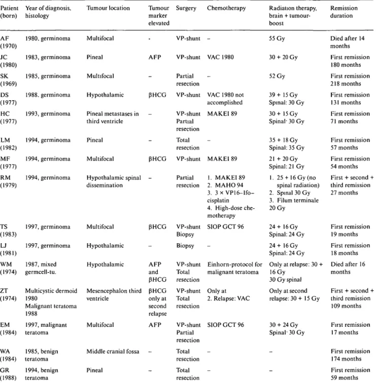
Table 3. Individual clinical characteristics, treatment and outcome of all patients. Patient (born) AF (1970) JC (1980) SK (1969) DS (1977) HC (1977) LM (1982) MF (1977) RM (1979) TS (1983) LJ (1981) WM (1974) ZT (1974) EM (1984) WA (1984) GR (1988) Year of diagnosis, histology 1980. germinoma 1983, germinoma 1985, germinoma 1988. germinoma 1993, germinoma 1994, germinoma 1994, germinoma 1994, germinoma 1997, germinoma 1997, germinoma 1987, mixed germcell-tu. Multicystic dermoid 1980 Malignant teratoma 1988 1997, malignant teratoma 1985, benign teratoma 1994, benign teratoma Tumour location Multifocal Pineal Multifocal Hypothalamic Pineal metastases in third ventricle Pineal Multifocal Hypothalamic spinal dissemination Multifocal Hypothalamic Hypothalamic Mesencephalon third ventricle Multifocal
Middle cranial fossa Pineal Tumour marker elevated -AFP -PHCG -PHCG pHCG -AFP and PHCG PHCG only at second relapse AFP -Surgery VP-shunt VP-shunt Partial resection VP-shunt VP-shunt Partial resection Total resection VP-shunt Partial resection VP-shunt Biopsy Biopsy VP-shunt Total resection VP-shunt Total resection VP-shunt Partial resection Total resection Total resection Chemotherapy -VAC 1980 -VAC 1980 not accomplished MAKEI 89 -MAKEI 89 1. MAKEI 89 2. MAHO94 3. 3 x VP16-Ifo-cisplatin 4. High-dose che-motherapy SIOP GCT 96 -Einhorn-protocol for malignant teratoma Only at 2. Relapse: VAC SIOP GCT 96 -Radiation therapy, brain + tumour-boost 55 Gy 30 + 20 Gy 52 Gy 39+ 15 Gy Spinal: 30 Gy 30+ 15 Gy Spinal' 30 Gy 35+ 18 Gy Spinal: 35 Gy 21 +20Gy Spinal: 21 Gy 1. 25 + 16Gy(no spinal radiation) 2. Spinal 30 Gy 3. Filum terminale 20 Gy 24+16Gy Spinal: 24 Gy 24+ 16 Gy Spinal: 24 Gy Only at relapse: 30 + 16 Gy 30 Gy spinal Only at second relapse: 30+ 15 Gy 30 + 24 Gy Spinal1 30 Gy -Remission duration Died after 14 months First remission 180 months First remission 218 months First remission 131 months First remission 71 months First remission 57 months First remission 54 months First + second + third remission 27 months First remission 19 months First remission 18 months Died after 16 months First + second + third remission 109 months First remission 17 months First remission 174 months First remission 59 months
of the spine (from C2 to S2) with 30 Gy was given. A second relapse in the conus medullaris region occurred 29 months after initial diagnosis and was treated with chemotherapy including high-dose treatment (carbo-platin and vepesid), autologous stem-cell transplantation and radiotherapy of the conus medullaris. The patient is currently in complete remission 27 months after end of treatment. A further patient (case ZT) was originally diagnosed 1980 with a benign multicystic dermoid in the third ventricle. Ninety-two months after the initial diag-nosis he developed a malignant teratoma in the recessus infundibularis. Again, thirteen months after resection of this tumour, a local relapse occurred. A third remission
was achieved following combined radiation- and chemo-therapy. PHCG was elevated only at second relapse. One hundred nine months later he is in continuous complete remission.
Direct therapy associated morbidity Neurosurgery
All 15 patients had either biopsy, partial or total tumour resection, ventriculo-peritoneal-shunt insertion or a combination of procedures. There was no associated mortality. One patient (WA) was operated at the age of six weeks because of a mature teratoma of the right
0 , 8 0 , 6 - 0,40 . 2 --4 U. tumours- 0,0-0,4. Other histologies 96 144 Progression-free survival (months)
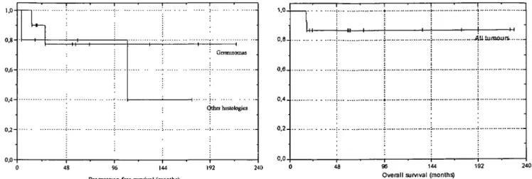
Figure 1. Kaplan-Meier analysis for progression-free survival in 15
patients with intracerebral germ-cell tumour. Analysis separately for germinoma (ft = 10) and all other histologies (ft = 5).
0,2-0,0.
96 144 192 Overall survival (monlhs)
Figure 2. Kaplan-Meier analysis for the overall survival in 15 patients
with intracerebral germ-cell tumour.
middle cranial fossa. After surgery she had left sided paresis of facial and hypoglossus nerves which persisted. In two patients Parinaud syndrome was observed only after neurosurgical intervention.
Shunt complications occurred in one of nine patients: Patient MF presented with meningitis due to clostridium diphteriae two months after insertion of a ventriculo-peritoneal shunt. Subsequently, three months later, he developed acute abdominal pain which was caused by a perforation of the distal part of the shunt into the duodenum. No peritoneal metastases have been ob-served.
Radiation therapy
Postactinic neuronal dysfunctions were found in two patients. One child, irradiated 1983 at the age of 2 10/12 years (patient JC) with 30 Gy to the whole brain and 20 Gy tumour boost, presented with ataxia only during the first year after radiotherapy. A 16-year-old adolescent treated according to the MAKEI 89 protocol developed spastic hemiplegia due to leukencephalopathy four months after radiotherapy with 20 x 1.5 Gy to the whole brain including spinal axis and 14 Gy tumour boost. He has residual findings of hemiparesis six years after the end of treatment.
Chemotherapy
One patient had severe gastrointestinal bleeding with acute abdomen and bowel obstruction under VAC-chemotherapy. There were no other severe infectious or bleeding complications reported. During long-term follow-up patients treated with cisplatin based chemo-therapy showed no detectable hearing deficit on audio-gramm or laboratory signs of impaired renal function.
Long-term follow-up Ophthalmologic symptoms
At presentation nine patients had abnormal eye move-ments. Complete Parinaud syndrome was found in six
patients. Four patients had decreased visual acuity and three patients showed visual field defects.
At the last follow-up, six children had persisting eye movement abnormalities. Complete Parinaud syndrome was found in five cases. Two patients had a restricted visual field and in four children a reduced visual acuity was found. One adolescent girl presented with headache and reduced visual acuity of the right eye. The initial MRI showed a tumour infiltrating the optic nerve near the chiasm. Her visual acuity decreased rapidly and 14 months after the end of therapy (cranial and spinal radiotherapy with 24 Gy and tumour boost of 16 Gy, fractionated into 25 doses of 1.6 Gy) she was blind on the right eye and had a visual acuity of 0.4 in the left eye.
Endocrine abnormalities
In nine patients (age 2 10/12-16 11/12 years; eight germinomas and one malignant teratoma) clinical and laboratory signs of endocrine dysfunction were found.
Eight patients had already diabetes insipidus on admission, which persisted at follow-up. All had tumour infiltration of the hypothalamus. Seven patients developed persistent panhypopituitarism. All had received radio-therapy with 40-55 (mean 46) Gy to the tumour region, fractionated into doses of 1.6-2 Gy.
GH deficiency was found in seven patients, but only in three patients decreased growth rate was documented and GH was substituted. In these patients radiation therapy was given at the age of 3, 11, and 14 1/2 years, respectively, with 40, 50 and 52 Gy total tumour dose, fractionated into doses of 1.8-2 Gy. At the time of the last visit all patients had body height above the 10th percentile.
Increased prolactine secretion (21-1310 mU/ml) was found in six children with hypothalamic or multifocal tumour. Two had initial tumour resection and in all radiation therapy had been applied (40-54 total tumour dose, fractionated into doses of 1.6-2 Gy/d). All six patients also had hypogonadism and decreased FSH and LH levels.
One patient (case JC), who received focal radiation therapy in 1983 (50 Gy total tumour dose, fractionated into 25 doses of 2 Gy) for germinoma of the pineal region at the age of 2 10/12 years, showed thyroid dysfunction, growth hormone deficiency and subnormal FSH not before 10 years after therapy completion.
Six out of fifteen children had no endocrine dysfunc-tion at last follow-up and showed normal growth and normal bone age. This includes both patients with a mature teratoma who were treated with tumour resection alone, 1 patient who died 6 months after radiotherapy for tumour relapse and 1 patient with a malignant teratoma of the pineal gland who is in first remission 17 months after partial tumour resection and radiotherapy with 30 Gy and 24 Gy tumour boost (dose per fraction 1.5-1.6 Gy). Two patients were followed for 57 and 71 months. Both had resection of a pineal germinoma and radiation therapy with a dose of 53 Gy (fractions of 1.6-1.8 Gy) and 45 Gy (fractions 30 x 1.5 Gy) at an age of 12 and 16 years, respectively.
Neuropsychological sequelae
Nine patients underwent neuropsychological evaluation 14-157 months after the end of treatment. They had undergone radiation therapy at an age of 2 10/12-16 11/12 (mean age 10 5/12 years), with total tumour doses of 40-52 Gy. Eight children received additional chemotherapy and five had partial or total tumour resection. All nine patients had mild to severe difficulties to cope with schooling demands. All were 'slower' and had attention deficits in comparison to their healthy peers. Three patients had fine motor dysfunctions, two patients had disturbed spatial processing. In four children planing and organising functions were inadequate.
Schooling outcome: at last control six patients at-tended a college or high school. Five patients served an apprenticeship or had already finished their professional training. Four patients had been integrated in a profes-sional training program for mentally disabled adolescents. All of the seve children treated after 1993 were going to the same class as before treatment for GCTC. Of them three had some concentration difficulties and three patients had difficulties coping with schooling demands. Only one patient had severe disabilities that did interfere with daily life.
In six patients (age at tumour diagnosis 10-16 10/12 years, mean 13 1/12 years) no neuropsychological abnor-malities or schooling difficulties were reported and routine neurological examination did not reveal any of such deficiencies. In these patients no neuropsychologi-cal testing was performed.
Among this group radiation therapy had been per-formed in four patients. The applied tumour dose (40-55 Gy, mean 47 Gy) was similar to that of the group of children with abnormalities in the neuropsychological testings.
Discussion
With regard to the total number of children with brain tumour treated at our institution the incidence of GCTC seems high in comparison to that previously published from centres in western countries [1-4]. An increasing incidence of GCTC during the last years (In our series 1980-1992: seven patients, 1993-1997: eight patients) has been noted by other authors too [1,2]. The incidence of pediatric brain tumours in general has slightly in-creased over the last decades [10]. Concerning our series of GCTC some increase of incidence might be explained by improved diagnostic procedures as well as the changes of referral pattern over time.
In agreement to the literature our data demonstrate an excellent survival outcome for germinomas and benign teratomas. Modern neurosurgical techniques have mini-mised damage to brain tissue and have reduced the high morbidity and mortality associated with past surgical practices. Furthermore, relapsed patients can be sal-vaged today by high-dose chemotherapy and autologous stem cell transplantation [1, 4, 8].
Our results indicate that, in order to reduce late morbidity, treatment should be tailored to histological subtype and the spread of the disease. There is discus-sion about the value of routine adjuvant spinal irradi-ation in patients with germinoma [2, 4, 11]. Controlled prospective studies are needed to determine its benefit
versus the attendant risk. Some authors state that
PHCG-producing germinomas have a higher recurrence rate [8, 12]. Among our patients with J3HCG producing germinoma (three patients) we saw no relapses although a test for detection of pHCG in CSF routinely [12] was not available at our institution.
Histology is still the most important prognostic factor in GCTC. In an unusual histological diagnosis one should always be aware of the possibility of a sampling error in the biopsy. Late tumour relapse in GCTC is unusual but has been described in teratomas [2], as demonstrated by one of our patients who suffered a relapse 92 months after initial diagnosis for multicystic dermoid tumour. As histology at relapse showed a malig-nant teratoma it is difficult to deduce retrospectively if tumour transformation happened, or if the initial biopsy missed part of the tissue of a mixed germ-cell tumour.
Eight out of ten patients with germinoma and one patient with malignant teratoma had endocrine abnor-malities, that were already present in the majority at diagnosis. Seven out of eight patients with a hypothalamic tumour developed panhypopituitarism during follow-up. There is continuing discussion whether radiotherapy is responsible for worsening pituitary and hypothalamic function. Most of our patients had already one to three endocrine axes involved when a CGTC was diagnosed. Panhypopituitarism emerged during therapy or time of follow-up in nearly all of them. Follow-up of endocrine functions is required as most will need lifelong hormone substitution. In seven patients hyperprolactinemia was found. With a disruption of hypothalamic-pituitary axis
after radiotherapy or surgery prolactin secretion is no longer inhibited [4,13] and in addition to gonadotropine deficiency this might contribute to hypogonadism and delayed puberty [14].
In this report seven patients had growth hormone deficiency. In three patients decreased growth rate had been documented. Unfortunately not all patients had been evaluated for growth hormone deficiency, but it is interesting to note that at last follow-up all patients had achieved a height above the 10th percentile. The majority of the patients had radiation therapy after 10 years of age, thus reducing the possible impact of radiation therapy on growth.
In our series 5 of 13 surviving patients have to date no endocrine dysfunction. None of these patients had tumour infiltrating the thalamic or hypothalamic region. Two of the patients with a pineal germinoma and without endocrine dysfunction had radiotherapy with a mean dose similar to that given to patients with endocrine dysfunction but without involvement of the pituitary region. So, here the favourable outcome might be due to tumour location (pineal gland) and better exclusion of the pituitary region from irradiation.
At this point we cannot draw any final conclusions as we could observe in our patients (and according to the literature) endocrine dysfunction presenting more than 10 years after therapy [13, 15].
Radiation therapy has an impact on neurodevelop-mental status and IQ, particularly when radiation therapy is applied before the age of 36 months. Older patients score lower in schooling outcome after radiation ther-apy [14, 16]. All but one patients of this series were much older than three years at time of cranial radiation therapy but 60% of the patients we studied show neuro-psychological deficits even years after completing the treatment. In our series we could not observe any differ-ence in applied mean radiation dose among patients with and without neuropsychological sequelae. However, the impact on IQ and schooling outcome seems to be less severe in children treated in the last decade in comparison to data from patients treated with radiation therapy during the 1980s [14]. For definite conclusions a longer follow-up is required, but the better outcome might be due to the reduction of the total applied doses and the reduced daily fractions of current protocols [2,16].
In the patients we studied no secondary neoplasms have to date been observed. Other authors reported (and predicted for future follow-up) high rates of secondary neoplasms due to intense radiation therapy [8].
In conclusion, today GCTC in children and adoles-cents are potentially curable diseases. As a consequence of tumour site and applied therapy a large proportion of patients will probably show life long sequelae and require further care. The aim of modern treatment pro-tocols is to tailor treatment according to the various subsets of GCTC. The goal is to increase the cure rate of the more unfavourable subsets of GCTC (i.e., secret-ing tumours) and to reduce late sequelae particularly in
those cases with a favourable prognosis (i.e., pure germinoma). These objectives are followed by the cur-rent treatment protocol of the International Society of Paediatric Oncology (SIOP). In the subset of germinoma two approaches of treatment strategies are compared. One group of germinoma is treated by a reduced dose of craniospinal irradiation and tumour boost and the other group will receive a combination of chemotherapy and restriction of radiotherapy to the primary site. The application of radiotherapy in a hyperfractionated manner might be an additional approach for further reducing long-term side effects [2, 16]. Achievement of complete remission of intracerebral germinomas by che-motherapy alone is possible [3]. However, whether such an approach of replacing radiotherapy completely by multiagent chemotherapy might become standard in at least some subgroups of intracerebral germ-cell tumours has to be shown in long-term follow-up studies.
Acknowledgements
We wish to thank D. Betts and M. Suter, for their help to prepare this manuscript, Dr M. Hany and Dr L. Nobile for providing clinical date of follow-up controls and Dr E. Martin and Dr U. Willi and other colleagues from the Radiology Department of our hospital for the CT and MRI studies. Finally we thank Dr P. Huguenin and the Department of Radiooncology of the University Hospital of Zurich for the collaboration in the care for our paediatric oncology patients.
References
1. Cohen ME. Brain Tumors in Children, 2nd ed. New York: Raven press 1993; 329-46.
2. Calaminus G, Gobel U. Intracranial germ-cell tumors: A com-prehensive update of the European data. Neuropediatrics 1994; 25: 26-32.
3. Balmaceda C, Finlay L. Chemotherapy without irradiation - a novel approach for newly diagnosed CNS germ-cell tumors: Results of an international trial. J Clin One 1996; 11: 2908-15. 4. Horowitz MB, Hall WA. Central nervous system germinomas. A
review. Arch Neurol 1991; 48: 652-7.
5. Kleihues PC, Cavenee WK. Pathology and Genetics of Tumours of the Nervous System. Lyon: International agency for research on cancer. Team Rush 1997; 164-9.
6. Kleihues PC, Burger PC. Scheithauer BW. Histological typing of tumor of the central nervous System. In Scheithauer BW (ed): WHO International Histology Classification of Tumors. 2nd edn. Berlin: Spnnger-Verlag 1993.
7. Hainsworth JD. Greco AF. Extragonadal germ-cell tumors and unrecognized germ-cell tumors. Semin Oncol 1992; 19: 119-27. 8. Savamura Y, Ikeda J. Shirato H et al. Germ-cell tumours of the
central nervous system: Treatment consideration based on 111 cases and their long-term clinical outcomes. Eur J Cancer 1998: 34:104-10.
9. Kaplan EL. Meier PJ. Nonparametric estimation from incom-plete observations. J Am Stat Assoc 1958; 53: 457-81.
10. Smith MA. Freidlin B. Ries LA, Simon R. Trends in reported incidence of primary malignant brain tumors in children in the United States. J Natl Cancer Inst 1998: 90 (17): 1269-77.
11. Linstadt D, Wara WM. Edwards MS et al. Radiotherapy of primary intracranial germinomas: The case against routine cranio-spinal irradiation. J Radiat Oncol Biol Phys 1988; 15: 291-7. 12. Inamura T, Nishio S, Ikezaki K, Fukui M. Human chorionic
gonadotropine in CSF, not serum predicts outcome in germinoma. J Neurol Neurosurg Psych 1999; 66: 654-7.
13. Constine LS, Woolf PD. Cann D et al. Hypothalamic-pituitary dysfunction after radiation for brain tumor. N Engl J Med 1993; 328: 87-94.
14. Duffner PK, Hororwitz M E, Krischer J P. The long-term effects of cranial irradiation on the central nervous system. Cancer 1985: 56:1841-6.
15. Oberfield SE, Allen JC, Pollack J et al. Long-term endocrine sequelae after treatment of medulloblastoma: Prospective study of growth and thyroid function. J Pediatr 1986; 108: 219-23. 16. Moor BD, Copeland DR, Ater JL. Long-term
neuropsycholog-ical effect of cranial radiation therapy on patients with brain tumors diagnosed in infancy. J Clin Exp Neuropsychol 1991; 13' 98a.
Received 2 November 1999; accepted 8 March 2000.
Correspondence to:
F. K. Niggli. MD
University Children's Hospital Zurich Division of Oncology
Steinwiesstr. 75 CH-8032 Zurich Switzerland