Induction of adenocarcinoma from hamster pancreatic islet cells
treated with N-nitrosobis(2-oxopropyl)amine in vitro
B.Schmied
1,2, G.Liu
1, M.P.Moyer
3, I.S.B.Hernberg
1,4,
W.Sanger
5, S.Batra
1,6and Parviz M.Pour
1,7,81UNMC/Eppley Cancer Center, University of Nebraska Medical Center, Omaha, NE, USA,2Visceral and Transplantation Surgery, Inselspital, Bern, Switzerland,3The University of Texas Health Science Center, Center for Human Cell Biotechnology, San Antonio, TX, USA,4Klinik fu¨r Gastroenterologie, Hepatologie und Infektiologie, Otto-von-Guericke Universita¨t Magdeburg, Magdeburg, Germany and5Munroe-Meyer Center for Human Genetics,6Department of Biochemistry and Molecular Biology and7Department of Pathology and Microbiology, University of Nebraska Medical Center, Omaha, NE, USA
8To whom correspondence should be addressed at: UNMC/Eppley Cancer Center, University of Nebraska Medical Center, 986805 Nebraska Medical Center, Omaha, NE 68198-6805, USA
Email: ppour@mail.unmc.edu
Our previous studies in the hamster pancreatic cancer
model have indicated that pancreatic ductal
adenocarcin-omas derive not only from ductal/ductular cells but also
from islets. To verify the presence of carcinogen-responsive
cells within islets, we tested the effect of the pancreatic
carcinogen
N-nitrosobis(2-oxopropyl)amine
(BOP)
on
recently established continuous hamster pancreatic islet
culture. Isolated pure pancreatic islets of hamsters were
treated in vitro with BOP at a concentration of 0.25 mM
three times a week for 19 weeks. Each treatment week was
designed as a stage. The growth of these cells, designated
KL5B, was compared with untreated cultured islets,
desig-nated KL5N. As in our previous study, between 14 and 21
days of culture, exocrine and intermediary cells developed
within both KL5N and KL5B islets, which were then
replaced by undifferentiated cells. No differences were
found in the growth patterns of KL5N and KL5B until
stage 4, when KL5B cells showed accelerated cell growth
and cell pleomorphism, which increased gradually at later
stages of treatment. Anchorage-independent and in vivo
growth did not appear until stage 19. Mutation of c-Ki-ras
at codon 12 (GGT
→
GAT) was detected in KL5B cells but
not in KL5N cells. In vivo KL5B cells formed anaplastic
invasive cancer with areas of glandular formation,
over-expressed TGF-
α
and EGFR, expressed cytokeratin,
vimen-tin, laminin and
α
-1 antitrypsin and reacted strongly with
L-phytohemagglutinin and tomato lectin. Some cells withinislets are responsive to the carcinogenic effects of BOP.
Whether these cells represent islet cell precursors (stem
cells) or malignant transdifferentiated islet cells remains
to be seen.
Introduction
The silent course of pancreatic cancer and its explosive
fatal outcome have hampered our understanding of tumor
histogenesis and early biochemical and genetic alterations,
Abbreviations: BOP, N-nitrosobis(2-oxopropyl)amine; SMG, submandibular
gland; PHA, phytohemagglutinin.
information that could help us diagnose the disease at a curable
stage. Although general opinion favors the hypothesis that
pancreatic tumors originate from ductal/ductular cells in
humans, the views are divided for experimental pancreatic
cancer. Some believe in ductal genesis, whereas others suggest
acinar cells are the precursor cells (1).
In the hamster model, which mimics human disease in many
clinical and biological aspects, we have demonstrated that
pancreatic adenocarcinoma originates not only from ducts and
ductules (2–6), but also from islets (7–11). In fact, the first
morphological change that occurs during pancreatic
carcino-genesis is the appearance of ductular structures within islets
(intra-insular ductules) or around the islets (peri-insular
ductules) (2,4,6,8). The intra-insular ductules proliferate
form-ing either benign patterns consistent with human pancreatic
microcystic adenomas or becoming increasingly hyperplastic,
dysplastic and atypical and culminating in the formation
of malignant glands that destroy the islets and invade the
surrounding tissues, even when they are of microscopic size
(4–6,8). The following observations support the role of islets
in pancreatic ductal carcinomas: (i) streptozotocin pretreatment,
which causes destruction of
β
-cells, inhibits the pancreatic
carcinogenicity of N-nitrosobis(2-oxopropyl)amine (BOP) at
low doses or prevents it at high doses (12–14); (ii) genetically
diabetic hamsters with atrophic islets are resistant to the
pancreatic carcinogenic effects of BOP, whereas the pancreas
of a non-diabetic strain with intact islets is susceptible (15);
(iii) induction of nesidioblastosis enhances pancreatic
carcino-genesis (16); and (iv) transplantation of homologous islets into
the submandibular gland (SMG) of recipient hamsters and
subsequent BOP treatment induces invasive and metastasizing
ductal-type adenocarcinomas, histological and
immunohisto-chemical analyses of which point to the derivation of tumors
from within islets (9–11).
Our limited studies in humans have indicated that cancer
cells also develop within islets (4,17). In a recent observation,
both of the microscopical pancreatic carcinomas which we
found incidentally at autopsy appeared to develop within islets
(18). These observations led us to believe that certain pancreatic
cells within islets are particularly sensitive to the
carcino-genic insult.
To test this hypothesis, we established, for the first time, a
continuous culture of hamster pancreatic islets (19). In this
culture, the initially pure islets were replaced temporarily by
ductal, acinar and intermediary cells (having the characteristics
of both acinar and islet cells) before giving rise to
undifferenti-ated cells, which seem to present as pancreatic stem cells.
Although presently the origin of these cells from islet cell
precursors or from transdifferentiation of islet cells is obscure,
the model provided a unique opportunity to test the effect of
BOP on cultured islets. Because it has been shown by us (20)
and others (21) that BOP can transform isolated pancreatic
ductal cells in vitro, we tested its effect on cultured hamster
islets.
Materials and methods
Animals
Eight- to 10-week-old male Syrian golden hamsters from the Eppley colony were used for transplantation experiments. Animals were housed in cages with Sani-cell bedding and were kept under standard laboratory conditions (temperature 20 6 2°C, 10 air changes/min, 12 h/12 h light/dark cycle) and received pelleted diet (Wayne, Indianapolis, IN). Water was provided ad libitum.
Chemicals
BOP was synthesized in our laboratories. The culture medium (M3:6) was a gift from InCell (San Antonio, TX). Penicillin, streptomycin, trypsin, EDTA, fetal bovine serum and RPMI 1640 were purchased from Life Technologies (Gaithersburg, MD). Purified agar used for soft agar assay was ordered from Difco (Detroit, MI). PCR core reagents were purchased from Perkin-Elmer Cetus (Norwalk, CT).
Islet culture
Two hundred freshly isolated islets, designated KL5N (no BOP treatment), and 250 islets designated KL5B (with BOP treatment), were cultured in M3:6 medium supplemented with 6% fetal bovine serum, 100 U/ml penicillin and 100µg/ml streptomycin. The islets were placed in a polystyrene Petri dish (Baxter, McGaw Park, IL) on a rocker (15 r.p.m./min) at 37°C in a humidified atmosphere of 5% CO2 in air for 14 days, as reported (19). In none of the islets that were examined immunohistochemically and electron microscopically after isolation was any exocrine cell contamination noticeable. There were no cells reactive to pancytokeratin, a marker for hamster pancreatic ductal/ ductular cells or that presented features of ductal or acinar cells. The floating islets were hand picked every day to separate them from the attached fibroblasts and were placed in a new dish with fresh M3:6 medium. On day 14 after the initial isolation, when no fibroblasts could be identified, the islets were plated into a six-well tissue culture plate, allowed to attach and continuously cultured without shaking. Thereafter, they were trypsinized and subcultured in M3:6 medium every week.
BOP treatment
Islets were treated with 0.25 mM BOP from the first day of islet culture, designated KL5B cells. A pilot study had shown that this concentration of BOP has no toxic effects and is well tolerated by islet cells. The BOP-containing medium were changed for fresh every day for the first 14 days, the period during which fibroblasts were eliminated. Thereafter, the medium was changed three times per week (Mondays, Wednesdays and Fridays) for fresh BOP. This treatment scheme was designated a stage, which was longitudinally continued in subsequent culture, as reported (20). After the end of each stage, the cells were transferred (passaged) into a new flask. For better comparison of data at each stage, the KL5N cells were also transferred (passaged) to a new flask whenever the KL5B cells were passaged. Con-sequently, the stage of KL5B cells corresponded to the passage of KL5N cells. Like KL5N cells, when islets were transferred to a flask, they attached to the bottom of the flask and at day 21 (stage three) a mass of cells radiated from islets into the surrounding culture. The cell aggregate representing the remnant of islets disappeared and at stage 4 the culture consisted of a monolayer of undifferentiated cells. These cells were trypsinized and counted. A portion of the cells (between 33103and 33104cells) were harvested for soft agar assay, for examination of the c-Ki-ras mutation and, if the cell number was sufficient, for immunohistochemistry and electron microscopy analysis. After stage (passage) 19, BOP treatment was discontinued and further cell transfer was defined as passage.
Anchorage-independent growth assay
This assay was performed as reported earlier (20). Colony formation was checked once a week and discarded after 45 days.
Electron microscopical examination
Transmission electron microscopical examination was performed according to our published technique (3,22).
Cell growth determination
The growth curve of the cells in culture was established by plating 50 000 and 200 000 cells into separate T-25 plastic flasks. The number of viable cells, determined by the dye exclusion test, was counted at 24 h intervals using a hemocytometer. Population doubling time was calculated by plotting the mean cell number versus days in culture on a semi-logarithmic graph, as reported (21,22).
Transplantation experiment
Approximately 1 000 000 KL5B cells at stage 19 were transplanted into the subcutaneous tissue, pancreas or SMGs of recipient hamsters (three hamsters per site), as reported (23). Subcutaneous masses and lesions in the SMG and
pancreas were removed at autopsy and all organs were examined grossly for metastases. The regional lymph nodes of these hamsters and the pancreas of three age-matched control hamsters were removed, fixed in buffered formalin and processed for histology by conventional methods.
Histochemical and immunocytochemical examination
All histochemical and immunocytochemical examinations, including the multilabeling technique, were performed as previously described (24). Anti-bodies used in the experiment are reported in our earlier study (19). The immunoreactivity was scored as none (–), weak (1), moderate (11) or strong (111). The cellular immunoreactive site was defined as cytoplasmic, luminal or cell membrane. To calculate the number of stained KL5N and KL5B cells, two observers counted 1000 cells on each slide and the number of cells immunoreactive with a given antibody was estimated as a percentage. The average value was considered representative.
Cytogenetic analysis
The karyotyping of KL5N and KL5B cells at passages 24 and 34 was performed, as reported (20,22). Fifty-one KL5B and 42 KL5N cells were examined.
DNA and RNA purification
DNA and total RNA from the pancreas, islet and KL5N cells were purified by minor modifications in the guanidine isothiocyanate/acid phenol method (27) published previously (28). Briefly, tissue or cells were homogenized in a buffer containing 4 M guanidine isothiocyanate, 25 mM sodium citrate, 0.5% sarcosyl and 0.1 M 2-mercaptoethanol. It is acidified by adding 2 M sodium acetate (pH 4.0) and then phenol/chloroform extracted and ethanol precipitated.
Examination of the c-Ki-ras mutation
Mutation of the c-Ki-ras gene was examined by RT–PCR, according to a previously described method (20).
RT–PCR
One microgram of total RNA was combined with 100 ng random hexamer primers and the solution was heated at 72°C for 5 min and then placed on ice. dNTPs (10 mM each), reverse transcriptase, RNasin, 103 PCR buffer, 25 mM MgCl2and water were added (as per the manufacturer’s instructions; Perkin-Elmer, Norwalk, CT) and the mixture was incubated at 42°C for 60 min. The cDNA synthesis reaction was terminated by heating at 95°C and the mixture was stored at –70°C. PCR was performed using 10µl cDNA in a total reaction volume of 50µl containing 2.5 U Taq DNA polymerase, Taq buffer containing 1.5 mM Mg21, 0.6µM forward primer, 0.6µM reverse
primer and 200µM dNTPs. A hot start technique was used. Thirty-five cycles of amplification were performed of 95°C for 1 min, 55°C for 90 s and 72°C for 290 s, with a final elongation for 10 min. A negative control lacking template was used in the reaction. Products were analyzed by electrophoresis on 1.5% agarose gels. Primers for PCR of IPF1, IEF1, NKx6.1, Neuro-D, Pax6, EGFR and TGF-α are shown below. These primer sequences were obtained from hamster (Nkx6.1), rat (IPF1, IEF1, EGFR and TGF-α) and mouse (NeuroD and Pax6). The primer sequences are: IPFI, forward 5 9-CTCGCTGGGAACGCTGGAACA-39, reverse 59-GCTTTGGTGGATTTCA-TCCACGG-39; IEF1, forward 59-ACCCTTCACCAATGACTCCTATG-39, reverse 59-ATGATGACTGCAAATCGC-39; Nkx6.1, forward 59-TCTTCTG-GCCCGGGGTGATG-39, reverse 59-AGCCGCGTGCTTCTTCCTCCA-39; NeuroD, forward 59-CTTGGCCAAGAACTACATCTGG-39, reverse 59-GGA-GTAGGGATGCACCGGGAA-39; Pax6, forward 59-TTGGGAAATCCGAG-ACAGAT-39, reverse 59-GGTACTGAAGTCCCGGGATA-39; EGFR, forward 59-ACTGGCCTTAGGGAACTGCCC-39, reverse 59-TCCTGTACACCCAG-CGGCACA-39; TGF-α, forward 59-ACCTGCAGGTTTTTGGTGCAG-39, reverse 59-GGAGGGCGCTGGGCTTCTCG-39.
Results
Islet culture
The growth patterns of both KL5N and KL5B islets were
similar to those observed in our previous study (19). The size
of islets varied between 0.12 and 0.32 mm and there were no
differences in the size of islets between BOP-treated and
untreated. At day 14 (stage 2), the number of floating islets
transferred to flasks was 90 and 180 for the KL5N and KL5B
cultures, respectively. As in our previous report (19), ductular,
acinar and intermediary cells appeared within both KL5N and
KL5B islets between stages 2 and 3. In both cultures, the
epithelial cells from islet cores spread out into the surrounding
Fig. 1. KL5N cells (islets without BOP treatment) in culture. (a) At passage
4, the cells that spread out of islets attach to each other to make island-like aggregates (H&E,340). (b) At later passages, they form uniform cells with a tendency to adhere to each other (H&E,3210).
area and attached to the bottom of the flask. At stage 4,
although KL5N cells tended to attach to each other and grew
in islet-like cell aggregates (Figure 1), KL5B cells formed a
monolayer of pleomorphic cells with abundant eosinophilic
cytoplasm and a few pleomorphic and hyperchromatic nuclei
(Figure 2a).
At stage 8, the growth of KL5B cells accelerated, showing
a doubling time of 36 h, compared with 48 h for KL5N cells.
Although KL5N cells retained their monomorphic phenotype,
KL5B cells presented as a small cell population with
hyperchro-matic nuclei between large cells (Figure 2b). The number of
small cells increased gradually and, at stage 19, the culture
was composed entirely of small cells with pleomorphic and
hyperchromatic nuclei and scanty eosinophilic cytoplasm. Fine
cytoplasmic attachments could be seen between some of these
cells (Figure 2c). Whereas the growth and phenotype of KL5N
cells at passage 19 remained fairly constant, that of KL5B
cells further accelerated (doubling time 26 h). Also in contrast
to KL5N cells, KL5B cells showed, for the first time,
anchor-age-independent growth and could be maintained in RPMI
1640 culture medium supplemented with fetal calf serum.
Electron microscopically, the patterns of KL5N cells were
similar to the cultured islet cells in our previous study (19)
and presented as undifferentiated cells poor in cell organelles.
KL5B cells, however, were pleomorphic and contained large
vesicles, small cystic spaces containing material of various
optical densities, microfilaments and a few intracytoplasmic
lumens (Figure 3). No endocrine granules were detected.
Fig. 2. KL5B cells (islets treated with BOP) in culture. (a) At stage 4, the
cells around the islets have large eosinophilic cytoplasm and hyperchromatic nuclei of various sizes. In a few cells, the nuclei occupied a larger portion of the cell (arrow). No c-Ki-ras mutation was found in these cells. (b) At stage 8, smaller cells appeared between the large cells. In some, the small cytoplasm was occupied by a large and hyperchromatic nucleus (arrows). No signs of malignancy and mutation were found. (c) At stage 19, small cells dominated the culture. The nuclei were hyperchromatic and
pleomorphic and a few showed mitotic figures (arrow). At high power view (inset), the cells showed fine cytoplasmic processes between the cells. All sections were stained with H&E; (a–c)3210; (c inset) 3400.
Fig. 3. Electron microscopical appearance of KL5B cells. (a) The small
cells at stage 8 had a narrow cytoplasmic rim containing many mitochondria (M) and cystic structures filled with material of medium electron density.
37100. (b) At stage 19, the malignant cells were pleomorphic with
irregular nuclei, cytoplasmic vesicles and a few intracytoplasmic lumens (upper left).33000.
Activation of the c-Ki-ras oncogene
Mutation of c-Ki-ras was found only in KL5B cells at stage
19 (Figure 4) The mutation was in codon 12 (GGT
→
GAT),
as found in primary pancreatic cancers, in cell lines derived
from them (25) and in BOP-induced tumors arising from islets
in vivo (10).
Immunohistochemical findings
In contrast to normal pancreatic islets, cultured islets did not
react with anti-pancytokeratin. With KL5N cells after stage 3,
only a few cells in islets but none in the monolayer showed
reactivity with antibodies against insulin, glucagon or
somato-statin. We did not use anti-chromogranin, because the antibody
does not recognize hamster cells. Because of the availability
of cells, further immunohistochemical examination was
performed only in cells of stage 4 and thereafter. The
immuno-reactivity of KL5N cells to the antibodies was similar to that
published earlier (19). No differences were found in the
staining of KL5N and KL5B cells, except that more KL5B
cells expressed laminin and TGF-
α
than KL5N (Table I).
KL5B cells were reactive with antibodies against laminin,
vimentin and
α
1-antitrypsin. In the normal pancreas, ductal,
ductular, centroacinar and islet cells stained strongly with
anti-pancytokeratin (Figure 5), whereas cytokeratins 14 and 18
Fig. 4. An electropherogram showing nucleotide sequences around codon 12
(GGT) of the c-Ki-ras gene (wild-type) in KL5N (a) and KL5B (b) cells. Two peaks (G and A) were seen in mutant-type (KL5B) cells at base 19. The codon GGT is mutated to the codon GAT.
Fig. 5. Immunoreactivity of the normal pancreatic tissue with antibody against pancytokeratin. Note the strong staining of a duct (D), ductules (arrowheads),
centroacinar cells (arrows) and islet cells (I). At stage 4 and later ~20% of KL5B cells reacted with this antibody (inset). ABC method,3120; inset, 3210.
were present only in ductal cells (Table II). The cells also
showed strong binding to tomato lectin and phytohemagglutinin
(PHA) (Figure 6).
In vivo growth patterns of KL5B cells
Subcutaneous tumors grew to a 20 mm mass within 2 weeks
(Figure 7). At this time, all nine hamsters with KL5B cell
transplants were autopsied. In all of these hamsters, invasive
tumors were found, depending on the inoculation site, in the
subcutaneous tissue, pancreas or SMGs. The subcutaneous
tumors were encapsulated but had invaded the abdominal
muscles (Figure 7). Tumors in the pancreas and SMGs had
invaded the surrounding tissues, but no metastases were
detected. Histologically, tumors of all three sites were
anaplas-tic with a focal area of glandular formation (Figure 8). Profound
vascularization, hemorrhage and necrosis were found within all
tumors. Immunohistochemical findings in tumors of different
transplantation sites are summarized in Table I. Except for the
antibodies listed in Table I, no reactivity was seen with the
remaining antibodies. A different degree of reactivity of the
tumors was seen with EGFR, tomato lectin and PHA.
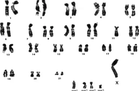
Cytogenetic analysis
Cytogenetic analysis of KL5N at passages 27 and 34 showed
the same pattern of the 45, XY normal hamster chromosome
complement (22,26), with no detectable numerical or structural
anomaly. Forty-five KL5B cells of passage 27 represented an
abnormal clone characterized by a missing Y, monosomy 7
and 11, one copy of two markers and two copies of another
marker (Figure 9). Six cells were a tetraploid version of this
clone. The same patterns were found in KL5B cells of
passage 34.
Expression of islet-specific genes
We analyzed the expression of insulin, IPF1, IEF1, NeuroD,
Pax6 and NKx6.1 in normal pancreas, freshly cultured islets
and undifferentiated islet culture KL5N and KL5B cells by
RT–PCR analysis, as shown in Table III. Expression of insulin,
IPF1, NeuroD, Pax6 and Nkx6.1 was seen in the normal
pancreas and islets. However, these genes were not expressed
in KL5B cells. Interestingly, the IEF1 gene was expressed in
Fig. 6. Reactivity of KL5B cells at passage 32 with PHA (a) and tomato
lectin (b). The reactivity of both lectins was mostly with the cell membrane although in a few cells it was diffuse cytoplasmic.
Fig. 7. Vertical section of a subcutaneous tumor in a hamster that was
inoculated with KL5B cells at stage 19. The 2 cm tumor was encapsulated, firm and showed areas of hemorrhage.
the normal pancreas, islets and KL5B cells. Tubulin expression
was seen in all the samples under study. EGFR and TGF
α
were expressed in pancreas, islets and undifferentiated cells.
Discussion
Several studies in our laboratories have focused on the role of
the islets in pancreatic carcinogenesis in the hamster model.
In particular, promotion of pancreatic carcinogenesis by
stimu-lating islet cell proliferation (16) and by inducing ductal-type
cancers in the SMGs of hamsters bearing homologous islet
transplantation (9–11) strongly supported this view. However,
the question as to whether the tumors arose from some
components attached to the transplanted islets (ductular or
acinar cells) could not be ruled out with certainty. However,
based on our observation of
.500 isolated islets, the possibility
of exocrine cell contamination cannot exceed one islet in 100.
Table I. Immunohistochemical reactivity of antibodies to normal hamster
pancreas, KL5N and KL5B cells (passage 32)
Antibody Normal pancreas KL5N KL5B
(% cells stained) (% cells stained) (% cells stained) Pancytokeratin 111a,b 111a(30) 111a (20) Cytokeratin 14 111*,a 11a(20) 11a (10) Cytokeratin 18 11*,a 111a(5) 111a(10) Carbonic 1** 1 (20) 1a(20) anhydrase II Laminin 11a,b(100) 111a(50) 111a(100) Vimentin 111c(100) 111a(70),1( 20) 111a(100) Tomato lectin 111e(100) 111a,d(100) 111d,f(100) PHA 111f(100) 111d(100) 111d(100) TGF-α 1g(20) 11a(30) 111a(70)
EGFR – 1d(60) 11a(80)
α1-Antitrypsin 1b(100) 1a,d(90) 111a,d(100) –, no staining;1, weak staining; 11, moderate staining; 111, strong staining.
*Staining of ductal, ductular, centroacinar and islet cells **Staining of ductal cell only.
aDiffuse cytoplasmic;bacinar, islet and ductal cells staining;cstaining of smooth muscles only;dcell membrane staining;ediffuse cytoplasmic staining of zymogen granules and of islet cells, luminal staining of ductal cells; fdiffuse cytoplasmic staining of zymogen granules and islet cells;gstaining of glucagon cells only.
This negligible level of impurity cannot explain the massive
cell migration from all islets that are attached to the bottom
of the flask into the surrounding culture (19). Moreover,
according to our experience the growth of cells deriving from
islets is significantly different from that of cultured ductal
cells, in that ductal cells form glandular structures even in
monolayer (27).
Because it has been shown that BOP can transform hamster
ductal cells in vitro (20,21), it was of interest to examine the
effect of BOP on isolated pure islets in vitro in the system we
have established (19). In this system, isolation and long-term
maintenance of pure islets free of exocrine cell contamination
was possible.
Confirming our previous study (19), the present experiment
demonstrates that islet cells in culture give rise temporarily to
exocrine cells but culminate in the formation of undifferentiated
cells, possibly representing stem cells. Although the dose of
BOP used is relatively high compared with doses that reach
the pancreas after in vivo treatment, no sign of toxicity was
seen when compared with the KL5N cells. This could be the
result of reduced levels of BOP-metabolizing enzymes in
cultured cells, a possibility supported by the requirement for
larger doses of BOP for in vitro transformation of ductal cells
(20,21), while in vivo a single treatment with BOP is sufficient
to induce tumors. There were no differences in the initial
growth of islets with or without treatment with BOP, although
it has been shown that carcinogens initially inhibit cell division
by interacting with cellular DNA (28). There were also no
differences in morphological and subcellular levels between
BOP-treated and untreated cells until stage 4, when
pleo-morphic epithelial cells appeared in the KL5B cultures. This
phenotypical change and the loss of carbonic anhydrase II was
obviously unrelated to the c-Ki-ras mutation, which was first
detected much later, at stage 19. It is likely that other as yet
unknown genetic abnormalities preceded the c-Ki-ras mutation.
Hence, it appears that differences exist between the malignancy
of human and hamster pancreatic cells, because in humans
mutation of c-Ki-ras occurs early and even in histologically
Fig. 8. The patterns of the subcutaneous tumor in Figure 7. Histologically, the tumor was anaplastic with large pleomorphic cells and many mitotic figures
(~20 mitotic cells in 253 objective). Large necrotic and hemorrhaging areas were present in the center and glandular formation at the edge of the tumor (inset). H&E,3210.
Table II. Comparison of the immunohistochemical reactivity of antibodies to KL5B cells (passage 32) injected s.c. into the submandibular gland or pancreas
and grown in vitro
Antibody Subcutaneous Submandibular gland Pancreas In vitro
(% cells stained) (% cells stained) (% cells stained) (% cells stained)
Pancytokeratin 1a(10) 1 to 11a(10) 1 to 11a(20) 111a(20) Cytokeratin 13 1a(5) 1a(5) 1a (2) – Cytokeratin 18 1a(10) 1a (10) 1a(10) 111a(10) Carbonic anhydrase – 1a(4) 1a(5) 1a(20) Laminin 1a(100) 1a(100) 1 to 111a,b(100) 111a(100) Vimentin 1 to 111c(100) 111c(100) 1 to 11c(80) 111a(100) Tomato lectin 11 to 111d(100) 1 to 111b(100) 1 to 11d(100) 111a(100) PHA 1e(100) 111c(100) 111e(100) 111a(100) EGFR 1a(30) 1a(60) 1 1a(80) 11a(80) α1-Antitrypsin 1a(70) 11a(80) 1a(60) 111a(100)
–, no staining;1, weak staining; 11, moderate staining; 111, strong staining.
aDiffuse cytoplasmic;bweak staining of tumor cells, strong staining of blood vessels;cgranular cytoplasmic staining;dcell membrane and vascular staining; ecell membrane staining.
Table III. Expression of insulin and insulin-associated transcriptional
factors in normal hamster pancreas, islets and KL5B cells derived from islets
Factor Pancreas Islets KL5N KL5B
IPF1 1 1 – – IEF1 1 1 1 1 Insulin 1 1 – – Neuro-D 1 1 – – Pax6 1 1 – – NKx6.1 1 1 – – EGFR 1 1 1 1 TGF-α 1 1 1 1
normal-appearing cells (29,30). Perhaps, certain environmental
factors lacking under the in vitro conditions are necessary for
this mutation.
The study nevertheless confirms that pancreatic islet cells
can give rise to ductal-type cancer cells. However, we do not
yet know whether the tumor progenitor cells derive from
stem cells within islets or from transdifferentiated islet cells.
Pancreatic exocrine cells as a contaminant of islets can be
ruled out because our immunohistochemical examination using
pancytokeratin, a marker for hamster pancreatic ductal cells,
and electron microscopic examination of isolated islets and
those examined 7 days later in culture did not show any
evidence of cell contamination. All the exocrine and
intermedi-ary cells developed within islets. The origin of the exocrine
cells from intra-insular ductules can also be ruled out, because
intra-insular ductules develop only in aged hamsters and under
some pathological conditions. In the present study, however,
we used islets of healthy and young hamsters, which do not
contain intra-insular ductules. Moreover, in our hands
BOP-transformed hamster ductal cells lack the c-Ki-ras mutation
(20). Consequently, the cells that develop from cultured islets
appear to be different from ductal/ductular cells.
What was remarkable was the in vivo growth pattern of
KL5B cells. They formed anaplastic invasive tumors, similar
to those induced by BOP in islets growing in the SMG of
hamsters (10,11) and to PC-1.0 cells, derived from a primary
BOP-induced pancreatic cancer (31). The anaplastic and
invas-ive nature of the KL5B tumor could be related to massinvas-ive
over-production of TGF-
α
, which was shown to be overexpressed in
metastasizing ILA cells (32) derived from tumors induced in
the SMG of hamsters after homologous islet transplantation
(9–11) and in human pancreatic cancer cells (33–35). The
remarkable vascularization of the tumors could also be related
to TGF-
α
(35). The expression of
α
1-antitrypsin in both KL5N
and KL5B cells but not in any adult hamster pancreatic cells
further supports the origin of these cells from a primitive
precursor cell. This acute phase reactant protein has been
found in human pancreatic tumors assumed to derive from
stem cells, including solid cystic (papillary) tumor (36) and
pancreatoblastoma (37). Hence, this protein, as well as
vimen-tin, not present in pancreatic parenchymal cells, also appears
to present as a marker for hamster pancreatic stem cells (38).
Interestingly,
α
-fetoprotein, another protein produced in fetal
tissues, has recently been found in human pancreatic cancer
(39), an indication that human pancreatic cancer cells also
derive from primitive pancreatic cells.
Simultaneous expression of vimentin and cytokeratin, also
found in aggressive human breast cancer (40), seems to be
associated with increased invasive behavior. The same seems
to apply to the lack of blood group A antigen expression. We
have shown that well-differentiated, BOP-induced tumors and
the slow growing PC-1.0 cell line derived from a primary
BOP-induced cancer (41) consistently express blood group A
antigen (42), whereas poorly differentiated, fast growing
PC-1.0 cells derived from an s.c. transplant of a primary hamster
pancreatic cancer (31) do not. However, in contrast to PC-1.0
cells, which express blood group A antigen expression when
transplanted into hamsters (23), KL5B cells failed to produce
this antigen in vivo. Hence, it appears that in KL5B cells the
genes involved in blood group A synthesis are inactivated.
Whether these genes were located in the missing or altered
chromosomes is unclear. Nevertheless, like ILA cells, KL5B
cells were also missing a Y chromosome, a finding that was
not seen in transformed ductal cells (20). Interestingly, the
missing sex chromosome has been found to be one of the
most frequent findings in human pancreatic cancer (43–45).
However, because deletion of a Y chromosome can occur in
other types of cancer, particularly often in metastases of colon
cancer (45), this abnormality is not specific for pancreatic
cancer and its role in the evolution of pancreatic cancer is
unknown. In human pancreatic cancer, enhanced expression
of EGFR has been found to correlate with alterations of
chromosome 7 (46), one of which was missing in KL5B cells.
Because expression of EGFR appeared early in both KL5B
and KL5N cells, a correlation between this chromosomal
damage and EGFR expression is unlikely.
There were some significant differences in the chromosomal
changes between TAKA-1-BOP tumors arising from hamster
pancreatic ductal cells (22), from tumors in SMGs of hamsters
bearing transplanted islets (32) and in KL5B cells. These
include deletion of one of the chromosomes 4 and 7 only in
KL5B cells, alteration of chromosome 3 only in ILA and
TAKA-1-BOP cells and many extra chromosomes in ILA cells
(32). However, because of a limited number of cells examined
from each cell line, it is unclear whether these abnormalities
are homogenous or merely heterogeneous.
The malignant alteration of islets by BOP in culture
unequi-vocally points to the ability of islet cells to metabolize BOP.
Consequently, it appears that both hamster ductal cells, which
can also be transformed by BOP in culture (20), and islets
have the necessary metabolizing enzyme and, thus, are direct
targets of BOP.
Acknowledgements
This work was supported by National Institutes of Health National Cancer Institute grant 5RO1 CA60479 and SPORE grant P50CA72712, National Cancer Institute Laboratory Cancer Research Center support grant CA367127 and an American Cancer Society Special Institutional Grant.
References
1. Flaks,B., Moore,M.A. and Flaks,A. (1980) Ultrastructural analysis of pancreatic carcinogenesis. III. Multifocal cystic lesions induced by N-nitroso-bis(2-hydroxypropyl)amine in the hamster exocrine pancreas. Carcinogenesis, 1, 693–706.
2. Pour,P.M. (1984) Histogenesis of exocrine pancreatic cancer in the hamster model. Environ. Health Perspect., 56, 229–243.
3. Pour,P.M. (1988) Mechanism of pseudoductular (tubular) formation during pancreatic carcinogenesis in the hamster model. Am. J. Pathol., 130, 335– 344.
4. Pour,P.M. and Wilson,R. (1980) Experimental pancreas tumor. In Moossa,A.R. (ed.) Cancer of the Pancreas. Williams and Wilkins, Baltimore, MD, pp. 37–158.
Fitzgerald,P.J. and Morrison,A.B. (eds) The Pancreas. William and Wilkins, Baltimore, MD, pp. 111–139.
6. Pour,P.M., Runge,R.G., Birt,D., Gingell,R., Lawson,T., Nagel,D., Wallcave,L. and Salmasi,S.Z. (1981) Current knowledge of pancreatic carcinogenesis in the hamster and its relevance to the human disease. Cancer,
47, 1573–1589.
7. Pour,P.M. (1981) The endocrine-exocrine pancreas: its clinical and morphological aspects and hyperplastic and neoplastic patterns. In Nagasawa,H. and Abe,K. (eds) Hormone Related Tumors. Japan Scientific Societies Press, Tokyo, Japan, pp. 103–120.
8. Pour,P.M. (1978) Islet cells as a component of pancreatic ductal neoplasms. I. Experimental study. Ductular cells, including islet cell precursors, and primary progenitor cells of tumors. Am. J. Pathol., 90, 295–316.
9. Pour,P.M., Weide,L., Liu,G., Kazakoff,K., Scheetz,M., Toshkov,I. and Sanger,W. (1997) Langerhans islets are the origin of ductal-type adenocarcinoma. In Malfertheiner,P. et al. (eds) Diagnostic Procedures in Pancreatic Disease. Springer Verlag, Heidelberg, Germany, pp. 331–339. 10. Pour,P.M., Weide,L., Liu,G., Kazakoff,K., Scheetz,M., Toshkov,I.
Ikematsu,I., Fienhold,M.A. and Sanger,W. (1997) Experimental evidence for the origin of ductal type adenocarcinoma from the islets of Langerhans. Am. J. Pathol., 150, 2167–2180.
11. Fienhold,M.A., Kazakoff,K. and Pour,P.M. (1997) The effect of Streptozotocin and high-fat diet on BOP-induced tumors in the pancreas and in the submandibular gland of hamsters bearing transplants of homologous islets. Cancer Lett., 117, 155–160.
12. Bell,R.H., Sayers,H.J., Pour,P.M., Ray,M.B. and McCullough,P.J. (1989) Importance of diabetes in inhibition of pancreatic cancer by Streptozotocin. J. Surg. Res., 46, 515–519.
13. Bell,R.H., McCullough,P.J. and Pour,P.M. (19880 Influence of diabetes on susceptibility to experimental pancreatic cancer. Am. J. Surg., 155, 159–164. 14. Pour,P.M., Kazakoff,K. and Carlson,K. (1990) Inhibition of Streptozotocin-induced islet cell tumors and BOP-Streptozotocin-induced exogenous pancreatic tumors in Syrian hamsters. Cancer Res., 50, 1634–1639.
15. Bell,R.H. and Pour,P.M. (1987) Induction of pancreatic tumors in genetically non-diabetic but not in diabetic Chinese hamsters. Cancer Lett., 34, 221–230. 16. Pour,P.M. and Kazakoff,K. (1996) Stimulation of islet cell proliferation enhances pancreatic ductal carcinogenesis in the hamster model. Am. J. Pathol., 149, 1017–1025.
17. Pour,P.M. and Morohoshi,T. (1994) Ductal adenocarcinoma. In Atlas of Exocrine Pancreatic Tumors. Morphology, Biology and Diagnosis with an International Guide for Tumor Classification. Springer Verlag, Tokyo, Japan, pp. 117–154.
18. Kimura,W., Morikane,K., Esaki,Y., Chan,W.C. and Pour,P.M. (1998) Histological and biological patterns of microscopical pancreatic ductal adenocarcinomaa detected incidentally at autopsy. Cancer, 82, 1839–1849. 19. Schmied,B., Liu,G., Matsuzaki,H., Hernberg,S., Moyer,M.P., Weide,L., Murphy,L., Batra,S.K. and Pour,P.M. (1999) Transformation of hamster pancreatic endocrine cells into exocrine cells. J. Cell. Sci., in press. 20. Ikematsu,Y., Liu,G., Fienhold,M.A., Cano,M., Adrian,T.E.,
Hollingsworth,M.A., Williamson,J.E., Sanger,W., Tomioka,T. and Pour,P.M. (1997) In vitro pancreatic ductal carcinogenesis. Int. J. Cancer, 72, 1095– 1103.
21. Mangold,K.A., Mangino,M.M., Laconi,S. and Scarpelli,D.G. (1994) In vitro carcinogenesis of hamster pancreatic duct cells: cellular and molecular alterations. Carcinogenesis, 15, 1979–1984.
22. Takahashi,T., Moyer,M.P., Cano,M., Wang,Q.J., Mountjoy,C.P., Sanger,W., Adrian,T.E., Sugiura,H., Katoh,H. and Pour,P.M. (1995) Differences in molecular biological, biological and growth characteristics between the immortal and malignant hamster pancreatic ductal cells. Carcinogenesis, 16, 931–939.
23. Egami,H., Tomioka,T., Tempero,M. and Pour,P.M. (1991) Development of intrapancreatic transplantable model of pancreatic duct adenocarcinoma in Syrian golden hamasters. Am. J. Pathol., 138, 557–561.
24. Pour,P.M., Kazakoff,K. and Dulany,K. (1993) A new multilabeling technique for simultaneous demonstration of different islet cells in permanent slides. Int. J. Pancreatol., 13, 139–142.
25. Fujii,H., Egami,H., Chaney,W., Pour,P.M. and Pelling,J. (1990) Pancreatic ductal adenocarcinomas induced in Syrian hamsters by N-nitrosobis(2-oxopropyl)amine contain a c-Ki-ras oncogene with a point-mutated codon 12. Mol. Carcinogen., 3, 296–301.
26. Takahashi,T., Moyer,M.P., Cano,M., Wang,Q.J., Adrian,T.E., Mountjoy,C.P.,
Sanger,W., Sugiura,H., Katoh,H. and Pour,P.M. (1995) Establishment and characterization of a new, spontaneously immortalized, pancreatic ductal cell line from the Syrian golden hamster. Cell Tissue Res., 282, 163–174. 27. Takahashi,T., Moyer,M.P., Cano,M., Wang,Q.J., Adrian,T.E., Mountjoy,C.P.,
Sanger,W., Sugiura,H., Katoh,H. and Pour,P.M. (1995) Establishment and characterization of a new, spotaneously immortalized, pancreatic ductal cell line from the Syrian golden hamster. Cell Tissue Res., 282, 163–174. 28. Mirvish,S.S., Chu,C. and Clayson,D.B. (1978) Inhibition of [3H]thymidine
incorporation into DNA of rat esophageal epithelim and related tissues by carcinogenic N-nitroso compounds. Cancer Res., 38, 458–466.
29. Fujii,H., Inagaki,M., Kasai,S., Miyokawa,N., Tokusashi,Y., Gabrielson,E. and Hruban,R.H. (1997) Genetic progression and heterogeneity in intraductal papillary-mucinous neoplasms of the pancreas. Am. J. Pancreatol., 151, 1447–1454.
30. Sugio,K., Molberg,K., Albores-Saavedra,J., Virmani,A.K., Kishimoto,Y. and Gazdar,A.F. (1997) K-ras mutations and allelic loss at 5q and 18q in the development of human pancreatic cancers. Int. J. Pancreatol., 21, 205–217. 31. Hirota,M., Egami,H., Corra,S., Fujii,H., Chaney,W.G., Rizzino,A. and Pour,P.M. (1993) Production of scatter factor-like activity by a nitrosamine-induced pancreatic cancer cell line. Carcinogenesis, 14, 259–264. 32. Toshkov,I., Schmied,B., Adrian,T.E., Murphy,L.O., Wahab,H.A.A.Y. and
Pour,P.M. (1998) Establishment of tumor cell line, ILA, from hamster islets treated with BOP. Int. J. Cancer, 78, 636–641.
33. Korc,M., Chandasekar,B., Yamanaka,Y., Friess,H., Bu¨chler,M. and Beger,H.G. (1992) Overexpression of the growth factor receptor in human pancreatic cancer is associated with concomitant increases in the levels of epidermal growth factor and transforming growth factor alpha. J. Clin. Invest., 90, 1352–1360.
34. Smith,J.J., Derynck,R. and Korc,R. (1987) Production of transforming growth factor α in human pancreatic cancer cells: evidence for a superagonistic autocrine cycle. Proc. Natl Acad. Sci. USA, 84, 7567–7570. 35. Screiber,A.B., Winkler,M.E. and Derynck,R. (1986) Transforming growth
factor-α: a more potent angiogenic mediator than epidermal growth factor. Science, 232, 1250–1253.
36. Yamaguchi,K., Morohoshi,T. and Zamboni,G. (1994) Solid cystic tumors. In Atlas of Exocrine Pancreatic Tumors. Morphology, Biology and Diagnosis with an International Guide for Tumor Classification. Springer Verlag, Tokyo, Japan, pp. 83–100.
37. Morohoshi,T., Kanda,M., Horie,A., Chotl,A., Dreyer,T., Klo¨ppel,G. and Heitz,P.U. (1987) Immunohistochemical markers of uncommon pancreatic tumors. Acinar cell carcinoma, pancreatoblastoma and solid cystic (papillary-cystic) tumor. Cancer, 59, 739–747.
38. Bouwens,L. and De Blay,E. (1996) Islet morphogenesis and stem cell markers in rat pancreas. J. Histochem. Cytochem., 44, 947–951.
39. Tanno,S., Obara,T., Shudo,R., Fujii,T., Sugawara,K., Nishino,N., Ura,H. and Kohgo,Y. (1997)α-Fetoprotein producing mucin-producing carcinoma of the pancreas. A case report with immunohistochemical study and lectin-affinity profile. Dig. Dis. Sci., 42, 2513–2518.
40. Hendrix,M.J.C., Seftor,E.A., Seftor,R.E.B. and Trevor,K.T. (1997) Experimental co-expression of vimentin and keratin intermediate filaments in human breast cancer cells results in phenotypic interconversion and increased invasive behavior. Am. J. Pathol., 150, 483–495.
41. Egami,H., Takiyama,Y., Cano,M., Houser,W.H. and Pour,P.M. (1989) Establishment of hamster pancreatic ductal carcinoma cell line (PC-1) producing blood group-related antigens. Carcinogenesis, 10, 861–869. 42. Pour,P.M., Uchida,E., Burnett,D.A. and Steplewski,Z. (1986) Blood-group
antigen expression during pancreatic cancer induction in hamsters. Int. J. Pancreatol., 1, 327–340.
43. Bardi,G., Johansson,B., Pandis,N., Mandahl,N., Bak-Jensen,E., Andre´n-Sandberg,Å., Mittelman,F. and Heim,S. (1993) Karyotypic abnormalities in tumors of the pancreas. Br. J. Cancer, 67, 1106–1112.
44. Johansson,B., Bardi,G., Heim,S., Mandahl,N., Mertens,F., Bak-Jensen,E., Andre´n-Sandberg,Å. and Mittelman,F. (1992) Nonrandom chromosomal rearrangements in pancreatic carcinomas. Cancer, 69, 1674–1681. 45. Bardi,G., Parada,L.A., Bomme,L., Pandis,N., Johansson,B., Wille´n,R.,
Fenger,C., Kronborg,O., Mittelman,F. and Heim,S. (1997) Cytogenetic findings in metastases from colorectal cancer. Int. J. Cancer, 72, 604–607. 46. Korc,M., Meltzer,P. and Trent,J. (1986) Enhanced expression of epidermal
growth factor receptor correlates with alterations of chromosome 7 in human pancreatic cancer. Proc. Natl Acad. Sci. USA, 83, 5141–5144.