Acta Genet Med Gemellol 34:175-178 (1985) © 1985 by The Mendel Institute, Rome
Received 2 November 1984 Final 15 March 1985
Premature Contractions: Are They Caused by
Maternal Standing?
K.T.M. Schneider, A. Huch, R. Huch
Department of Obstetrics, University of Zurich
Abstract. In 33 out of 51 women studied in late gestation, the uterus was found to
phasic-ally compress the pelvic vessels and impede the venous blood flow during quiet standing. This caused a reduction of the cardiac stroke volume with resultant reduction of systemic blood pressure and a compensatory increased heart rate (range of increases 9-51 beats/ min). In all cases uterine contractions (mostly subclinical) coincided with the phase of circulatory readjustment. Apparently, the contracting uterus, by changing its position and/or shape, relieves the venous obstruction and prevents decompensation. In the women displaying the uterine compression syndrome (UCS), uterine activity was marked-ly increased in standing compared to the left recumbent position.
It was also investigated whether the UCS appeared more often and earlier in gestation in women with twins. In all 9 women with twin pregnancies (mean gestational age 28 5/7 weeks) the UCS associated with uterine contractions was apparent in the standing posture. Although at present no definite conclusions can be reached on the effect on the cervix of these contractions, quiet standing especially in twin pregnancies seems to provoke an increased uterine activity and should therefore be avoided.
Key words: Standing posture, Uterine compression, Venous return, Premature contrac-tions, Twin pregnancy, Prematurity
INTRODUCTION
In 1933 Ahltorp [1] published his clinical observation on the supine hypotensive shock syndrome in late pregnancy. His hypothesis that the venous return in the vena cava was impeded by the weight of the gravid uterus in the supine position and that the shocklike symptoms were induced by this was later confirmed by the angiographic studies perfor-med by Kerr and Bieniarz [2,4] in the mid 1960s.
Until now, mechanical obstruction of the venous return in other body postures was
available at https:/www.cambridge.org/core/terms. https://doi.org/10.1017/S0001566000004694
176 Schneider et al
not considered. Since we have systematically studied cardiopulmonary functions of pregnant women [5], our attention was focused on this possibility as a result of an upright posture [6].
MATERIALS AND METHODS
Fifty one healthy pregnant women between gestational weeks 36 and 41 with singleton pregnancies were investigated in the quiet standing posture. The left lateral recumbent position was used as a reference in each case. The duration of the measurements was 10 min in each position. Nineteen of these women were rechecked within 7 days after delivery.
In addition, 9 healthy women with twin pregnancies between 21 3/7 and 33 0/7 gestational weeks were also investigated under the same conditions. None of the women were in labor or under any treatment.
We continuously recorded the maternal beat-to-beat heart rate by means of ECG electrodes fixed on the thorax; these electrodes were also used to measure the transthoracic impedance (Hewlett Packard 78203 A, 78202 B). The blood pressure (Dynamap) and the cardiac output (indirect Fick principle, Ergostar) were recorded every 30 sec.
In 23 women we measured the fetal beat-to-beat heart frequency (Hewlett Packard 8040 A) by ultrasound and the external tocogram. In 12 subjects, the venous flow velocity was measured using a continuous-wave Doppler technique (Directional Doppler, Parks Electronics, 806 A, 8 MHz) over the maternal femoral vein together with plethysmography of the lower thigh (strain gauge plethysmo-gram). The variables thus continuously recorded were charted on a six-channel recorder in parallel with the original cardiotocogram (CTG) tracing.
RESULTS
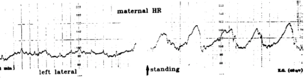
As shown in Fig. 1, an excerpt of a maternal beat-to-beat heart rate recording, the change from the lying to the standing position resulted in consistent periodic increases in the heart rate; in each case, there was a subsequent return to the normal baseline. Of the 51 women with singletons, this phenomenon of periodic tachycardia in the resting standing posture was demostrable in 33 (65%) of the subjects during the whole investigation period. The average cycle length of tachycard phases was in all these cases 105 s (range 85-240 s). The average amplitude was 27 beats per minute (range 9-51). While the heart rate increased, the cardiac output, as measured by the indirect Fick principle, decreased and the blood pressure fell.
Simulated walking with.activation of the "muscle pump" weakened the phenome-non. The same reaction was seen when the woman was leaning forward, or when the uterus was lifted upwards by the investigator. In the 19 postpartum controls, the cyclical tachycardia at standing was not present, although in some cases the interval between the pre- and postpartum measurements amounted to only a few hours [6].
In simultaneous recordings of maternal heart rate, qualitative flow over the femoral vein, leg plethysmogram and transthoracic impedance during quiet standing, we observed the following.
The venous flow velocity decreased remarkably coinciding with each phase of tachy-cardia; in contrast, with each increase of the venous flow, maternal heart rate returned to normal level [6]. In addition, the plethysmogram showed a continuous venous pooling in the leg with a periodic release of venous blood coinciding with the onset of maternal heart rate renormalisation. The transthoracic impedance showed, on the one hand, that the venous flow variations were not induced by respiration, and on the other hand, that
available at https:/www.cambridge.org/core/terms. https://doi.org/10.1017/S0001566000004694
Premature Contractions and Maternal Standing 177
the tracing had a spindle-shaped form with an increase of the breathing movements during the increase in heart rate [6].
In women who did not have tachycardia in the standing posture, the venous flow and the other parameters were unrestricted [6].
Although our patients were not in labor, the external tocogram revealed slight uteri-ne activity in women displaying this uteriuteri-ne compression syndrome (UCS) while standing in all cases investigated (Fig. 2): there was an obvious relationship between the onset of contractions and the beginning of the heart rate renormalisation. Concomitant with each contraction, maternal heart rate returned to normal level while the contraction persisted. In women with the UCS, the uterine activity upon standing was markedly increased compared to the left lateral position.
In the 9 twin pregnancies (mean gestational age 28 5/7 weeks; range 21 3/7 - 33 0/7 weeks), UCS occurred in all cases in the standing posture but not in the left lateral position. Fig. 2 shows a recording from a woman with twins in the 32nd week. Compared with the left lateral position, during quiet standing a markedly increased uterine activity coincided with the normalisation of the maternal heart rate.
T 1 — J maternal HR ^ ^ ^ ^ ^ ^ ^ ^ ^ ^ f left lateral •us , ,/v 4 s t a n d i n g " -77";-" r "" 1 A 1?'
•
; ! / • Us
•
\* t o . <«•*»)Fig. 1 - Recording of maternal beat-to-beat heart rate in left lateral and standing postures in late
preg-nancy (40 2/7 weeks). The change from lying to standing resulted in periodic heart rate accelerations (from Schneider et al [6]).
standing I'left lateral
Fig. 2 - Simultaneous recording of maternal beat-to-beat heart rate and tocogram in standing and left lateral postures in a twin pregnancy (31 2/7 weeks). The arrows mark the simultaneous onset of contractions and maternal heart rate normalisation.
available at https:/www.cambridge.org/core/terms. https://doi.org/10.1017/S0001566000004694
178 Schneider et al
DISCUSSION
Our investigations show that in motionless standing posture the gravid uterus seems to compress the pelvic vessels and to impede periodically the venous return, as this was observed in two thirds of the women with singletons and in all women with twins investi-gated to date. This maternal disturbance, eg, the decrease in cardiac output and blood pressure and the increase in heart rate, is analogous with the initial symptoms of the so-called supine hypotensive shock syndrome. Apart from an incidental reference in 1942 [3] it is surprising that this phenomenon has not become clinically evident earlier. Perhaps this is due to the fact that, as far as out investigations are concerned, no patient fainted nor did any of them experience marked subjective symptoms.
It was logical to expect that the UCS should appear earlier in the gestational period and more frequently in women with a "heavier and larger" uterus, ie, one containing twins, for example. However, it was surprising to see how universal this circulatory phenomenon was in twin pregnancies. UCS could be observed in all studied mothers of twins and in one case, as early as in the 21 3/7 week. In our department mothers of twins are treated with prophylactic bed rest; routine antenatal monitoring is therefore perfor-med in the horizontal position. It is tempting to think of the possible clinical and thera-peutic consequences when interpreting the tocogram from the standing mothers.
The observation that the maternal heart frequency returns phasically to normal levels after each increase leads to the conclusion that an effective autoregulation system prevents decompensation. The strict coincidence between the onset of contractions and the normalisation of the circulatory parameters may be the key to the understanding of this phenomenon. Changes in volume or position of the contracted uterus seem to reduce the pressure upon the pelvic vessels and to release the venous return.
Therefore, this type of contractions might play an essential role in the orthostatic regulation in pregnant women.
REFERENCES
1. Ahltorp G (1933): In Rueckenlage eintretende Beschwerden bei Graviden. Acta Obstet Gynecol Scand 15:295-341.
2. Bieniaiz J, Crottogini JJ, Curuchet E, Romero-Salinas G, Yoshida T, Poseiro JJ, Caldeiro-Barcia R (1968): Aortocaval compression by the uterus in late human pregnancy. Am J Obstet Gynecol 15:203-217.
3. Hansen R (1942): Ohnmacht und Schwangerschaft. KlinWschi 11:241-245.
4. Kerr MG, Scott DB, Samuel E (1964: Studies of the inferior vena cava in late pregnancy. Br Med J 1:532-533.
5. Schneider KTM, Spaetling L, Huch R, Huch A (1983): Einfluss der Koerperhaltung auf die Lun-gen- und Kreislauffunktion in der Spaetschwangerschaft. Atemw Lungenkrkh 9:205-208. 6. Schneider KTM, Bollinger A, Huch A, Huch R (1984): The oscillating "vena cava syndrome"
during quiet standing. An unexpected observation in late pregnancy. Br J Obstet Gynecol 91: 776-780.
Correspondence: Dr. K.T.M. Schneider, Frauenklinikstr. 10, CH-8091 Zurich, Switzerland.
available at https:/www.cambridge.org/core/terms. https://doi.org/10.1017/S0001566000004694